1
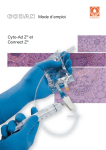
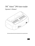
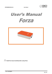
Product Manual CytoSelect™ 96-Well In Vitro Tumor Sensitivity Assay (Soft Agar Colony Formation) Catalog Number CBA-150 96 assays CBA-150-5 5 x 96 assays FOR RESEARCH USE ONLY Not for use in diagnostic procedures Introduction Tumor sensitivity assays are intended to help predict the sensitivity of various tumors to chemotherapeutic agents, with the intent of identifying the most effective treatment with the fewest side effects. With this information, physicians can devise tailor made chemotherapy regiments and eliminate ineffective drugs, sparing patients of unnecessary toxicity. Ideally, an in vitro tumor sensitivity assay must be reliable, sensitive, and resemble the 3-D, in vivo environment (such as culturing in collagen gel or soft agar). Traditionally, the soft agar colony formation assay is a common method to monitor anchorageindependent growth, which measures proliferation in a semisolid culture media after 3-4 weeks by manual counting of colonies. Cell Biolab’s CytoSelect™ 96-well In Vitro Tumor Sensitivity Assay does not involve subjective manual counting of colonies or require a 3-4 week incubation period. Instead cells are incubated only 6-8 days in a proprietary semisolid agar media before being solubilized, transferred and detected by the provided MTT Solution in a microtiter plate reader (see Assay Principle below). The CytoSelect™ 96-well In Vitro Tumor Sensitivity Assay provides a stringent, anchorageindependent model for chemosensitivity testing and potential anticancer drug screening. Each kit provides sufficient quantities to perform 96 tests in a microtiter plate. 2 Assay Principle Related Products 1. CBA-100: CytoSelect™ 24-Well Cell Migration Assay (8μm, Colorimetric) 2. CBA-106: CytoSelect™ 96-Well Cell Migration Assay (8µm, Fluorometric) 3. CBA-106-C: CytoSelect™ 96-Well Cell Migration and Invasion Assay (8µm, Fluorometric) 4. CBA-112: CytoSelect™ 96-Well Cell Invasion Assay (Basement Membrane, Fluorometric) 5. CBA-130: CytoSelect™ 96-Well Cell Transformation Assay (Soft Agar Colony Formation) 6. CBA-135: CytoSelect™ 96-Well Cell Transformation Assay (Cell Recovery, Colorimetric) 7. CBA-140: CytoSelect™ 96-Well Cell Transformation Assay (Cell Recovery, Fluorometric) 8. CBA-145: CytoSelect™ 384-Well Cell Transformation Assay 9. CBA-155: CytoSelect™ Clonogenic Tumor Cell Isolation Kit 10. CBA-320: CytoSelect™ 96-Well Hematopoietic Colony Forming Cell Assay 3 Kit Components 1. 10X CytoSelect™ Agar Matrix Solution (Part No. 114001): One sterile bottle – 10.0 mL 2. CytoSelect™ Matrix Diluent (Part No. 114002): One sterile bottle – 4.0 mL 3. 5X DMEM Solution (Part No. 113002): Three sterile tubes – 1.5 mL each 4. 1X Matrix Solubilization Buffer (Part No. 115001): One sterile bottle – 20.0 mL 5. Detergent Solution (Part No. 113501): One bottle – 10.0 mL 6. MTT Solution (Part No. 113502): One tube – 1.0 mL Materials Not Supplied 1. Tumor Cells (cancer cell line or cells prepared from solid tumor) 2. Anticancer Agents (e.g. Taxol, 5-Fluorouracil, anticancer mAb or siRNA) 3. 37ºC Incubator, 5% CO2 Atmosphere 4. Light Microscope 5. 96-well Microtiter Plate Reader 6. 37ºC and boiling water baths Storage Store all components at 4ºC until their expiration dates. Preparation of Reagents • 2X DMEM/20% FBS Medium: In a sterile tube, dilute the provided 5X DMEM in sterile cell culture grade water to 2X containing 20% FBS. For example, to prepare a 5 mL solution, add 2 mL of 5X DMEM, 1 mL of FBS and 2 mL of sterile cell culture grade water. Sterile filter the 2X media to 0.2 µm. • 10X CytoSelect™ Agar Matrix Solution: Heat the Agar Matrix Solution bottle to 90-95ºC in a water bath for 30 minutes, or until agarose liquefies (microwaving is optional). Transfer the bottle to a 37ºC water bath for 20 minutes and maintain until needed. Assay Protocol (must be under sterile conditions) I. Preparation of Base Agar Matrix Layer 1. Heat the 10X CytoSelect™ Agar Matrix Solution to 90-95ºC in a water bath for 30 minutes, or until agarose liquefies (microwaving is optional). Transfer the bottle to a 37ºC water bath for 20 minutes and maintain until needed. 2. Warm the 2X DMEM/20% FBS medium (see Preparation of Reagents section) to 37ºC in a water bath. Allow at least 30 minutes for the temperature to equilibrate. 4 3. According to Table 1 (below), prepare the desired volume of Base Agar Matrix Layer in the following sequence: a. In a sterile tube, add the appropriate volume of 2X DMEM/20% FBS medium. b. Next, add the corresponding volume of sterile water. Mix well. c. Finally, add the corresponding volume of 10X CytoSelect™ Agar Matrix Solution. Mix well. Note: The 10X CytoSelect™ Agar Matrix Solution is slightly viscous; care should be taken in accurately pipetting the appropriate volume. 2X DMEM/20% FBS Medium (mL) 2.5 1.25 0.5 Sterile Water (mL) 10X Total Volume of # of Tests in 96CytoSelect™ Base Agar Matrix well Plate (50 Agar Matrix Layer (mL) µL/test) Solution (mL) 2 0.5 5 100 1 0.25 2.5 50 0.4 0.1 1 20 Table 2. Preparation of Base Agar Matrix Layer 4. After mixing, maintain the Base Agar Matrix Layer at 37ºC to avoid gelation. 5. Dispense 50 μL of Base Agar Matrix Layer into each well of a 96-well sterile flat-bottom microplate (samples should be assayed in triplicate). Gently tap the plate a few times to ensure the Base Agar Matrix Layer evenly covers the wells. Notes: • Work quickly with the layer to avoid gelation. Also, try to avoid adding air bubbles to the well. • To avoid fast and uneven evaporation that leads to aberrant results, we suggest not using the wells on the plate edge, or filling the edge wells with medium to reduce evaporation. 6. Transfer the plate to 4ºC for 30 minutes to allow the Base Agar Matrix Layer to solidify. 7. Prior to adding the Cell Suspension/Agar Matrix Layer (Section II), allow the plate to warm to room temperature for 30 minutes. II. Addition of Cell Suspension/Agar Matrix Layer (under sterile conditions) 1. Heat the 10X CytoSelect™ Agar Matrix Solution to 90-95ºC in a water bath for 30 minutes, or until agarose liquefies (microwaving is optional). Transfer the bottle to a 37ºC water bath for 20 minutes and maintain until needed. 2. Warm the 2X DMEM/20% FBS medium (see Preparation of Reagents section) and CytoSelect™ Matrix Diluent to 37ºC in a water bath. Allow at least 30 minutes for the temperature to equilibrate. 5 3. Harvest and resuspend cells in culture medium at 0.1 - 1 x 10⁶ cells/mL. Keep the cell suspension warm in a 37ºC water bath. 4. According to Table 2 (below), prepare the desired volume of Cell Suspension/Agar Matrix Layer in the following sequence: a. In a sterile tube, add the appropriate volume of 2X DMEM/20% FBS medium. b. Next, add the corresponding volume of CytoSelect™ Matrix Diluent. Mix well. c. Next, add the corresponding volume of 10X CytoSelect™ Agar Matrix Solution. Mix well. d. Finally, add the corresponding volume of cell suspension. Mix well. Note: The CytoSelect™ Matrix Diluent and 10X CytoSelect™ Agar Matrix Solution are slightly viscous; care should be taken in accurately pipetting the appropriate volumes. 2X DMEM/20% FBS Medium (mL) 3.5 1.75 0.875 CytoSelect™ Matrix Diluent (mL) 10X Cell Total Volume of CytoSelect™ Suspension Cell Suspension/ Agar Matrix (mL) Agar Matrix Solution (mL) Layer (mL) 2.75 0.75 0.5 7.5 1.375 0.375 0.25 3.75 0.688 0.188 0.125 1.875 Table 3. Preparation of Cell Suspension/Agar Matrix Layer # of Tests in 96-well Plate (75 µL/test) 100 50 25 5. After mixing, incubate the Cell Suspension/Agar Matrix Layer at room temperature for 5 minutes. 6. Immediately dispense 75 μL of Cell Suspension/Agar Matrix Layer into each well of the 96well plate, already containing the Base Agar Matrix Layer (Section I). Notes: • Work quickly with the layer to avoid gelation, but gently pipette as not to disrupt the base layer integrity. Also, try to avoid adding air bubbles to the well. • Always include negative control wells that contain no cells in the Cell Suspension/Agar Matrix Layer. 7. Transfer the plate to 4ºC for 20 minutes to allow the Cell Suspension/Agar Matrix Layer to solidify. 8. Allow the plate to warm to room temperature for 30 minutes. 9. Add 50 μL of culture medium containing anticancer agents (e.g. Taxol, 5-Fluorouracil, mAb, etc.) to each well. 10. Incubate the cells for 6-8 days at 37ºC and 5% CO2. Examine the colony formation under a light microscope. 6 III. Quantitation of Anchorage-Independent Growth 1. Add 125 μL of the 1X Matrix Solubilization Buffer to each well. 2. Pipette the entire volume of the well 10-12 times to mix thoroughly and solubilize the agar matrix completely. 3. Transfer 100 µL of the mixture to a 96-well microtiter plate. 4. Add 10 µL of MTT Solution to each well. Pipette each well 7-10 times to ensure a homogeneous mixture. 5. Incubate the plate for 2-4 hours at 37ºC and 5% CO2. Note: Under the microscope, a purple precipitate should be visible within the cells. 6. Add 100 µL of Detergent Solution to each well. 7. Incubate the plate in the dark for 2-4 hours at room temperature. 8. Pipette each well 7-10 times to ensure a homogeneous mixture. 9. Measure the absorbance at 570 nm in a 96-well microtiter plate reader. Cell Dose Curve (optional) 1. Heat the 10X CytoSelect™ Agar Matrix Solution to 90-95ºC in a water bath for 30 minutes, or until agarose liquefies (microwaving is optional). Transfer the bottle to a 37ºC water bath for 20 minutes and maintain until needed. 2. Warm the 2X DMEM/20% FBS medium (see Preparation of Reagents section) and CytoSelect™ Matrix Diluent to 37ºC in a water bath. Allow at least 30 minutes for the temperature to equilibrate. 3. Harvest and resuspend cells in culture medium at 5 - 10 x 106 cells/mL. 4. Prepare a serial 2-fold dilution in culture medium, including a blank without cells. 5. Transfer 50 µL of each dilution to a 96-well plate. 6. According to Table 3 (below), prepare the desired volume of Cell Dose Curve Solution in the following sequence: a. In a sterile tube, add the appropriate volume of 2X DMEM/20% FBS medium. b. Next, add the corresponding volume of sterile water. Mix well. c. Next, add the corresponding volume of CytoSelect™ Matrix Diluent. Mix well. d. Finally, add the corresponding volume of 10X CytoSelect™ Agar Matrix Solution. Mix well. Note: The CytoSelect™ Matrix Diluent and 10X CytoSelect™ Agar Matrix Solution are slightly viscous; care should be taken in accurately pipetting the appropriate volumes. 7 2X Sterile Water CytoSelect™ 10X DMEM/20% (mL) Matrix Diluent CytoSelect™ FBS Medium (mL) Agar Matrix (mL) Solution (mL) 1.25 0.45 0.55 0.25 0.625 0.225 0.275 0.125 Table 4. Preparation of Cell Dose Curve Solution Total Volume of Cell Dose Curve Solution (mL) 2.5 1.25 7. Immediately dispense 125 µL of Cell Dose Curve Solution into the wells of the 96-well plate, already containing the cell serial dilution (from step 5). 8. Add 125 µL of 1X Matrix Solubilization Buffer to each well. Pipette each well 10-12 times to mix thoroughly. 9. Transfer 100 µL of the mixture to a 96-well microtiter plate. 10. Add 10 µL of MTT Solution to each well. Pipette each well 7-10 times to ensure a homogeneous mixture. 11. Incubate the plate for 2-4 hours at 37ºC and 5% CO2. Note: Under the microscope, a purple precipitate should be visible within the cells. 12. Add 100 µL of Detergent Solution to each well. 13. Incubate the plate in the dark for 2-4 hours at room temperature. 14. Pipette each well 7-10 times to ensure a homogeneous mixture. 15. Measure the absorbance at 570 nm in a 96-well microtiter plate reader. Example of Results The following figures demonstrate typical results with the CytoSelect™ Cell Transformation Assay Kit. Absorbance measurements were performed on a Microplate Autoreader EL311 (Bio-Tek Instruments Inc.) with a 570 nm filter. One should use the data below for reference only. This data should not be used to interpret actual results. 1.6 OD 570nm OD 570nm 1.6 1.2 1.2 0.8 0.8 0.4 0.4 0 0 0 2000 4000 6000 0 Cells/mL (x 1000) 2000 4000 6000 Cells/mL (x 1000) Figure 1. HeLa Cell Dose Curve. Cervical carcinoma HeLa cells were resuspended at 6 x 10⁶ cells/mL and titrated 1:2 in culture medium, followed by addition of Cell Dose Curve Solution, Matrix Solubilization Solution, MTT Solution, and Detergent Solution (as described in the Cell Dose Section). Results are shown by cell concentration or by actual cell number in MTT Detection. 8 0.60 OD 570nm 0.50 0.40 0.30 0.20 0.10 0.00 -11 -9 -7 -5 -3 Log [5-FU] (M) Figure 2. Inhibition of Hela Cell Transformation by 5-Fluorouracil. HeLa cells were seeded at 5000 cells/well and cultured 7 days at various 5-FU concentrations. Cell transformation was determined according to the assay protocol. IC50 value of 5-Fluorouracil on HeLa cell anchorageindependent growth was determined to be ~ 1 μM. Figure 3. Inhibition of HeLa Cell Anchorage-Independent Growth by Taxol. HeLa cells were cultured for 7 days in the absence (left) or presence (right) of 1 nM Taxol according to the assay protocol. 9 Calculation of Anchorage-Independent Growth 1. Compare OD570nm values with the Cell Dose Curve and extrapolate the cell concentration. 2. Calculate the Total Transformed Cell Number/Well Total Transformed Cells/Well = cells/mL x 0.050 mL/well For example: If you extrapolate your OD570nm value from your cell dose curve and determine you have 500,000 cells/mL in your sample. Total Transformed Cells/Well = 500,000 cells/mL x 0.050 mL/well = 25,000 cells/well References 1. Shin SI, Freedman VH, Risser R, and Pollack R. (1975) Proc Natl Acad Sci U S A. 72:4435-9. 2. Hahn WC, Counter CM, Lundberg AS, Beijersbergen RL, Brooks MW and Weinberg RA. (1999) Nature 400:464-8. Recent Product Citations 1. Li, C. et al. (2012).The Root Bark of Paeonia moutan is a Potential Anticancer Agent in Human Oral Squamous Cell Carcinoma Cells. Anticancer Res. 32:2625-2630. 2. Itamochi, H. et al. (2011). Inhibiting the mTOR Pathway Synergistically Enhances Cytotoxicity in Ovarian Cancer Cells Induced by Etoposide through Upregulation of c-Jun. Clin. Cancer Res. 17:4742-4750. 3. Kang, D.W. et al. (2010). Phospholipase D1 drives a positive feedback loop to reinforce the Wnt/ßcatenin/TCF signaling axis. Cancer Res. 70: 4233-4242. Warranty These products are warranted to perform as described in their labeling and in Cell Biolabs literature when used in accordance with their instructions. THERE ARE NO WARRANTIES THAT EXTEND BEYOND THIS EXPRESSED WARRANTY AND CELL BIOLABS DISCLAIMS ANY IMPLIED WARRANTY OF MERCHANTABILITY OR WARRANTY OF FITNESS FOR PARTICULAR PURPOSE. CELL BIOLABS’ sole obligation and purchaser’s exclusive remedy for breach of this warranty shall be, at the option of CELL BIOLABS, to repair or replace the products. In no event shall CELL BIOLABS be liable for any proximate, incidental or consequential damages in connection with the products. Contact Information Cell Biolabs, Inc. 7758 Arjons Drive San Diego, CA 92126 Worldwide: +1 858-271-6500 USA Toll-Free: 1-888-CBL-0505 E-mail: [email protected] www.cellbiolabs.com 10 2006-2012: Cell Biolabs, Inc. - All rights reserved. No part of these works may be reproduced in any form without permissions in writing. 11