1
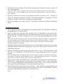
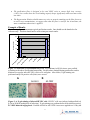
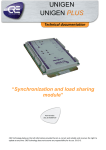
Product Manual ViraBind™ Adenovirus Purification Kit, Trial Size Catalog Number VPK-100-T 4 preps FOR RESEARCH USE ONLY Not for use in diagnostic procedures Introduction Recombinant adenoviruses have tremendous potential in both research and therapeutic applications. There are numerous advantages in using an adenovirus to introduce genetic material into host cells. The permissive host cell range is very wide. The virus has been used to infect many mammalian cell types (both replicative and non-replicative) for high expression of the recombinant protein. Recombinant adenoviruses are especially useful for gene transfer and protein expression in cell lines that have low transfection efficiency with liposome. After entering cells, the virus remains epichromosomal (i.e. does not integrate into the host chromosome so does not activate or inactivate host genes). Recently, recombinant adenoviruses have been used to deliver RNAi into cells. HEK 293 cells or their variants are used as host cells for viral amplification. Recombinant adenoviruses can be grown at high titer (1010 VP (viral particles)/mL, which can be concentrated up to 1013 VP/mL). The concentrated viral supernatant is subjected to CsCl ultracentrifugation to separate the viruses from the cellular proteins and media components. Following ultracentrifugation, CsCl is then removed by dialysis. The CsCl procedure is both tedious and time consuming (16-24 hrs). ViraBind™ Adenoviral Purification Kit does not involve ultracentrifugation, instead it’s based on the unique properties of adenoviral capsid proteins. The entire procedure takes about 30 minutes. Each preparation has a capacity up to 2.5 x 10¹² VPs. ViraBind™ Adenovirus Purification Kit provides an efficient system for quick adenoviral purification with high recovery (>90%). The system may be adapted to purification of other viral types, such as retrovirus and lentivirus. The optimal purification conditions for retrovirus are currently under investigation. Related Products 1. AD-100: 293AD Cell Line 2. AD-200: ViraDuctin™ Adenovirus Transduction Reagent 3. VPK-099: ViraBind™ Adenovirus Miniprep Kit 4. VPK-109: QuickTiter™ Adenovirus Titer Immunoassay Kit 5. VPK-110: QuickTiter™ Adenovirus Titer ELISA Kit 6. VPK-111: Rapid RCA Assay Kit 7. VPK-252: RAPAd® CMV Adenoviral Expression System 8. VPK-254: RAPAd® CMV Adenoviral Bicistronic Expression System (GFP) Kit Components 1. Purification Filter (Part No. 90001-T): Two filters* 2. 10X Wash Buffer (Part No. 90002-T): One 30 mL bottle 3. 2X Elution Buffer (Part No. 90003-T): One 15 mL bottle of 50 mM Tris, pH 7.5, 5 mM Mg2Cl, 2 M NaCl 2 4. 1X Regeneration Solution (Part No. 90004-T): One 25 mL bottle *Note: Each filter is regenerated one time after initial use, allowing each filter to be used twice. Materials Not Supplied 1. Recombinant adenovirus of interest 2. HEK 293 cells and cell culture growth medium 3. Cell culture centrifuge 4. Glycerol 5. 0.45 µm filter 6. 10 or 30-mL size syringe with a Luer-Lok® tip Storage Store all kit components at room temperature. Safety Considerations Remember that you will be working with samples containing infectious virus. Follow the recommended NIH guidelines for all materials containing BSL-2 organisms. Preparation of Reagents 1X Wash Buffer: Prepare a 1X Wash Buffer by diluting the provided 10X stock 1:10 in deionized water. Store the diluted solution at room temperature. 1X Elution Buffer: Prepare a 1X Elution Buffer by diluting the provided 2X stock 1:2 in deionized water. Store the diluted solution at room temperature. Harvesting Infected Cell Lysate The following procedure is suggested for T75 flasks, use only up to 4 x T75 flasks for each purification prep. 1. Use HEK 293 cells that have been passaged regularly 2-3 times prior to the infection. Culture these cells until the monolayer is 90% confluent. 2. Replace the cell culture media with new growth media, 15 mL per flask. Next, add either crude or purified viral stock to the culture. Use a multiplicity of >0.5 PFU (plaque forming units) or enough viruses that cells demonstrate cytopathic effects (CPEs) within 48 hrs. CPEs on adenovirus infected 293 cells consist of cell rounding, swelling, loss of cell-cell contact. 3. During 24 - 48 hr infection, examine the monolayer twice per day under the microscope for CPE. When CPE is nearly complete (i.e. most cells rounded but not yet detached from the flask), harvest cells by pipetting media up and down to wash the infected cells from the flask into the media. Note: To achieve the maximal yield for the purification steps, it is critical to harvest the infected 293 cells when most of the cells show cytopathic effects but not yet detached from the flask. 3 4. Pool infected cells and medium. Pellet cells by centrifugation at 1000 g for 5 minutes. Remove all but 15 mL supernatant. Note: When CPE is nearly complete, most viruses are still inside the infected cells before freeze and thaw cycles. After centrifugation, the excess low titer supernatant may be stored at -80ºC for future infection. 5. Release the adenoviruses from the cell suspension with three freeze/thaw cycles. Centrifuge at 3000 g for 10 minutes to pellet the cell debris. Discard the pellet and save supernatant. If a large amount of cell debris is still visible, centrifuge the supernatant again. 6. The viral supernatant can be stored at -80ºC or immediately purified (see purification instructions below). Purification Protocol 1. Prior to application of the viral supernatant to the purification filter, the supernatant is clarified by passing through a 0.45 µm sterile filter. 2. Attach a syringe to the purification filter, and add 5 mL of 1X Wash Buffer to pre-rinse the filter. Slowly allow the viral supernatant to pass through the purification filter by gravity flow, and save the flow-through. To ensure maximal recovery, pass the flow-through through the same filter again. Note: When the flow through noticeably slows down during loading viral sample or reapplying the first flow-through for the second time, gently apply pressure with syringe plunger. To ensure maximal virus binding, try to keep the flow rate at less than 10 mL/min, we recommend flow through at dropwise (3-5ml/min). 3. Thoroughly wash the purification filter with 10 mL of 1X Wash Buffer using gravity flow or gently applying moderate pressure with syringe plunger. Repeat the wash step twice, using 10 mL of 1X Wash Buffer each wash. 4. Position a collection tube under the purification filter, add 2-3 mL of 1X Elution Buffer (25 mM Tris, pH 7.5, 2.5 mM Mg2Cl, 1 M NaCl) and allow it to pass through (gravity flow or moderate pressure). Note: Air bubbles between syringe and filter will slow down the elution, to avoid them by pipetting a few times, be careful not stab the membrane filter. When apply moderate pressure, it’s critical to keep the flow rate slow at drop by drop to ensure the maximal yield. 5. Add glycerol to a final concentration of 10% to the purified virus or dialyze the viral solution into a desired buffer. Aliquot and store the final purified virus solution at -80ºC. Before infect target cells, we recommend adding the needed amount of purified virus to 5-10 mL culture medium of your target cells and filter through a 0.2 µm sterile filter before infection. 6. The purification filter used in step 4 can be used twice. Upon completion of the purification, add 10 mL of 1X Regeneration Solution to the filter, followed by 5 mL of 1X Wash Buffer. Wrap the regenerated filter in parafilm and store at 4ºC until the next use. Notes: 4 The purification filter is designed to be used ONLY twice to ensure high virus recovery (>90%), our results show the virus-binding capacity drops significantly when used more than two times. The Regeneration Solution should remove any virus or protein remaining on the filter, however to avoid cross-contamination, we suggest that when the filter is used for the second time, the same recombinant adenovirus is applied. Example of Results The following figures demonstrate typical purification results. One should use the data below for reference only. This data should not be used to interpret actual results. 100 Relative VP (%) 80 60 40 20 Vi ra l io n El ut W as h 3r d as h W as h 2n d W 1s t Su pe rn at 1s an tF t lo w -t h 2n ro d ug Fl h ow - th ro ug h 0 Figure 1: Purification of recombinant Ad-β Gal. Recombinant Ad-β Gal viruses were purified according to the above Purification Instructions. Each fraction, obtained during purification, and its dilution were used to infect HUVEC cells in a 24-well plate. After 48 hrs, X-gal staining was performed and β Gal positive cells (blue) were scored. Figure 2: A. X-gal staining of infected HUVEC cells. HUVEC cells were infected with purified Adβ Gal at 50 MOI (multiplicity of infection). X-gal staining was performed after 48 hr infection period. B. VEGF-induced Angiogenesis. Purified Ad-Null or Ad-VEGF viruses were applied to a 10-day old 5 CAM (chick chorioallanoic membrane). After three days, the blood vessels on the CAM were examined under a stereomicroscope. References 1. Stewart, P. L., Fuller, S. D., and Burnett, R. M. (1993) EMBO J. 12:2589-2599. 2. Robbins, P. D., Tahara, H., and Ghivizzani, S. C. (1998) Trends Biotechnol. 16:35-40. 3. Huang, S., Stupack, D., Mathias, P., Wang, Y., and Nemerow, G. (1997) Proc. Natl. Acad. Sci. U. S. A. 94:8156-8161. 4. Bergelson, J. M., J. A. Cunningham, G. Droguett, E. A. Kurt-Jones, A. Krithivas, J. S. Hong, M. S. Horwitz, R. L. Crowell, and R. W. Finberg. (1997) Science 275:1320-1323. 5. Von Seggern, D. J., C. Y. Chiu, S. K. Fleck, P. L. Stewart, and G. R. Nemerow. (1999) J. Virol. 73:1601-1608. 6. Kleinerman, D. I., W. W. Zhang, S. H. Lin, N. T. Van, A. C. von Eschenbach, and J. T. Hsieh. (1995) Cancer Res. 55:2831-2836. Recent Product Citations 1. García-Pascual, C. M. et al. (2015). Evaluation of the potential therapeutic effects of a doublestranded RNA mimic complexed with polycations in an experimental mouse model of endometriosis. Fertil Steril. doi:10.1016/j.fertnstert.2015.07.1147. 2. Miklavc, P. et al. (2015). Actin depolymerisation and crosslinking join forces with myosin II to contract actin coats on fused secretory vesicles. J Cell Sci. 128:1193-1203. 3. Nuche-Berenguer, B. et al. (2015). Elucidation of the roles of the src kinases in pancreatic acinar cell signaling. J Cell Biochem. 116:22-36. 4. Westermeier, F. et al. (2015). Insulin requires normal expression and signaling of insulin receptor a to reverse gestational diabetes-reduced adenosine transport in human umbilical vein endothelium. FASEB J. 29:37-49. 5. Lakshmanan, J. et al. (2015). Glycogen synthase kinase 3 regulates IL-1β mediated iNOS expression in hepatocytes by down-regulating c-Jun. J Cell Biochem. 116:133-141. 6. Brock, S. E. et al. (2014). MIF family members cooperatively inhibit p53 expression and activity. PLoS One. 9:e99795. 7. Kim, S. M. et al. (2014). A recombinant adenovirus bicistronically expressing porcine interferon-α and interferon-γ enhances antiviral effects against foot-and-mouth disease virus. Antiviral Res. 104:52-58. 8. Zhang, J. et al. (2014). Propofol exerts anti-hepatocellular carcinoma by microvesicle-mediated transfer of miR-142-3p from macrophage to cancer cells. J Transl Med. 12:279. 9. Sanchez-Lugo, Y. E. et al. (2014). CXCL10/XCL1 fusokine elicits in vitro and in vivo chemotaxis. Biotechnol Lett. doi:10.1007/s10529-014-1746-4. Fang, J. et al. (2014). PAX6 downregulates miR-124 expression to promote cell migration during embryonic stem cell differentiation. Stem Cells Dev. 23:2297-2310. 10. Yang, M et al. (2014). Over‐expression of JAZF1 reduces proinflammatory cytokines release via inhibition of stress kinases and NF‐КB. FEBS J. doi:10.1111/febs.12853. 6 11. Cheng, L. et al. (2014). Induction of specific humoral and cellular immune responses in a mouse model following gene fusion of HSP70C and Hantaan virus Gn and S0. 7 in an adenoviral vector. PLoS One. 9:e88183. 12. Cyphert, H. A. et al. (2014). Glucagon stimulates hepatic FGF21 secretion through a PKA-and EPAC-dependent posttranscriptional mechanism. PLoS One. 9:e94996. 13. Obokata, H. et al. (2014). Stimulus-triggered fate conversion of somatic cells into pluripotency. Nature. 505:641-647. 14. Haidari, M. et al. (2014). Disruption of endothelial adherens junctions by high glucose is mediated by protein kinase C-β-dependent vascular endothelial cadherin tyrosine phosphorylation. Cardiovasc Diabetol. 13:112. 15. Wu, D. et al. (2014). Hypothalamic nesfatin-1/NUCB2 knockdown augments hepatic gluconeogenesis that is correlated with inhibition of mTOR-STAT3 signaling pathway in rats. Diabetes 63:1234-1247. 16. Araki, K. et al. (2013). Cytoplasmic translocation of the retinoblastoma protein disrupts sarcomeric organization. eLife Sci. 2:e01228. 17. Scallan, C. et al. (2013). An Adenovirus-based vaccine with a double-stranded RNA adjuvant protects mice and ferrets against H5N1 avian influenza in oral delivery models. Clin. Vaccine Immunol. 20:85-94. 18. Haidar, M. et al. (2012). Integrin α2β1 mediates tyrosine phosphorylation of vascular endothelial cadherin induced by invasive breast cancer Cells. J. Biol. Chem. 287: 32981-32992. 19. Chen, F. et al. (2011). Dynamic regulation of PDX-1 and FoxO1 expression by FoxA2 in dexamethasone-induced pancreatic ß-cells dysfunction. Endocrinology. 152:1779-1788. 20. Prasad, S.S. et al (2011). Enzymatic activities of the human AGPAT isoform 3 and isoform 5: localization of AGPAT5 to mitochondria. J. Lipid Res. 52:451-462. 21. Triulzi, C. et al. (2010). Antibody-dependent natural killer cell-mediated cytotoxicity engendered by a kinase-inactive human HER2 adenovirus-based vaccination mediates resistance to breast tumors. Cancer Res. 70:7431-7441. 22. Lam, Y.W. et al. (2010). Proteomics analysis of the nucleolus in adenovirus-infected cells. Mol. Cell. Proteomics 9:117-130. 23. Sabbatini, M. E. et al. (2010). CCK activates RhoA and Rac1 differentially through G-alpha-13 and G-alpha-q in mouse pancreatic acini. Am. J. Physiol. Cell Physiol. 298:C592-C605. 24. Kothari, H. el at. (2010) Cystine 186-cystine 209 disulfide bond is not essential for the procoagulant activity of tissue factor or for its de-encryption. Blood. 115:4273-4283. 25. Sen, P. et al. (2010). Zinc modulates the interaction of protein C and activated protein C with endothelial cell protein C receptor. J. Biol. Chem. 285:20410-20420. 26. Agarwal, A.K. et al. (2010). Enzymatic activity of the human 1-acylglycerol-3-phosphate-Oacyltransferase isoform 11: upregulated in breast and cervical cancers. J. Lipid Res. 51:2143-2152. 27. Li, H. et al. (2009). Nuclear translocation of Ad/EYFP-hCAR: a novel tool for screening human CAR activators in human primary hepatocytes. Drug Metab. Dispos. 37:1098-1106. 28. Meng, Z. et al. (2009). Forkhead box O1/pancreatic and duodenal homeobox 1 intracellular translocation is regulated by c-Jun N-terminal kinase and involved in prostaglandin E2-induced pancreatic ß-cell dysfunction. Endocrinology 10.1210/en.2009-0671. 29. Gosselin, K. et al. (2009). Senescence-associated oxidative DNA damage promotes the generation of neoplastic cells. Cancer Res. 69:7917-7925. 7 Warranty These products are warranted to perform as described in their labeling and in Cell Biolabs literature when used in accordance with their instructions. THERE ARE NO WARRANTIES THAT EXTEND BEYOND THIS EXPRESSED WARRANTY AND CELL BIOLABS DISCLAIMS ANY IMPLIED WARRANTY OF MERCHANTABILITY OR WARRANTY OF FITNESS FOR PARTICULAR PURPOSE. CELL BIOLABS’ sole obligation and purchaser’s exclusive remedy for breach of this warranty shall be, at the option of CELL BIOLABS, to repair or replace the products. In no event shall CELL BIOLABS be liable for any proximate, incidental or consequential damages in connection with the products. Contact Information Cell Biolabs, Inc. 7758 Arjons Drive San Diego, CA 92126 Worldwide: +1 858-271-6500 USA Toll-Free: 1-888-CBL-0505 E-mail: [email protected] www.cellbiolabs.com 2013-2015: Cell Biolabs, Inc. - All rights reserved. No part of these works may be reproduced in any form without permissions in writing. 8