1
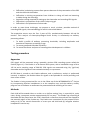
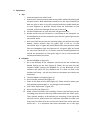
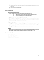
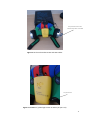
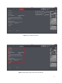
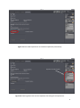
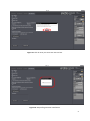
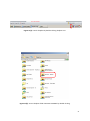
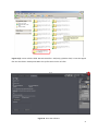
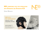
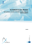
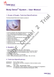
INTERGROWTH-21st International Fetal and Newborn Growth Standards for the 21st Century The International Fetal and Newborn Growth Consortium CORTICAL AUDITORY ERP MANUAL – June 2014 December 2013 1 Please read this manual carefully and refer to it throughout the study if any clarification is needed This Operations Manual was produced by the INTERGROWTH-21st Infant Development Group, based on the 1st Meeting of the Group, Oxford, March 2012. This document reflects the consensus reached by members of the group regarding the selection of tests to be included in the INTERGROWTH-21st Neurodevelopment Package to be implemented by all centers taking part in the INTERGROWTH-21st follow-up study. INTERGROWTH-21st is a large project involving health institutions from eight geographically diverse countries. It is therefore essential that all participating institutions follow a standardized neurodevelopment protocol. 2 Table of Contents Credits ........................................................................................................................................ 4 Source ........................................................................................................................................ 5 Introduction ............................................................................................................................... 6 Testing procedure ............................................................................................................................... 7 Apparatus ........................................................................................................................................ 7 Methods .......................................................................................................................................... 7 Auditory Novelty Paradigm ............................................................................................................. 9 Experimental Protocol ............................................................................................................. 10 Assessment Kit .................................................................................................................................. 10 Set Up and Method of Assessment................................................................................................... 10 Maintenance ............................................................................................................................ 25 Safety and Regulatory Information.......................................................................................... 27 Use environment .............................................................................................................................. 27 Safety Warnings ................................................................................................................................ 27 Conditions of use .............................................................................................................................. 28 Technical Specifications .................................................................................................................... 28 Product Description .......................................................................................................................... 29 References ............................................................................................................................... 31 3 Credits This manual was prepared by members of the INTERGROWTH-21st Infant Development Group and reflects the general consensus reached during the Infant Development Group Meeting, Oxford, 23 March 2012 regarding the selection of tests to be included in the INTERGROWTH-21st Neurodevelopment Package. The following people made important contributions to this final version, for which we thank them: Michelle Fernandes – Group Coordinator (Nuffield Department of Obstetrics and Gynaecology, the John Radcliffe Hospital, University of Oxford, Oxford, UK) Alan Stein – Senior Advisor (Section of Child and Adolescent Psychiatry, Department of Psychiatry, Warneford Hospital, University of Oxford, Oxford, UK) Charles Newton – Senior Advisor (Department of Psychiatry, Warneford Hospital, University of Oxford, Oxford, UK and Senior Clinical Research Fellow, KEMRI-Wellcome Trust Research Programme, Kilifi, Kenya) Michael Kihara – Expert Advisor (Associate Professor, Department of Psychology, United States International University, Nairobi, Kenya) Michelle de Haan – Expert Advisor (Reader in Developmental Cognitive Neuroscience, Institute of Child Health, University College London, London, UK) Aureli Soria Frisch – Technical & Signal Processing Expert (Neuroelectrics, STARLAB, Barcelona, Spain) David Ibanez – Technical & Signal Processing Expert (Neuroelectrics, STARLAB, Barcelona, Spain) Javier Acedo – Technical & Signal Processing Expert (Neuroelectrics, STARLAB, Barcelona, Spain) Guillem Mitja – Technical Expert (Neuroelectrics, STARLAB, Barcelona, Spain) Morten Kringelbach – Expert Advisor (Director of Hedonia: Trygfonden Research Group, Department of Psychiatry, Warneford Hospital, University of Oxford, Oxford, UK) 4 Source Kihara M, Hogan AM, Newton CR, Garrashi HH, Neville BR, de Haan M. Auditory and visual novelty processing in normally-developing Kenyan children. Clinical Neurophysiology. 2010; 121: 564-576. Further information: Dr. Michelle Fernandes, E:[email protected], T: +44(0)1865222936 © 2012 INTERGROWTH-21st Project The Nuffield Department of Obstetrics & 5Gynaecology The John Radcliffe Hospital, Oxford OX3 9DU, United Kingdom www.intergrowth-21st.org.uk 5 Introduction An estimate of the prevalence for auditory processing disorders (APD) in childhood is 7%.1 Hearing impairment(s) arising from pathology of the brain may have detrimental consequences on a child's life if untreated; however, diagnostic and management strategies for these “central” hearing impairments in childhood are rarely implemented. These auditory deficits have been collectively termed “auditory processing disorders”, in order to incorporate in the term the interaction between peripheral and central pathways. 1 Disturbances in auditory processing are an important indicator of disturbances in sensory neurodevelopment in children and measurement of a child’s ability to detect and assimilate novel auditory stimuli may be used to examine subtle differences in auditory processing. 2 In addition, higher prevalence of auditory processing disorders are reported in preterm babies, children with a family history of deafness, intra-uterine infections and epilepsy.1 A number of tests to assess auditory function in children were considered. These included the use of audiometry, acoustic reflex thresholds, otoacoustic emissions, communication inventories and the measurement of evoked response potentials (ERPs) to auditory stimuli. It was decided that for use in the follow up study of the INTERGROWTH-21st Project, the test of auditory assessment should: Be suitable for use on children at 24 months of age. Assess the entire auditory pathway and not just elements of the same. Yield information about an auditory threshold, amplitude and latency of responses. Assess subtle changes in auditory processing between individuals. Be easy to administer without specialist training. Be internationally valid. Be rapid to administer (<15 minutes). Of the tests of auditory processing considered, the measurement of ERPs to auditory stimuli was found to meet the criteria mentioned above. Measurement of ERPs to an auditory novelty paradigm was found to have a high degree of sensitivity in detecting subtle differences in auditory processing in children. Audiometry was not found to be suitable for children less than 4 years of age. The measurement of acoustic reflex thresholds and otoacoustic emissions assessed solely middle ear function, and not the entire auditory pathway. In addition, these tests required a probe to be placed in the child’s ear for up to 3 minutes. This was found to pose considerable difficulties in compliance in the field. The measurement of ERPs to auditory stimuli was selected to measure auditory processing in 2 year olds in the follow up study of the INTERGROWTH-21st Project. This was selected because ERPs response to deviance and/or novelty reflects the child’s ability to detect and assimilate novel events, and this has been found to be fundamental to cognitive development.2 Technical issues related to the measurement of electroencephalography (EEG) in the field included: 6 Difficulties in obtaining consent from parents because of the presentation of the EEG and the child with electrodes. Difficulties in eliciting co-operation from children in sitting still and not becoming irritable during the recording. Specialist training required for setting up the electrodes and recording EEG signals. Difficulties in preparation of the child’s scalp using gel. Difficulties in the placement of electrodes. In order to meet these challenges, we propose a novel, wireless, portable method of recording EEG signals, the methodology for which is described below. The acquisition occurs over the first 2 years of life, predominantly between 18 and 24 months. Their analysis of electrophysiological brain activity in relationship to auditory processing allows: I. To build a profile of auditory processing thresholds, including amplitudes and latencies of responses, according to age. II. To assess group and individual variability. III. To assess the nature and pace of neurocognitive development in children. Testing procedure Apparatus EEG signals will be measured using a portable, wireless EEG recording system called the Enobio (Figure 1).3 The Enobio is an 8 channel EEG system, with a bandwidth range of 0 to 125 Hz and a sampling range of 500 SPS. EEG signals are measured using ‘dry’ (gel-free) electrodes and the signals are transmitted wirelessly to a processor. All EEG data is stored on the Enobio software, and a preliminary analysis is performed instantly. In addition, the Enobio allows for signals to be uploaded to a cloud providing real time access of data. The Enobio is being customized for use in the follow up of the INTERGROWTH-21st Project by incorporating wireless headphones in to the neoprene cap, and the auditory novelty program into its software to automate the presentation and recording of the same. Methods Each child will be seated either on a chair or on his/her mother’s lap, in a partially lit, quiet room, facing a computer monitor approximately 50 cm away.2 The Enobio cap containing 8 electrodes will be placed over the child’s head and a reference electrode will be clipped to the mastoid region of the Enobio cap. The Enobio was customized for the purpose of this study to (a) fit the cranial dimensions of a two year old child and (b) integrate wireless headphones into the cap. 7 The Enobio cap will contain dry EEG placed at sites Fz, Cz, Pz, T3, T4, T5 and T6 according to the international 10-20 system of electrode placement (Figure 2).4 Eye movements and blinks will be monitored by the Fp2 electrooculogram electrode also integrated into the Enobio cap. The child will be asked to watch a series of cartoon video clips on the computer screen. The cartoons will have their audio clips removed and instead of the cartoon’s audio, the experimental auditory stimuli will be presented to the child via the wireless earphones. The presentation of these stimuli, and the recording, digitization and processing of the EEG data will be carried out automatically with the Enobio software. If the child prefers not to watch the video, a colouring book and crayons will be provided. Care will be taken to ensure noise levels are kept to a minimum in the room and the child/mother will be encouraged to not speak loudly. Electrical sources of sounds, like ceiling fans, will be switched off. The Enobio’s electrodes take 2 minutes to calibrate when placed on the child’s head, during which a baseline EEG is recorded for 2 minutes before the auditory novelty paradigm is presented. The EEG data will be recorded at a sampling rate of 500 Hz with a system band pass between 0 and 100 Hz. Figure 1. The Enobio wireless EEG monitoring system (Lt: posterior view, Rt: lateral view). 8 Figure 2. Schematic representation of electrode placement on the Enobio for the assessment of auditory processing. Auditory Novelty Paradigm The auditory paradigm is composed of 3 types of sounds presented through the wireless headphones attached to the Enobio. 10% of these sounds are infrequent tones (2kHz, 200 ms long, 5 ms rise and fall time and 70 dB SPL), 10% are novel noises such as a dog bark, bell ring, drum roll etc., while the remaining 80% are frequent sinusoidal tones (1.5 kHz, 200 ms long, 5 ms rise and fall time, 70 dB SPL).2,5 Two blocks of 700 stimuli each were presented (560 frequent, 70 infrequent and 70 novels). There were 14 different novel stimuli, and these were repeated no more than 5 times each during the course of the experiment. The total duration of the stimuli presentation is 11 minutes. 9 Experimental Protocol Assessment Kit The assessment kit consists of: 1. The Enobio – i.e. the Neuroelectrics Control Box (NECBOX), 10 wire cables with backends, electrode font-ends, battery charger, NE headband, NE cap, USB connector. 2. A laptop containing the Enobio software. 3. A computer (laptop or desktop), a television or a portable DVD player on which the silent cartoons can be viewed. A colouring book and crayons in case the child doesn’t want to watch the cartoons. 4. A comfortable chair for the mother and child. Set Up and Method of Assessment 1. Pre-experiment tasks a. Ensure battery of NECBOX is charged. b. Install front-ends and back-ends into cap according to pre-defined format in the table below (Figure 3). Back-end number Position 1 2 3 4 5 6 7 8 Fp2 Fz Cz Pz T4 T6 T3 T5 c. Attach stick on electrodes to DRL and CMS cables (Figure 4). d. Ensure headphones and antenna are fixed on channel 1. e. Ensure the antenna/transmitter is connected to the computer through the headphone jack. f. Ensure computer volume is set on level 1 or 30. g. Ensure the computer is connected to the internet either via a broadband cable or Wifi. 10 2. Experiment a. Cap i. Place the cap on the child’s head. ii. Attach the 2 reference electrodes to the child’s mastoid by peeling off the back sticker. Both electrodes should be on the same mastoid (so both on right or both on the left mastoid) and they should ideally be as close together as possible. Please paste the electrodes on the mastoid, and not on the angle of the jaw or the neck. iii. Put the headphones on (you will see a red light come on). iv. Double check that the transmitter is connected to the computer via the headphone jack of the computer (you will see a green light on the transmitter base). v. One and a half minute into the recording (after you perform the steps below), please recheck that the green light is still on on the transmitter base. If it goes off, please disconnect the transmitter cable from the headphone jack and reconnect it, the green light will come on. You must check this at 1.5 minutes into the recording before the sounds are presented, as if the green light is not on, the sounds will not be presented to the child. b. Software i. Put the NECBOX on (Figure 5). ii. Go to the desktop of the computer and launch the NIC software by double clicking on the icon (Figure 6). (Note: Do not open the NIC software before putting the NECBOX on, as the Bluetooth of the software will search for the NECBOX and if it does not find it the software will hang – you will then have to shut down and restart the computer). iii. The NIC software will open (Figure 7). iv. Go to the ERP protocol tab (Figure 8). v. Enter all the data of the child (Figure 9). Please mention where you have placed the electrodes i.e. on the right or left mastoid. vi. Click “Start Experiment” (Figure 10). vii. Go to the EEG tab (Figure 11). viii. Make sure all 8 channels are recording. If one or two channels are not recording press them so that they make contact with the child’s scalp. Then recheck whether all 8 channels are recording. If the signals do not look correct, please check the reference electrodes to make sure they have not fallen off or are stuck to the child’s hair rather than the child’s skin – it is important that these electrodes are in the right 11 ix. x. xi. xii. xiii. xiv. xv. xvi. xvii. position on the child’s mastoid and are in contact completely with the skin. If you need to terminate the experiment prematurely, click “STOP EXPERIMENT” under the mastoid data entry tab. Do not stop recording on the grey right hand column, as this column is not related to the ERP experiment (Figure 11). If you do not terminate the experiment, it will run for 10 minutes, you can see how much time has elapsed and how much time is remaining on the software (Figure 13). Once the experiment is over (because 10 minutes are up or because you clicked “STOP EXPERIMENT”) you will be presented with a text box (Figure 14 a and b) and asked to add ‘POST-EEG NOTES’. You will have to type something in this text box otherwise the software will not allow you to proceed with the upload. Please include all comments such as ‘the child was eating during the assessment’, ‘the child was crying’, ‘the mother was talking loudly and the room was noisy’. If the assessment was uneventful, please write ‘NONE’ or ‘UNEVENTFUL’ in the box. Once you type something in, click “OK” (Figure 15). Once you click “OK” in the text box, an output file will be automatically generated and a pop up box will appear to tell you about this (Figure 16). The zipped files will be saved in a specific folder location within the NIC program. Copy the files from this folder. Go to the dropbox folder called EEG Data – Nagpur. Inside this open the folder called ‘EEG data’ ‘Zipped files’. Create a folder called ‘EEG extracted files – dd/mm/yy (add the date)’. Paste the zipped files into this folder. Please paste ONLY the ‘.zip’ files and not the ‘.info’ files (Figure 17 a to e). Close the software (Figure 18). Put the NECBOX off. 3. Post-experiment tasks a. b. c. d. e. Take off the reference electrodes from the child’s skin with care. Turn the NECBOX off and remove it from the cap. Take the cap off the child’s head. Turn the headphones off on the cap (Red light must go off). Disconnect the cables carefully. Disentangle any tangled cables and stored the cables carefully in the black box. 12 f. Remove the front and back ends of the electrodes and store them in the black box. g. Place the cap in the black box. Other points to note Cleaning of electrodes and cap 1. Clean and dry the electrodes immediately after assessment by: (i) Wiping with a clean paper towel dipped in distilled water. (ii) Drying immediately with a soft paper towel. 2. Wipe cap and cover clean using disinfectant wipe. 3. The electrodes are very delicate – they corrode in contact with metal, air, water, light and solvents. They are also very expensive. Ensure that they are stored in their own envelope and placed in the black box. Charging equipment 1. Charge the NECBOX via the MicroUSB connector located at the rear part of the NECBOX when the battery falls below 50%. You can check the battery strength on the software. 2. Charge the headphones via the transmitter once in 4 assessments. Supplies Please inform Michelle Fernandes if the stock of disposable electrodes is running low. Please give her 6 weeks to pace the order and courier them to you. Test Duration Details Set-up: 1-3 minutes. Test duration: 10 minutes. Termination: <1 minute. Total duration: 12-14 minutes. 13 Enobio EEG placements 1 2 Right Left 3 7 8 4 5 6 DRL CMS Figure 3. Electrode placements. 14 Disposable electrodes attached to DRL and CMS Figure 4. Stick-on electrodes to DRL and CMS cables. On/off switch Figure 5. NECBOX on (yellow light comes on when you put it on). 15 Double click this icon to launch the NIC software (make sure the NECBOX is on first) Figure 6. Launching NIC software on desktop. Figure 7. NIC software opening after double clicking icon. 16 This yellow bar tells you the Bluetooth is connected This tells you how much % of the battery is left Figure 8. Go to ERP protocol tab. Figure 9. Entering the data of the child in the ERP tab. 17 Figure 10. Click start experiment. Figure 11. Go to EEG tab. Make sure all eight channels (blue circles) are recording. 18 Figure 12. Click ‘STOP experiment’ to terminate experiment prematurely. Elapsed and remaining time Figure 13. Checking how much time has elapsed/is remaining for the experiment. 19 Figure 14(a). Text box for post-EEG notes. Figure 14(b). Entering text into the text box (the ok button appears). 20 Figure 15. Click ok once you enter text into text box. Figure 16. Output file generation notification. 21 Figure 17(a). Launch dropbox by double-clicking dropbox icon. Figure 17(b). Access dropbox folder ‘EEG DATA-NAGPR’ by double-clicking. 22 Figure 17(c). Access folder called ‘EEG data’. Figure 17(d). Access folder called ‘zipped files’ by double-clicking. 23 Figure 17(e). Create a folder called ‘EEG extracted files – dd/mm/yy (add the date)’. Paste the zipped files into this folder. Please paste ONLY the .zip files and not the .info files. Figure 18. Close the software. 24 Maintenance Silver-Silver Chloride (Ag/AgCl) electrode instructions Precautions for handling electrodes Electrodes should not come into direct contact with metals as this may cause corrosion. Avoid touching or contaminating the bare electrode surface as dirt can increase calibration times. Use of corrosive chemicals will damage the electrodes. Conditioning To minimize DC offset and drift, place the electrodes in the working position 5 minutes before starting the measurement. The electrodes need some time to achieve electrochemical equilibrium with the skin and electrolyte in the human body. Cleaning Silver/silver-chloride (Ag/AgCl) electrodes, if wet, will corrode. This corrosion process will, in the long run, make the electrodes noisier. It is important to clean and dry the electrodes immediately after use. Wipe the electrodes clean with distilled water and dry the electrodes will a soft paper towel. Never use solvents (e.g. acetone), acids or alkaline; or soap water to clean the electrodes. Sterilization When sterilization is required, only gas or liquid agents should be employed. Disinfectants containing mercury, phenols, bromine, iodine, zinc, tin and organo-metallic compounds should be avoided. The electrodes and cable insulation will not withstand steam sterilization. Darkening Silver chloride is light-sensitive. Exposure to light will darken the electrode surface. This will not impair the electrode performance since the largest interstitial Ag/AgCl matrix is not reached by light. Storage Electrodes should be handled with care. They must not be stored in contact with metals, especially such active metals as iron or aluminium. Also, substances that could affect the electrochemical characteristics should be avoided. Bare electrodes should be stored in a clean, dry and dark container (plastic or glass). Malfunction If an electrode is not operating as specified and a noisy signal is experienced, please follow the directions below concerning noisy electrodes. Noisy electrodes A continuously noisy electrode generally means that it has reached its end of life. The life of the electrode can be extended a little by using a grain 600 or higher waterproof abrasive 25 paper to polish the electrode tip. Use very soft circular movements, preferably no more than 2-3 times on the same area, removing an even very thin layer across the entire surface. Life expectancy Ag/AgCl sintered electrodes have a limited life span. This is caused by several processes such as corrosion, the dissolving of the chloride in the pellets and the wearing of the pellet during the cleaning process. Eventually the colour of the pellets will change from grey/brown (silver–chloride) to silver, due to the loss of the chloride. The AgCl slowly dissolves in gel and water during the cleaning. Eventually, this leaves only silver behind. The resulting pure silver electrode has much higher drift and noise characteristics than the original Ag/AgCl electrode. At this point replacement is recommended. Neoprene Cap and NECBOX cleaning and disinfecting instructions After each use the Neoprene Cap and NECBOX should be cleaned and disinfected. The surfaces of the neoprene cap and NECBOX may be wiped clean using a disinfectant wipe, and left to air dry. Battery operating/safety instructions Charging the battery The battery can only be charged when the power ON switch is at the OFF position. To charge the battery, the following specifications have to be met: 5V DC (4,25 – 7)V. 1 A current output. Power input: 5W. Battery charger complies according to standard IEC-EN 60601-1. It has to be connected to the microUSB connector located at the rear part of the NECBOX (Fig. 11). Charging with a higher voltage than specified may cause damage to the cell. The usual time to charge a battery from the cut off voltage to the maximum capacity is around 2 hours. The expected life cycle is > 500 cycles with > 70 % of initial capacity. The battery state of charge is measured by the software while the device is powered ON. It should be noted that the battery will be over-discharged if the device is not used for a long time. In order to prevent over-discharging, the battery should be charged periodically. Overdischarging may cause loss of cell performance, or damage battery function. 26 Safety and Regulatory Information Enobio is a class IIa device according to the classification in the Council Directive 93/42/CEE for medical devices. It conforms to the following Standards and Directives: Directive 93/42/CEE for medical devices. UNE-EN 60601-1:1993 + A1:1996 + A2:1996 + A3:1997 + A2/ERR:1999. UNE-EN 60601-1-2:2008. UNE-EN 60601-1-26:2004. Manufacturer: Neuroelectrics Barcelona SL C/ Teodor Roviralta, 45 08022 Barcelona Spain Telephone: + 34 93 254 03 70 Brand: Enobio Models: Enobio 8 Channels, Enobio 20 Channels Use environment The Enobio system has been designed for the use in a clinical environment, hospital or research centre. Safety Warnings 1) 2) 3) 4) 5) 6) 7) 8) The results of the measurements must be analysed by a doctor or specialist. No self-medication should be done based on these results. The device must never be opened or damaged. The battery can only be replaced by authorized personnel. Before using, please check that the device is undamaged and the packaging has not been affected by the transport or storage. Before using the device, please inform the prescribing clinician or operator of the presence of any pacemakers, intracranial electrodes, implanted defibrillators, cranial pathologies (e.g., holes, plaques) or any other prosthesis. The use of the device could result, in these cases, unsafe. In the case of malfunction, immediately contact the manufacturer or the distributor. The device is not protected against excessive moisture or immersion in liquid. In the case of the device becoming wet or damp, do not use it and immediately contact the manufacturer. 27 9) 10) 11) 12) 13) 14) 15) 16) 17) Do not touch the device while EEG monitoring is on. Always unplug the USB power supply from the device prior connecting electrodes to the subject. The device will not work when charging the battery. Never use the device or install the electrodes on the head of the patient while connected to the power network. The device must be used only with Ag/AgCl electrodes recommended by the manufacturer. The device is not protected against other high frequency devices. To avoid risks place the CMR/DRL as far as possible from the s and return electrode of the high frequency device. The device is wireless and might be affected by other RF devices. The device needs special EMC precautions. It needs to be used according to user manual information regarding EMC. The EMC emissions and immunity has been tested using the 10 wires or 12 wires 34 cm cables provided with the system. The use of other cables or electrodes than the ones delivered with the product might produce higher EMC emissions and less EMC immunity. The device cannot be beside used or piled under other equipment. If such usage is needed, check the normal configuration. The device can only be used in healthy skin without wounds. Conditions of use Enobio must be used in normal temperature and pressure conditions (T range:10-45°C, humidity 20-90%, atmospheric pressure 700- 1000 hPa). Technical Specifications EEG functionality: Number of Channels: 8 or 20 Channels. Bandwidth: 0 to 125 Hz. Sampling rate: 500 SPS. Resolution: 24 bits – 0,05 microVolt. Measurement Noise: < 1 uV RMS. Input impedance 1000 Mohm minimum. Allows for Stimulation simultaneously. Other Technical Specifications: Communication: Bluetooth 2.1. Output: EDF+, ASCII data files or tcp/ip raw data streaming. 3 axes accelerometer. MicroSD card for off-line storage. 28 Operating time: 8 hours for EEG only. Dimensions: 60 x 85 x 20 mm. Weight: 65 g. Compatible with Windows 7, Windows Vista, Windows XP, MAC OS X. Product Description See Figure 19 to 21 below for product description. 1. USB connector for charging battery Must not be on when EEG recording is in progress. 2. Power ON switch O indicates OFF position and allows for charging of the battery. I indicated ON position (no charging possible). Figure 19. NECBOX USB connector and power switch. 4. 12 EEG connector and reference signals 5. 8 EEG connector and reference signals. 3. MicroSD card socket Figure 20. NECBOX EEG connector, reference signals and MicroSD card socket. 29 8. Technical specifications label 6. LED indicates normal operation 7. Velcro to attach device to cap Figure 21. LED light, label and velcro support. 30 References 1. Bamiou D-E, Musiek FE, Luxon LM. Aetiology and clinical presentations of auditory processing disorders—a review. Archives of Disease in Childhood. 2001; 85(5): 361-5. 2. Kihara M, Hogan AM, Newton CR, Garrashi HH, Neville BR, de Haan M. Auditory and visual novelty processing in normally-developing Kenyan children. Clinical Neurophysiology. 2010; 121(4): 564-76. 3. Neuroelectrics. Enobio. Available Accessibility verified June 18, 2014. 4. Attias J, Urbach D, Gold S, Shemesh Z. Auditory event related potentials in chronic tinnitus patients with noise induced hearing loss. Hearing Research. 1993; 71(1–2): 106-13. 5. Haenschel C, Baldeweg T, Croft RJ, Whittington M, Gruzelier J. Gamma and beta frequency oscillations in response to novel auditory stimuli: A comparison of human electroencephalogram (EEG) data with in vitro models. Proceedings of the National Academy of Sciences. 2000; 97(13): 7645-50. at: http://www.neuroelectrics.com/enobio. 31