1
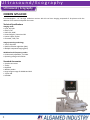
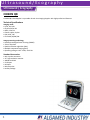
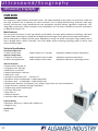
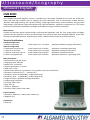
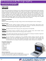
Ultrasound Ecography 0 Ultrasound/Ecography Ultrasound / Ecography CHISON IVIS 60 Ecocolourdoppler - 15" flat high resolution monitor in VGA format. Technical Specifications Imaging mode B, 2B, 4B, B/M CFM, B / BC PW mode, HPRF Power Doppler / Directional PD Instant triplex, duplex Chroma B / PW / CW Image processing technology THI on all probes Speckle reduction algorithm (SRA) Multiple compound imaging (MCI) Wideband multi-frequency probes Continuously adjustable, 2-13 MHz Operating voltage 110V-220Va Standard Accessories 3 probe connectors Trolley Keyboard Without probes Large image storage of 160GB Hard Disk 2 port USB DVD-RW 1 Ultrasound/Ecography Ultrasound / Ecography CHISON IVIS 60 4D Ecocolourdoppler - 15" flat high resolution monitor with 4D real time imaging composed of 3D pictures with the addition of live motion and probe 4D convex. Technical Specifications Imaging mode B, 2B, 4B, B/M CFM, B / BC PW mode, HPRF Power Doppler / Directional PD Instant triplex, duplex Chroma B / PW / CW Image processing technology THI on all probes Speckle reduction algorithm (SRA) Multiple compound imaging (MCI) Wideband multi-frequency probes Continuously adjustable, 2-13 MHz Operating voltage 110V-220Va Standard Accessories 3 probe connectors Trolley Keyboard Without probes Large image storage of 160GB Hard Disk 2 port USB DVD-RW 2 Ultrasound/Ecography Ultrasound / Ecography Sonotouch 20 Lightweight, portable, small size, touchscreen ultrasound devices. Main Features Display mode: B, B/B, 4B, B/M, M and Colour mode. In the M or B/M mode, 4 steps sweep speeds. Multi-step display magnification, depth enhancement Setting adjustment of total gain, and 8 segments of TGC slides for selection and adjustment Multifocal firing focus, edge enhancement, frame averaging, compound Image freezing and storage function, built-in 16 GB high-speed memory and external USB memory disk can be connected to the system for mass storage through USB port. Stored images can be retrieved for analysis. 256 frames of real-time images can be stored in Cine-memory Probe scanning direction can be changed and the image can be reversed in left/right, up/down direction Measurements of distance, area, circumference, volume, OB etc. Automatic calculation of OB and cardiology. Direct display of gestation age, expected date of child delivery and direct measure of heart rate Real-time clock displays date and time automatically Display of body marks with corresponding probe position indication Annotation function in image area of the screen. Special annotation terms for different exam-mode can be added according to user's requirement Battery operated, battery life up to 2.5 hours Technical Specifications Button free screen and waterproof front panel Select your applications from visual icons Take measure with your fingers, no trackball (accuracy 1 mm) Adjustable angle stand converts into carrying handle Refine the image with easy, touch driven TGG, focus and depth 3 Ultrasound/Ecography Ultrasound / Ecography CHISON Q5 CHISON Q5 Colourdoppler is a portable digital colour Doppler with high professional features. Technical Specifications Imaging mode B, 2B, 4B, B/M PD, directional PD B/BC, CFM-TSS Instant triplex, duplex PW, HPRF, CW Chroma B / M / PW / CW Image processing technology Double phase digital beam forming (DPDBF) THI on all probes Speckle reduction algorithm (SRA) Multiple compound imaging (MCI) Operating voltage: 110V - 230V, 50-60 Hz Standard Accessories with 2 probe connector 15" high resolution monitor 80GB Hard Disk 2 USB port VGA port without probe 4 Ultrasound/Ecography Ultrasound / Ecography CHISON Q8 CHISON Q8 Colourdoppler is a portable 4D real time imaging Doppler with high professional features. Technical Specifications Imaging mode B, 2B, 4B, B/M PD, directional PD B/BC, CFM-TSS Instant triplex, duplex PW, HPRF, CW Chroma B/M/PW/CW Image processing technology Double phase digital beam forming (DPDBF) THI on all probes Speckle reduction algorithm (SRA) Multiple compound imaging (MCI) Operating voltage: 110V - 230V, 50-60 Hz Standard Accessories with 2 probe connector 15" high resolution monitor 160GB Hard Disk 2 USB port VGA port without probe cardiac package 5 Ultrasound/Ecography Ultrasound / Ecography CMS 280C Main Features Advanced probe technology and special protection for probe Various image process function: 4 background colour for adjustment 8 degree dynamic range 8 degree boundary enhancement 4 for adjustment 4 framed of correlation Option: Multi-frequency probe, transvaginal probe, 7.5MHz Linear probe, Cine loop memory, double sockets Technical Specifications Measuring Distance Circumference Area Volume Heart rate Pregnant week Foetal weight Character display ID number Time Date Body make probe position, Focus Frame rate Zoom Gray scale Puncture guide line Menu Zoom ×1 ×1.2 ×1.5 ×2 (according to the selected probe) 6 Ultrasound/Ecography Ultrasound / Ecography CMS 600A This equipment is high resolution ultrasound scanner. It adopts 4-sector dynamic focusing and digital scan converter (DSC), dynamic logarithm compress, TGC control and wave filtering, high-frequency beam-former. The device has been widely used in examining abdomen and obstetrics, urology, cardiology, gynaecology, small parts etc, in various hospitals at all level. Main Features Optional wide-frequency electronic convex array probe, electronic linear array probe and transvaginal probe The image can be uploaded to computer through the USB port The machine software can be upgraded by U-disk, and store or load image on the U-disk Light-touch keyboard and trackball Direct operation keys for easy and quick operation This device is attractively designed, plastic injection, small-sized, lightweight Technical Specifications Standard configuration 3.5 MHz Convex Probe; Probe Frequency: 2.5-5.0 MHz; Applications: Abdominal organs examination Optional configuration 6.5 MHz HF Linear Probe; Probe Frequency: 5.0-7.5 MHz; Applications: Small part examination 6.5 MHz Transvaginal Probe; Probe Frequency: 5.0-7.5 MHz; Applications: Obstetrics and gynaecology examination Main Performance Display mode: B, 2B, BM, M Image gray scale: 256 Scale Monitor size: 10 Inch CRT Depth of penetration: ≥ 170 mm Dead zone: ≤ 4 mm Geometric: Horizontal ≤ 4 %; Vertical ≤ 4 % Resolution: Lateral ≤2 mm, Axial ≤1 mm Measurement: Distance, area, volume(ellipse method), heart rate, slope, weight and Obstetrics Image conversion: Up/down, left/right, black/white Image storage: 16 Frame Cine loop: 256 Frame Body mark: 38 Software: Obstetrics, gynaecology, urology, cardiology Interface: USB2.0, VIDEO Physical Identity Dimension: 335mm (L) × 465mm (W) × 380mm (H) Weight: 12 kg 7 Ultrasound/Ecography Ultrasound / Ecography CMS 600B The CMS600B ultrasound diagnostic system is a portable type. The images displayed by the system are crystal clear, stable and with high resolution due to adopting the latest techniques, such as continuously variable aperture, automatic multi-stage focusing, TGC, dynamic filtering, image edge enhancement, frame correlation, 256 gray scales image display, wide dynamic range and wide-band low noise preamplifiers, logarithmic compression etc. The system can be used for obstetrics examination and diagnosis of abdominal organs, and other small parts. Main Features Portable and attractive plastic injection design: small-sized and lightweight, with clip. Finer image display and higher resolution due to the application of the latest technologies. Easy operation with newly designed keyboard. Screen filter ensuring a comfortable operation. Supporting the variety’s probe and four kinds of scan center frequency. Technical Specifications Standard configuration 3.5 MHz Convex Probe; Probe Frequency: 2.5-5.0 MHz; Applications: Abdominal organs examination Optional configuration 3.2 MHz Micro-Convex Probe; Probe Frequency: 2.0-5.0 MHz; Applications: Cardiology examination 7.5 MHz HF Linear Probe; Probe Frequency: 6.5-8.5 MHz; Applications: Small part examination 6.5 MHz Transvaginal Probe; Probe Frequency: 5.0-8.0 MHz; Applications: Obstetrics and gynecology examination 7.5 MHz Endorectal Linear Probe; Probe Frequency: 6.0-6.9 MHz; Applications: Animal examination 5.0MHz Linear Probe; Probe Frequency: 4.06.0 MHz; Application: Animal examination Main Performance Display mode: B, 2B, BM, M, 4B Image gray scale: 256 Scale Monitor size: 10 Inch CRT Depth of penetration: ≥ 170 mm Dead zone: ≤ 4 mm Geometric: Horizontal ≤ 10 % Vertical ≤ 5 % Resolution: Lateral ≤3mm (Depth≤80) ≤4 mm(80<Depth≤130) Axial ≤1 mm(Depth≤80) ≤2 mm(80<Depth≤130) Image conversion: Up/down, left/right, black/white Image storage: 192 Frame Cine loop: 1024 Frame Body mark: 35 Software : Obstetrics Interface: USB2.0, VIDEO, VGA, COM Measurement: Distance, circumference, area, volume, heart rate and obstetrics Physical Identity Dimension: 300mm (L) × 404mm (W) × 262mm (H) Weight: 9.6 kg 8 Ultrasound/Ecography Ultrasound / Ecography CMS 600B-2 This equipment is high resolution linear/convex ultrasound scanner. It adopts micro-computer control and digital scan converter (DSC), digital beam-forming (DBF), real time dynamic aperture (RDA), real time dynamic receiving apodization, real time dynamic receiving focusing (DRF), digital frequency scan (DFS), frame correlation technologies. The device is suitable for ultrasonic examination on abdominal, obstetric, cardiac, small parts. Main Features PAL-D video output offers connection to external video image printer and big display and other equipments. High speed USB port provides real time image transfer to the PC. Adoption of folded soft push keyboard and trackball provides immediate, convenient and flexible operation. Field programmable gate array and surface mounted technology make this equipment compact and light in weight. Jet molding enclosure and portable structure. Technical Specifications Standard configuration 3.5 MHz Convex Probe Optional configuration 7.5 MHz HF Linear Probe 5.0 MHz Micro-Convex Probe 6.5 MHz Transvaginal Probe 6.5 MHz Endorectal Linear Probe Probe Frequency: 2.5-5.0 MHz Applications: Abdominal organs examination Probe Frequency: 6.5-8.5 MHz Probe Frequency: 4.0-5.5 MHz Probe Frequency: 5.5-7.5 MHz Probe Frequency: 5.0-7.5 MHz Applications: Small part examination Applications: Cardiology examination Applications: Obstetrics + gynaecology examin. Applications: Animal examination Main Performance Display mode: B, B/B, BM, M, B+M/M,4B Image gray scale: 256 Scale Monitor size: 10 Inch CRT Depth of penetration: 40mm-240mm Dead zone: ≤ 3 mm Geometric: Horizontal ≤ 15 % Vertical ≤ 10 % Resolution: Lateral ≤2 (Depth≤80) ≤3 (80<Depth≤130); Axial ≤1mm (Depth≤80) Image conversion: Up/down, left/right, black/white Image storage: 64 Frame Cine loop: ≥ 500 Frame Body mark: 40 Software: Obstetric, cardiology Interface: USB2.0, VIDEO, COM Measurement: Distance, circumference, area, volume, heart rate, GA, FW, EDD Physical Identity Dimension: 291mm (L) × 365mm (W) × 300mm (H) Weight: 6.4 kg Qualification Passed CE 9 Ultrasound/Ecography Ultrasound / Ecography CMS 600B-3 This equipment is high resolution linear/convex ultrasound scanner. It adopts micro-computer control and digital scan converter (DSC), digital beam-forming (DBF), real time dynamic aperture (RDA), real time dynamic receiving apodization, real time dynamic receiving focusing (DRF), digital frequency scan (DFS), frame correlation technologies. The device is suitable for ultrasonic examination on abdominal, obstetric, cardiac, small parts. Main Features PAL-D video output offers connection to external video image printer and big display and other equipments. High speed USB port provides real time image transfer to the PC. Adoption of folded soft push keyboard and trackball provides immediate, convenient and flexible operation. Field programmable gate array and surface mounted technology make this equipment compact and light in weight. Jet molding enclosure and portable structure. Technical Specifications Standard configuration 3.5 MHz Convex Probe Optional configuration 7.5 MHz HF Linear Probe 5.0 MHz Micro-Convex Probe 6.5 MHz Transvaginal Probe Probe Frequency: 2.5-5.0 MHz Applications: Abdominal organs examination Probe Frequency: 6.5-8.5 MHz Probe Frequency: 4.0-5.5 MHz Probe Frequency: 5.5-7.5 MHz Applications: Small part examination Applications: Cardiology examination Applications: Obstetrics + gynaecology examin. Main Performance Display mode: B, B/B, B/M, M, 4B,B+M/M Image gray scale: 256 Scale Monitor size: 12.1 Inch LCD Depth of penetration: 40 mm-240 mm Dead zone: ≤ 3 mm Geometric: Horizontal ≤ 15 % Vertical ≤ 10 % Resolution: Lateral: ≤2mm (Depth≤80) ≤3mm (80<Depth≤130); Axial: ≤1mm (Depth≤80) Image conversion: Up/down, left/right, black/white Image storage: 64 Frame Cine loop: ≥500 Frame Body mark: 40 Software: Obstetric, cardiology Interface: USB2.0, VIDEO, COM Measurement: Distance, circumference, area, volume, heart rate, GA, FW, EDD Physical Identity Dimension: 304mm (L) × 222mm (W) × 289mm (H) Weight: 6.1 kg Qualification Passed CE 10 Ultrasound/Ecography Ultrasound / Ecography CMS 600C The CMS600C ultrasound diagnostic system is a trolley type. The images displayed by the system are crystal clear, stable and with high resolution due to adopting the latest techniques, such as continuously variable aperture, automatic multi-stage focusing, TGC, dynamic filtering, image edge enhancement, frame correlation, 256 gray scales image display, wide dynamic range and wide-band low noise preamplifiers, logarithmic compression etc. The system can be used for examination and diagnosis of abdominal organs, and other small parts. Main Features Finer image display and higher resolution due the application of the latest technologies. High resolution monitor for clean sharp images. Easy operation with newly designed keyboard. Screen filter ensuring a comfortable operation. Supporting the variety's probe and four kinds of scan center frequency. Technical Specifications Standard configuration 3.5 MHz Convex Probe Optional configuration 3.2 MHz Micro-Convex Probe 7.5 MHz HF Linear Probe 6.5 MHz Transvaginal Probe 7.5 MHz Endorectal Linear Probe Probe Frequency: 2.5-5.0 MHz Application: Abdominal organs examination Probe Frequency:2.0-5.0 MHz Probe Frequency:6.0-9.0 MHz Probe Frequency:5.0-8.0 MHz Probe Frequency: 6.0-6.9 MHz Application: Cardiology examination Application: Small part examination Application: Obstetrics + gynecology examin. Applications: Animal examination Main Performance Display mode: B, 2B, BM, M, 4B Image gray scale: 256 Scale Resolution: Lateral ≤ 3 mm (Depth≤80) ≤ 4 mm (80<Depth≤130), Axial ≤ 1 mm (Depth≤80) ≤ 2 mm (Depth≤80) Monitor size: 14 Inch CRT Depth of penetration: ≥ 170 mm Dead zone: ≤ 4 mm Geometric: Horizontal ≤ 10 % Vertical ≤ 5% Image conversion: Up/down, Left/right, black/white Image storage: 192 Frame Cine loop: 1024 Frame Body mark: 35 Software: Obstetrics Interface: USB2.0, VIDEO, VGA, COM Measurement: Distance, circumference, area, volume, heart rate and obstetrics Physical Identity Dimension: 375mm (L) × 470mm (W) × 1292mm (H) Weight: 38.7 kg Qualification None 11 Ultrasound/Ecography Ultrasound / Ecography CMS 600C2 The CMS600C2 is a high resolution full digital B/W ultrasound diagnostic instrument. It adopts micro-computer control and digital scan converter (DSC), digital beam-forming (DBF), real time dynamic aperture (RDA), real time dynamic receiving apodization, real time dynamic receiving focusing (DRF) technologies. The device is suitable for ultrasonic examination on abdominal, obstetric, cardiac, small parts, urology. Main Features Full digital beam forming technology. High resolution monitor for clean sharp images. Comfortable operating station: special designed keyboard greatly helps doctors from repetitive jobs. Using latest chipsets brings most stable systems. Support for hard disk, CD-RW, U-disk, CF cards, SD cards and other storage. Compatible with VGA, PAL, NTSC and other display mode. Technical Specifications Standard configuration 3.5 MHz Convex Probe Optional configuration 3.2 MHz Micro-Convex Probe 7.5 MHz HF Linear Probe 6.5 MHz Transvaginal Probe Probe Frequency: 2.5-5.0 MHz Application: Abdominal organs examination Probe Frequency:2.0-5.0 MHz Probe Frequency:5.0-10.0 MHz Probe Frequency:5.0-8.0 MHz Application: Cardiology examination Application: Small part examination Application: Obstetrics + gynecology examin. Main Performance Display mode: B, 2B, BM, M, 4B Image gray scale: 256 Scale Monitor size: 14 Inch CRT Depth of penetration: ≥ 180 mm Dead zone: ≤ 3 mm Software package: Obstetric, cardiology, gynaecology, urology, small part Interface: USB2.0, VIDEO, COM, RJ-45 Measurement: Distance, circumference, area, volume, angle, ratio, heart rate, slope, intervals and obstetric Resolution: Lateral ≤2mm (Depth≤80) ≤3mm (80<Depth≤130) Resolution: Axial ≤1mm (Depth≤80) ≤2mm (80<Depth≤130) Image conversion: Up/down, left/right, black/white Image storage: Thousands of frames Cine loop: 512 Frames × N Body mark: 43 Physical Identity Dimension: 390mm (L) × 480mm (W) × 1155mm (H) Weight: 42.5 kg Qualification None 12 Ultrasound/Ecography Ultrasound / Ecography CMS 600E This equipment is high resolution ultrasound scanner. The images displayed by the system are crystal clear, stable and with high resolution due to adopting the latest techniques, such as digital beam-forming, automatic multi-stage focusing, wide dynamic range, wide-band low noise preamplifier, dynamic filtering, logarithmic compression, TGC, image edge enhancement, frame correlation, linear interpolation, etc. The device is suitable for ultrasonic examination on abdominal, obstetric, etc. Main Features The structure of this device is brief, high efficiency and stability. The latest probe production technology, multi-layer matched sound, wide-frequency. Adopt the full digital beam technology to have good received signal and resolution Switch steady power is adopted, and the power adaptability is strong. Low power consumption, high reliability and no heat is brought when it works continually. The circuit adopts surface mounting technology (SMT) to ensure small volume and light weight. Technical Specifications Standard configuration 3.5 MHz Convex Probe Optional configuration 7.5 MHz HF Linear Probe 6.5 MHz Transvaginal Probe Probe Frequency: 2.5-5.0 MHz Application: Abdominal organs examination Probe Frequency:5.0-10.0 MHz Probe Frequency:5.0-8.0 MHz Application: Small part examination Application: Obstetrics + gynecology examin. Main Performance Display mode: B, 2B, BM, M Image gray scale: 256 Scale Monitor size: 10 Inch CRT Depth of penetration: ≥ 180 mm Dead zone: ≤ 4 mm Geometric: Horizontal ≤ 10 % Vertical ≤ 5 % Resolution: Lateral ≤3 (Depth≤80) ≤4 (80<Depth≤130) Resolution: Axial ≤1 (Depth≤80) ≤2 (80<Depth≤130) Measurement: Distance, circumference, area, volume, angle, heart rate, slope and obstetric Image conversion: Up/down, left/right, black/white Image storage: External USB storage Cine loop: 256 Frame Body mark: 27 Software: Obstetric Interface: USB2.0, VIDEO, VGA Physical Identity Dimension: 310mm (L) × 450mm (W) × 300mm (H) Weight: 9.5 kg Qualification None 13 Ultrasound/Ecography Ultrasound / Ecography CMS 600H The CMS600H ultrasound diagnostic system is a portable type. The images displayed by the system are crystal clear, stable and with high resolution due to adopting the latest techniques, such as continuously variable aperture, automatic multi-stage focusing, TGC, dynamic filtering, image edge enhancement, frame correlation, 256 gray scales image display, wide dynamic range and wide-band low noise preamplifiers, logarithmic compression etc. The system can be used for obstetrics examination and diagnosis of abdominal organs, and other small parts. Main Features Portable and attractive plastic injection design: small-sized and lightweight, with clip. Finer image display and higher resolution due the application of the latest technologies. Easy operation with newly designed keyboard. Screen filter ensuring a comfortable operation. Supporting the variety' s probe and four kinds of scan centre frequency. Technical Specifications Standard configuration 3.5 MHz Convex Probe Optional configuration 3.2 MHz Micro-Convex Probe 7.5 MHz HF Linear Probe 7.5 MHz HF Endorectal Linear Pr. 6.5 MHz Transvaginal Probe Probe Frequency: 2.5-5.0 MHz Application: Abdominal organs examination Probe Frequency:2.0-5.0 MHz Probe Frequency:5.0-10.0 MHz Probe Frequency:6.0-6.9 MHz Probe Frequency:5.0-8.0 MHz Application: Cardiology examination Application: Small part examination Application: Animal examination Application: Obstetrics + gynecology exam. Main Performance Display mode: B, 2B, BM, M, 4B Image gray scale: 256 Scale Monitor size: 10 Inch CRT Depth of penetration: ≥ 170 mm Dead zone: ≤ 4 mm Measurement: Distance, circumference, area, volume, heart rate and obstetrics Geometric: Horizontal ≤ 15 % Vertical ≤ 10 % Resolution: Lateral ≤3 (Depth≤80) ≤4 (80<Depth≤130) Resolution: Axial ≤1 (Depth≤80) ≤2 (80<Depth≤130) Image conversion: Up/down, left/right, black/white Image storage: 192 Frame Cine loop: 1024 Frame Body mark: 35 Software : Obstetrics Interface: USB2.0, VIDEO, VGA, COM Physical Identity Dimension: 300mm (L) × 404mm (W) × 262mm (H) Weight: 9.6 kg Qualification Passed CE 14 Ultrasound/Ecography Ultrasound / Ecography CMS 600P2 The notebook type CMS600P2 is a full digital B-Ultrasound diagnostic system. It adopts embedded operating system, which greatly optimizes the product performance. The system is taken more conveniently for its high effective data processing ability, pop-up menu and keyboard design. It includes rich measuring software packages that satisfy the clinic diagnostic needs fully. Main Features The device which adopts Linux embedded operating system, has good compatibility, transplantable character, and flexible expansibility. It adopts advanced full digital beam-forming, real time dynamic aperture, real time dynamic receiving apodization, real time dynamic receive focusing, frame correlation, modern image processing technology etc., which improves image quality. The system can be taken conveniently for notebook type, built-in battery, high integration of unit, small volume, light weight. It adopts touch type folding keyboard, flexible and shortcut trackball operation, which greatly quicken the test speed. Neat and convenient image managing function which can print and export report. It includes rich measuring software packages: obstetrics, gynaecology, cardiology, urology, small parts. Many probes can be chosen, and wide application can satisfy the clinic diagnostic need fully. Near gain, far gain and total gain can be adjustable alone. Possess external memory function. Can be connected to the video printer, inkjet printer, laser jet printer. Technical Specifications Main Performance Standard Configuration Display mode: B, 2B, 4B, B/M, M 3.5 MHz convex probe, 2.0 MHz~5.0 MHz Image gray scale: 256 Monitor: 10.1 inch TFT LCD Optional Configuration Display resolution: 1024*600 6.5 MHz endo-vaginal probe, 5.0 MHz~8.5 MHz Lateral resolution: 2 mm (near field) 4 mm (far field) 7.5 MHz linear probe, 5.0 MHz~10.0 MHz Axial resolution: 2 mm (near field) 2 mm (far field) Dead zone: ≤3 mm Physical Identity Display depth: ≤240 mm Dimension: 292mm x 232mm x 45mm Horizontal geometric position precision: ≤5% Weight: 2.3 kg (include probe) Vertical l geometric position precision: ≤5% Zoom: 0.9, 1.0, 1.1, 1.2, 1.3, 1.5, 2.0 Qualification Cine loop: 600 frames None Image storage: 2048 frames Body mark: 43 Focus: The number and position can be adjustable. Assistant tool: Puncture guide and histogram Measurement: Distance, circumference, area, volume, angle, ratio, slope Language Chinese and English Image processing: Controllable frame correlation, gamma correction, histogram Support USB storage; Update software though USB Interface: USB, Video, VGA Comment: Date, time, name, hospital, number, frame rate, depth, gain, dynamic range, frame correlation, frequency 15 Ultrasound/Ecography Ultrasound / Ecography CMS 600P3 The notebook type CMS600P3 is a high resolution linear/convex B-ultrasound diagnostic system. It adopts digital beam-forming, real-time dynamic receiving apodization, real-time dynamic aperture, real-time dynamic receiving focusing, dynamic filtering, edge enhancement, frame correlation technologies. The device is suitable for ultrasonic examination on abdominal, obstetric, cardiac etc. Main Features The image is clear, stable and high-resolution. The system can be taken conveniently for notebook type, built-in battery, small volume, light weight. Through the special-purpose image gathering software, the system implements the real-time image upload to the computer. Realized read-write function through USB connection and SD connection It can connect the mouse, ink-jet printer. Many probes can be chosen, and wide application can satisfy the clinic diagnostic need fully. Power save mode. Technical Specifications Standard Configuration 3.5 MHz Convex Optional Configuration 3.5 MHz Micro-Convex 5.0 MHz Micro-Convex 7.5 MHz HF Linear 6.0 MHz Transvaginal 7.5 MHz Endorectal Linear 5.0MHz Endorectal Linear Probe Frequency: 2.5-5.0 MHz Application: Abdominal organs examination Probe Frequency: 2.0-5.0 MHz Probe Frequency: 4.0-6.0 MHz Probe Frequency: 6.0-9.0 MHz Probe Frequency: 5.0-8.0 MHz Probe Frequency: 6.0-9.0 MHz Probe Frequency:4.0-6.0 MHz Application: Cardiology examination Application: Cardiology examination Application: Small part examination Application: Obstetrics + gynaecology examin. Application: Animal examination Application: Animal examination Main Performance Scanning mode: Convex/linear Display depth: ≤240 mm Display mode: B, 2B, 4B, B/M, M Image storage: 1024 frames Image gray scale: 256 Body mark: 35 Monitor size: 10.1 inch TFT LCD Software: Obstetrics, cardiology IP settings: 8 Acoustic output: 8 Image conversion: Up and down, left and right, black and white It has the functions that multi-step display magnifications, depth change, zoom, scroll control and puncture guide. Measurement: Distance, circumference, area, volume, scanning angle, heart rate and obstetrics. Comment: Date, time, hospital name, patient name, ID, gain, frequency etc., full screen note function Preset: Hospital name, date, time, image format, foetal weight formula, pseudo-colour etc. The device supports the read-write in Bmp form or Dicom form. Support Chinese, English language interface display and input Interface: USB port, SD card port Focus: The number and position can be adjustable. Physical Identity Dimension: 255mm x 185mm x 35mm Weight: 1.5 kg (include probe) 16 Ultrasound/Ecography Ultrasound / Ecography CMS 1900 Main Features Display mode’s, B/B, 4B, B/M, CF, PDI/DPDI, PW, THI. It adopts continuous dynamic receiving aperture (CDA), continuous dynamic receiving focusing (CDF) technologies, which improve the resolution of images. Clear layout for functional keyboard and humane care design make the system. It adopts advanced image processing technologies, such as: frame correlation, wall filter, colour code figure, image enhance. Scientific probe match: Can avoid wastage of ultrasonic energy, accordingly improve detection capability and image definition. Advanced probe technology supports multifrequency probe and can adjust to the needed depth of penetration through the probe frequency conversion. High effective Doppler technology: Doppler frame correlation, Doppler apace optimize, transmit coding control. Expanded interface: VGA, VIDEO, AUDIO,USB2.0, RJ-45. Power supply: AC 100 V~240 V, 50 Hz/60 Hz Technical Specifications Application Abdomen, obstetrics, gynaecology, small organs, urology, incretion, blood vessel/peripheral vessels, cardiology etc Main Performance Image conversion: up/down, left/right; 8 segments TGC control; Dynamic Range, image enhance, frame B Mode correlation, gray scale, image rejection can be adjustable; Cine loop: ≥ 500 frame; Automatically review, Plus/contrary review with single pace, segment review, cine memory. M Mode Sampling line speed, dynamic range, gray scale, image rejection can be adjustable. CF Mode Pulse repetition frequency adjustable; Colour PRI; Colour Threshold control; Colour baseline control; Doppler frequency selection; Colour frame average; Colour Transparency PDI Mode Linear Doppler angle adjustable; Colour PRI; The size and position of sampling box adjustable; Image frame correlation, frame frequency adjustable; Wall filter adjustable PW mode The size and position of sampling box can be adjustable, Doppler frequency conversion; Wall filter adjustable; Triple mode (B+CFM/PDI+PWD); Doppler SV range can be adjustable; Support Doppler angle correction; Base line move; Colour frequency spectrogram sweep speed can be adjustable; Can automatically obtain the envelope of frequency spectrum graph; Volume in PW mode can be adjustable Standard Configuration 3.5MHz Convex Probe 9.0 MHz Linear Probe Power Cable Fuse Coupling Gel User Manual Packing List Inspection Report Optional Configuration: ● 3.5 MHz Micro-Convex Probe ● 6.5 MHz Transvaginal Probe ● 7.5 MHz Transrectal Probe ● 7.5 MHz Linear Probe; Monitor ● Ground Cable ● Laser printer ● ink jet printer ● video printer ● Probe puncture support 17 Ultrasound/Ecography Probes Ecograph 4D Convex Probe 3.5 MHz Probe for CHISON IVIS 60 EXPERT 4D Ecocolourdoppler Scanning method: 4D convex Main frequency: 3.5 4-Step multifrequency: 2.0/6.8 Main application: abdominal, ob/gyn 18 Ultrasound/Ecography Probes Ecograph 4D Convex Probe 4.0 MHz Probe for CHISON Q8 Colourdoppler Scanning method: 4D convex Main frequency: 4.0 4-Step multifrequency: 2.0/6.8 Main application: abdominal, ob/gyn 19 Ultrasound/Ecography Probes Ecograph Cardiac (Phased Array) Probe 3.0 MHz Probe for CHISON Q8 Colourdoppler Scanning method: cardiac (phased array) Main frequency: 3.0 4-Step multifrequency: 2.0/5.4 Main application: cardiology 20 Ultrasound/Ecography Probes Ecograph Cardiac (Phased Array) Probe 6.0 MHz Probe for CHISON IVIS EXPERT 4D Ecocolourdoppler Scanning method: cardiac (phased array) Main frequency: 3.0 4-Step multifrequency: 2.0/4.4 Main application: cardiology 21 Ultrasound/Ecography Probes Ecograph Convex Probe 3.5 MHz Probe for CHISON IVIS 60 EXPERT 4D Ecocolourdoppler Scanning method: convex Main frequency: 3.5 4-Step multifrequency: 2.0/5.8 Main application: abdominal, ob/gin, urology 22 Ultrasound/Ecography Probes Ecograph Convex Probe 3.5 MHz Probe for CHISON Q8 Colourdoppler Scanning method: convex Main frequency: 3.5 4-Step multifrequency: 2.0/6.8 Main application: abdominal, ob/gin, urology 23 Ultrasound/Ecography Probes Ecograph Linear Probe 7.5 MHz Probe for CHISON IVIS 60 EXPERT 4D Ecocolourdoppler Scanning method: linear Main frequency: 7.5 4-Step multifrequency: 4.0/13.0 Main application: small parts, vessels 24 Ultrasound/Ecography Probes Ecograph Linear Probe 7.5 MHz Probe for CHISON Q8 Colourdoppler Scanning method: linear Main frequency: 7.5 4-Step multifrequency: 4.0/15.0 Main application: small parts, vessels 25 Ultrasound/Ecography Probes Ecograph Micro Convex (Transvaginal) Probe Probe for CHISON IVIS 60 EXPERT 4D Ecocolourdoppler Scanning method: micro convex (transvaginal) Main frequency: 6.0 4-Step multifrequency: 4.0/9.9 Main application: ob/gin 26 Ultrasound/Ecography Probes Ecograph Micro Convex (Transvaginal) Probe 6.0 MHz Probe for CHISON Q8 Colourdoppler Scanning method: micro convex (transvaginal) Main frequency: 6.0 4-Step multifrequency: 4.0/12.0 Main application: ob/gin , urology, endocavity 27 Ultrasound/Ecography Probes Ecograph Paediatric Probe 5.0 MHz Probe for CHISON IVIS 60 EXPERT 4D Ecocolourdoppler Scanning method: paediatric Main frequency: 5.0 4-Step multifrequency: 3.0/8.5 Main application: paediatrics 28 Ultrasound/Ecography Probes Ecograph Paediatric Probe 5.0 MHz Probe for CHISON Q8 Colourdoppler Scanning method: paediatric Main frequency: 5.0 4-Step multifrequency: 3.0/9.3 Main application: paediatrics 29