1
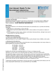
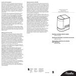
Product Manual QuickTiter™ Adenovirus Titer Immunoassay Kit Catalog Number VPK-109 100 assays FOR RESEARCH USE ONLY Not for use in diagnostic procedures Introduction Recombinant adenoviruses have tremendous potential in both research and therapeutic applications. There are numerous advantages they provide when introducing genetic material into host cells. The permissive host cell range is very wide. The virus has been used to infect many mammalian cell types (both replicative and non-replicative) for high expression of the recombinant protein. Recombinant adenoviruses are especially useful for gene transfer and protein expression in cell lines that have low transfection efficiency with liposome. After entering cells, the virus remains epichromosomal (i.e. does not integrate into the host chromosome so does not activate or inactivate host genes). Recently, recombinant adenoviruses have been used to deliver RNAi into cells. HEK 293 cells or their variants are used as host cells for viral amplification. Recombinant adenoviruses can be grown at high titer (1010 VP (viral particles)/mL, which can be concentrated up to 1013 VP/mL) and purified by Cell Biolabs ViraBind™ Adenoviral Purification Kit or traditional CsCl ultracentrifugation. A particular challenge in the delivery of a gene by a viral vector is the accurate measurement of virus titer. Traditionally, infectivity particles are measured in culture by a plaque-forming unit assay (PFU) that scores the number of viral plaques as a function of dilution. These methods are time-consuming (10 days), require a long infection period, and suffer from a high degree of inter-assay variability and are affected by virus-cell interactions. Cell Biolabs QuickTiter™ Adenovirus Titer Immunoassay Kit utilizes an antibody against adenovirus hexon proteins to visualize infected cells by immunocytochemistry staining. The hexon proteins are the largest and most abundant of the structural proteins in the adenovirus capsid, and they are distributed symmetrically to form capsid facets. Cell Biolabs’ QuickTiter™ Adenovirus Titer Immunoassay Kit provides a quick and complete system to functionally titer virus infectivity; it provides sufficient reagents for up to 100 titrations in 24-well plate. In contrast to the 10-day infection of a classical plaque assay, the kit only requires a 2-day infection. The kit antibody against hexon protein recognizes all 41 serotypes of adenovirus by immunocytochemistry and can be used with any adenovirus system as long as the virus is able to amplify in HEK 293 cells. 2 Assay Principle Related Products 1. VPK-106: QuickTiter™ Adenovirus Quantitation Kit 2. VPK-110: QuickTiter™ Adenovirus Titer ELISA Kit 3. VPK-111: Rapid RCA Assay Kit 4. VPK-252: RAPAd® CMV Adenoviral Expression System 5. AD-100: 293AD Cell Line 6. VPK-099: ViraBind™ Adenovirus Miniprep Kit 7. VPK-100: ViraBind™ Adenovirus Purification Kit 8. AD-200: ViraDuctin™ Adenovirus Transduction Reagent 9. VPK-112: QuickTiter™ Lentivirus Quantitation Kit 10. VPK-120: QuickTiter™ Retrovirus Quantitation Kit Kit Components 1. Anti-Hexon Antibody (1000X) (Part No. 10901): One tube – 30 µL. 2. Secondary Antibody, HRP Conjugate (1000X) (Part No. 10902): One tube – 50 µL. 3. DAB Substrate (25X) (Part No. 10903): One tube – 1.5 mL. 4. Diluent (10X) (Part No. 10905): Three tubes – 1.5 mL each. 5. Ad-β gal Positive Control (Part No. 10904): One tube – 50 µL at 1.0 x 10⁹ ifu/mL. 3 Materials Not Supplied 1. Recombinant adenovirus of interest 2. HEK 293 cells and cell culture growth medium 3. Methanol 4. 1% BSA/PBS 5. H2O2 6. Light Microscope 7. (optional) β-Galactosidase Staining Kit (Cat.# AKR-100) Storage Upon receipt, store the Ad-β-gal Positive Control at -80ºC. Store all other kit components at 4ºC until their expiration dates. Safety Considerations Remember that you will be working with samples containing infectious virus. Follow the recommended NIH guidelines for all materials containing BSL-2 organisms. Preparation of Reagents The table below is suggested for tests in a 24-well plate. Use twice the amount of reagents for samples in a 12-well plate. • 1X Anti-Hexon antibody solution: Prepare a 1X anti-hexon antibody solution by diluting the provided 1000X Anti-Hexon antibody stock 1:1000 in 1% BSA/PBS. Store the diluted solution on ice. • 1X Secondary antibody solution: Prepare a 1X Secondary antibody solution by diluting the provided 1000X stock 1:1000 in 1% BSA/PBS. Store the diluted solution on ice. • 1X DAB working solution: Prior to use, FRESHLY prepare a 1X DAB working solution. First dilute the provided 10X Diluent to 1X with ddH2O, and add H2O2 to a final concentration of 0.01%. Then dilute the 25X DAB stock to 1X with 1X Diluent/ H2O2 and use the 1X DAB working solution immediately. Note: When dilute 10X diluent, use ddH2O. Heavy metals in impure H2O will cause DAB precipitation. Reagents 1000X Anti-hexon Antibody 1000X Secondary Antibody 25X DAB Final Volume (Each Reagent) 24 tests (24-well plate) 6 μL 6 μL 240 μL 48 tests (24-well plate) 12 μL 12 μL 480 μL 96 tests (24-well plate) 24 μL 24 μL 960 μL 6 mL 12 mL 24 mL Table 1. Preparation of antibody and DAB solutions. 4 Preparation of Adenoviral Samples 1. Immediately before infection, create a 10-fold serial dilution of viral sample from 10-3 to 10-7. First, dilute original viral sample 1:100. For example, adding 10 µL of viral sample to a sterile tube containing 990 µL of culture medium. 2. Label six sterile tubes #1 to #6, and add 900 µL of culture medium to each tube. Add 100 µL of 1:100 diluted viral sample to tube #1, mix tube #1 well. Transfer 100 µL of the mixture (1:1000 dilution) to the next tube. Repeat the steps until tube #5 and use tube #6 as a blank. Note: An Ad-β gal Positive Control is provided as an assay control, you may include 1:104 and 1:105 dilutions of this stock in your assay. Ad-β gal expression can also be visualized by X-gal staining. Assay Protocol The instructions below are suggested for assays performed in 24-well plate. Use twice as much the amount of cells and reagents for assays performed in 12-well plate. I. Virus Infection 1. Harvest HEK 293 cells and resuspend cells in culture medium at 2.5 x 10⁵ cells/mL. Seed 1 mL in each well of a 24-well plate and incubate at 37ºC, 5% CO2 for 1 hr. Note: Adenovirus titer assay is critically dependent of the firm attachment of cells. If the cells look thin and easy to come off during immunostaining steps, you won’t get consistent result. Only use low passage 293 cells with flattened morphology or 293AD (Cat.# AD-100), a selected 293 cell line for plasmid transfection, adenovirus amplification and tittering. To improve cell adhesion, you can also precoat plate with polylysine or extracellular matrix. 2. Prepare a 10-fold serial dilution of your viral sample in culture medium. Dropwise add 100 µL of diluted viral sample to each well of the 24-well assay plate (note: a negative control should be performed simultaneously). To ensure accuracy, perform each sample in duplicate. 3. Incubate infected cells at 37ºC, 5% CO2 for 2 days. II. Immunostaining 1. Slowly remove medium from the wells by tilting the plate and aspirating from the edge, then fix infected 293 cells by gently adding 0.5 ml of cold methanol down the side of each well of the 24-well assay plate, taking care not to dislodge the cells. Incubate 20 minutes at -20ºC. 2. Gently wash the fixed cells three times with 1X PBS, five minutes each wash. 3. Block for 1 hr with 1% BSA in PBS at room temperature on an orbital shaker. 4. Add 0.25 mL of diluted 1X anti-Hexon antibody solution to each well and incubate for 1 hr at room temperature on an orbital shaker. 5. Gently wash the fixed cells three times with 1X PBS, five minutes each wash. 6. Add 0.25 mL of diluted 1X Secondary antibody solution (HRP-conjugated) to each well and incubate for 1 hr at room temperature on an orbital shaker. 7. Gently wash the fixed cells five times with 1X PBS, five minutes each wash. 5 8. Add 0.25 mL of freshly diluted 1X DAB working solution to each well and incubate for 10 minutes at room temperature on an orbital shaker. Note: Adenovirus infected cells should show dark brown staining within 5 minutes. During incubation, excess DAB starts to form light precipitates in solution, and this will not affect the staining results. 9. Aspirate DAB, wash twice with 1X PBS and add 1 mL of 1X PBS to each well. 10. Count positive stained cells (brown) for at least five separate fields per well using a light microscope and 10X objective. 11. Calculate the average number of positive cells per well and viral titer (infectious units/mL). Example of Results The following figures demonstrate typical titration results. One should use the data below for reference only. This data should not be used to interpret actual results. Figure 2: Ad-β gal Titration. Different dilutions of the Ad-β gal positive control were used to infect HEK 293 cells for 48 hrs. Anti-Hexon immunostaining was performed as described in Assay Instructions and X-gal staining is done by using β-Galactosidase Staining Kit (Cat. # AKR-100). 6 Calculation of Adenovirus Titer (Infectious Units/mL) 1. Calculate the average number of positive cells per field. Ideally, choose a dilution with 5-50 positive cells/field and count at least five fields. 2. Determine the number of fields per well For most microscopes, a standard 10X objective lens with 10X eyepiece lens has a field diameter (D) of 1.8 mm, then: Area per field = 3.14 x (D/2)2 = 3.14 x 0.92 = 2.54 mm2 For 24-well plate, area of a well (standard 24-well plate) is 2.0 cm2, therefore, Fields/well = 2.0 cm2/2.54 mm2 = 2.0 cm2/2.54 x 10-2 cm2 = 79 For 12-well plate, area of a well (standard 12-well plate) is 3.8 cm2, therefore, Fields/well = 3.8 cm2/2.54 mm2 = 3.8 cm2/2.54 x 10-2 cm2 = 150 Note: If you are not sure about the field diameter of the 10X objective lens you are using or you are using objective lenses other than 10X, the field diameter can be determined by aligning with the grids of hemacytometer (Figure 2), or referring to Table 2. Figure 2. Hemacytometer Grid Dimensions. Objective Lenses Eyepiece Lenses (10X) Total Field Field Area Magnification Diameter (mm2) 4X 40X 5 mm 19.6 10X 100X 1.8 mm 2.54 20X 200X 0.9 mm 0.64 Table 2. Field sizes of objective lenses. 7 Fields/Well 12-well 24-well Plate Plate 19 10 150 79 594 313 3. Calculate viral titer (Infectious Units or ifu/mL) Tests in 24-well: Viral Titer (ifu/mL) = (average positive cells/field) x (79 fields/well) x (dilution factor) (0.1 mL) Tests in 12-well: Viral Titer (ifu/mL) = (average positive cells/field) x (150 fields/well) x (dilution factor) (0.1 mL) Calculation Example: A serial of 10-fold dilutions of the provided Ad-β gal Positive Control was made and its titer was determined in a 24-well plate as described in assay instruction. Ten fields were counted and the average positive cells/field is 12 for 1/105 dilution under a standard 10X objective, therefore: Viral Titer (ifu/mL) = (average positive cells/field) x (79 fields/well) x (dilution factor) (0.1 mL) Viral Titer (ifu/mL) = (12/field) x (79 fields/well) x (105) = 0.95 x 10⁹ (ifu/mL) (0.1 mL) References 1. Bewig, B., and W. E. Schmidt (2000) Accelerated titering of adenoviruses. BioTechniques 28:870873. Recent Product Citations 1. Hisamitsu, T. et al. (2012). Na+/H+ Exchanger 1 Directly Binds to Calcineurin A and Activates Downstream NFAT Signaling, Leading to Cardiomyocyte Hypertrophy. Mol.Cell Biol. 32:32653280. 2. Lee, S. et al. (2012). Adiponectin Abates Diabetes-Induced Endothelial Dysfunction by Suppressing Oxidative Stress, Adhesion Molecules, and Inflammation in Type 2 Diabetic Mice. Am J Heart Circ Physiol. 303: H106-H115. 3. Polling, J. et al. (2011). Induction of Smooth Muscle Cell Migration during Arteriogenesis is Mediated by Rap 2. Arterioscler Thromb Vasc Biol 31:2297-2305. 4. Triulzi, C. et al. (2010). Antibody-Dependent Natural Killer Cell-Mediated Cytotoxicity Engendered by a Kinase-Inactive Human HER2 Adenovirus-Based Vaccination Mediates Resistance to Breast Tumors. Cancer Res. 70:7431-7441. 5. Peled, M. et al. (2009). Systemic Administration of a Conditionally Replicating Adenovirus, Targeted to Angiogenesis, Reduced Lung Metastases Burden in Cotton Rats. Clin. Cancer Res. 15:1664-1673. 6. Troidl, K. et al. (2009). Actin-binding Rho Activating Protein (Abra) is Essential for Fluid Shear Stress-Induced Arteriogenesis. Arterioscler. Thromb. Vasc. Biol. 10.1161/ATVBAHA.109.195305. 8 7. Soesanto, Y. et al. (2008). Regulation of Akt signaling by OGlcNAc by euglycemia. Am J. Physiol. Endocrinol. Metab. 295:E974-E980. 8. Ivanov, A.I. (2008). Exocytosis and Endocytosis, Chapter 13, Humana Press. 9. Black, S.A. et al. (2008). TGFß1 Stimulates Connective Tissue Growth Factor (CCN2/CTGF) Expression in Human Gingival Fibroblasts through a RhoA-Independent, Rac1/Cdc42-dependent Mechanism: Statins with Forskolin Block TGFß1-Induced CCN2/CTGF Expression. J. Biol. Chem. 283:10835-10847. 10. Akai, S. et al. (2007). Knock Down of Gamma-Glutamylcysteine Synthetase in Rat Causes Acetaminophen-Induced Hepatotoxicity. J. Biol. Chem. 282:23996-24003. 11. Saavedra, L. et al. (2007). Internalization of Beta Amyloid Peptide by Primary Neurons in the Absence of apoE. J. Biol. Chem. 282:35722-35732. 12. Chang, C-T. et al. (2007). Transforming Growth Factor-Beta1 Decreases Epithelial Sodium Channel Functionality in Renal Collecting Duct Cells using a Smad4-Dependent Pathway. Nephrol. Dial. Transplant 23:1126-1134. Warranty These products are warranted to perform as described in their labeling and in Cell Biolabs literature when used in accordance with their instructions. THERE ARE NO WARRANTIES THAT EXTEND BEYOND THIS EXPRESSED WARRANTY AND CELL BIOLABS DISCLAIMS ANY IMPLIED WARRANTY OF MERCHANTABILITY OR WARRANTY OF FITNESS FOR PARTICULAR PURPOSE. CELL BIOLABS’ sole obligation and purchaser’s exclusive remedy for breach of this warranty shall be, at the option of CELL BIOLABS, to repair or replace the products. In no event shall CELL BIOLABS be liable for any proximate, incidental or consequential damages in connection with the products. Contact Information Cell Biolabs, Inc. 7758 Arjons Drive San Diego, CA 92126 Worldwide: +1 858-271-6500 USA Toll-Free: 1-888-CBL-0505 E-mail: [email protected] www.cellbiolabs.com 2004-2012: Cell Biolabs, Inc. - All rights reserved. No part of these works may be reproduced in any form without permissions in writing. 9