1
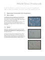
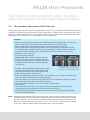
Microdissection from Carl Zeiss
PALM User Protocols
Cell
Capture
Cell Culture,
Culture,non-contact
non-contactLaser
Laser
Capture
Microdissection
Microdissection(LCM)
(LCM)and
andRecultivation
Recultivation
PALM Laboratories
A unique service of Carl Zeiss
PALM User Protocols
Cell Culture, non-contact Laser Capture
Microdissection (LCM) and Recultivation
1
Sample preparation
3
1.1
Cultivation of adherent cells
3
1.2
Preparation of cells in suspension –
Coating of DuplexDishes and FrameSlides
4
1.2.1
Poly-L-Lysin
4
1.2.2
Cell-Tak
4
1.2.3
Fibronectin
4
1.2.4
Gelatin
5
1.2.5
Laminin
5
1.3
Immunostaining of murine stem cell line P19 with α-SSEA-1-antibody 6
2
Improvement of visualization in live cell applications
7
2.1
Phase contrast
7
2.2
PlasDIC
7
3
Non-contact LCM
8
3.1
Holders for PALM RoboStage
8
3.2
Collection devices
8
3.2.1
CapMover
8
3.2.2
RoboMover
8
3.2.3
LiveCell Collector
8
3.3
The procedure: Non-contact LCM of live cells
9
4
Recultivation of collected live cells - clonal expansion
10
5
Time-lapse
11
6
Incubator
11
7
Optical Tweezers and PALM CombiSystem
11
2
PALM User Protocols
Cell Culture, non-contact Laser Capture
Microdissection (LCM) and Recultivation
1
Sample preparation
1.1
Cultivation of adherent cells
Adherently growing cells are seeded into a DuplexDish 35 (Order No. 415101-4400-551) or DuplexDish 50
(Order No. 415101-4400-550) and are cultivated as usual up to the desired cell density.
DuplexDishes are especially adapted for non-contact LCM.
The growth-surface of these culture dishes is optimized for
attachment of many different cell types.
Alternatively LumoxTM Dish 35 (Greiner bio-one, # 76077331)
combined with MembraneRing 35 (Order No. 415101-4400-581)
and LumoxTM Dish 50 (Greiner bio-one, # 76077410) with
MembraneRing 50 (Order No. 415101-4400-580) can be used.
The cells are growing directly on top of the membrane in
an appropriate cell culture medium including all necessary
supplements. Depending on the proliferation rate of the
cell type the cells are ready for laser microdissection after
1-2 days.
DuplexDishes
Adherently growing cells can also be cultivated on
FrameSlides PET (Order No. 415101-4401-055).
These slides are recommended if weak fluorescence signals
have to be detected, e.g. due to low signal to noise ratio.
FrameSlides are inserted into a quadriPerm cell culture plate
(Greiner bio-one, # 96077307) with the membrane-side
facing the bottom of the plate (Zeiss symbol visible).
Cells are seeded into the cavity of the FrameSlide and covered
with approximately 0.5 to 1 ml medium of choice.
LumoxTM Dish with MembraneRing
quadriPerm with FrameSlides
3
PALM User Protocols
Cell Culture, non-contact Laser Capture
Microdissection (LCM) and Recultivation
1.2
Preparation of cells in suspension –
Coating of DuplexDishes and FrameSlides
Prior non-contact LCM procedure the cells in suspension have to be attached to the bottom of the dish.
It is necessary to optimize the preparation protocol for each cell type. Immobilization of cells in
suspension can be performed by e.g. coating of the membrane.
1.2.1 Poly-L-Lysin treatment
Nearly all types of human and animal cells adhere to these polymers of basic amino acids.
Protocol:
• Distribute a drop of Poly-L-Lysin (0.15 w/v, e.g. SIGMA, # P8920) on top of the membrane.
• Avoid any leakage underneath the membrane, as this might result in impairment of LCM.
• Let air-dry at room temperature.
1.2.2 Coating with Cell-Tak
BD Cell-Tak (BD Biosciences, # 354240) is a specially formulated protein solution which is particularly
suitable to immobilize cells, if recultivation is intended after laser microdissection.
Protocol:
• Please follow manufacturer´s protocol.
1.2.3 Fibronectin
Fibronectin is an extracellular matrix for the culture of endothelial cells, fibroblasts, neurons and CHO cells.
It also can be used to improve attachment of cells onto the membrane.
Protocol:
• Stock solution is prepared by dissolving 1 mg/ml Fibronectin in PBS.
• Filter sterilize and freeze in aliquots.
• Dilute stock solution to 50-100 μg/ml in basal medium or PBS.
• Add enough solution to distribute over the surface of the DuplexDish.
• Incubate for 30-45 minutes at room temperature.
• Remove excess of Fibronectin solution by aspiration and rinse with medium or PBS.
• Immediately add cell suspension or growth medium into the dish. Do not allow coating to dry.
4
PALM User Protocols
Cell Culture, non-contact Laser Capture
Microdissection (LCM) and Recultivation
1.2.4 Gelatin
Gelatin is a protein produced by partial hydrolysis of collagen. It improves attachment of cells on slides
and prevents movement during the laser capture microdissection procedure.
Protocol:
• Dissolve 10 mg Gelatin in 100 ml water (final: 0.01 % Gelatin).
• Autoclave to sterilize.
• While hot, thoroughly mix Gelatin solution.
• Add approximately 2 ml of Gelatin solution to each DuplexDish or about 1 ml to
each FrameSlide, respectively.
• Leave the Gelatin solution on the membrane overnight at 4 °C, covered with lids.
• Remove Gelatin solution by aspiration
• Add sterile water. Do not let dishes or slides run dry before adding the cells.
• Dishes and slides can be stored for up to one week at 4 °C.
• Remove water immediately before use for cell culture.
1.2.5 Laminin
Laminin is used especially for the culture of neurons, epithelial cells, leukocytes, myoblasts and CHO cells.
Protocol:
• Stock solution is prepared by following manufacturer´s protocol.
• Filter sterilize and freeze in aliquots.
• Dilute stock solution to 10-100 μg/ml in basal medium or PBS.
• Add enough solution to distribute over the surface of DuplexDish or FrameSlide.
• Incubate about 8 hours at 4 °C, covered with lid.
• Remove excess of Laminin solution by aspiration and rinse with medium or PBS.
• Immediately add cell suspension or growth medium. Do not allow coating to dry.
• Coating firstly with Poly-L-Lysin or Poly-Ornithine and subsequently with Laminin
may increase the concentration of bound Laminin.
5
PALM User Protocols
Cell Culture, non-contact Laser Capture
Microdissection (LCM) and Recultivation
1.3
Immunostaining of murine stem cell line P19 with α-SSEA-1-antibody
a)
b)
a) HeLa and P19 cells grown in co-culture and visualized with PlasDIC.
b) Very same image in the corresponding fluorescence channel.
Only SSEA-1 expressing P19 cells showing Texas Red fluorescence.
Material:
• Culture medium: α-MEM supplement with 10 % fetal calf serum (FCS) and
1 % Penicillin/Streptomycin (10.000 IE/10.000 μg/ml)
• Trypsin/EDTA solution (0.05/0.02 μg/ml (w/v) in PBS)
• PBS pH 7,4 (without CaCl2 and MgCl2)
• Incubation medium: α-MEM with 2 % FCS
• Primary antibody: Directed against h/mSSEA-1 (IgM; R&D Systems # MAB2155)
Dilutions of anti-h/mSSEA in incubation buffer:
5 μg/ml (1 : 200; 1 μl ad 200 μl) => weak staining result
10 μg/ml (1 : 100; 2 μl ad 200 μl) => intermediate staining result
20 μg/ml (1 : 50; 4 μl ad 200 μl) => intermediate staining result
40 μg/ml (1 : 25; 8 μl ad 200 μl) => strong staining result
• Secondary antibody: TexasRed-labelled Donkey anti-mouse IgM, Jackson Immunoresearch,
# 715-075-140 (1.5 mg/ml), dilution 1:200 in incubation buffer.
Protocol:
Day 1
• Trypsinize a confluent P19 stem cell line grown in a T25-flask and wash
once with PBS. Resuspend the cells in 10 ml fresh culture medium.
• Seed out 10-50 μl of the resulting suspension (depending on the desired cell density)
onto a DuplexDish 35 (see product information for details) and incubate the cells over
night in 1 ml culture medium.
Day 2
• Remove all medium and wash the cells gently with 1 ml incubation medium.
• Incubate with the primary antibody at the desired dilution in a total volume
of 400 μl incubation medium for 1 h at 37 °C, 5 % CO2.
• Gently wash the cells 3x with incubation buffer.
• Incubate with the secondary antibody labelled with TexasRed in a total volume
of 400 μl incubation medium for 1 hour at 37 °C, 5 % CO2.
• Gently wash the cells 3x with incubation buffer.
• Cover the cells with 1 ml incubation buffer and let them revive for 30 minutes.
• Now the cells are ready for microscopic analysis and laser microdissection.
6
PALM User Protocols
Cell Culture, non-contact Laser Capture
Microdissection (LCM) and Recultivation
2
Improvement of visualization in live cell applications
2.1
Phase contrast
In optical microscopy many objects such as live cells and
microorganisms are nearly fully transparent unless stained.
Phase contrast microscopy is a type of light microscopy
that enhances contrasts of transparent and colorless objects
by influencing the optical path of light.
Thus, components in cells or bacteria can be shown, which
would be very difficult to see without phase contrast.
This technique is widely utilized to examine dynamic events
in live cells.
2.2
PlasDIC
Phase contrast
PlasDIC (DIC=Differential Interference Contrast) is the first
DIC method, which allows the use of plastic dishes and
membrane coated DuplexDishes for microscopic examinations
of unstained live cells.
It provides more information and details about the cells to
be examined and allows better visualization of thick cells.
PlasDIC is ideal for microdissection or micromanipulation of
live cells on the microscope with better depth of focus than
conventional differential interference contrast methods.
Visualization in brightfield...
...and in PlasDic
7
PALM User Protocols
Cell Culture, non-contact Laser Capture
Microdissection (LCM) and Recultivation
3
Non-contact LCM
Please, additionally have a look into the PALM MicroBeam user manual.
3.1
Holders for PALM RoboStage
Cells can be cultivated in DuplexDish 35 and 50, in LumoxTM Dish with appropriate
MembraneRing 35 or 50, as well as on FrameSlide PET (please see chapter 1: Sample preparation).
Depending on whether dishes or FrameSlides are used DishHolder 35 respectively 50
or SlideHolder 3x1.0 has to be applied. These holders are adapted to the PALM RoboStage II.
3.2
Collection devices
PALM RoboMover, PALM CapMover and LiveCell Collector are collection devices for positioning
collection vessels above the sample.
3.2.1 PALM CapMover
For PALM CapMover, SingleTube Collectors for dishes and slides are available. These Collectors are
designed to be equipped with 200 μl or 500 μl caps in order to enable the direct lifting of cells
from a dish or a slide.
3.2.2 PALM RoboMover
PALM RoboMover is a device for highly automated harvesting and sorting of different kinds of
microdissected specimen in a higher throughput process. The non-contact LCM process can be
operated automatically or manually, respectively. PALM RoboMover is completely controlled by
PALM RoboSoftware and can be equipped with different collectors, e.g. SingleTube Collector and
CapturePlate Collector.
3.2.3 LiveCell Collector
LiveCell Collector (Order No. 415101-2100-310) is designed
for harvesting live cells from DuplexDish 50.
Non-contact LCM now can be performed in an entirely
enclosed compartment and thus under sterile conditions.
Due to optimal humidity conditions time for cell harvesting
is elongated.
LiveCell Collector
8
PALM User Protocols
Cell Culture, non-contact Laser Capture
Microdissection (LCM) and Recultivation
3.3
The procedure: Non-contact LCM of live cells
Adherent growing cells are seeded into DuplexDish or LumoxTM Dish combined with MembraneRing and
cultivated as usual up to the desired cell density. Usually after 1-2 days the cells are ready for laser microdissection. Cells in suspension have to be attached to the bottom of the dish using an adequate coating
depending on the cell type (see 1.2 Preparation of cells in suspension).
Protocol:
• Please arrange all necessary items for recultivation before starting the non-contact LCM
steps to limit the time the cells will stay with minimum amount of medium.
• Before starting laser microdissection excess of medium has to be removed.
Due to evaporation of the residual medium, time for harvesting of the cells is limited.
For example using a SingleCap Collector there are about 15 minutes with an open dish.
Using the LiveCell Collector with a closed DuplexDish 50 more than 30 minutes are
available to collect the cells.
• Find optimal laser settings.
Please note: Dish shall be closed for this step.
• For use of LD 20x, LD 40x, or LD 63x objective
adapt the working distance to different glass slides
or DuplexDish by turning the correction ring on
the objective.
Regular glass slide (1 mm thick) => 1,
thin slide (0.17 mm thick) => dot,
DuplexDish => between dot and 0.
• Set Delta Laser Energy (difference between cutting
and lifting energy) to 20-40, depending on the
objective used and the size of the selected area.
• Adjust cutting speed to a lower level.
• Prepare an appropriate collection vessel, e.g. a sterilized cap of a 500 μl microfuge tube
filled with 40 μl of appropriate cell culture medium.
• Place the prepared cap in the LiveCell Collector or in the SingleTube Collector for dishes.
• Center the cap directly above the region of interest.
• Select the cells of interest by any of the software marking tools.
• Start the laser.
• The marked cells or areas are cut together with the underlying membrane and are
subsequently lifted by a single laser pulse into the collection vial.
• After non-contact LCM the collected cells can be inspected by moving the RoboStage
to the check-point position.
Note: Having adjusted optimal settings even large areas can be harvested easily. Induced by the
residual humidity, it may occur that the separated cell areas cannot be lifted with the laser
function “RoboLPC”. In this case first cut around with the laser function “Close&Cut” and
then lift in a second step with the function “LPC”. The “LPC-dot”, the lifting pulse, should
be set into a cell-free region within the outlined area on the membrane.
9
PALM User Protocols
Cell Culture, non-contact Laser Capture
Microdissection (LCM) and Recultivation
4
Recultivation of collected live cells – clonal expansion
Protocol:
• Attach the cap containing the collected cells onto a sterile microfuge tube.
• Spin down immediately at 140 g (800 rpm Hettich Universal 32R) for 2 minutes.
• Transfer the cells with a pipette into e.g. a 24-well culture plate filled with 1 ml
medium per well.
• The lifted cells can be also transferred directly from the cap into a culture
dish by pipetting, without a centrifugation step. To be sure that all captured
cells are transferred, it is advisable to rinse the cap several times.
• Cultivate the cells under usual cell culture conditions. The harvested cells enter
the cell cycle and start to proliferate within a few days, depending on cell type
and amount of selected cells.
• The originating DuplexDish from which the isolation took place can be put back
into the incubator for further experiments.
Note: Non-contact LCM has no influence on the viability, the proliferation rate, and the genetic
stability of isolated cells. Even frequently recultivated cell colonies are still viable and
unaltered after several non-contact LCM isolation procedures.
a
b
a) selected area after transfer to a fresh culture vessel
b) Clonal expansion of the selected cells
10
PALM User Protocols
Cell Culture, non-contact Laser Capture
Microdissection (LCM) and Recultivation
5
Time-lapse
With time-lapse photography very slow motions can be observed, e.g. motion of cells in culture, which
are unable to view in real time. The images are captured by taking photos from just a few seconds to a
minute or more apart, they are put together as a sequence allowing to view the event over a short time
period. With the time-lapse function in the PALM RoboSoftware pictures in series or a video sequence
can be saved.
6
Incubator
PALM Incubator XL PALM-3 S1 is a tool for
working in a very clean surrounding and/or
long time experiments (e.g. time-lapse)
with live cells and organisms, which demand
temperature control.
PALM CombiSystem with Incubator
7
Optical Tweezers and PALM CombiSystem
Carl Zeiss MicroImaging GmbH
Location Munich
Kistlerhofstr. 75
81379 München, Germany
Phone: +49 (0) 89 90 9000-900
E-Mail: [email protected]
Web:
www.zeiss.de/microdissection
September 2008
With PALM MicroTweezers, also known as laser traps, it is possible to catch, hold and move microscopic
particles such as beads, cells or even sub-cellular particles. The high intensity gradient of the focused,
continuously working laser beam causes microscopically small objects in liquid to be assembled and kept
within the laser focus by means of radiation pressure forces. Application fields of MicroTweezers are
sorting, i.e. picking single cells, bacteria or protozoa out of a bulk or a mixture of different specimen and
to separate them, e.g. for subsequent molecular biological analysis. Bringing different species in close
contact to each other, or to any nearby surface, and studying cell membrane formation and elasticity is
feasible as well as working inside live cells.
PALM CombiSystem combines the cutting and lifting properties of PALM MicroBeam with trapping and
manipulation capabilities of PALM MicroTweezers. Only the forces of light are used to cut, lift, trap, sort
or position with high precision.