1
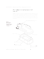
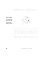
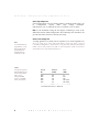
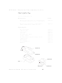
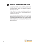
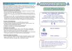
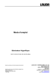
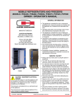
HE 33 Mini Submarine
Electrophoresis Unit
User Manual
80-6273-42
HE33-IM/Rev. E1/7-99
HE 33 M i n i
Submarine
Electrophoresis
Unit
Mini Submarine Electrophoresis Unit Function . . 1
Important Information . . . . . . . . . . . . . 2
Unpacking
. . . . . . . . . . . . . . . . . 3
Specifications . . . . . . . . . . . . . . . . 3
Operating Instructions
. . . . . . . . . . . . 4
Care and Maintenance
. . . . . . . . . . . . 9
Troubleshooting . . . . . . . . . . . . . . . 10
Notes, Buffers, and Volumes . . . . . . . . . 11
Bibliography . . . . . . . . . . . . . . . . 15
Customer Service Information . . . . . . . . . 16
A m e r s h a m
B i o s c i e n c e s
HE 33 M i n i
Submarine
Electrophoresis
Unit
Mini Submarine Electrophoresis Unit
Function
The HE 33 horizontal agarose unit is intended for rapid electrophoresis of small
quantities of nucleic acids in agarose gels. A gel is cast in the gel caster, which holds
one or two combs. (Eight different combs are available; a maximum of 32 samples
can be run if two 16-well combs are used.) After the gel sets, the running tray is
transferred to the platform of the horizontal unit. The base of the unit holds coolant
that can be chilled before the run. This passive cooling capacity allows fast, high
voltage runs.
Color-coded leads connect electrodes in
the unit base to the power supply.
Figure 1.
Main components
(See Figure 2 for an
illustration of the
casting kit.)
Buffer chamber
Lid assembly
Electrode connectors (2)
Running platform
(supports the running tray)
Fill the base with 50/50
ethylene glycol/ water
A m e r s h a m
B i o s c i e n c e s
1
HE 33 M i n i
Submarine
Electrophoresis
Unit
Important Information
Informations importantes
➧
➧
➧
➧
➧
➧
➧
➧
➧
2
The safety lid must be in place before
connecting the power leads to a
power supply.
Turn all power supply controls off and
disconnect the power leads before
removing the safety lid.
➧
Before the first use, fill the base with
about 600 ml 50/50 ethylene glycol/water to prevent irreparable damage to the unit. (See instructions on
page 4.) The base must be filled even
if no cooling is required.
➧
Do not use pure water, commercial
anti-freeze, or any organic solvent to
fill the base. Water expands as it
freezes. Because the water is trapped
in the base, it may crack. Organic solvents will cause irreparable chemical
damage to the unit!
➧
Do not chill the base below -20 °C
(-4 °F). Chill the base in a bucket of
ice, refrigerator, or in a freezer.
Do not operate with gel or buffer temperature above 50 °C.
Prevent overheating by chilling the
base prior to use. To prevent overheating during prolonged high-voltage
runs, exchange the first base with an
extra prechilled second base if one is
available. Overheating will cause
irreparable damage to the unit!
➧
➧
If this equipment is used in a manner
not specified by the manufacturer,
the protection provided by the equipment may be impaired.
➧
Only accessories and parts approved
or supplied by Amersham Biosciences
may be used for operating,
maintaining, and servicing this product.
➧
A m e r s h a m
Le couvercle de sécurité doit être en place
avant de brancher les prises au générateur.
Eteindre le générateur et débrancher les
prises avant d’enlever le couvercle de sécurité.
Avant la premiére utilisation, remplir le
socle avec environ 600 ml 50/50 d’éthylène
glycol et d’eau afin de ne pas endommager
l’instrument. (Voir page 4 du mode d’emploi). Le socle doit être rempli même si le
refroidissement n’est pas nécessaire.
Ne pas utiliser seulement de l’eau, de l’antigel commercial ou tout autre solvant
organique pour remplir le socle. L’eau augmente en volume lorsqu’elle gêle. Du fait
que l’eau est enfermée, le socle peut se
fendre. Les solvants organiques peuvent
causer des dommages chimiques irréparables.
Ne pas refroidir le socle en dessous de
-20 °C (-4 °F). Refroidir le socle dans un
seau rempli de glace ou un dans un
réfrigérateur.
Ne pas utiliser avec un gel ou un tampon à
plus de 50 °C. Eviter le surchauffement en
refroidissant le socle avant l’emploi. Afin
d’éviter le surchauffement durant de
longues utilisations sous haut voltage,
échanger si possible, le socle avec un second socle déjà refroidi.Un surchauffement
peut causer des dommages irréparables à
l’instrument.
Si l'instrument n'est pas utilisé en conformité avec les recommandations du fabriquant, les protections de sécurité qui
équipent cet appareil peuvent être rendues
inéfficaces.
Seulement les accessoires et piéces
detachées approuvés ou fournis par
Amersham Biosciences sont recommandés pour l’utilisation, l’entretien et
réparation de cet appareil.
B i o s c i e n c e s
HE 33 M i n i
Submarine
Electrophoresis
Unit
Unpacking
Unwrap all packages carefully and compare contents with the packing list, making
sure all items arrived. If any part is missing, contact your local sales office. Inspect
all components for damage that may have occurred while the unit was in transit. If
any part appears damaged, contact the carrier immediately. Be sure to keep all
packing material for damage claims or for repacking should it become necessary to
return the unit.
Specifications
Max. voltage
500 V for 5 minutes or less
Max. wattage
15 W
Max. current
500 mA
Max. operating temperature
50 °C
Max. buffer volume
250 ml
Coolant required
≈600 ml 50/50 water/ethylene glycol
Gel size
Environmental operating conditions
7 × 10 cm
Indoor use: 4–40 °C
Humidity up to 80%
Altitude up to 2000 m
Installation category
II
Pollution degree
2
Dimensions
width × depth × height
24×13×7 cm (9.5×5.2×2.8 in.)
Weight (base, lid, and leads only)
Product certifications
0.4 kg (0.9 lbs)
EN61010–1, UL3101–1, CSA C22.2
1010.1, CE
This declaration of conformity is only valid for the instrument when it is:
◗
used in laboratory locations,
◗
used as delivered from Amersham Biosciences except for alterations
described in the User Manual, and
◗
connected to other CE labeled instruments or products recommended or
approved by Amersham Biosciences.
A m e r s h a m
B i o s c i e n c e s
3
HE 33 M i n i
Submarine
Electrophoresis
Unit
Operating Instructions
Agarose gels are first prepared using the gel casting kit. Samples are then loaded
into wells and electrophoretically separated. The fluorescent dye ethidium bromide
can be added to the gel or electrophoresis buffer or both to track separation
progress. After electrophoresis, the gel may be stained and photographed, blotted,
or dried for autoradiography.
Fill the base with coolant
Important
Do not fill the base
with commercial
antifreeze, organic
solvents, or pure
water.
Even if no cooling is required, it is important to fill the base with the proper coolant
solution before the first use because the solution provides a necessary heat sink.
1
Prepare 600 ml of 50/50 ethylene glycol/water.
Optional: To help see wells better while loading the sample, add a drop or
two of soluble dye or food color to the coolant solution.
Locate the inlet hole on the bottom of the base, near the edge. Fill the base
cavity as full as possible with coolant using a 50-ml syringe or pump.
2
Fit the threaded, white plastic plug into the hole and, taking care that the
thread is properly engaged, screw it in place until it is snug. Tighten another
1/8 to 1/4 turn to prevent leaks.
3
Place the prepared base in an ice bucket or into a refrigerator or freezer set no
lower than -20 °C for about an hour before use. (The base will always be ready
if you store it in the refrigerator or freezer.)
Note
It is not necessary to
replace the coolant.
4
A m e r s h a m
B i o s c i e n c e s
HE 33 M i n i
Submarine
Electrophoresis
Unit
Prepare solutions
1
Prepare 250 ml of running buffer. (Refer to p. 13 for recipes of commonly
used electrophoretic running buffers.)
2
Prepare the sample loading buffer. (Refer to p. 14 for a recipe and a table of
volume requirements for each comb size.)
3
Prepare approximately 7 ml agarose solution per mm of gel thickness.
(For example, a 3-mm gel requires 0.3 cm x 7 cm x 10 cm = 21 ml)
Caution
Dissolve agarose in running buffer, heat according to instructions accompanying the agarose, and allow the solution to cool to 50 °C before pouring into
the casting tray.
Ethidium bromide is
a known mutagen.
Always wear gloves
when handling.
Optional: Add 0.5 µg/ml ethidium bromide to the gel solution to observe separation during electrophoresis.
Cast the gel
1
Install the running tray. Firmly grasp the casting tray with one hand. With the
other hand, place one end of the running tray against the foam pad at the bottom edge, press the tray against the pad, and then lower it to rest on the bottom of the casting tray, seating the other end of the tray against the opposite
foam pad.
Figure 2.
Gel casting kit
Approach the foam
pad with one end of
the running tray and
then gently press the
tray edge against the
pad, compressing it
enough to allow the
opposite end of the
running tray to drop
fully into the casting
tray before sealing
against the foam
pad.
UV-transparent running tray
(Cast the gel on this tray, then
transfer gel to the horizontal
unit base for electrophoresis.)
Foam pads (2)
A m e r s h a m
B i o s c i e n c e s
Gel casting tray
5
HE 33 M i n i
Submarine
2
Electrophoresis
Unit
Prepare the comb(s). Fit the two slots in the comb between the (loosened)
thumb screw heads and the comb back. Tighten the screws until the comb is
just supported. Seat the comb assembly on the rim of the casting tray and
adjust the bottom of the comb so that it is about 1.0 mm from the running
tray. Tighten the screws to secure the comb. To run twice as many samples,
prepare two combs.
Figure 3.
A comb back, which
fits onto the rim of
the casting tray, positions the comb in the
gel. Two screws hold
the adjustable comb.
For twice as many
wells, a second comb
can be placed in the
center of the gel.
Comb back
6
Screws (2)
Comb
3
Remove the comb assembly. Place the casting assembly on a leveling surface
and level, using the spirit level on the running tray as a guide.
4
Pour the agarose solution (cooled to 50 °C) into the casting tray. Orient the
comb assembly so that the comb faces the nearest foam pad and seat it on the
tray rim. Check that the comb is vertical to prevent well shape distortions. To
run twice as many samples, place the second comb assembly in the center of the
tray. Allow a minimum of 30 minutes for the gel to set.
5
Once the gel is set, remove the comb carefully. Partially lift and slightly tilt the
comb at one end and then slowly withdraw it from the gel. (Pulling the comb
straight up creates a vacuum in the wells that may lift the gel out of the tray.)
6
Remove the running tray and gel by grasping the handles of the tray and pressing against one of the foam pads. Once the tray clears the opposite pad, lift it
out. Transfer the running tray and gel to the chilled base.
A m e r s h a m
B i o s c i e n c e s
HE 33 M i n i
Submarine
Electrophoresis
Unit
The electrophoresis run
Refer to the Notes, Buffers, and Volumes section for additional information
and guidelines.
1
Caution
Chill the base before use, especially when higher voltage settings will be used
or when the separation will require more than 30 minutes.
Note: To monitor separation progress, either add 0.5 µg/ml (final conc.) of
ethidium bromide to the running buffer now, or, add 50 µg/ml (final conc.)
ethidium bromide to the sample buffer. To visualize progress, turn off the
power supply, remove the lid assembly, and hold a portable UV lamp near the
gel.
Ethidium bromide is
a known mutagen.
Always wear gloves
when handling.
Wear UV safety goggles and protect skin
when using a UV
lamp.
Adding ethidium bromide to the running or sample buffer slows migration
slightly. Detection by this method is not as sensitive as staining and viewing
on a transilluminator. (See DNA detection, p. 15.)
2
Fill both buffer chambers with running buffer until the gel is submerged under
≈1 mm of solution. (This requires about 220 ml.)
Note
3
If no dye was added
to the coolant, place
the base on a dark
background to see
the wells more easily.
Load the samples. Add sample to 5X sample loading buffer and mix
(1/5 of the final volume is loading buffer, see p. 14). Use a micro-pipette to load
each sample, taking care to avoid puncturing the well bottom or entrapping
any bubbles.
4
Place the lid so that the cathode (–, black lead) is at the end nearest the sample
well. (Nucleic acid samples migrate toward the anode, +, red lead.) Connect
the color-coded leads (red to red, and black to black) to an approved power
supply, such as the EPS 2A200. Set the voltage level and timer (if available) according to the degree of resolution sought.
A m e r s h a m
B i o s c i e n c e s
7
HE 33 M i n i
Submarine
Electrophoresis
Unit
Quick, high-voltage runs
Certain applications, such as screening samples or checking sample purity, can
be
accomplished
quickly
under
high
voltage
conditions.
Chill the base (-20 °C) and limit the run to 5 minutes or less at 500 V.
Note: At the maximum setting the unit begins overheating as soon as the
chilled base reaches ambient temperature. If overheating is not controlled, the
gel will melt and/or the base of the unit will warp!
Note
To calculate the voltage gradient, divide
the voltage setting
by the distance
between the electrodes (12.7 cm).
Slower, lower voltage runs
A voltage gradient of 12 V/cm (150 V) separates 0.1 to 23 kb fragments of a
Hind III digest of l DNA in 30 to 40 minutes (using 1% agarose gel and
0.5X TBE running buffer). Alternatively, using the same solutions, this sample
could also be run at 24 V/cm (300 V) with acceptable band resolution in
20 to 30 minutes. Chill the base before use.
Table 1
Voltage settings and
the maximum recommended separation
time, using
1% Agarose NA
agarose, 0.5X TBE,
and a chilled base.
Voltage
(V)
Gradient
(V/cm)
Time
(min)
500
400
300
200
150
40
31
24
16
12
5*
10*
20*
30 to 40
30 to 60
*For rapid runs of 20 minutes or less, use 0.5X TBE
and chill the base to -20 °C before use.
8
A m e r s h a m
B i o s c i e n c e s
HE 33 M i n i
Submarine
Electrophoresis
Unit
After the separation
1
Turn off the power supply, disconnect the leads, and remove the lid.
2
If no ethidium bromide was added to the gel or sample before the run, stain
the gel in a solution of 0.5 to 1.0 µg/ml ethidium bromide in water or buffer.
3
Clean the unit as described below.
Care and Maintenance
Cleaning
Important:
Never autoclave any component of the electrophoresis unit or casting
kit.
After each use, clean the unit with a mild detergent and water, rinse thoroughly
with distilled water, and allow to air dry. Never use abrasive cleansers. Do not
expose the unit to solutions or vapors of aromatic or halogenated hydrocarbons,
ketones, esters, alcohols (over 30%), or concentrated acids (over 25%).
To reduce DNase and RNase contamination, soak the buffer chamber or casting kit
for 10 minutes in a 3% hydrogen peroxide (H2O2) solution, then rinse thoroughly
with DEPC-treated, autoclaved, deionized water. (Sambrook, et al. 1:7:40)
Replacing foam pads
Remove worn foam pads. Peel off the adhesive cover on a new foam pad. Align the
pad so that it will rest on the bottom of the tray along a short
(7-cm) side, adhesive side toward the inside wall, and then press it in place. Repeat
with second pad on the wall opposite the first pad.
Replacing the electrode
It is recommended that electrodes be replaced only by Amersham
technicians. Call your local representative for advice.
Amersham
Biosciences
9
HE 33 M i n i
Submarine
Electrophoresis
Unit
Troubleshooting
Deformed sample well
✓
Allow the gel to set for a minimum of 30 minutes and make sure it is at room
temperature before removing the comb.
✓
When removing the comb, hold it at a slight angle and lift very slowly to prevent
the gel from breaking.
✓
Take care to not damage the well with the pipet while loading the sample; aim for
the center of the well and do not puncture the bottom with the pipet tip.
Samples not running along a straight path
✓
If a comb or running tray is warped, replace.
✓
Reduce the voltage.
✓
Choose a buffer with the appropriate ionic strength and buffering capacity.
(The buffering capacity of TBE, for example, is higher than that of TAE.)
If the buffer is depleted, stop the run, remove the lid, and pipette the buffer from
each chamber into the opposite chamber to replenish the buffer.
✓
If the gel is uneven, level the casting tray before pouring the gel.
Double-banded pattern
✓
The comb must be vertical to prevent well shape distortion.
✓
Decrease the buffer level to 1 mm above the top of the gel, to reduce the vertical
temperature gradient.
Poor band resolution
✓
Add Ficoll, glycerol, or sucrose to the sample loading buffer to ensure that the sample sinks to the bottom of the well. (Ficoll is preferred.)
✓
Make sure the sample is completely dissolved.
✓
Reduce the voltage.
✓
Reduce the sample concentration.
✓
Reduce the sample volume.
✓
At least 1 mm of gel below the bottom of the comb is required to prevent samples from leaking out of the well bottom.
✓
Reduce the salt concentration of the sample.
✓
Check enzyme activity; the sample may require longer digestion or a different
restriction buffer.
✓
Prepare fresh sample if you suspect nuclease contamination.
✓
Choose agarose with a low endosmosis value.
✓
Install the running tray as as described on page 4; do not press straight down into
place.
Foam pads peel off
10
A m e r s h a m
B i o s c i e n c e s
HE 33 M i n i
Submarine
Electrophoresis
Unit
Notes, Buffers, and Volumes
Agarose gel electrophoresis notes
Agarose gel electrophoresis can be used to separate DNA fragments as small as 0.1
kb or less. Polyacrylamide gels are usually used for fragments smaller than 1 kb.
DNA mobility
Suggested agarose concentration for separating fragments of various sizes is given
in Table 2 below. Other factors affecting separation results include the running
buffer, voltage setting, temperature, conformation, and the presence of ethidium
bromide. Special agaroses are available that can extend resolution ranges.
A common standard is a Hind III digest of lambda phage, which gives eight fragments
ranging in size from 0.1 to 23 kb pairs. For good resolution, run 45 minutes on a
10-cm long, 1% agarose gel in 0.5X TBE gel at 150 V.
Table 2.
Agarose concentrations
for separating
DNA fragments of
various sizes
Agarose (%)
0.5
0.7
1.0
1.2
1.5
Effective range of resolution
of linear DNA fragments* (kb)
1.0
0.8
0.5
0.4
0.2
—
—
—
—
—
30
12
10
7
3
*Current Protocols in Molecular Biology, p 2.5.2 (1993)
A m e r s h a m
B i o s c i e n c e s
11
HE 33 M i n i
Submarine
Electrophoresis
Unit
RNA mobility
RNA can also be separated on the basis of size. To avoid irregularities due to secondary structure, RNA is denatured either before or during electrophoresis. For
example, RNA fragments previously denatured with glyoxal and dimethylsulfoxide
can be separated on neutral agarose gels, or RNA can be fractionated on agarose
gels containing methylmercuric hydroxide or formaldehyde.
RNA samples usually require longer runs or buffers that are easily depleted, and so
require circulation. The HE 100 horizontal unit is recommended for this
application rather than the HE 33.
12
A m e r s h a m
B i o s c i e n c e s
HE 33 M i n i
Submarine
Electrophoresis
Unit
Running buffers for DNA in agarose gels
Recipes for the two most commonly used running buffers for DNA electrophoresis are
listed below. The ionic strength of these buffers is appropriate for the application; do
not adjust the pH of these buffers once they are prepared according to the recipe!
Important
1
10X Tris-borate-EDTA (TBE) stock buffer†
(0.89 M Tris, 0.89 M boric acid, 20 mM EDTA, pH ≈8.2, 1000 ml)
Do not adjust the pH of
these buffers once they
are prepared according
to the recipe!
Tris base (FW 121.1)
0.89 M
Boric acid (FW 61.8)
0.89 M
EDTA solution (0.5 M, pH 8.0, soln. 3) 0.02 M
Deionized H2O
108.0
55.0
40.0
to 1000.0
g
g
ml
ml
Stir. Do not adjust pH.
BEFORE
USE DILUTE EITHER TO:
0.5X, to yield 45 mM Tris base, 45 mM boric acid, and 1 mM EDTA. This dilution is often
used because current remains low, resulting in less heat.
—or—
1X, to yield 89 mM Tris base, 89 mM boric acid, and 2 mM EDTA.
2
10X Tris-acetate-EDTA (TAE) stock buffer†
(0.4 M Tris, 0.2 M acetic acid, 10 mM EDTA, pH ≈8.4, 1000 ml)
Tris base (FW 121.1)
0.40 M
Acetic acid (99.5%)
0.20 M
EDTA solution (0.5 M, pH 8.0, soln. 3) 0.01 M
Deionized H2O
48.4
11.4
20.0
to 1000.0
g
ml
ml
ml
Stir. Do not adjust pH. Dilute to 1X before use to yield 40 mM Tris base, 20 mM acetic acid,
and 1 mM EDTA.
3
EDTA solution (ethylenediamine tetraacetic acid)†
(0.5 M, pH 8.0, 100 ml)
Na2EDTA·2H2O, (FW 372.2)
Deionized H2O
NaOH (10 M) to pH 8.0
Deionized H2O
†
0.5 M
18.6
to 70.0
≈5.0
to 100.0
g
ml
ml
ml
Modified from Sambrook, J., Molecular Cloning: A Laboratory Manual, p. B.23 (1989). See also
Current Protocols in Molecular Biology, p. A.2.1 (1993).
A m e r s h a m
B i o s c i e n c e s
13
HE 33 M i n i
Submarine
Electrophoresis
Unit
Sample loading buffer
Loading buffer
(5X, 25% Ficoll 400, 0.25% Bromphenol blue†, 10 ml)
Deionized H2O
Ficoll 400
Bromphenol blue (FW 691.9)
Deionized H2O
to 7.0
2.5
25.0
to 10.0
ml
g
mg
ml
Add to sample in proportion so that 1/5 of the final volume is loading buffer.
(Loading buffer increases solution density.)
Note 1
Sucrose or glycerol may be used instead of Ficoll 400.
Note 2
Xylene cyanol (0.25%), which migrates more slowly than bromophenol blue, can
be added as an additional marker if desired. The agarose concentration determines
the position of the dye bands relative to a polynucleotide.
†
Tracking dyes may be omitted to eliminate obscuring and dragging effects caused by comigration with smaller nucleic acids.
Well volume
Table 3.
Comb
specifications
Comb
Code no.
no. of
wells
thickness
(mm)
well width
(mm)
sample vol. per
1 mm depth (µl)
80-6051-88
80-6052-07
1 prep/2 ref
1 prep/2 ref
1.0
1.5
44/6
44/6
44/6*
66/9*
80-6051-50
80-6051-69
8
8
1.0
1.5
6.5
6.5
6.5
9.7
80-6050-74
80-6050-93
12
12
1.0
1.5
3.9
3.9
3.9
5.8
80-6051-12
80-6051-31
16
16
1.0
1.5
2.6
2.6
2.6
3.9
*The preparative combs form two reference wells (for MW standards), one on each side of the
preparative well. The first number is sample volume/mm in the preparative well; the second is
volume/mm in the reference well.
14
A m e r s h a m
B i o s c i e n c e s
HE 33 M i n i
Submarine
Electrophoresis
Unit
DNA detection
DNA can be detected either by the fluorescence of bound ethidium bromide or by
autoradiography of radio-labeled DNA.
Caution!
Ethidium bromide is
a known mutagen.
Always wear gloves
when handling.
Wear UV safety goggles and protect skin
when using any UV
light source.
Ethidium bromide (0.5 µg/ml) can be added to running buffer to monitor sample
progress because the dye's fluorescence under a UV lamp reveals band location. (To
check progress, turn off the power supply and remove the lid of the agarose unit.
Hold a portable UV-lamp near the running tray. Replace the lid and turn on the
power again to resume electrophoresis.) Note: Ethidium bromide slows DNA migration by about 15%.
Alternatively, after electrophoresis, stain the gel in an ethidium bromide solution
(0.5 µg/ml H2O) for 15 to 60 minutes and then view or photograph the sample on
a UV transilluminator. Note: Minimize the staining time to prevent small nucleic
acid fragments from diffusing out of the gel.
To photograph the gel, either place the running tray on the transilluminator surface
or slide the gel onto the surface for maximum exposure. (The running tray is 95%
transparent to 302-nm light and 40% transparent to 254 nm light.) View the sample under 366-nm UV light or reduced intensity 302-nm UV light to reduce photoknicking.
To reduce the background fluorescence of unbound ethidium bromide, the gel can
be destained by soaking it for 5 minutes in 0.01 M MgCl2, or for 1 hour in
0.001 M MgSO4. Destaining makes it easier to detect small quantities (less than
10 ng) of DNA. (Sambrook, section 6.15)
Bibliography
Ausubel, et al., (eds). Current Protocols in Molecular Biology. Greene Publishing and WileyInterscience. New York (1993).
Sambrook, J., Fritsch, E.F., and Maniatis, T., Molecular Cloning: A Laboratory Manual. Cold
Spring Harbor Laboratory Press (1989).
A m e r s h a m
B i o s c i e n c e s
15
HE 33 M i n i
Submarine
Electrophoresis
Unit
Customer Service Information
Technical Service and Repair
Amersham Biosciences offers complete technical support for all our products. If you have any questions about how to use this product, or would like to
arrange to repair it, please call or fax your local Amersham Biosciences sales
office or representative.
Important: Request a copy of the Amersham “Health and Safety
Declaration” Form before returning the item. No items can be accepted for servicing or return unless this form is properly completed.
16
A m e r s h a m
B i o s c i e n c e s
HE 33 M i n i
Submarine
Electrophoresis
Unit
Ordering Information
All quantities are 1 except where indicated.
Basic unit and kit
Code No.
Mini Submarine Electrophoresis Unit, basic. Includes gel running tray,
gel casting tray and bubble level. (Order comb and comb back separately.)
80-6052-64
Mini Submarine Electrophoresis Unit, kit. Same as above plus
one 8-well, 1.5 mm thick comb, comb back, and screws.
80-6052-45
Replacement parts
Buffer chamber assembly
80-6050-17
Lid with power cables
80-6052-83
Fill plug, bottom
80-6053-02
Fill plug, top
80-6412-31
Gel running tray, UVT, 7×10 cm
80-6053-40
Casting tray, 7×10 cm
80-6053-59
Gel casting kit with gel casting tray and running tray, 7×10 cm
80-6053-78
Foam gaskets (pk/2)
80-6053-97
High voltage leads
80-6177-09
Electrode replacement kit
80-6053-21
Bubble level
80-6194-19
80-6177-09
80-6052-83
80-6053-40
80-6050-17
80-6053-97
A m e r s h a m
B i o s c i e n c e s
80-6053-59
17
HE 33 M i n i
Submarine
Electrophoresis
Unit
Combs
Specify thickness and number of wells, as tabulated below.
(Order a comb back separately.)
Thickness
(mm)
1.0
1.0
1.0
1.0
1.5
1.5
1.5
1.5
No. of wells
Code No.
Preparative
8
12
16
Preparative
8
12
16
80-6051-88
80-6051-50
80-6050-74
80-6051-12
80-6052-07
80-6051-69
80-6050-93
80-6051-31
Comb back with 2 screws
80-6050-36
Replacement screws for comb backs (pk/12)
80-6050-55
Reagents
Ethidium bromide
10 ml
Agarose, NA
100 g
17-1328-01
17-0554-02
Agarose, prep
50 g
80-1130-07
Ficoll 400
100 g
17-0400-01
Bromophenol Blue
10 g
17-1329-01
Tris
500 g
17-1321-01
EDTA, disodium salt
100 g
17-1324-01
Boric acid
500 g
17-1322-01
Companion products
EPS 2A200 power supply
80-6406-99
EPS 301 power supply
18-1130-01
MacroVue UV-20 Transilluminator
115 V~
230 V~
18
A m e r s h a m
80-6245-11
80-6245-30
B i o s c i e n c e s
Notes
Notes
Amersham
Amersham
Amersham
Amersham
Biosciences
Biosciences
Biosciences
Biosciences
UK Limited Amersham Place Little Chalfont Buckinghamshire England HP7 9NA
SE-751 84 Uppsala Sweden
800 Centennial Avenue PO Box 1327 Piscataway NJ 08855 USA
Europe GmbH Postfach 5480 D-79021 Freiburg
Ficoll are trademarks of Amersham Biosciences Limited or its subsidiaries.
Amersham Biosciences is a trademark of Amersham plc
Tris is a trademark of Union Carbide Chemicals and Plastics Co.
All goods and services are sold subject to the terms and conditions of sale of the company within the Amersham Biosciences
group which supplies them. A copy of these terms and conditions is available on request.
© Amersham Biosciences 1999—All rights reserved.
Printed in the USA