1
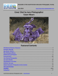
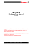
B-Bridge International, Inc.
Glutathione S-Transferase
Fluorescent Detection Kit
User Manual
Catalog # K3008-1
1
B-Bridge International, Inc.
Glutathione S-Transferase 110629
TABLE OF CONTENTS
Intended Use
3
Background
3
Assay Principle
3
Kit Components
3
Materials Required
4
Precautions
4
Reagent Preparation
4
Sample Preparation
5
Assay Protocol
5
Calculations
5
Typical Standard Curve Example
6
Notice to Purchaser
This product is to be used for Research Purposes Only. It is not to be used for Drug or
Diagnostic Purposes, nor is it intended for Human Use. B-Bridge products may not be
resold, modified for resale, or used to manufacture commercial products without the
express written consent of B-Bridge International, Inc.
EXCEPT AS OTHERWISE EXPRESSLY SET FORTH IN THIS USER MANUAL, B-BRIDGE
DOES NOT MAKE ANY REPRESENTATION OR WARRANTIES OR CONDITIONS OF ANY
KIND, EITHER EXPRESSED OR IMPLIED, WITH RESPECT TO THE PRODUCTS, OR
INFORMATION DISCLOSED HEREUNDER, INCLUDING, BUT NOT LIMITED TO, THE IMPLIED
WARRANTIES OF MERCHANTABILITY, FIT FOR A PARTICULAR PURPOSE, OR
NONINFRINGEMENT OF THE INTELLECTUAL PROPERTY RIGHTS OF THIRD PARTIES.
B-Bridge International, Inc. All Rights Reserved.
2
B-Bridge International, Inc.
Glutathione S-Transferase 110629
INTENDED USE
The B-Bridge Glutathione S-Transferase Fluorescent Detection Kit (cat.# K3008-1) quantitatively measures
Glutathione S-Transferase (GST) in human urine, serum, EDTA, heparin plasma, toadfish liver (Opsanus
tau), oyster hemolymph samples, and most cell lysates.
BACKGROUND
The Glutathione S-Transferase (GST) family of isozymes function to detoxify and neutralize a wide variety of
electrophilic molecules by mediating their conjugation with reduced glutathione. Human GSTs are encoded
by 5 gene families, expressing in almost all tissues as four cytosolic and one microsomal forms. Dividing the
family by isoelectric points, the basic alpha (pI 8–11), the neutral mu (pI 5–7) and acidic pi (pH<5) classes are
populated by additional subclasses, each isozyme displaying differential specificity for given electrophilic
molecules.
Given its pivotal role in ameliorating oxidative stress/damage, GST activity has been repeatedly investigated
as a biomarker for arthritis, asthma, COPD, and multiple forms of cancer, as well as an environmental marker.
Examination of GST isoforms and activity in human cancers, tumors and tumor cell lines has revealed the
predominance of the acidic pi class. Furthermore, this activity is thought to substantially contribute to the
innate or acquired resistance of specific neoplasms to anticancer therapy.
ASSAY PRINCIPLE
The Glutathione S-Transferase Fluorescent Activity kit is designed to quantitatively measure the
concentration of GST present in a variety of samples. Please read the complete kit insert before performing
this assay.
1. Sample or standard added to well in microtiter plate.
2. The reaction is initiated with the addition of the glutathione fluorescent detection reagent.
3. Incubate for 30 minutes at room temperature and read fluorescent signal at 460 nm with 390 nm excitation
wavelength.
4. Calculate Glutathione S-Transferase concentration from standard curve.
KIT COMPONENTS
Black Half-Area 96-well plate
1 plate
Glutathione S-Transferase Standard (10 U/mL)
50 uL
GST Detection Reagent
- Stored in a desiccator.
1 vial
Assay Buffer
45 mL
Glutathione (20mM)
300 uL
Store above components at 4°C
Dry DMSO
2 mL
- Dry Dimethyl sulfoxide solvent over molecular sieves
Store DMSO tightly capped at room temperature
3
B-Bridge International, Inc.
Glutathione S-Transferase 110629
MATERIALS REQUIRED BUT NOT SUPPLIED
-
96-well plate reader capable of reading fluorescent emission at 510 nm with excitation at 390 nm
-
Software for converting raw relative fluorescent unit (FLU) readings from the plate reader and carrying
out four parameter logistic curve (4PLC) fitting
PRECAUTIONS
This kit should only be used by qualified personnel who have had laboratory safety instruction. The complete
User Manual should be read and understood before using this product.
Dimethyl sulfoxide is a powerful aprotic organic solvent that has been shown to enhance the rate of skin
absorption of skin-permeable substances. Wear protective gloves when using the solvent especially when it
contains dissolved chemicals.
In all cases, please consult your institution’s safety procedures for working with hazardous chemicals.
REAGENT PREPARATION
Standard Preparation
GST Standards are prepared by labeling seven test tubes as #1 through #7. Briefly spin vial of standard in a
microcentrifuge to ensure contents are at bottom of vial. Pipet 380 µL of Assay Buffer into tube #1 and 200 µL
into tubes #2 to #7. Carefully add 20 µL of the Glutathione S-Transferase Standard to tube #1 and vortex
completely. Take 200 µL of the GST solution in tube #1 and add it to tube #2 and vortex completely. Repeat
these serial dilutions for tubes #3 through #7. The concentration of GST in tubes 1 through 7 will be 500, 250,
125, 62.5, 31.25, 15.61 and 7.81 mU/mL.
Use all Standards within 1 hour of preparation.
Standard Standard Standard
1
2
3
Reagent
Buffer Volume
380 µl
200 µl
200 µl
GST Standard
20 µl
Standard 1
200 µl
Standard 2
200 µl
Standard 3
Standard 4
Standard 5
Standard 6
Final
Concentration
(mU/mL)
500
250
125
Standard
4
200 µl
Standard
5
200 µl
Standard
6
200 µl
Standard
7
200 µl
200 µl
200 µl
200 µl
200 µl
62.5
31.25
15.625
7.81
Detection Reagent
Remove the vial of Detection Reagent from the desiccator and add 300 µL of the dry DMSO to the vial. Vortex
thoroughly. Store any unused reconstituted Detection Reagent at 4°C in the desiccator for no longer than 2
weeks.
Dilute one part of reconstituted Detection Reagent 1:10 into nine parts of Assay Buffer. 150 µL of Detection
Reagent should be diluted with 1.35 mL of Assay Buffer to use half the plate. Discard any excess diluted
Detection Reagent.
Glutathione
Dilute one part Glutathione stock provided 1:10 into nine parts of Assay Buffer. 150 µL of Glutathione stock
should be diluted with 1.35 mL of Assay Buffer to have enough GSH to be able to read half the plate. Discard
any excess diluted glutathione.
4
B-Bridge International, Inc.
Glutathione S-Transferase 110629
SAMPLE PREPARATION
Any samples requiring larger dilutions or with GST activities outside the standard curve range should be
diluted further with Assay Buffer to obtain readings within the standard curve.
Urine Samples
Samples that are not clear or that contain visible particulate should be centrifuged prior to using.
Urine samples should be diluted ≥ 1:2 in Assay Buffer by adding one part of urine to one part of Assay Buffer.
Sample values should be normalized for urinary volume to urinary creatinine levels. Our Urinary Creatinine
Detection Kit, K302-1. performs simple, safe and accurate determination of urinary creatinine levels.
Serum and Plasma Samples
Samples that are not clear or that contain visible particulate should be centrifuged prior to using. Fresh serum
or EDTA and heparin plasma are separated by centrifugation at 600 x g for 10 minutes. Transfer the serum or
plasma from the red blood cells into fresh tubes. The serum or plasma may be stored at -80°C or analyzed
immediately.
Serum or plasma should be diluted with assay Buffer at a dilution of ≥ 1:2.
Cell Lysates
6
Washed cell pellets are resuspended at 10-40x10 cells/mL in Assay Buffer (we used Jurkats at 10
6
x 10 cells/mL) and are lysed by vigorous vortexing, freeze/thaw cycling or other suitable disruption method.
Resulting centrifuged lysate supernatents are measured at appropriate dilutions. The protocol might require
adjustment for other cell types. If protein determinations are to be made on the samples, we would
recommend using higher number of cells and lysing in your normal PBS-based lysis buffer and determining
protein concentration, prior to additional dilutions in Assay Buffer to measure GST activity.
Use all samples within 2 hours of dilution.
ASSAY PROTOCOL- FREE AND TOTAL GSH
1. Pipet 50 µL of treated samples or standards into duplicate wells in the plate.
2. Pipet 50 µL of Assay Buffer into duplicate wells as the Zero Standard.
3. Add 25 µL of the Detection Reagent to each well using a repeater or multichannel pipet.
4. Add 25 µL of GSH to each well using a repeater or multichannel pipet.
5. Gently tap the sides of the plate to ensure adequate mixing of the reagents.
6. Incubate at room temperature for 30 minutes.
7. Read the fluorescent signal from each well in a plate reader capable of reading the fluorescent
emission at 460 nm with excitation at 370-410 nm. Please contact your plate reader manufacturer for
suitable filter sets.
8. Use the plate reader’s built-in 4PLC software capabilities to calculate GST activities for each
sample.
CALCULATIONS
Average the duplicate FLU readings for each standard and sample. Create a standard curve by
reducing the data using the 4PLC fitting routine on the plate reader, after subtracting the mean
FLUs for the zero standard. The concentrations obtained should be multiplied by the dilution factor to obtain
neat sample values.
5
B-Bridge International, Inc.
Glutathione S-Transferase 110629
TYPICAL STANDARD CURVE
6
B-Bridge International, Inc.
Glutathione S-Transferase 110629