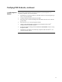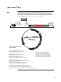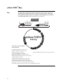
1
TOPO® Reporter Kits Five-minute Cloning of PCR Products for Analysis of Promoter Function in Mammalian Cells Catalog nos. K4830-01, K4831-01 Version K 23 February 2006 25-0235 A Limited Label License covers this product (see Purchaser Notification). By use of this product, you accept the terms and conditions of the Limited Label License. Corporate Headquarters Invitrogen Corporation 1600 Faraday Avenue Carlsbad, CA 92008 T: 1 760 603 7200 F: 1 760 602 6500 E: [email protected] For country-specific contact information visit our web site at www.invitrogen.com User Manual ii Table of Contents Table of Contents.............................................................................................................................iii Kit Contents and Storage .................................................................................................................iv Accessory Products..........................................................................................................................vi Methods ......................................................................................................................... 1 Overview...........................................................................................................................................1 General Cloning Considerations .......................................................................................................3 Cloning into pBlue-TOPO® ..............................................................................................................4 Cloning into pGlow-TOPO® .............................................................................................................5 Producing PCR Products...................................................................................................................6 TOPO® Cloning Reaction and Transformation.................................................................................7 Optimizing the TOPO® Cloning Reaction ......................................................................................11 Transfection ....................................................................................................................................12 Detection of β-Galactosidase Activity ............................................................................................13 Detection of GFP ............................................................................................................................14 Creation of Stable Cell Lines ..........................................................................................................16 Appendix...................................................................................................................... 17 TOPO® Reporter Control Reactions ...............................................................................................17 Purifying PCR Products..................................................................................................................20 Addition of 3´ A-Overhangs Post-Amplification............................................................................22 pBlue-TOPO® Map.........................................................................................................................23 pGlow-TOPO® Map........................................................................................................................24 Features of the TOPO® Reporter Vectors .......................................................................................25 Recipes............................................................................................................................................26 Technical Service............................................................................................................................27 Purchaser Notification ....................................................................................................................28 Product Qualification ......................................................................................................................30 References.......................................................................................................................................31 iii Kit Contents and Storage Shipping and Storage Each TOPO® Reporter Kit is shipped on dry ice. Each kit contains a box with TOPO® Reporter reagents (Box 1) and a box with One Shot® TOP10 chemically competent cells (Box 2). Store Box 1 at -20°C and Box 2 at -80°C. TOPO® Reporter Kits Ordering information for the TOPO® Reporter Kits is provided below. Kit Vector pBlue TOPO® TA Expression Kit pBlue-TOPO® pGlow TOPO® TA Expression Kit pGlow-TOPO® TOPO® Reporter Reagents Reactions Catalog no. 20 K4831-01 20 K4830-01 TOPO® Reporter reagents (Box 1) are listed below. Please note that the user must supply Taq polymerase. Store Box 1 at -20°C. Item ® Concentration Amount pBlue-TOPO or pGlow-TOPO® 10 ng/µl plasmid DNA in: 50% glycerol 50 mM Tris-HCl, pH 7.4 (at 25°C) 1 mM EDTA 2 mM DTT 0.1% Triton X-100 100 µg/ml BSA phenol red 20 µl 10X PCR Buffer 100 mM Tris-HCl, pH 8.3 (at 42°C) 500 mM KCl 25 mM MgCl2 0.01% gelatin 100 µl dNTP Mix 12.5 mM dATP 12.5 mM dCTP 12.5 mM dGTP 12.5 mM dTTP neutralized at pH 8.0 in water 10 µl Salt Solution 1.2 M NaCl; 0.06 M MgCl2 50 µl T7 Sequencing Primer 0.1 µg/µl in TE Buffer 20 µl LacZ Reverse or GFP Reverse Sequencing Primer 0.1 µg/µl in TE Buffer 20 µl Control PCR Template 0.05 µg/µl in TE Buffer 10 µl Control PCR Primers 0.1 µg/µl each in TE Buffer 10 µl Sterile Water -- 1 ml Continued on next page iv Kit Contents and Storage, continued One Shot® Reagents The table below describes the items included in the One Shot® TOP10 chemically competent cell kit. Store at -80°C. Item Sequencing Primers Amount SOC Medium (may be stored at +4°C or room temperature) 2% Tryptone 0.5% Yeast Extract 10 mM NaCl 2.5 mM KCl 10 mM MgCl2 10 mM MgSO4 20 mM glucose 6 ml TOP10 cells -- 21 x 50 µl pUC19 Control DNA 10 pg/µl in 5 mM Tris-HCl, 0.5 mM EDTA, pH 8 50 µl The table below provides the sequence and pmoles of the T7, LacZ Reverse, and GFP Reverse sequencing primers. Primer Genotype of TOP10 Cells Composition Sequence Amount T7 5´-TAATACGACTCACTATAGGG-3´ 328 pmoles LacZ Reverse 5´-CAGTCATGCTAGCCATACC-3´ 350 pmoles GFP Reverse 5´-GGGTAAGCTTTCCGTATGTAGC-3´ 296 pmoles TOP10: Use this strain for general cloning. Please note that this strain cannot be used for single-strand rescue of DNA. F- mcrA ∆(mrr-hsdRMS-mcrBC) Φ80lacZ∆M15 ∆lacΧ74 recA1 araD139 ∆(araleu)7697 galU galK rpsL (StrR) endA1 nupG v Accessory Products Additional Products The table below lists additional products available from Invitrogen which you may use in conjunction with the TOPO® Reporter Kit. Item One Shot® Kit (TOP10 Electrocompetent Cells) Catalog no. 10 reactions C4040-50 20 reactions C4040-52 10 reactions C4040-10 20 reactions C4040-03 40 reactions C4040-06 T7 Promoter Primer 2 µg (328 pmoles) N560-02 S.N.A.P.™ MiniPrep Kit 20 reactions K1900-01 MidiPrep Kit 20 reactions K1910-01 Anti-Xpress™ Antibody 25 Westerns R910-25 β-galactosidase Antiserum 25 Westerns R901-25 GFP Antiserum 25 Westerns R970-01 β-Gal Assay Kit 100-400 reactions K1455-01 β-Gal Staining Kit 1 kit K1465-01 Shot® One Kit (TOP10 Chemically Competent Cells) S.N.A.P.™ vi Amount Methods Overview Introduction TOPO® Reporter Kits provide a highly efficient, 5 minute, one-step cloning strategy ("TOPO® Cloning") for the direct insertion of promoter sequences amplified by Taq polymerase into a reporter vector. Recombinant vectors can then be transfected into mammalian cells or transformed into E. coli (pGlow-TOPO only) and tested for promoter function in vivo or in vitro. No ligase, post-PCR procedures, or PCR primers containing specific sequences are required. Two types of TOPO® Reporter Kits are discussed in this manual: • pBlue TOPO® TA Expression Kit containing pBlue-TOPO® for insertion of promoter sequences upstream of the E. coli β-galactosidase gene (lacZ) for in vitro or in vivo assay. • pGlow TOPO® TA Expression Kit containing pGlow-TOPO® for insertion of promoter sequences upstream of GFP for in vitro or in vivo assay of promoter function. How It Works Each reporter vector (pBlue-TOPO® or pGlow-TOPO®) is supplied linearized with: • Single 3´ thymidine (T) overhangs for TA Cloning® • Topoisomerase I covalently bound to the vector (referred to as "activated vector") Taq polymerase has a nontemplate-dependent terminal transferase activity that adds a single deoxyadenosine (A) to the 3´ ends of PCR products. The linearized vector supplied in this kit has single, overhanging 3´ deoxythymidine (T) residues. This allows PCR products to ligate efficiently into the vector. TOPO® Cloning exploits the ligation activity of topoisomerase I by providing an "activated", linearized TA vector using proprietary technology (Shuman, 1994). Ligation of the vector with a PCR product containing 3´ A-overhangs is very efficient and occurs spontaneously within 5 minutes at room temperature. The TOPO® Cloning Reaction can be transformed into chemically competent cells (provided) or electroporated directly into electrocompetent cells (available separately, see page vi). Topoisomerase Tyr-274 P CCCTT GGGA A O OH PCR Product HO Tyr-274 O A AGGG TTCCC P Topoisomerase Continued on next page 1 Overview, continued Experimental Outline The flow chart below outlines the experimental steps necessary to clone and test your promoter sequences. Determine strategy for PCR Produce PCR product TOPO Cloning Reaction: Mix together PCR product and TOPO Reporter vector Incubate 5 minutes at room temperature Transform into TOP10 E. coli cells Select and analyze colonies Prepare purified plasmid for transfection Transfect mammalian cell line and test for expression of reporter 2 General Cloning Considerations Introduction In general, promoter reporter vectors can be used to analyze-• Tissue and cell-specific promoter function • Transcriptional enhancers in a known promoter • Deletions within a promoter Please note that each TOPO® Reporter vector contains not only a TOPO® Cloning site but also additional unique restriction sites upstream of the TOPO® Cloning site. These may be exploited to analyze promoter function. When analyzing promoters in a reporter vector, it is important to realize that sequences within the native gene can influence regulation of its own promoter. In addition, sequences within the reporter gene can also affect transcription from the promoter under study. We recommend that you verify any observations of transcriptional control of the fusion gene with expression of the native gene. S1 mapping (Current Protocols in Molecular Biology, pages 4.6.1 to 4.6.13) can be used to confirm that the subcloned promoter initiates transcription at the correct site. Important PCR Primer Design Since initiation of translation in eukaryotes occurs at the first available AUG codon, it is important that there are no AUG codons between the start of transcription and the AUG of the reporter gene. Use the diagrams on pages 4 and 5 and the sequence of your promoter to design PCR primers. Unique restriction sites may be included in the 5´ and 3´ primers to excise the fragment or facilitate analysis once it is TOPO® Cloned. For analysis of promoter function in E. coli, please use pGlow-TOPO and read page 5 before designing your primers. Once you have decided on the sequence of your PCR primers, do not add 5´ phosphates to your primers. Phosphates will inhibit topoisomerase I and the synthesized PCR product will not ligate into the TOPO® Reporter vectors. Please note that cloning efficiencies may vary depending on the 5´ nucleotide in the primers (see page 19). 3 Cloning into pBlue-TOPO® There is a cryptic prokaryotic promoter upstream of the lacZ reporter gene. E. coli transformants may appear to be light blue when screened on plates containing X-Gal. We do not recommend using pBlue-TOPO® to evaluate promoter function in E. coli. Please use pGlow-TOPO® for these studies (next page). Please note that background expression of β-galactosidase does not occur in mammalian cells. Important TOPO® Cloning Site of pBlueTOPO® Restriction sites are labeled to indicate the actual cleavage site. The vector is supplied linearized between base pair 116 and 117. This is the TOPO® Cloning site. Please note that the full sequence of pBlue-TOPO® may be downloaded from our Web site (www.invitrogen.com) or requested from Technical Service (see page 27). A map of pBlue-TOPO® is provided on page 23. Bgl II* T7 promoter/priming site Afl II Hind III* BamH I 1 GACGGATCGG GAGATCTAAT ACGACTCACT ATAGGGAGAC CCAAGCTGGC TAGCGTTTAA ACTTAAGCTT GGTACCGAGC 81 TCGGATCCAC TAGTCCAGTG TGGTGGAATT GCCCTT ... CGGGA A 149 GGT TCT CAT CAT CAT CAT CAT CAT GGT ATG GCT AGC ATG ACT GGT GGA CAG CAA ATG GGT CGG GAT Gly Ser His His His His His His Gly Met Ala Ser Met Thr Gly Gly Gln Gln Met Gly Arg Asp 215 CTG TAC GAC GAT GAC GAT AAG GTA CCT AAG GAT CAG CTT GGA GTT GAT CCC GTC GTT TTA CAA CGT Leu Tyr Asp Asp Asp Asp Lys Val Pro Lys Asp Gln Leu Gly Val Asp Pro Val Val Leu Gln Arg 281 CGT GAC TGG GAA AAC CCT ... Arg Asp Trp Glu Asn Pro ... Spe I Pst I* Hind III* Nco I* Polyhistidine region Xpress epitope PCR Product Start of lacZ fusion A AGG GCAATTCTGC AGAAAGCTTA CC ATG GGG TTCC ... Met Gly LacZ Reverse priming site lacZ ORF EK Recognition site *These sites are not unique, but they may be used to excise the promoter sequence after TOPO® Cloning. 4 Cloning into pGlow-TOPO® Using pGlowTOPO in E. coli To use pGlow-TOPO as a reporter in E. coli, you must engineer your PCR product to ensure expression of GFP in the event that the sequences you are testing contain a promoter. Please note that there is no prokaryotic ribosomal binding site upstream of the GFP initiation codon. Your reverse PCR primer must include a ribosomal binding site (-AGGA-) and an initiation codon (ATG) in frame with the GFP initiation codon. Allow 8-12 nucleotides between the ribosomal binding site and the initiation codon to ensure proper spacing. There should not be any palindromic sequences within this region. Successful expression of GFP will result in additional amino acids at the N-terminus. This has been shown not to affect fluorescence. We recommend that you use a known promoter as a positive control and DNA sequences that do not contain a promoter as a negative control. TOPO® Cloning Site of pGlowTOPO® Restriction sites are labeled to indicate the actual cleavage site. The vector is supplied linearized between base pair 116 and 117. This is the TOPO® Cloning site. Please note that the full sequence of pGlow-TOPO® may be downloaded from our Web site (www.invitrogen.com) or requested from Technical Service (see page Error! Bookmark not defined.). A map of pGlow-TOPO® is provided on page 24. Bgl II* T7 promoter/priming site Pme I* Afl II Asp718 I Kpn I 1 GACGGATCGG GAGATCTAAT ACGACTCACT ATAGGGAGAC CCAAGCTGGC TAGCGTTTAA ACTTAAGCTT GGTACCGAGC 81 TCGGATCCAC TAGTCCAGTG TGGTGGAATT GCCCTT ... CGGGA A Spe I BstX I* Bgl II* Pst I* Xba I* PCR Product GFP ORF A AGG GCAATTCTGC AGATCTAGA ATG GCT AGC TTCC ... Met Ala Ser 149 AAA GGA GAA GAA CTT TTC ACT GGA GTT GTC CCA ATT CTT GTT GAA TTA GAT GGT GAT GTT AAT GGG Lys Gly Glu Glu Leu Phe Thr Gly Val Val Pro Ile Leu Val Glu Leu Asp Gly Asp Val Asn Gly GFP Reverse priming site 215 CAC AAA TTT TCT GTC AGT GGA GAG GGT GAA GGT GAT GCT ACA TAC GGA AAG CTT ACC CTT AAA TTT His Lys Phe Ser Val Ser Gly Glu Gly Glu Gly Asp Ala Thr Tyr Gly Lys Leu Thr Leu Lys Phe 281 ATT TGC ACT ACT GGA AAA ... Ile Cys Thr Thr Gly Lys ... *These sites are not unique, but they may be used to excise the promoter sequence after TOPO® Cloning. 5 Producing PCR Products Introduction Once you have decided on a PCR strategy and have synthesized the primers you are ready to produce your PCR product. Materials Supplied You will need the following reagents and equipment. by the User • Taq polymerase • • Polymerase Mixtures Thermocycler DNA template and primers for PCR product If you wish to use a mixture containing Taq polymerase and a proofreading polymerase, Taq must be used in excess of a 10:1 ratio to ensure the presence of 3´ A-overhangs on the PCR product. If you use polymerase mixtures that do not have enough Taq polymerase or a proofreading polymerase only, you can add 3′ A-overhangs using the method on page 22. Producing PCR Products 1. Set up the following 50 µl PCR reaction. Use less DNA if you are using plasmid DNA as a template and more DNA if you are using genomic DNA as a template. Use the cycling parameters suitable for your primers and template. Be sure to include a 7 to 30 minute extension at 72°C after the last cycle to ensure that all PCR products are full length and 3´ adenylated. DNA Template 10X PCR Buffer 50 mM dNTPs Primers Sterile water Taq Polymerase (1 unit/µl) Total Volume 2. 10-100 ng 5 µl 0.5 µl 100-200 ng each add to a final volume of 49 µl 1 µl 50 µl Check the PCR product by agarose gel electrophoresis. You should see a single, discrete band. If you do not see a single band, please refer to the Note below. If you do not obtain a single, discrete band from your PCR, you may gel-purify your fragment before using the TOPO® Reporter Kits (see page 20). Take special care to avoid sources of nuclease contamination and long exposure to UV light. Alternatively, you may optimize your PCR to eliminate multiple bands and smearing (Innis et al., 1990). The PCR Optimizer™ Kit (Catalog no. K1220-01) from Invitrogen can help you optimize your PCR. Please call Technical Service for more information (page Error! Bookmark not defined.). 6 TOPO® Cloning Reaction and Transformation Introduction TOPO® Cloning technology allows you to produce your PCR products, ligate them into either pBlue-TOPO® or pGlow-TOPO®, and transform the recombinant vector into E. coli all in one day. It is important to have everything you need set up and ready to use to ensure you obtain the best possible results. If this is the first time you have TOPO® Cloned, you may wish to perform the control reactions on pages 17-19 in parallel with your samples. Recent experiments at Invitrogen demonstrate that inclusion of salt (200 mM NaCl, 10 mM MgCl2) in the TOPO® Cloning reaction increases the number of transformants 2to 3-fold. We have also observed that in the presence of salt, incubation times of greater than 5 minutes can also increase the number of transformants. This is in contrast to earlier experiments without salt where the number of transformants decreases as the incubation time increases beyond 5 minutes. Inclusion of salt allows for longer incubation times because it prevents topoisomerase I from rebinding and potentially nicking the DNA after ligating the PCR product and dissociating from the DNA. The result is more intact molecules leading to higher transformation efficiencies. Important Because of the above results, we recommend adding salt to the TOPO® Cloning reaction. A stock salt solution is provided in the kit for this purpose. Please note that the amount of salt added to the TOPO® Cloning reaction varies depending on whether you plan to transform chemically competent cells (provided) or electrocompetent cells (see below). For this reason two different TOPO® Cloning reactions are provided to help you obtain the best possible results. Please read the following information carefully. Chemically Competent E. coli For TOPO® Cloning and transformation into chemically competent E. coli, adding sodium chloride and magnesium chloride to a final concentration of 200 mM NaCl, 10 mM MgCl2 in the TOPO® Cloning reaction increases the number of colonies over time. A Salt Solution (1.2 M NaCl; 0.06 M MgCl2) is provided to adjust the TOPO® Cloning reaction to the recommended concentration of NaCl and MgCl2. Electrocompetent E. coli For TOPO® Cloning and transformation of electrocompetent E. coli, salt must also be included in the TOPO® Cloning reaction, but the amount of salt must be reduced to 50 mM NaCl, 2.5 mM MgCl2 to prevent arcing. The Salt Solution is diluted 4-fold to prepare a 300 mM NaCl, 15 mM MgCl2 solution for convenient addition to the TOPO® Cloning reaction (see next page). Materials Supplied In addition to general microbiological supplies (i.e. plates, spreaders), you will need the following reagents and equipment. by the User • • • • 42°C water bath (or electroporator with cuvettes, optional) LB plates containing 50-100 µg/ml ampicillin (two for each transformation) Reagents and equipment for agarose gel electrophoresis 37°C shaking and non-shaking incubator Continued on next page 7 TOPO® Cloning Reaction and Transformation, continued There is no blue-white screening for the presence of inserts. Individual recombinant plasmids need to be analyzed by restriction analysis or sequencing for the presence and orientation of insert. Sequencing primers included in the kit can be used to sequence across an insert in the multiple cloning site to confirm orientation and reading frame. Preparation for Transformation For each transformation, you will need one vial of competent cells and two selective plates. • • • • • Setting Up the TOPO® Cloning Reaction Equilibrate a water bath to 42°C (for chemical transformation) or set up your electroporator if you are using electrocompetent E. coli. For electroporation, dilute a small portion of the Salt Solution 4-fold to prepare Dilute Salt Solution (e.g. add 5 µl of the Salt Solution to 15 µl sterile water) Warm the vial of SOC medium from Box 2 to room temperature. Warm selective plates at 37°C for 30 minutes. Thaw on ice 1 vial of One Shot® cells for each transformation. The table below describes how to set up your TOPO® Cloning reaction (6 µl) for eventual transformation into either chemically competent TOP10 One Shot® E. coli (provided) or electrocompetent E. coli. Additional information on optimizing the TOPO® Cloning reaction for your needs can be found on page 11. Note: The red or yellow color of the TOPO® vector solution is normal and is used to visualize the solution. Reagent* Chemically Competent E. coli Electrocompetent E. coli Fresh PCR product 0.5 to 4 µl 0.5 to 4 µl Salt Solution 1 µl -- Dilute Salt Solution -- 1 µl Sterile Water add to a final volume of 5 µl Add to a final volume of 5 µl TOPO® vector 1 µl 1 µl *Store all reagents at -20°C when finished. Salt solutions and water can be stored at room temperature or +4°C. Performing the TOPO® Cloning Reaction 1. Mix reaction gently and incubate for 5 minutes at room temperature (22-23°C). Note: For most applications, 5 minutes will yield plenty of colonies for analysis. Depending on your needs, the length of the TOPO® Cloning reaction can be varied from 30 seconds to 30 minutes. For routine subcloning of PCR products, 30 seconds may be sufficient. For large PCR products (> 1 kb) or if you are TOPO® Cloning a pool of PCR products, increasing the reaction time will yield more colonies. 2. Place the reaction on ice and proceed to One Shot® Chemical Transformation (next page) or Transformation by Electroporation (next page). Note: You may store the TOPO® Cloning reaction at -20°C overnight. Continued on next page 8 TOPO® Cloning Reaction and Transformation, continued One Shot® Chemical Transformation 1. Add 2 µl of the TOPO® Cloning reaction from Step 2 previous page into a vial of One Shot® TOP10 Chemically Competent E. coli and mix gently. Do not mix by pipetting up and down. 2. Incubate on ice for 5 to 30 minutes. Note: Longer incubations on ice seem to have a minimal effect on transformation efficiency. The length of the incubation is at the user’s discretion (see page 11). Transformation by Electroporation 3. Heat-shock the cells for 30 seconds at 42°C without shaking. 4. Immediately transfer the tubes to ice. 5. Add 250 µl of room temperature SOC medium. 6. Cap the tube tightly and shake the tube horizontally (200 rpm) at 37°C for 1 hour. 7. Spread 25-200 µl from each transformation on a pre-warmed selective plate and incubate overnight at 37°C. We recommend that you plate two different volumes to ensure that at least one plate will have well-spaced colonies. 8. An efficient TOPO® Cloning reaction will produce hundreds of colonies. Pick ~10 colonies for analysis (see Analysis of Positive Clones, next page). 1. Add 2 µl of the TOPO® Cloning reaction into a 0.1 cm cuvette containing 50 µl of electrocompetent E. coli and mix gently. Do not mix by pipetting up and down. Avoid formation of bubbles. 2. Electroporate your samples using your own protocol and your electroporator. Note: If you have problems with arcing, see below. 3. Immediately add 250 µl of room temperature SOC medium. 4. Transfer the solution to a 15 ml snap-cap tube (i.e. Falcon) and shake for at least 1 hour at 37°C to allow expression of the antibiotic resistance gene. 5. Spread 10-50 µl from each transformation on a pre-warmed selective plate and incubate overnight at 37°C. To ensure even spreading of small volumes, add 20 µl of SOC. We recommend that you plate two different volumes to ensure that at least one plate will have well-spaced colonies. 6. An efficient TOPO® Cloning reaction will produce hundreds of colonies. Pick ~10 colonies for analysis (see Analysis of Positive Clones, next page). Addition of the Dilute Salt Solution in the TOPO® Cloning Reaction brings the final concentration of NaCl and MgCl2 in the TOPO® Cloning reaction to 50 mM and 2.5 mM, respectively. To prevent arcing of your samples during electroporation, the volume of cells should be between 50 and 80 µl (0.1 cm cuvettes) or 100 to 200 µl (0.2 cm cuvettes). If you experience arcing during transformation, try one of the following suggestions: • • • Reduce the voltage normally used to charge your electroporator by 10% Reduce the pulse length by reducing the load resistance to 100 ohms Ethanol-precipitate the TOPO® Cloning reaction and resuspend in water prior to electroporation Continued on next page 9 TOPO® Cloning Reaction and Transformation, continued Analysis of Positive Clones 1. 2. 3. Pick 10 colonies and culture them overnight in LB medium containing 50-100 µg/ml ampicillin (3-5 ml). Note: In cells transformed with pGlow-TOPO, a hand-held UV light can be used to detect fluorescence. Isolate plasmid DNA using your method of choice. If you need ultra-pure plasmid DNA for automated or manual sequencing, we recommend the S.N.A.P.™ MiniPrep Kit (Catalog no. K1900-01). Please note that PCR products will clone bidirectionally. Analyze the plasmids for insertion and orientation by restriction analysis or by sequencing. Use the sequencing primers included in the kit to help you sequence your insert. Please refer to the diagrams on page 4 and page 5 for restriction sites and sequence surrounding the TOPO Cloning® site. For the complete sequence of either vector, please see our Web site (www.invitrogen.com) or contact Technical Service (page 27). If you need help with setting up restriction enzyme digests or DNA sequencing, refer to general molecular biology texts (Ausubel et al., 1994; Sambrook et al., 1989). Alternative Method You may wish to use PCR to directly analyze positive transformants. Use a combination of either the T7 or LacZ Reverse (or GFP Reverse) sequencing primer and a primer that binds of Analysis within your insert as PCR primers. You will have to determine the amplification conditions. If this is the first time you have used this technique, we recommend that you perform restriction analysis in parallel to confirm that PCR gives you the correct result. Artifacts may be obtained because of mispriming or contaminating template. The following protocol is provided for your convenience. Other protocols are suitable. 1. Prepare a PCR cocktail consisting of PCR buffer, dNTPs, primers, and Taq polymerase. Use a 20 µl reaction volume. Multiply by the number of colonies to be analyzed (e.g. 10). 2. Pick 10 colonies and resuspend them individually in 20 µl of the PCR cocktail. (Don't forget to make a patch plate to preserve the colonies for further analysis.) 3. Incubate the reaction for 10 minutes at 94°C to lyse the cells and inactivate nucleases. 4. Amplify for 20 to 30 cycles using parameters previously determined (see text, above). 5. For the final extension, incubate at 72°C for 10 minutes. Store at +4°C. 6. Visualize by agarose gel electrophoresis. Important Long-Term Storage 10 If you have problems obtaining transformants or the correct insert, perform the control reactions described on pages 17-19. These reactions will help you troubleshoot your experiment. Once you have identified the correct clone, be sure to isolate a single colony and prepare a glycerol stock for long term storage. We recommend that you store a stock of plasmid DNA at -20°C. 1. Streak the original colony on LB plates containing 50-100 µg/ml ampicillin. 2. Isolate a single colony and inoculate into 1-2 ml of LB containing 50-100 µg/ml ampicillin. Grow until culture reaches stationary phase. 3. Mix 0.85 ml of culture with 0.15 ml of sterile glycerol and transfer to a cryovial. 4. Store at -80°C. Optimizing the TOPO® Cloning Reaction Introduction The information below will help you optimize the TOPO® Cloning reaction for your particular needs. Faster Subcloning The high efficiency of TOPO® Cloning technology allows you to streamline the cloning process. If you routinely clone PCR products and wish to speed up the process, consider the following: • Incubate the TOPO® Cloning reaction for only 30 seconds instead of 5 minutes. • You may not obtain the highest number of colonies, but with the high efficiency of TOPO® Cloning, most of the transformants will contain your insert. After adding 2 µl of the TOPO® Cloning reaction to chemically competent cells, incubate on ice for only 5 minutes. Increasing the incubation time to 30 minutes does not significantly improve transformation efficiency. More Transformants If you are TOPO® Cloning large PCR products, toxic genes, or cloning a pool of PCR products, you may need more transformants to obtain the clones you want. To increase the number of colonies: • Incubate the salt-supplemented TOPO® Cloning reaction for 20 to 30 minutes instead of 5 minutes. Increasing the incubation time of the salt-supplemented TOPO® Cloning reaction allows more molecules to ligate, increasing the transformation efficiency. Addition of salt appears to prevent topoisomerase from rebinding and nicking the DNA after it has ligated the PCR product and dissociated from the DNA. Cloning Dilute PCR Products To clone dilute PCR products, you may: • • • Increase the amount of the PCR product Incubate the TOPO® Cloning reaction for 20 to 30 minutes Concentrate the PCR product 11 Transfection MEND ION AT RECOM Introduction Once you obtain the desired construct, you are ready to transfect the plasmid into the mammalian cells of choice. Please note the following guidelines for transfection. We recommend that you include a positive and a negative control to evaluate expression of the reporter genes. A negative control can be either a mock transfection, or TOPO® Clone a PCR product that does not contain a promoter (stuffer DNA) into the desired TOPO® Reporter vector. For a positive control, we recommend cloning a known promoter that is active in your cell line. Plasmid Preparation Plasmid DNA for transfection into eukaryotic cells must be very clean and free from phenol and sodium chloride. Contaminants will kill the cells and salt will interfere with lipid complexing decreasing transfection efficiency. We recommend isolating plasmid DNA (up to 200 µg) using the S.N.A.P.™ MidiPrep Kit (Catalog no. K1910-01) or CsCl gradient centrifugation. Methods of Transfection For established cell lines (e.g. HeLa), please consult original references or the supplier of your cell line for the optimal method of transfection. It is recommended that you follow exactly the protocol for your cell line. Pay particular attention to medium requirements, when to pass the cells, and at what dilution to split the cells. Further information is provided in Current Protocols in Molecular Biology (Reference section, page 31). Methods for transfection include calcium phosphate (Chen and Okayama, 1987; Wigler et al., 1977), lipid-mediated (Felgner et al., 1989; Felgner and Ringold, 1989) and electroporation (Chu et al., 1987; Shigekawa and Dower, 1988). Invitrogen offers the Calcium Phosphate Transfection Kit (Catalog no. K2780-01) and a large selection of reagents for transfection. For more information on the reagents available, please visit our Web site (www.invitrogen.com) or call Technical Service (see page 27). Detection of Reporter 12 Once you have transfected your cell line with one of the TOPO® Reporter vectors and the appropriate controls, you are ready to assay for reporter function. See the next page for information on how to assay for β-galactosidase activity and page 14 for GFP assays. Detection of β-Galactosidase Activity Introduction β-galactosidase is one of the most versatile reporters available. It can be assayed both in vitro and in vivo and a wide variety of substrates are available for detection. A few assays and substrates are described below. Other assays and substrates may be used. In addition to its use as a reporter for uncharacterized promoters, constitutive promoters may be cloned upstream of the lacZ gene for use as an internal control to normalize variability with other promoter reporter assays (Alam and Cook, 1990). Choosing an In Vitro Assay β-galactosidase activity can be detected using cell-free lysates and o-nitrophenyl-β-Dgalactopyranoside (ONPG). This colorimetric assay is easy to perform and useful for determining whether or not a promoter is active, but it lacks the sensitivity needed for promoter analysis (detects about 100 pg of β-galactosidase). Invitrogen offers a β-Gal Assay Kit (Catalog no. K1455-01) which contains ONPG and all the buffers necessary to assay for β-galactosidase activity. Instructions are also included for a 96-well format (see page vi for ordering information). If you need greater sensitivity for promoter analysis, we recommend using chemiluminescent 1,2-dioxetane substrates (i.e. Galacton, Tropix) (Beale et al., 1992; Jain and Magrath, 1991). Use of these substrates increases the sensitivity of the assay and extends the range of detection (Bronstein et al., 1994). If endogenous enzyme activity is minimized, sensitivity is enhanced (Young et al., 1993). As little as 2 fg of β-galactosidase can be detected using chemiluminescent substrates. For more information on this assay, please see the references cited above and Current Protocols in Molecular Biology, pages 9.7.15 to 9.7.21. Choosing an In Vivo Assay In vivo detection systems are defined as those in which the reporter gene is detected in live cells or tissues or in cells or tissues fixed for histochemical staining. This is a less quantitative approach but provides important information about cell-type specificity, temporal and tissue expression patterns, and distribution of transcription factors. The precipitating substrate X-Gal may be used to determine in vivo levels of βgalactosidase in eukaryotic cells, tissue sections, and intact embryos (Alam and Cook, 1990). Please note that staining with X-Gal requires that the cells or tissue be fixed. Invitrogen offers the β-Gal Staining Kit (Catalog no. K1465-01) to stain cells expressing β-galactosidase (see page vi for ordering information). Alternatively, detection in live cultured cells may be achieved with the substrate fluorescein di-β-D-galactopyranoside (FDG) (Jongkind et al., 1986). Using hypotonic loading, FDG is introduced into the cell and cleaved by β-galactosidase. The resulting fluorescein compound is trapped in the cell because of its hydrophobic nature and easily assayed using fluorescence. Detection of β-galactosidase If you do not detect activity of β-galactosidase, check for expression by Western blot. You may use antibody to β-galactosidase (see page vi for ordering information) , or, since β-galactosidase is expressed as a fusion to an N-terminal peptide containing the Xpress™ epitope, use the Anti-Xpress™ Antibody (see page vi for ordering information). 13 Detection of GFP Introduction Green fluorescent protein (GFP) is very useful for in vivo or in vitro assay of promoter function. In vivo assays, while less quantitative than in vitro assays, provide information regarding cell-type specificity of promoters/enhancers and the tissue distribution of specific transcription factors. Use of pGlow-TOPO® allows you to monitor transcriptional changes in real time. Please note that detection of GFP in vivo will depend on the strength of the promoter. For low-level expression it may be necessary to prepare cell lysates and assay in a fluorimeter. For detection in E. coli, assay cell lysates. GFP Gene Used in pGlow-TOPO® The GFP gene used in pGlow-TOPO® is described in Crameri et al., 1996. The codon usage was optimized for expression in E. coli and three cycles of DNA shuffling were used to generate a collection of mutants. The GFP mutant that exhibited the greatest fluorescence in mammalian cells is utilized in pGlow-TOPO®. This mutant form of GFP has the following characteristics: • Excitation and emission maxima that are the same as wild-type GFP (395 nm and 478 nm for primary and secondary excitation, respectively, and 507 nm for emission) • High solubility in E. coli for visual detection of transformed cells (if expressed from a promoter recognized by E. coli) • >40-fold increase in fluorescent yield over wild-type GFP This GFP protein will be subsequently referred to as Cycle 3 GFP to differentiate it from wild-type GFP. To detect fluorescent cells, it is important to pick the best filter set to optimize detection. The primary excitation peak of Cycle 3 GFP is at 395 nm. There is a secondary excitation peak at 478 nm. Excitation at either of these wavelengths yields a fluorescent emission peak with a maximum at 507 nm, as shown in the figure below. Please note that the quantum yield can vary as much as 5- to 10-fold depending on the wavelength of light that is used to excite the GFP fluorophore. 507 600 500 400 300 395 200 478 630 610 590 570 550 530 510 490 470 450 430 410 390 370 0 350 100 330 For general information about GFP fluorescence and detection, refer to Current Protocols in Molecular Biology, pages 9.7.22 to 9.7.28 (Ausubel et al., 1994). Excitation and Emission Spectra for GFP 700 310 Use of the best filter set will ensure that the optimal regions of the Cycle 3 GFP spectra are excited and passed (emitted). For best results, use a filter set designed to detect fluorescence from wild-type GFP (e.g. XF76 filter from Omega Optical, www. omegafilters.com). FITC filter sets can also be used to detect Cycle 3 GFP fluorescence. For example, the FITC filter set that we use excites Cycle 3 GFP with light from 460 to 490 nm, which covers the secondary excitation peak. The filter set passes light from 515 to 550, allowing detection of most of the Cycle 3 GFP fluorescence. Relative Fluorescence In Vivo Detection of Cycle 3 GFP Fluorescence Wavelength (nm) Continued on next page 14 Detection of GFP, continued Detection of Transformed E. coli After transformation of E. coli, screen colonies using a hand-held UV light and select glowing cells. To quantitatively assay fluorescence, prepare cell lysates (108 to 109 cells/ml) from mid-log phase cells using your method of choice. Pellet cell debris and assay supernatant for fluorescence. Be sure to include positive and negative controls. Detection of Transfected Cells After transfection, allow the cells to recover and monitor the cells by fluorescence for expression of Cycle 3 GFP. Please note that the CMV promoter is a strong promoter and usually allows detection of Cycle 3 GFP by 24 hours posttransfection. If your promoter is not as strong as CMV, it will take longer to observe fluorescence. Most media fluoresce because of the presence of riboflavin (Zylka and Schnapp, 1996) and may interfere with detection of Cycle 3 GFP fluorescence. Medium can be removed and replaced with PBS during the assay to alleviate this problem. If cells will be cultured further after assaying, do not keep cells in PBS for a prolonged time. Remove PBS and replace with fresh medium prior to re-incubation. In Vitro Detection of Cycle 3 GFP Detection of Cycle 3 GFP by Western If promoter activity is too low to be detected in vivo, you may prepare mammalian cell lysates and assay fluorescence in a fluorimeter if available. A sample protocol is provided below to prepare lysates. 1. Wash cell monolayers (~106 cells) two times with PBS. 2. Scrape cells into 1 ml PBS and pellet the cells at 1500 x g for 5 minutes. 3. Resuspend in 100 µl Cell Lysis Buffer (see recipe on page 26). 4. Incubate cell suspension on ice or at room temperature for 5 to 10 minutes to lyse the cells. 5. Centrifuge the cell lysate at 10,000 x g for 10 minutes to pellet nuclei and transfer the supernatant to a fresh tube. Assay the lysate for protein concentration. Note: Do not use protein assays utilizing Coomassie Blue or other dyes. NP-40 interferes with the binding of the dye with the protein. 6. Assay 20 to 100 µg protein in 0.5-1 ml of PBS. Excite at 395 nm and detect at 510 nm. If you do not detect fluorescence activity, check for expression of Cycle 3 GFP by Western blot. Antiserum to Cycle 3 GFP is available from Invitrogen as a rabbit polyclonal antibody (see page vi for ordering information). 15 Creation of Stable Cell Lines Introduction If you wish to create stable cell lines, select for foci using Geneticin® Selective Antibiotic. General information and guidelines are provided below for your convenience. Geneticin® Selective Antibiotic Geneticin® Selective Antibiotic blocks protein synthesis in mammalian cells by interfering with ribosomal function. It is an aminoglycoside, similar in structure to neomycin, gentamycin, and kanamycin. Expression in mammalian cells of the bacterial aminoglycoside phosphotransferase gene (APH), derived from Tn5, results in detoxification of Geneticin® Selective Antibiotic (Southern and Berg, 1982). Geneticin® Selection Guidelines Geneticin® Selective Antibiotic is available from Invitrogen (Catalog no. 11811-031). Use as follows: 1. Prepare Geneticin® Selective Antibiotic in a buffered solution (e.g. 100 mM HEPES, pH 7.3). 2. Use 100 to 1000 µg/ml of Geneticin® Selective Antibiotic in complete medium. 3. Calculate concentration based on the amount of active drug. 4. Test varying concentrations of Geneticin® Selective Antibiotic on your cell line to determine the concentration that kills your cells (kill curve). Cells differ in their susceptibility to Geneticin® Selective Antibiotic. Note: Cells will divide once or twice in the presence of lethal doses of Geneticin® Selective Antibiotic, so the effects of the drug take several days to become apparent. Complete selection can take from 2 to 4 weeks of growth in selective medium. To obtain stable transfectants, you may choose to linearize your vector before transfection. Possible Linearization Sites While linearizing your vector may not improve your chances of obtaining stable transfectants, it ensures that the vector does not integrate in a way that disrupts the gene of interest. The table below lists some unique sites that can be used to linearize your construct prior to transformation. Other sites are possible. Be sure that your insert does not contain the restriction enzyme site you wish to use to linearize your vector. Enzyme Aat II Afl II Site in pBlue-TOPO® Site in pGlow-TOPO® -- 5333 63 63 Location Backbone Supplier Many ® Upstream of TOPO Cloning site Many AlwN I -- 3934 pUC origin BamH I 84 -- Upstream of TOPO® Cloning site Invitrogen, Cat. no. 15201-023 Bgl II 13 -- Upstream of TOPO® Cloning site Invitrogen, Cat. no. 15213-010 4411 Ampicillin gene Eam1105 I 6871 Many AGS*, Fermentas, Takara ® Hind III 66 -- Upstream of TOPO Cloning site Invitrogen, Cat. no. 15207-012 Kpn I -- 76 Upstream of TOPO® Cloning site Invitrogen, Cat. no. 15232-010 Pvu I -- 4781 Ampicillin gene Invitrogen, Cat. no. 25420-019 Sap I 5862 3402 Backbone New England Biolabs Sca I 7351 4891 Ampicillin gene Spe I 16 90 90 Invitrogen, Cat. no. 15436-017 ® Upstream of TOPO Cloning site Invitrogen, Cat. no. 15443-013 Appendix TOPO® Reporter Control Reactions Introduction We recommend performing the following control TOPO® Cloning reactions the first time you use the kit to help you evaluate your results. Performing the control reactions using the reagents included in the kit involves producing a control PCR product containing the lac promoter and the LacZα protein. Successful TOPO® Cloning of the control PCR product will yield blue colonies on LB agar plates containing ampicillin and X-gal. Before Starting Be sure to prepare the following reagents before performing the control reaction: • 40 mg/ml X-gal in dimethylformamide (see page 26 for recipe) • LB plates containing 100 µg/ml ampicillin and X-gal (two per transformation) To add X-gal to previously made agar plates, warm the plate to 37°C. Pipette 40 µl of the 40 mg/ml stock solution onto the plate, spread evenly, and let dry 15 minutes. Protect plates from light. Producing the Control PCR Product 1. To produce the 500 bp control PCR product containing the lac promoter and LacZα, set up the following 50 µl PCR: Control DNA Template (50 ng) 1 µl 10X PCR Buffer 5 µl 50 mM dNTPs 0.5 µl Control PCR Primers (0.1 µg/µl each) Sterile Water Taq Polymerase (1 unit/µl) 1 µl Total Volume 50 µl 2. Overlay with 70 µl (1 drop) of mineral oil. 3. Amplify using the following cycling parameters: Step 4. 2 µl 40.5 µl Time Temperature Initial Denaturation 2 minutes 94°C Denaturation 1 minute 94°C Annealing 1 minute 60°C Extension 1 minute 72°C Final Extension 7 minutes 72°C Cycles 1X 25X 1X Remove 10 µl from the reaction and analyze by agarose gel electrophoresis. A discrete 500 bp band should be visible. Proceed to the Control TOPO® Cloning Reactions, next page. Continued on next page 17 TOPO® Reporter Control Reactions, continued Control TOPO® Cloning Reactions Using the control PCR product produced on the previous page and either the pBlueTOPO® or the pGlow-TOPO® vectors set up two 6 µl TOPO® Cloning reactions as described below. 1. Set up control TOPO® Cloning reactions: Reagent "Vector Only" "Vector + PCR Insert" Sterile Water 4 µl 3 µl Salt Solution or Dilute Salt Solution 1 µl 1 µl Control PCR Product -- 1 µl 1 µl 1 µl ® TOPO vector Analysis of Results 2. Incubate at room temperature for 5 minutes and place on ice. 3. Transform 2 µl of each reaction into separate vials of TOP10 One Shot® cells (page 9). 4. Spread 10-50 µl of each transformation mix onto LB plates containing 50-100 µg/ml ampicillin and X-Gal (see page 26). Be sure to plate two different volumes to ensure that at least one plate has well-spaced colonies. For plating small volumes, add 20 µl of SOC to allow even spreading. 5. Incubate overnight at 37°C. Hundreds of colonies from the vector + PCR insert reaction should be produced. Greater than 85% of these will be blue and contain the 500 bp insert. Very few colonies (<10% of the vector + PCR insert) will be present on the vector only plate. These colonies should be white. Note: pBlue-TOPO® will yield dark blue colonies on the "vector + PCR insert" plate and light blue colonies on the "vector only" plate. This is apparently because of a cryptic prokaryotic promoter upstream of the reporter. Please note that no expression of β-galactosidase has been detected in mammalian cells without a promoter. Transformation Control pUC19 plasmid is included to check the transformation efficiency of the One Shot® competent E. coli. Transform one vial of One Shot® TOP10 cells with 10 pg of pUC19 using the protocol on page 9 Plate 10 µl of the transformation mixture plus 20 µl SOC on LB plates containing 100 µg/ml ampicillin. Transformation efficiency should be ~1 x 10⁹ cfu/µg DNA. Continued on next page 18 TOPO® Reporter Control Reactions, continued Factors Affecting Cloning Efficiency Please note that lower transformation and/or cloning efficiencies will result from the following variables. Most of these are easily corrected, but if you are cloning large inserts, you may not obtain the expected 85% (or more) cloning efficiency. Variable Solution pH>9 Check the pH of the PCR amplification reaction and adjust with 1 M Tris-HCl, pH 8. Incomplete extension during PCR Be sure to include a final extension step of 7 to 30 minutes during PCR. Longer PCR products will need a longer extension time. Cloning large inserts (>3 kb) Gel-purify as described on page 20. Excess (or overly dilute) PCR product Reduce (or concentrate) the amount of PCR product. Please note that you can add up to 4 µl of the PCR to the TOPO® Cloning reaction. Cloning blunt-ended fragments Add 3´ A-overhangs by incubating with Taq polymerase (page 22). PCR cloning artifacts ("false positives") TOPO® Cloning is very efficient for small fragments (< 100 bp) present in certain PCR reactions. Gel-purify your PCR product (page 20). PCR product does not contain sufficient 3´ A-overhangs even though you used Taq polymerase Increase the time of the final extension to ensure that the 3´ ends are adenylated. Size of promoter sequences cloned For large plasmids, you may have to use electroporation to transform into E. coli. Do not use the chemically competent TOP10 cells included in the kit for electroporation. Use electrocompetent TOP10 cells (see page vi for ordering information). Please note that Taq polymerase is less efficient at adding a nontemplate 3´ A next to another A. Taq is most efficient at adding a nontemplate 3´ A next to a C. You may have to redesign your primers so that they contain a 5´ G instead of a 5´ T (Brownstein et al., 1996). 19 Purifying PCR Products Introduction Smearing, multiple banding, primer-dimer artifacts, or large PCR products (>1 kb) may necessitate gel purification. If you intend to purify your PCR product, be extremely careful to remove all sources of nuclease contamination. There are many protocols to isolate DNA fragments or remove oligonucleotides. Please refer to Current Protocols in Molecular Biology, Unit 2.6 (Ausubel et al., 1994) for the most common protocols. Three simple protocols are provided below for your convenience. Please note that cloning efficiency may decrease with purification of the PCR product. You may wish to optimize your PCR to produce a single band (see Producing PCR Products, page 6). Using the S.N.A.P.™ Gel Purification Kit The S.N.A.P.™ Gel Purification Kit (Catalog no. K1999-25) allows you to rapidly purify PCR products from regular agarose gels. 1. Electrophorese amplification reaction on a 1 to 5% regular TAE agarose gel. Note: Do not use TBE. Borate will interfere with the NaI step (Step 2.) 2. Quick S.N.A.P.™ Method Cut out the gel slice containing the PCR product and melt it at 65°C in 2 volumes of 6 M NaI. 3. Add 1.5 volumes of Binding Buffer. 4. Load solution (no more than 1 ml at a time) from Step 3 onto a S.N.A.P.™ column. Centrifuge 1 minute at 3000 x g in a microcentrifuge and discard the supernatant. 5. If you have solution remaining from Step 3, repeat Step 4. 6. Add 900 µl of the Final Wash Buffer. 7. Centrifuge 1 minute at full speed in a microcentrifuge and discard the flow-through. 8. Repeat Step 7. 9. Elute the purified PCR product in 40 µl of TE or sterile water. Use 4 µl for the TOPO® Cloning reaction and proceed as described on page 8. An even easier method is to simply cut out the gel slice containing your PCR product, place it on top of the S.N.A.P.™ column bed, and centrifuge at full speed for 10 seconds. Use 1-2 µl of the flow-through in the TOPO® Cloning reaction (page 8). Be sure to make the gel slice as small as possible for best results. Continued on next page 20 Purifying PCR Products, continued Low-Melt Agarose Method Please note that gel purification will result in a dilution of your PCR product. Use chemically competent cells for transformation. 1. Electrophorese as much as possible of your PCR reaction on a low-melt agarose gel (0.8 to 1.2%) in TAE buffer. 2. Visualize the band of interest and excise the band. 3. Place the gel slice in a microcentrifuge tube and incubate the tube at 65°C until the gel slice melts. 4. Place the tube at 37°C to keep the agarose melted. 5. Add 4 µl of the melted agarose containing your PCR product to the TOPO® Cloning reaction as described on page 8. 6. Incubate the TOPO® Cloning reaction at 37°C for 5 to 10 minutes. This is to keep the agarose melted. 7. Transform 2 to 4 µl directly into chemically competent TOP10 One Shot® E. coli using the method on page 9. 21 Addition of 3´ A-Overhangs Post-Amplification Introduction Direct cloning of DNA amplified by Vent® or Pfu polymerases into TOPO TA Cloning® vectors is often difficult because of very low cloning efficiencies. These low efficiencies are caused by the 3´ to 5´ exonuclease activity associated with proofreading polymerases which removes the 3´ A-overhangs necessary for TA Cloning®. A simple method is provided below to clone these blunt-ended fragments. Before Starting You will need the following items: • Taq polymerase • A heat block equilibrated to 72°C • Phenol-chloroform (optional) • 3 M sodium acetate (optional) • 100% ethanol (optional) • 80% ethanol (optional) • TE buffer (optional) Procedure This is just one method for adding 3´ adenines. Other protocols may be suitable. 1. After amplification with Vent® or Pfu polymerase, place vials on ice and add 0.71 unit of Taq polymerase per tube. Mix well. It is not necessary to change the buffer. 2. Incubate at 72°C for 8-10 minutes (do not cycle). 3. Place the vials on ice. The DNA amplification product is now ready for ligation into pBlue-TOPO® or pGlow-TOPO® Note: If you plan to store your sample(s) overnight before proceeding with TOPO® Cloning, you may want to extract your sample(s) with phenol-chloroform to remove the polymerases. After phenol-chloroform extraction, precipitate the DNA with ethanol and resuspend the DNA in TE buffer to the starting volume of the amplification reaction. You may also gel-purify your PCR product after amplification with Vent® or Pfu (see previous page). After purification, add Taq polymerase buffer, dATP, and 0.5 unit of Taq polymerase and incubate 10-15 minutes at 72°C. Use 4 µl in the TOPO® Cloning reaction. Vent® is a registered trademark of New England Biolabs. 22 pBlue-TOPO® Map The figure below summarizes the features of the pBlue-TOPO® vector. The vector is supplied linearized between base pairs 116 and 117. This is the TOPO® Cloning site. The complete nucleotide sequence is available for downloading from our Web site (www.invitrogen.com) or from Technical Service (see page 27.). T7 Pme I* Afl II Hind III* BamH I Spe I Bgl II A A T T ATG 6xHis Xpress epitope BGH pA f1 EK Site lacZ Not I Xho I Xba I Apa I Pme I* PCR Product Pst I* Hind III* Nco I* Map or i ri 40 o SV Neomy cin A m p i c i ll i pBlue-TOPO® 7793 bp n Comments for pBlue-TOPO® 7793 nucleotides p U C or i 40 SV pA T7 promoter/priming site: bases 17-36 TOPO® Cloning site: bases 116-117 * These sites are not unique but may ATG initiation codon: bases 143-145 be used to excise the PCR product. Polyhistidine region: bases 155-172 The Pme I sites may be used to excise LacZ Reverse priming site: bases 173-191 the reporter cassette, providing there Xpress epitope: bases 212-235 are no Pme I sites in the PCR product. Enterokinase recognition site: bases 221-235 LacZ ORF: bases 264-3313 BGH polyadenylation sequence: bases 3386-3613 f1 origin: bases 3659-4087 SV40 promoter and origin: bases 4141-4423 Neomycin resistance gene: bases 4498-5292 SV40 polyadenylation sequence: bases 5466-5596 pUC origin: bases 5979-6652 (complementary strand) Ampicillin resistance gene: bases 6797-7657 (complementary strand) 23 pGlow-TOPO® Map The figure below summarizes the features of the pGlow-TOPO® vector. The vector is supplied linearized between base pairs 116 and 117. This is the TOPO® Cloning site. The complete nucleotide sequence is available for downloading from our Web site (www.invitrogen.com) or from Technical Service (see page 27.). T7 Pme I* Afl II Asp718 I Kpn I Spe I BstX I* Bgl II* A PCR Product A Cycle 3 GFP T ATG Cycle 3 GFP BstX I* Not I Apa I Pme I* Map T BGH pA f1 or i ri 40 o SV Ne o m y cin A m p i c i ll i pGlow-TOPO® 5333 bp n Comments for pGlow-TOPO® 5333 nucleotides pU C ori 40 SV pA T7 promoter/priming site: bases 17-36 TOPO® Cloning site: bases 116-117 *pGlow-TOPO has only two Pme I sites as shown. Initiation ATG: bases 140-142 Cycle 3 GFP ORF: bases 140-859 GFP Reverse priming site: bases 251-272 BGH polyadenylation sequence: bases 926-1153 f1 origin: bases 1199-1627 SV40 promoter and origin: bases 1681-1963 Neomycin resistance gene: bases 2038-2832 SV40 polyadenylation sequence: bases 3006-3136 pUC origin: bases 3519-4192 (complementary strand) Ampicillin resistance gene: bases 4337-5197 (complementary strand) 24 Features of the TOPO® Reporter Vectors Features The TOPO® Reporter vectors pBlue-TOPO® (7793 bp) and pGlow-TOPO® (5333 bp) contain the following elements. All features have been functionally tested. Feature Benefit T7 promoter/priming site Allows for in vitro transcription in the sense orientation and sequencing through the insert TOPO® Cloning site Allows insertion of your PCR product containing the promoter of interest upstream of the reporter gene. β-galactosidase (pBlue-TOPO®) or Cycle 3 GFP (pGlow-TOPO®) Allows assay of promoter function either in vitro or in vivo. LacZ Reverse priming site (pBlue-TOPO®) or GFP Reverse priming site (pGlow-TOPO®) Permits sequencing through the insert Bovine growth hormone (BGH) polyadenylation signal Efficient transcription termination and polyadenylation of mRNA (Goodwin and Rottman, 1992) f1 origin Allows rescue of single-stranded DNA SV40 early promoter and origin Allows efficient, high-level expression of the neomycin resistance gene and episomal replication in cells expressing the SV40 large T antigen Neomycin resistance gene Selection of stable transfectants in mammalian cells (Southern and Berg, 1982) SV40 polyadenylation signal Efficient transcription termination and polyadenylation of mRNA pUC origin High-copy number replication and growth in E. coli Ampicillin resistance gene (β-lactamase) Selection of vector in E. coli 25 Recipes LB (Luria-Bertani) Medium and Plates Composition: 1.0% Tryptone 0.5% Yeast Extract 1.0% NaCl pH 7.0 1. For 1 liter, dissolve 10 g tryptone, 5 g yeast extract, and 10 g NaCl in 950 ml deionized water. 2. Adjust the pH of the solution to 7.0 with NaOH and bring the volume up to 1 liter. 3. Autoclave on liquid cycle for 20 minutes at 15 psi. Allow solution to cool to 55°C and add antibiotic (100 µg/ml ampicillin) if needed. 4. Store at room temperature or at +4°C. LB agar plates X-Gal Stock Solution Cell Lysis Buffer 1. Prepare LB medium as above, but add 15 g/L agar before autoclaving. 2. Autoclave on liquid cycle for 20 minutes at 15 psi. 3. After autoclaving, cool to ~55°C, add antibiotic (100 µg/ml of ampicillin), and pour into 10 cm plates. 4. Let harden, then invert and store at +4°C, in the dark. 1. To make a 40 mg/ml stock solution, dissolve 400 mg X-Gal in 10 ml dimethylformamide. 2. Protect from light by storing in a brown bottle at -20°C. 3. To add to previously made agar plates, warm the plate to 37°C. Pipette 40 µl of the 40 mg/ml stock solution onto the plate, spread evenly, and let dry 15 minutes. Protect plates from light. 50 mM Tris-HCl, pH 7.8 150 mM NaCl 1% Nonidet P-40 1. This solution can be prepared from the following common stock solutions. For 100 ml, combine: 1 M Tris base 5 ml 5 M NaCl 3 ml Nonidet P-40 1 ml 2. Bring the volume up to 90 ml with deionized water and adjust the pH to 7.8 with HCl. 3. Bring the volume up to 100 ml. Store at room temperature. Note: Protease inhibitors may be added at the following concentrations: 1 mM PMSF; 1 µg/ml pepstatin; 1 µg/ml leupeptin. 26 Technical Service Web Resources Contact Us Visit the Invitrogen Web site at www.invitrogen.com for: • Technical resources, including manuals, vector maps and sequences, application notes, MSDSs, FAQs, formulations, citations, handbooks, etc. • Complete technical service contact information • Access to the Invitrogen Online Catalog • Additional product information and special offers For more information or technical assistance, call, write, fax, or email. Additional international offices are listed on our Web page (www.invitrogen.com). Corporate Headquarters: Invitrogen Corporation 1600 Faraday Avenue Carlsbad, CA 92008 USA Tel: 1 760 603 7200 Tel (Toll Free): 1 800 955 6288 Fax: 1 760 602 6500 E-mail: [email protected] Japanese Headquarters: Invitrogen Japan LOOP-X Bldg. 6F 3-9-15, Kaigan Minato-ku, Tokyo 108-0022 Tel: 81 3 5730 6509 Fax: 81 3 5730 6519 E-mail: [email protected] European Headquarters: Invitrogen Ltd Inchinnan Business Park 3 Fountain Drive Paisley PA4 9RF, UK Tel: +44 (0) 141 814 6100 Tech Fax: +44 (0) 141 814 6117 E-mail: [email protected] Material Data Safety Sheets (MSDSs) MSDSs are available on our Web site at www.invitrogen.com. On the home page, click on Technical Resources and follow instructions on the page to download the MSDS for your product. Limited Warranty Invitrogen is committed to providing our customers with high-quality goods and services. Our goal is to ensure that every customer is 100% satisfied with our products and our service. If you should have any questions or concerns about an Invitrogen product or service, contact our Technical Service Representatives. Invitrogen warrants that all of its products will perform according to specifications stated on the certificate of analysis. The company will replace, free of charge, any product that does not meet those specifications. This warranty limits Invitrogen Corporation’s liability only to the cost of the product. No warranty is granted for products beyond their listed expiration date. No warranty is applicable unless all product components are stored in accordance with instructions. Invitrogen reserves the right to select the method(s) used to analyze a product unless Invitrogen agrees to a specified method in writing prior to acceptance of the order. Invitrogen makes every effort to ensure the accuracy of its publications, but realizes that the occasional typographical or other error is inevitable. Therefore Invitrogen makes no warranty of any kind regarding the contents of any publications or documentation. If you discover an error in any of our publications, please report it to our Technical Service Representatives. Invitrogen assumes no responsibility or liability for any special, incidental, indirect or consequential loss or damage whatsoever. The above limited warranty is sole and exclusive. No other warranty is made, whether expressed or implied, including any warranty of merchantability or fitness for a particular purpose. 27 Purchaser Notification Limited Use Label License No: 5 Invitrogen Technology The purchase of this product conveys to the buyer the non-transferable right to use the purchased amount of the product and components of the product in research conducted by the buyer (whether the buyer is an academic or for-profit entity). The buyer cannot sell or otherwise transfer (a) this product (b) its components or (c) materials made using this product or its components to a third party or otherwise use this product or its components or materials made using this product or its components for Commercial Purposes. The buyer may transfer information or materials made through the use of this product to a scientific collaborator, provided that such transfer is not for any Commercial Purpose, and that such collaborator agrees in writing (a) not to transfer such materials to any third party, and (b) to use such transferred materials and/or information solely for research and not for Commercial Purposes. Commercial Purposes means any activity by a party for consideration and may include, but is not limited to: (1) use of the product or its components in manufacturing; (2) use of the product or its components to provide a service, information, or data; (3) use of the product or its components for therapeutic, diagnostic or prophylactic purposes; or (4) resale of the product or its components, whether or not such product or its components are resold for use in research. Invitrogen Corporation will not assert a claim against the buyer of infringement of patents owned or controlled by Invitrogen Corporation which cover this product based upon the manufacture, use or sale of a therapeutic, clinical diagnostic, vaccine or prophylactic product developed in research by the buyer in which this product or its components was employed, provided that neither this product nor any of its components was used in the manufacture of such product. If the purchaser is not willing to accept the limitations of this limited use statement, Invitrogen is willing to accept return of the product with a full refund. For information on purchasing a license to this product for purposes other than research, contact Licensing Department, Invitrogen Corporation, 1600 Faraday Avenue, Carlsbad, California 92008. Phone (760) 603-7200. Fax (760) 602-6500. Email: [email protected] Limited Use Label License No: 22 Vectors and Clones Encoding Histidine Hexamer This product is licensed under U.S. Patent Nos. 5,284,933 and 5,310,663 and foreign equivalents from Hoffmann-LaRoche, Inc., Nutley, NJ and/or Hoffmann-LaRoche Ltd., Basel, Switzerland and is provided only for use in research. Information about licenses for commercial use is available from QIAGEN GmbH, Max-Volmer-Str. 4, D-40724 Hilden, Germany. Continued on next page 28 Purchaser Notification, continued Limited Use Label License No: 55 Cycle 3 GFP The ‘cycle 3’ mutant GFP was produced by Maxygen, Inc. using DNA shuffling technology. Commercial licensing inquiries should be directed to: Affymax Research Institute, 4001 Miranda Avenue, Palo Alto, CA 94304, U.S.A. Limited Use Label License No: 127 GFP with Heterologous Promoter This product and its use is the subject of one or more of U.S. Patent Nos. 5,491,084 and 6,146,826, and foreign equivalents. This product is sold under license from Columbia University. Rights to use this product are limited to research use only, and expressly exclude the right to manufacture, use, sell or lease this product for use for measuring the level of toxicity for chemical agents and environmental samples in cells and transgenic animals. No other rights are conveyed. Not for human use or use in diagnostic or therapeutic procedures. Inquiry into the availability of a license to broader rights or the use of this product for commercial purposes should be directed to Columbia Innovation Enterprise, Columbia University, Engineering Terrace-Suite 363, New York, New York 10027. 29 Product Qualification Introduction Invitrogen qualifies the TOPO® Reporter Kits as described below. Restriction Digest The parental supercoiled pBlue and pGlow vectors are qualified by restriction digest prior to adaptation with topoisomerase. The table below lists the restriction enzymes and the expected fragments. Restriction Enzyme TOPO® Cloning Efficiency pBlue pGlow EcoR I 7777 bp (linearizes) 5317 bp (linearizes) BamH I 7777 bp (linearizes) -- Xho I 7777 bp (linearizes) 323, 4994 bp Kpn I -- 5317 bp (linearizes) Once the vectors have been adapted with topoisomerase I, they are lot-qualified using the control reagents included in the kit. Under conditions described on pages 17-19, a 500 bp control PCR product was TOPO® Cloned into each vector and subsequently transformed into the One Shot® competent E. coli included with the kit. Each lot of vector should yield greater than 85% cloning efficiency. Primers Both primers have been lot-qualified by DNA sequencing experiments using the dideoxy chain termination technique. One Shot® Chemically Competent E. coli All competent cells are tested for transformation efficiency using the control plasmid included in the One Shot® kit. Transformed cultures are plated on LB plates containing 100 µg/ml ampicillin and the transformation efficiency is calculated. Test transformations are performed in duplicate. Transformation efficiency should be greater than 1 x 10⁹ cfu/µg plasmid DNA. In addition, untransformed cells are tested for the appropriate antibiotic sensitivity and lack of phage contamination. 30 References Alam, J., and Cook, J. L. (1990). Reporter Genes: Application to the Study of Mammalian Gene Transcription. Anal. Biochem. 188, 245-254. Ausubel, F. M., Brent, R., Kingston, R. E., Moore, D. D., Seidman, J. G., Smith, J. A., and Struhl, K. (1994). Current Protocols in Molecular Biology (New York: Greene Publishing Associates and Wiley-Interscience). Beale, E. G., Deeb, E. A., Handley, R. S., Akhaven-Tafti, H., and Schaap, A. P. (1992). A Rapid and Simple Chemiluminescent Assay for Escherichia coli β-galactosidase. BioTechniques 12, 320-322. Bronstein, I., Fortin, J., Stanley, P. E., Stewart, G. S., and Kricka, L. J. (1994). Chemiluminescent and Bioluminescent Reporter Gene Assays. Anal. Biochem. 219, 169-181. Brownstein, M. J., Carpten, J. D., and Smith, J. R. (1996). Modulation of Non-Templated Nucleotide Addition by Taq DNA Polymerase: Primer Modifications that Facilitate Genotyping. BioTechniques 20, 1004-1010. Chen, C., and Okayama, H. (1987). High-Efficiency Transformation of Mammalian Cells by Plasmid DNA. Molec. Cell. Biol. 7, 2745-2752. Chu, G., Hayakawa, H., and Berg, P. (1987). Electroporation for the Efficient Transfection of Mammalian Cells with DNA. Nucleic Acids Res. 15, 1311-1326. Crameri, A., Whitehorn, E. A., Tate, E., and Stemmer, W. P. C. (1996). Improved Green Fluorescent Protein by Molecular Evolution Using DNA Shuffling. Nature Biotechnology 14, 315-319. Felgner, P. L., Holm, M., and Chan, H. (1989). Cationic Liposome Mediated Transfection. Proc. West. Pharmacol. Soc. 32, 115-121. Felgner, P. L. a., and Ringold, G. M. (1989). Cationic Liposome-Mediated Transfection. Nature 337, 387-388. Goodwin, E. C., and Rottman, F. M. (1992). The 3´-Flanking Sequence of the Bovine Growth Hormone Gene Contains Novel Elements Required for Efficient and Accurate Polyadenylation. J. Biol. Chem. 267, 16330-16334. Innis, M. A., Gelfand, D. H., Sninsky, J. J., and White, T. S. (1990) PCR Protocols: A Guide to Methods and Applications. Academic Press, San Diego, CA. Jain, V. K., and Magrath, I. T. (1991). A Chemiluminescent Assay for Quantitation of β-galactosidase in the Femtogram Range: Application to Quantitation of β-galactosidase in lacZ-transfected Cells. Anal. Biochem. 199, 119-124. Jongkind, J. F., Verkerk, A., and Sernetz, M. (1986). Detection of Acid-β-galactosidase Activity in Viable Human Fibroblasts by Flow Cytometry. Cytometry 7, 463-466. Sambrook, J., Fritsch, E. F., and Maniatis, T. (1989). Molecular Cloning: A Laboratory Manual, Second Edition (Plainview, New York: Cold Spring Harbor Laboratory Press). Shigekawa, K., and Dower, W. J. (1988). Electroporation of Eukaryotes and Prokaryotes: A General Approach to the Introduction of Macromolecules into Cells. BioTechniques 6, 742-751. Shuman, S. (1994). Novel Approach to Molecular Cloning and Polynucleotide Synthesis Using Vaccinia DNA Topoisomerase. J. Biol. Chem. 269, 32678-32684. Continued on next page 31 References, continued Southern, P. J., and Berg, P. (1982). Transformation of Mammalian Cells to Antibiotic Resistance with a Bacterial Gene Under Control of the SV40 Early Region Promoter. J. Molec. Appl. Gen. 1, 327-339. Wigler, M., Silverstein, S., Lee, L.-S., Pellicer, A., Cheng, Y.-C., and Axel, R. (1977). Transfer of Purified Herpes Virus Thymidine Kinase Gene to Cultured Mouse Cells. Cell 11, 223-232. Young, D. C., Kingsley, S. D., Ryan, K. A., and Dutko, F. J. (1993). Selective Inactivation of Eukaryotic βgalactosidase in Assays for Inhibitors of HIV-1 TAT using Bacterial β-galactosidase as a Reporter Enzyme. Anal. Biochem. 215, 24-30. Zylka, M. J., and Schnapp, B. J. (1996). Optimized Filter Set and Viewing Conditions for the S65T Mutant of GFP in Living Cells. BioTechniques 21, 220-226. ©1999-2006 Invitrogen Corporation. All rights reserved. For research use only. Not intended for any animal or human therapeutic or diagnostic use. 32 Corporate Headquarters Invitrogen Corporation 1600 Faraday Avenue Carlsbad, CA 92008 T: 1 760 603 7200 F: 1 760 602 6500 E: [email protected] For country-specific contact information visit our web site at www.invitrogen.com User Manual