1
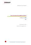
NanoLink™ Streptavidin Magnetic Beads 05142010 NanoLink™ Streptavidin Magnetic Beads ______________________________________________________ User Manual Catalog# M-1002 Note: This protocol can be downloaded from the appropriate category in the Solulink Library at http://www.solulink.com/library. NanoLink™ Streptavidin Magnetic Beads User Manual 1. 2. 3. 4. Product Description Applications Particle Characteristics Using NanoLink™ Magnetic Beads 4.1 NanoLink™ Microsphere Washing Procedure 4.2 Immobilization of Biotinylated PCR Products 4.3 Dissociation of unbiotinylated PCR Strand from Immobilized PCR Product 4.4 Capture and Immobilization of Biotinylated Antibody 4.5 Binding and Wash Buffers/Microsphere Blocking Solution 4.6 Microsphere Blocking Procedure 5. Examples 2 6 6 7 8 9 10 10 11 3 4 5 1. Product Description NanoLink™ Streptavidin Magnetic Microspheres are nanometer-sized, polymerencapsulated (no exposed iron), super-paramagnetic particles containing covalently cross-linked streptavidin. NanoLink streptavidin magnetic microspheres are made by covalently cross-linking streptavidin to a hydrophilic surface using SoluLink’s proprietary HydraLink conjugation chemistry. The high surface area of these paramagnetic microspheres when combined with SoluLink’s efficient linking chemistry produces the most consistent and highest biotin binding capacity per unit mass of any streptavidin magnetic microsphere on the market. Features Highest free biotin binding capacity of any microsphere (>12 nmol/mg) Binds > 2.5 nmol/mg of a biotinylated oligonucleotide Binds > 1.7 nmol/mg of a biotinylated-IgG (250 µg/mg) @ 3 biotins/IgG Encapsulated (no exposed iron) microspheres Microspheres size is less than 875 nm Fast magnetic response time (25 electromagnetic unit) Paramagnetic (no residual magnetism) The particles are supplied at 1% solids (10 mg/ml) in nuclease-free water with 0.05% sodium azide. There are no surfactants present. Particles can be washed prior to use to remove residual azide if desired but is not necessary for most applications. Covalently cross-linked streptavidin Outer core hydrophilic polymer surface Iron magnetite central layer Polystyrene core 3 2. Applications NanoLink™ streptavidin magnetic microspheres possess the highest biotin-binding capacity of any commercially available polymer-encapsulated streptavidin particle. These microspheres are particularly suited for high throughput robotic applications where high biotin loads must be immobilized and separated using a suitably strong magnet. The microspheres can be used to immobilize: Biotinylated antibodies and other proteins Biotinylated dsDNA (gDNA, PCR products) or biotinylated aRNA Biotinylated oligonucleotides The main advantage of using these ultra-high capacity microspheres includes reduction in the overall particle mass required to immobilize a biotinylated sample. This leads to reduced costs and lower non-specific background (NSB). Applications include separation of biotin-labeled biomolecules including biotinlabeled antibodies, genomic DNA, RNA, PCR products, oligonucleotides (e.g. biotinylated oligo (dT) or peptides. NanoLink™ streptavidin magnetic microspheres are also ideal for generating single-stranded PCR templates (by removal of the unbiotinylated competing PCR strand) to dramatically increase hybridization efficiency to complementary targets. NanoLink™ streptavidin magnetic microspheres are an affordable alternative for automated, high throughput immobilization processes using 96-well magnets to affect multiplex binding and separation of nucleic acid or immunoassay biomolecules. The particles are supplied as 1% solids (10 mg/mL) in nuclease-free water with 0.05% sodium azide. There are no surfactants present. Particles can be washed with water prior use to remove residual azide if desired. 3. Particle Characteristics Percent Solids: NanoLink streptavidin magnetic particles are packaged nominally at 1% solids (10 mg/mL) as measured using spectroscopic analysis set by their optical density at 600 nm versus a known mass standard of the same size. Biotin Binding Capacity: The biotin binding capacity of NanoLink streptavidin magnetic particles is measured in nmol/mg. Biotin binding is quantitatively measured by incubating a known mass of particles (0.5 mg) with a fluorescein-biotin standard solution for 60 minutes and quantifying the amount of residual unbound fluorescein-biotin left in solution versus negative control microspheres. 4 Size Distribution by Scanning Electron Microscopy (SEM): Scanning electron microscopy confirms a bimodal size distribution for NanoLink Magnetic Microspheres. The streptavidin particles consist of a core polymeric particle ~ 850 nm surrounded by a population of similar but smaller polymeric microspheres ~ 150 nm. Quality control: A sample of each NanoLink batch is retained for biotin binding capacity and % solids analysis. The remainder is packaged, refrigerated, and preserved in nuclease-free water containing 0.05% sodium azide to prevent microbial contamination. Cleaning: Surfactants are not added to this product and the particles are thoroughly washed with nuclease-free water containing 0.05% sodium azide prior to packaging. For some applications it may be desirable to remove residual azide using a brief wash. Stability: Particles should be stored at 2-8o C. Do not freeze. If particles are settled, resuspend by suitable methods including: vortexing, rotary mixing, or swirling. NanoLink™ streptavidin magnetic microspheres remain stable when stored at 2-8o C for 1 year. Washing: NanoLink™ streptavidin magnetic microspheres are washed by magnetic separation using commercially available magnetic stands. Niobium magnetic stands are available in 50 mL, 15 mL, 1.5 mL, and 96-well plate formats from various vendors. NanoLink™ microspheres are placed on a magnetic stand for 2-3 minutes and the clarified supernatant removed without disturbing the pellet. Re-suspension: After long-term storage and settling of particles, it is best to resuspend the particles thoroughly to avoid any mild particle-to-particle aggregation. 5 4. Using NanoLink™ Magnetic Microspheres In order to decide how much NanoLink™ Streptavidin Magnetic Microspheres you will need for your specific application, please refer to Table 1. Ligand Free biotin Biotinylated oligo (23-mer) Biotinylated IgG (3 biotins per IgG) NanoLink (0.8um) Binding Capacity Competitor's 1um Bead Binding Capacity >14nmol/mg > 1300 pmol/mg >2.5nmol/mg >1.7 nmol/mg NA 0.12 nmol/mg (250 µg/mg) (20 ug/mg) Table 1. NanoLink™ Streptavidin Magnetic Beads binding capacity vs other competitive bead of similar size. 4.1. NanoLink™ Microsphere Washing Procedure 1. Resuspend NanoLink™ streptavidin magnetic microsphere in their original vial with the aid of a vortex mixer. Mix vigorously for 1 minute to fully resuspend the microspheres. Pipette up and down if necessary to fully disperse the microspheres. 2. Transfer the desired volume of NanoLink™ streptavidin magnetic microspheres to a new 1.5 mL tube or other suitable vessel. For example, 5 µL @ 10 mg/mL (50 µg) is sufficient to bind 125 pmol biotinylatedoligonucleotide (~80 µg) or a biotinylated PCR product (~ 40 µg @ 500 bp). Note- always work with a suitable quantity of microsphere, for example 50 µg in a volume of 250 µL of 1x Nucleic Acid Wash and Binding Buffer. Never use less than 10 µg of beads since beads are difficult to visualize and track below this level. 3. Add sufficient Nucleic Acid Binding and Wash Buffer (see recipe below) to bring the final volume to 0.25 mL, mix gently to resuspend and wash the microspheres. 4. Place the tube on a magnet for 2 min., discard the supernatant. 5. Remove the tube from the magnet and resuspend the washed beads in 0.25 mL of Nucleic Acid Binding and Wash Buffer. 6 6. NanoLink™ streptavidin microspheres are now ready for immobilization of biotinylated PCR products or other biotinylated nucleic acids. 4.2. Immobilization of Biotinylated PCR Products 1. Determine the mass of NanoLink™ streptavidin magnetic microspheres required for your specific application and wash as described in section 3.2, leaving the microspheres in 250 µL of Nucleic Acid Binding and Wash Buffer. 2. Add a volume (5-50 µL) of purified PCR product in water or 1xT10E1 (free of excess biotinylated primers) to 0.25 mL of washed microspheres in Nucleic Acid Binding and Wash Buffer. 3. Vortex gently to mix. 4. Incubate for 30 minutes at room temperature preferably on a platform shaker (e.g. Titer-Tek Platform shaker, Lab-Line Instruments, at a setting of 5). The platform shaker is used to keep the microspheres fully resuspended during the binding process. Do not allow the microspheres to settle during binding for maximum capture efficiency. Note- For biotinylated oligonucleotides and DNA fragments smaller than 1 kb capturing for 30 minutes is suitable, but for larger fragments (e.g. 5 kb or larger) binding at 40oC for 60 minutes may be required. Inefficient biotinylation of the amplicon or the presence of excess, free biotinylated primers will lead to reduced amplicon capture efficiency. 5. After immobilization, place the tube on a magnet for 2 min. and carefully remove the supernatant. Note- Take care not to disrupt the pellet on the sides of the vessel during wash and aspiration steps. For some applications, the optical density of the supernatant can be used to quantify the amount of unbound DNA remaining (e.g. 1 absorbance unit DNA = 50 µg/mL/OD260 for double-stranded DNA) 6. Wash the immobilized, biotinylated amplicon using 0.25 mL Nucleic Acid Binding and Wash Buffer and vortex gently to mix. 7. Using a magnetic stand wash the microspheres two additional times. After the final wash remove the supernatant and immediately proceed to the next section. 7 4.3 Dissociation of Un-biotinylated PCR Strand from Immobilized PCR Product 1. Immediately after step 8 (Section 3.3), resuspend the DNA coated NanoLink™ microspheres in exactly 50 µL of *freshly prepared 100 mM NaOH. Note- Prepare daily from a 10N NaOH stock solution and molecular grade water. 2. Incubate the microspheres in 100 mM NaOH at room temperature for 1 minute. 3. Place the tube back on the magnetic stand for an additional 1 minute and transfer the supernatant to a new 1.5 mL tube. This supernatant contains the nonbiotinylated DNA strand. 4. Immediately neutralize the non-biotinylated strand by addition of 5.2 µL 1 M glacial acetic acid. Confirm the pH of the neutralized solution by spotting 1 µL on 0-14 pH paper. After neutralization store the solution at 4o C for later use. Note- Neutralization using acetic acid requires a calibrated P-10 pipette and a known glacial acetic acid molarity (1 M) to neutralize the pH. If necessary after adding 5.2 µl of 1 M glacial acetic acid, small incremental volumes (e.g. 0.5 µl) of either 100 mM NaOH or 1 M glacial acetic acid may need to be added to achieve neutrality. Always confirm neutrality of the solution (~ pH 7.0) using a 1 µl aliquot of the neutralized sample on colored pH paper. 5. With the aid of a magnetic stand, immediately, wash the immobilized biotinylated nucleic acid strand on the NanoLink™ microspheres three times with 0.25 mL Nucleic Acid Binding and Wash Buffer (50 mM Tris-HCl, 150 mM NaCl, 0.5% Tween-20, pH 8). Discard supernatants between washes. 6. Resuspend the NanoLink™ microspheres coated with the immobilized biotinylated strand in 0.25 mL Nucleic Acid Binding and Wash Buffer. Leave the microspheres in this solution until used in other down-stream applications. 4.4. Capture and Immobilization of Biotinylated Antibody 1. Refer to Section 3.1 (Table 1) to determine the mass of NanoLink™ Streptavidin Magnetic Microspheres required to capture and immobilize a given quantity of biotinylated-IgG. 8 Note- For example, 1 milligram of NanoLink™ Streptavidin Magnetic Microspheres will quantitatively bind 250 µg of biotinylated IgG at a biotin molar substitution ratio (MSR) of 3. 2. Transfer the desired volume of pre-blocked NanoLink™ Streptavidin microspheres @ 10 mg/mL into a 1.5 mL microfuge tube. (Refer to Microsphere Blocking Procedure, Section 3.7) 3. Place the tube containing the microspheres on a magnet stand for 2 min. and carefully remove the supernatant. 4. Wash the microspheres once with 0.25 mL of 1x Antibody Binding and Wash Buffer. 5. Place the tube containing the microspheres on a magnet stand for 2 min. and carefully remove the supernatant 6. Add 0.125 mL of 2x Antibody Binding and Wash Buffer and 0.125 mL of biotinylated IgG containing sample to the microsphere pellet. 7. Mix the beads well, and incubate the tube on a platform shaker (e.g. Titer-Tek Platform shaker, Lab-Line Instruments, setting of 5) at room temperature for 30 minutes to capture the biotinylated antibody. 8. Place the tube containing the immobilized antibody on a magnetic stand for 2 min. and carefully remove the supernatant. 9. Wash the microsphere pellet twice using 0.25 mL 1x Antibody Binding and Wash Buffer. 10. The immobilized biotinylated IgG is now ready for other downstream applications such as capture and/or release of cognate antigen. 4.5 Binding and Wash Buffers/Microsphere Blocking Solution Nucleic Acid Binding and Wash Buffer 50 mM Tris-HCl, 150 mM NaCl, 0.05% Tween 20, pH 8.0 1X Antibody Binding and Wash Buffer 50 mM Tris-HCl, 150 mM NaCl, 0.05% Tween 20, pH 8.0 2X Antibody Binding and Wash Buffer 100 mM Tris-HCl, 300 mM NaCl, 0.1% Tween 20, pH 8.0 9 Microsphere Blocking Solution BlockerTM Casein in TBS (Trademark of Pierce Chemical, Cat. # 37532). Filter the casein block solution through a 0.45 µ filter before using. Use Hammersten-grade casein for blocking streptavidin microspheres 4.6 . Microsphere Blocking Procedure (1 milligram microspheres) 1. Transfer 100 ul of NanoLink™ Streptavidin Magnetic Microspheres @ 10 mg/ml to a new 1.5 ml microfuge tube. 2. Place the tube on a magnetic stand for 2 minutes, carefully remove and discard the supernatant. 3. Add 1 mL of Blocker™ Casein in TBS (filtered) to the microspheres to resuspend the microspheres. 4. Place the tube on a platform shaker (e.g. LabLine Titer-TeK @ setting of 5) for 30 minutes to block. 5. Place the tube on a magnetic stand for 2 minutes and completely remove the blocking solution. 6. Wash the microspheres 4 X with 1 ml 1X Antibody Binding and Wash Buffer (50 mM Tris-HCl, 150 mM NaCl, 0.05% Tween 20, pH 8.0). Discard the wash solution between washes. 7. After the final wash, resuspend the blocked microspheres at 10 mg/ml using 100 ul 1X Antibody Binding and Wash Buffer. 8. Microspheres are now ready for capture and immobilization of biotinylated antibody. 10 5. Examples 5.1. Capture of Biotinylated Antibody Using NanoLink™ Streptavidin Microspheres 11 The products offered here are for research use only. Any commercial application will require a license from Solulink. The Solulink Conjugation System is patented and has multiple patents pending. Please contact Solulink for information regarding licensing information. Solulink products and methods may be covered by one or more of the following United States patents Nos. 6,686,461, 6,800,728, 7,102,024, 7,173,125, 7,462,689 and other pending patent applications. Information in this manual is subject to change without notice and does not constitute a commitment on the part of Solulink, Inc. It is supplied on an “as is” basis without any warranty of any kind, either explicit or implied. Information may be changed or updated in this manual at any time. This document may not be copied, transferred, reproduced, disclosed, or duplicated, in whole or in part, without the prior written consent of Solulink, Inc. This documentation is proprietary information and protected by the copyright laws of the United States and international treaties. The manufacturer of this documentation is Solulink, Inc. 12