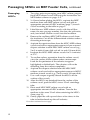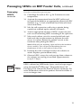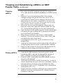
1
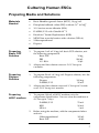
GIBCO® Mouse Embryonic Fibroblasts (Irradiated) Catalog nos. S1520-100, S1520-250 Revision date: 9 February 2010 Manual part no. A11560 MAN0001756 Corporate Headquarters Invitrogen Corporation 1600 Faraday Avenue Carlsbad, CA 92008 T: 1 760 603 7200 F: 1 760 602 6500 E: [email protected] For country-specific contact information visit our web site at www.invitrogen.com User Manual Table of Contents Contents and Storage........................................................................................... iv GIBCO® Mouse Embryonic Fibroblasts (Irradiated) ........................................1 Culturing MEF Feeder Cells .............................................................. 3 Preparing Medium and Culture Vessels ............................................................3 Thawing and Establishing MEFs.........................................................................4 Expected Results.....................................................................................................6 Culturing Human ESCs...................................................................... 7 Preparing Media and Solutions ...........................................................................7 Thawing and Establishing Human ESCs on MEF Feeder Cells.....................8 Expected Results...................................................................................................10 Passaging Human ESCs on MEF Feeder Cells................................................11 Culturing Mouse ESCs .................................................................... 14 Thawing and Establishing Mouse ESCs on MEF Feeder Cells.....................14 Expected Results...................................................................................................16 Passaging Mouse ESCs on MEF Feeder Cells..................................................17 Passaging Mouse ESCs using Mouse ESC Medium with FBS......................19 Troubleshooting ...................................................................................................20 Appendix ........................................................................................... 22 Accessory Products..............................................................................................22 Technical Support ................................................................................................24 Purchaser Notification.........................................................................................25 References..............................................................................................................27 iii Contents and Storage Catalog no. S1520-100 includes cells only. Kit Configurations Catalog no. S1520-250 includes cells plus media. Shipping GIBCO® Mouse Embryonic Fibroblasts (Irradiated) and KnockOut™ Serum Replacement are shipped on dry ice. Kit Contents and Storage Kit components and storage conditions for S1520-100 and S1520-250 are listed in the table below. S1520-100 Amount Storage GIBCO Mouse Embryonic Fibroblasts (Irradiated) (1 × 10⁶ cells/mL in freezing medium*) 1 mL Liquid nitrogen S1520-250 Amount Storage GIBCO Mouse Embryonic Fibroblasts (Irradiated) (1 × 10⁶ cells/mL in freezing medium) 1 mL Liquid nitrogen KnockOut™ Serum Replacement 100 mL –5 to –20°C ® ® *Freezing medium: 60% Dulbecco’s Modified Eagle medium containing 4.5 g/L glucose, 30% Fetal Bovine Serum, and 10% DMSO. Handle cells as potentially biohazardous material under at least Biosafety Level 1 (BL-1) containment. This product contains Dimethyl Sulfoxide (DMSO), a hazardous material. Review the Safety Data Sheet (SDS) before handling. Safety Data Sheets (SDSs) are available on our website at www.invitrogen.com/sds. Intended Use iv GIBCO® Mouse Embryonic Fibroblasts (Irradiated) are for research use only. They are not intended for any animal or human therapeutic or diagnostic use. GIBCO® Mouse Embryonic Fibroblasts (Irradiated) Uses of GIBCO® Mouse Embryonic Fibroblasts (Irradiated) GIBCO® Mouse Embryonic Fibroblasts (Irradiated) are used as feeder layers for culturing embryonic stem cells (ESCs), including mouse and human ESCs, in their undifferentiated state. The growth-arrested feeder layer supports the ESC culture by providing nutrients, growth factors, and matrix components, and it enables ESCs to survive and proliferate more readily in culture. Source of GIBCO® Mouse Embryonic Fibroblasts (Irradiated) GIBCO® Mouse Embryonic Fibroblasts (Irradiated) were isolated from ICR mouse embryos at day 13.5 of gestation under sterile conditions, expanded for up to two passages in D-MEM medium containing 4.5 g/L glucose supplemented with 10% FBS, and mitotically inactivated by -irradiation. After -irradiation, cells were cryopreserved in a cryopreservation medium composed of 60% D-MEM (high glucose), 30% FBS, and 10% DMSO. Each vial of GIBCO® Mouse Embryonic Fibroblasts (Irradiated) contains approximately 1 × 10⁶–1.5 × 10⁶ live cells. Characteristics of GIBCO® Mouse Embryonic Fibroblasts (Irradiated) Isolated from mouse primary cell cultures Mitotically inactivated by -irradiation Frozen at passage number up to 2 (≤ P2) Support the growth of embryonic stem cells (ESCs), including mouse and human ESCs in their undifferentiated state Continued on next page 1 GIBCO® Mouse Embryonic Fibroblasts (Irradiated), continued Guidelines for Using MEFs Media Requirements Follow the guidelines below to use inactivated mouse embryonic fibroblasts (MEFs) as feeder layers to culture mouse and human embryonic stem cells (ESCs). All solutions and equipment that come in contact with the cells must be sterile. Always use proper aseptic technique and work in a laminar flow hood. Make sure to start preparing MEF feeder layers two days before culturing ESCs. After thawing, transfer MEFs into pre-warmed medium. Plate MEFs on culture vessels coated with 0.1% gelatin solution (see page 3). Use MEF dishes or plates within one week after preparation. Before starting experiments, ensure that ESCs have been established (at least 1 passage), and also have some frozen ESC stocks on hand. For best results, we recommend using Dulbecco’s Modified Eagle medium (D-MEM) containing 4.5 g/L glucose, and supplemented with 10% FBS for establishing GIBCO® Mouse Embryonic Fibroblasts (Irradiated) (see page 22 for ordering information). Prepare the medium prior to use. As with other mammalian cell lines, when working with GIBCO® Mouse Embryonic Fibroblasts (Irradiated), handle as potentially biohazardous material under at least Biosafety Level 1 (BL-1) containment. For more information on BL-1 guidelines, refer to Biosafety in Microbiological and Biomedical Laboratories, 5th ed., published by the Centers for Disease Control, which is available for downloading at: www.cdc.gov/od/ohs/biosfty/bmbl5/bmbl5toc.htm. 2 Culturing MEF Feeder Cells Preparing Medium and Culture Vessels Materials Needed For gelatin coating culture vessels: AF solution (Attachment Factor) Note: AF is a sterile 1X solution containing 0.1 % gelatin available from Invitrogen (see page 22 for ordering information). Sterile culture vessels For preparing MEF medium: Gelatin Coating Culture Vessels Dulbecco’s Modified Eagle medium (D-MEM) containing 4.5 g/L glucose Fetal bovine serum (FBS), ES-Cell Qualified 1. Cover the whole surface of each culture vessel with AF solution and incubate the vessels for 30 minutes at 37°C or for 2 hours at room temperature. 2. Using sterile technique in a laminar flow culture hood, completely remove the AF solution from the culture vessel by aspiration. Note: It is not necessary to wash the culture surface before adding cells or medium. 3. Preparing MEF 1. Medium Coated vessels may be used immediately or stored at room temperature for up to 24 hours. Prepare 500 mL of MEF medium by mixing the following components (pre-warmed to 37°C): D-MEM FBS 450 mL 50 mL 3 Thawing and Establishing MEFs Materials Needed Thawing MEFs GIBCO® Mouse Embryonic Fibroblasts (Irradiated), frozen MEF medium (see previous page) Gelatin coated culture vessels (see previous page) Phosphate buffered saline (PBS) without Ca2+ or Mg2+ 37°C water bath 70% ethanol Disposable, sterile 0.5 mL and 15-mL tubes Microcentrifuge Hemacytometer, cell counter and Trypan Blue, or the Countess™ Automated Cell Counter 1. Remove the cryovial containing inactivated MEFs from the liquid nitrogen storage tank. 2. Briefly roll the vial between hands to remove frost, and swirl it gently in a 37°C water bath. 3. When only a small ice crystal remains in the vial, remove it from water bath. Spray the outside of the vial with 70% ethanol before placing it in the cell culture hood. 4. Pipet the thawed cells gently into a 15-mL conical tube. 5. Rinse the cryovial with 1 mL of pre-warmed MEF medium. Transfer the medium to the same 15-mL tube containing the cells. 6. Add 4 mL of pre-warmed MEF medium dropwise to the cells. Gently mix by pipetting up and down. Note: Adding the medium slowly helps the cells to avoid osmotic shock. 7. Centrifuge the cells at 200 × g for 5 minutes. 8. Aspirate the supernatant and resuspend the cell pellet in 5 mL of pre-warmed MEF medium. 9. Remove 20 μL of the cell suspension and determine the viable cell count using your method of choice. Invitrogen’s Countess™ Automated Cell Counter is a benchtop counter designed to accurately measure cell count and viability in less than a minute per sample, using the standard Trypan Blue technique (see page 23 for ordering information). Continued on next page 4 Thawing and Establishing MEFs, continued Plating MEFs 1. Centrifuge the remaining cell suspension (step 9, previous page) at 200 × g for 5 minutes at room temperature. 2. Aspirate the supernatant. Resuspend the cell pellet in MEF medium to a density of 2.5 × 10⁶ cells/mL. 3. Aspirate the gelatin solution from the gelatin coated culture vessel (step 4, page 3), and wash the vessels once with PBS. 4. Add the appropriate amount of MEF medium into each culture vessel (refer to the table below). 5. Into each of these culture vessels, add the appropriate amount of MEF suspension (refer to the table below). Note: The recommended plating density for GIBCO® Mouse Embryonic Fibroblasts (Irradiated) is 2.5 × 10⁴ cells/cm2. 6. Move the culture vessels in several quick back-and-forth and side-to-side motions to disperse the cells across the surface of the vessels. 7. Incubate the cells in a 37°C incubator with a humidified atmosphere of 5% CO2. 8. Use the MEF culture vessels within 3–4 days after plating. Vessel Size Growth Area Volume of Media 96-well plate 0.32 cm2/well 0.1 mL 2 24-well plate 2 cm /well 0.5 mL 12-well plate 3.8 cm2/well 1 mL 6-well plate 2 9.6 cm /well 2 2 mL 60-mm dish 19.5 cm 5 mL 100-mm dish 58.95 cm2 10 mL 2 25-cm flask 2 75-cm flask Important 25 cm 2 75 cm 2 5 mL 15 mL Number of MEFs Volume of MEF Suspension 1.0 × 10⁴/well 4 μL 4 20 μL 5 40 μL 5.0 × 10 /well 1.0 × 10 /well 5 2.5 × 10 /well 0.1 mL 5 0.2 mL 6 0.6 mL 5 0.25 mL 6 0.75 mL 5.0 × 10 1.5 × 10 6.3 × 10 1.9 × 10 The number of cells and the volume of cell suspension given in the table above are optimized for MEFs only. For plating other cell types (e.g., mESCs), calculate the number of cells to be plated using the growth area and the recommended plating density for the specific cell type you are using. 5 Expected Results Images of MEF Culture 6 The bright field image below shows GIBCO® Mouse Embryonic Fibroblasts (Irradiated) plated at the recommended density on culture dishes coated with 0.1% gelatin. The image was taken with a 10X objective. Culturing Human ESCs Preparing Media and Solutions Materials Needed Preparing Basic FGF Solution Preparing Dispase Solution Basic fibroblast growth factor (bFGF), 10 μg/mL Phosphate buffered saline (PBS) without Ca2+ or Mg2+ 10% bovine serum albumin (BSA) D-MEM/F-12 with GlutaMAX™-I Knockout™ Serum Replacement (KSR) MEM Non-essential amino acids solution (NEAA) 2-Mercaptoethanol Dispase 1. To prepare 1 mL of 10 μg/mL basic FGF solution, mix the following components: Basic FGF PBS 10% BSA 2. Aliquot and the solution store at –20°C for up to 6 months. 1. To prepare 50 mL of 2 mg/mL Dispase solution, mix the following components: 2. Preparing hESC medium 10 μg 980 μL 10 μL 1. Dispase 100 mg D-MEM/F-12 50 mL Aliquot and the solution store at 4°C for up to 2 weeks or at –20°C for up to 6 months. To prepare 100 mL of hESC medium, mix the components listed below. You can store the medium at 4°C for up to 7 days. D-MEM/F-12 KSR NEAA 2. 79 mL 20 mL 1 mL Before using the medium, add the components below and mix. 2-Mercaptoethanol Basic FGF 182 μL 40 μL 7 Thawing and Establishing Human ESCs on MEF Feeder Cells Materials Needed Thawing hESCs Human embryonic stem cells (hESCs) MEF culture vessels (step 7, page 5) hESC medium (see previous page) 70% ethanol Disposable, sterile 15-mL tubes 37°C water bath 37°C incubator with a humidified atmosphere of 5% CO2 Microcentrifuge Hemacytometer, cell counter and Trypan Blue, or the Countess™ Automated Cell Counter 1. 3–4 hours before plating hESCs, aspirate the MEF medium from the 60-mm culture dish containing the MEFs and add 4 mL of hESC culture medium. Note: You will plate the hESC into this dish (see next page). 2. Remove a cryovial containing hESCs from the liquid nitrogen storage tank. 3. 4. Roll the vial between your gloved hands briefly to remove frost, and swirl it gently in a 37°C water bath. When only a small ice crystal remains in the vial, remove it from water bath. Spray the outside of the vial with 70% ethanol before placing it in the cell culture hood. 5. Pipet the thawed cells gently into a 15-mL conical tube. 6. Rinse the cryovial with 1 mL of pre-warmed hESC medium. Transfer the medium to the same 15-mL tube containing the cells (from step 5). 7. Add 4 mL of pre-warmed hESC medium dropwise to the cells. While adding the medium, gently move the tube back and forth to mix the hESCs. Note: Adding the medium slowly helps the cells to avoid osmotic shock. 8. Centrifuge the cells at 200 × g for 5 minutes at room temperature. 9. Aspirate the supernatant and resuspend the cell pellet in 5 mL of pre-warmed hESC medium. Continued on next page 8 Thawing and Establishing hESCs on MEF Feeder Cells, continued Plating hESCs 1. 2. 3. Aspirate the hESC medium from the 60-mm culture dish containing the MEFs (step 1, page 8). Slowly add the hESC suspension into the MEF culture dish. Move the vessel in several quick back-and-forth and side-to-side motions to disperse the hESCs across the surface of the dish. 4. Place the culture dish gently in a 37°C incubator with a humidified atmosphere of 5% CO2. 5. Replace the spent medium and examine the cells under a microscope daily. It may take up to a week for colonies to become visible. 6. Observe the hESCs every day and passage them whenever the colonies are too big or crowded. The split ratio depends on the total number of hESCs in the culture dish (approximately 1:1 to 1:3 at the first passage after recovery). 9 Expected Results The image below shows H9 human embryonic stem cells Images of hESCs on MEF (hESCs) cultured on a layer MEF feeder cells. The bright field image of the hESCs at passage 1 was obtained with a 10X. Feeder Cells 10 Passaging Human ESCs on MEF Feeder Cells Guidelines for Passaging hESCs Materials Needed Passage your hESCs when the first of the following occurs: o MEF feeder layer is two weeks old. o hESC colonies are becoming too dense or too large. o Increased differentiation occurs. The split ratio is varies, but it is generally between 1:4 and 1:6. Occasionally hESCs grow at a different rate, requiring the split ratio to be adjusted. A general rule is to observe the last split ratio and adjust the ratio according to the appearance of the hESC colonies. If the cells look healthy and colonies have enough space, split them using the same ratio as the previous passage; if they are overly dense and crowded, increase the ratio, and if the cells are sparse, decrease the ratio. Generally, hESCs need to be split every 5–7 days based upon their appearance. Confluent hESCs growing on MEF feeder culture Fresh MEF culture vessels (see pages 3–5) hESC medium (see page 7) Dispase solution (see page 7) Disposable, sterile 15-mL tubes Cell scraper Dissecting microscope 37°C incubator with humidified atmosphere of 5% CO2 Optional: StemPro® EZPassage™ Disposable Stem Cell Passaging Tool (see Note on page 13) Continued on next page 11 Passaging hESCs on MEF Feeder Cells, continued Passaging hESCs 1. Two days prior to passaging your hESC culture, prepare fresh MEF culture vessels following the instructions for MEF feeder cultures on pages 3–5. 2. 3–4 hours before plating the hESCs, aspirate the MEF medium from each MEF culture vessel, and add an appropriate amount of hESC medium (page 7) to each vessel according to the table on page 5. 3. Label the new MEF culture vessels with the cell line name, the new passage number, the date, the split ratio, and your initials. Return vessels into the incubator. 4. Remove the confluent hESC-MEF culture vessels from the incubator. Cut off the differentiated colonies under a dissecting microscope. 5. Aspirate the spent medium from the hESC-MEF culture vessels and add an appropriate amount of pre-warmed dispase solution to each hESC-MEF culture vessel (e.g., 2 mL to each 60-mm dish or 4 mL to each 100-mm dish). 6. Incubate the hESC-MEF culture vessels for 5–6 minutes at 37°C. 7. To confirm colony separation from the culture vessel, view the surface of the culture under a microscope. Look for the perimeter of the colonies to appear highlighted or folded back. The colonies will not be detaching from the surface completely. 8. Aspirate the dispase solution from the hESC-MEF culture vessels and add an appropriate amount of hESC medium to each vessel (e.g., 5 mL to each 100-mm dish). 9. Use a cell scraper to gently detach the hESCs off the surface of the vessels. 10. After the hESCs are detached from the surface of the culture vessel, pool the hESCs into a 15-mL centrifuge tube. 11. Rinse each hESC-MEF culture vessel with an appropriate amount of hESC medium. Transfer the medium to the same 15-mL tube containing the hESCs (from step 10). 12. Gently pipet the cells up and down a few times in the tube to further break-up the cell colonies. Procedure continued on next page Continued on next page 12 Passaging hESCs on MEF Feeder Cells, continued Passaging hESCs, continued Procedure continued from previous page 13. Centrifuge the cells at 200 × g for 5 minutes at room temperature. 14. Aspirate the supernatant from the hESC pellet, and resuspend the pellet in an appropriate amount of hESC medium (e.g., 1–2 mL of medium for all the cells from one 60-mm dish). 15. Mix the cell suspension well using a pipette, being careful not to break up the colonies too much.. 16. Add an appropriate amount of hESC suspension into each vessel containing MEFs according to the split ratio. 17. Move each culture vessel in several quick back-andforth and side-to-side motions to disperse the cells across its surface. Return the culture vessels to the incubator after plating the hESCs. Note: While cells are attaching, open and close the incubator doors carefully. This will prevent disturbing the even distribution of cells on the surface of the vessels. 18. Incubate the cells overnight to allow the colonies to attach. Replace spent medium daily. 19. Observe hESCs every day and passage them whenever the colonies are too big or crowded (approximately every 5–7 days). hESCs cultured on MEF feeder layers can also be passaged using the StemPro® EZPassage™ Disposable Stem Cell Passaging Tool without the need for dispase treatment. For more information, refer to the product manual available at www.invitrogen.com. 13 Culturing Mouse ESCs Thawing and Establishing Mouse ESCs on MEF Feeder Cells Materials Needed Preparing mESC medium Mouse embryonic stem cells (mESCs) MEF culture vessels (see page 5) MEF medium (see page 4) Knockout™ D-MEM Knockout™ Serum Replacement (KSR) MEM Non-essential amino acids solution (NEAA) 2-Mercaptoethanol Recombinant human LIF (leukemia inhibitory factor) L-Glutamine 70% ethanol Disposable, sterile 15-mL tubes 37°C water bath 37°C incubator with a humidified atmosphere of 5% CO2 Microcentrifuge Hemacytometer, cell counter and Trypan Blue, or the Countess™ Automated Cell Counter 1. To prepare 50 mL of mESC culture medium, mix the components listed below. You can store the medium at 4°C for up to 1 week. Knockout™ D-MEM KSR NEAA L-Glutamine 2. 41.5 mL 7.5 mL 0.5 mL 0.5 mL Before using the medium, add the components below and mix. LIF (10 μg/mL) 50 μL 2-Mercaptoethanol 91 μL Continued on next page 14 Thawing and Establishing mESCs on MEF Feeder Cells, continued Thawing mESCs 1. Pre-warm the mESC medium (see previous page) to 37°C, and add 9 mL of mESC medium to a 15-mL conical tube 2. Remove a cryovial containing mESCs from liquid nitrogen storage and quickly thaw the vial in a 37°C water bath. Be careful not to submerge the entire vial. Note: Maximum cell viability is dependent on the rapid and complete thawing of frozen cells. Thawing the cells for longer than 3 minutes results in decreased viability. 3. When the last ice crystal disappears, remove the vial from the water bath. Spray the outside of the vial with 70% ethanol before placing it in the cell culture hood. 4. Pipet the thawed cells gently into the 15-mL conical tube containing the pre-warmed mESC medium. Be careful not to introduce any bubbles. 5. Rinse the cryovial with 1 mL of pre-warmed mESC medium. Transfer the medium to the same 15-mL tube containing the cells (from step 4). 6. Centrifuge the cells at 250 × g for 5 minutes at room temperature. 7. Aspirate the supernatant and resuspend the cell pellet in an appropriate amount of pre-warmed mESC medium. 8. Remove 20 μL of the cell suspension and determine the viable cell count using your method of choice. Plating mESCs 1. Aspirate the MEF medium from each culture vessel containing MEFs, and add an appropriate amount of mESC medium according to the table on page 5. 2. Plate the mESC suspension into MEF culture vessels at about 4 × 10⁴ cells/cm2. 3. Add sufficient amount of mESC medium (e.g., a total of 2.5 mL of medium for each well of a 6-well plate). Gently rock the culture vessels to evenly distribute the cells. 4. Incubate the cells in a 37°C incubator with a humidified atmosphere of 5% CO2. 5. Replace the spent medium with fresh pre-warmed mESC medium every day until mouse ESC colonies become confluent to be split. 15 Expected Results Images of mESCs on MEF Feeder Cells 16 The images below show C57 mouse embryonic stem cells (mESCs) cultured on a layer MEF feeder cells. The bright field images of the mESCs were taken 2 days after plating with a 10X objective. Passaging Mouse ESCs on MEF Feeder Cells Guidelines for Passaging mESCs Materials Needed Plate mESCs at a density that provides an even distribution of colonies over the surface of culture vessel, but does not result in contact between the colonies. If colonies are plated too densely or too sparsely, they may differentiate. Do not over-passage mESCs from the C57BL/6 strain. Minimize the number of passages and the length of time the cells are kept in culture. This will ensure optimal and reproducible experimental results. Passage mESCs before the colonies become too large and dense. When plated at the optimal density, mESC should be passaged every 48 hours. Split ratios for mESCs can vary from 1:5 to 1:15. Confluent mESCs growing on MEF feeder culture Fresh MEF culture vessels (see pages 3–5) mESC medium (see page 14) Phosphate buffered saline (PBS) without Ca2+ or Mg2+ StemPro® Accutase® Cell Dissociation Reagent Disposable, sterile 15-mL tubes 37°C incubator with humidified atmosphere of 5% CO2 Optional: StemPro® EZPassage™ Disposable Stem Cell Passaging Tool Continued on next page 17 Passaging mESCs on MEF Feeder Cells, continued Passaging mESCs 1. Pre-warm the mESC medium, PBS, and Accutase® solution to 37°C. 2. Aspirate the spent MEF medium from each MEF culture vessel, and rinse the MEFs with PBS (3 mL for one well of a 6-well plate). 3. Aspirate the PBS from the MEF culture vessels and add an appropriate amount of pre-warmed mESC medium into the vessels (e.g., 2.5 mL for each well of a 6-well plate). 4. Return the MEFs to the 5% CO2 humidified incubator. Note: Be careful not to disturb the monolayer of MEFs during steps 2–4. 5. Carefully aspirate the spent medium from culture vessels containing mouse ESCs, and rinse the cells with PBS (e.g., 3 mL for one well of a six-well plate). 6. Aspirate the PBS from the culture vessels. 7. Add an appropriate amount of Accutase® solution to cover the surface of culture vessels and incubate the vessels for 1–2 minutes until mESCs are dissociated. Gently tap the side of the culture vessels to detach the majority of cells from the surface of culture vessels. 8. Add mouse ESC medium (e.g., 3 mL for each well of a 6-well plate) to stop the dissociation reaction and gently pipet the cells up and down sufficiently to disperse the colonies into a single-cell suspension. Note: Be careful not to introduce any bubbles. 9. Transfer the mESC suspension into a 15-mL conical tube and centrifuge the tube at 250 × g for 5 minutes to pellet the cells. 10. Carefully aspirate as much of supernatant as possible and add an appropriate amount of mESC medium to the tube. Gently resuspend the mESCs. 11. Plate the mESCs into the culture vessels containing MEFs (step 4). Split ratios for mESCs can vary from 1:5 to 1:15. 12. Incubate mESCs at 37°C in a 5% CO2 humidified incubator and change the medium every day. Mouse ESC can be split every other day. 18 Passaging Mouse ESCs using Mouse ESC Medium with FBS Materials Needed Preparing mESC medium with FBS Confluent mESCs growing on MEF feeder culture Fresh MEF culture vessels (see pages 3–5) Dulbecco’s Modified Eagle Medium (D-MEM), low glucose Fetal Bovine Serum (FBS), ES-Cell Qualified MEM Non-essential amino acids solution (NEAA) 2-Mercaptoethanol Recombinant mouse LIF (leukemia inhibitory factor) Disposable, sterile 15-mL tubes Phosphate buffered saline (PBS) without Ca2+ or Mg2+ StemPro® Accutase® Cell Dissociation Reagent, Trypsin/EDTA Solution or TrypLE™ Express Dissociation Reagent Disposable, sterile 15-mL tubes 37°C incubator with humidified atmosphere of 5% CO2 Optional: StemPro® EZPassage™ Disposable Stem Cell Passaging Tool37°C water bath 1. To prepare 50 mL of mESC culture medium with FBS, mix the components listed below. You can store the medium at 4°C for up to 1 week. 2. Passaging mESCs using mESC Medium with FBS D-MEM 44.4 mL FBS 5 mL NEAA 0.5 mL Before using the medium, add the components below and mix. Mouse LIF (10 μg/mL) 10 μL 2-Mercaptoethanol 91 μL To passage mESC using mESC medium with FBS, follow the protocol on page 18, but replace mESC medium with mESC medium with FBS (see above). To detach the cells (Step 7 on page 18), you may use StemPro® Accutase® Cell Dissociation Reagent or Trypsin/EDTA solution (0.125% final concentration). 19 Troubleshooting Culturing MEFs The table below lists some potential problems and solutions that help you troubleshoot your MEF feeder cultures. Problem Cause Solution Cells have low viability after thawing Stock not stored correctly Order new stock and store in liquid nitrogen. Keep in liquid nitrogen until thawing. Use pre-warmed MEF medium, prepared as described on page 3. Thawing medium not correct Cells too diluted Cell not handled gently. Cells not adherent after initial thaw Did not use highquality FBS. The recommended plating density for GIBCO® Mouse Embryonic Fibroblasts (Irradiated) is 2.5 × 10⁴ cells/cm2. GIBCO® Mouse Embryonic Fibroblasts (Irradiated) are fragile; treat your cells gently, do not vortex, bang the flasks to dislodge the cells, or centrifuge the cells at high speeds. Be sure to prepare your culture medium using ESC-qualified FBS (see page 22 for ordering information). Continued on next page 20 Troubleshooting, continued ESC Culture The table below lists some potential problems and solutions that help you troubleshoot your embryonic stem cell (ESC) culture on on MEFs MEF feeder layers. Problem Cause Solution No viable cells after thawing ESCs Stock not stored correctly Order new stock and store in liquid nitrogen. Keep in liquid nitrogen until thawing. Use pre-warmed ESC culture medium, prepared as described. Thawing medium not correct Cell not handled gently Poor quality ESCs Cells grow slowly Growth medium not correct Cells too old Use pre-warmed ESC culture medium, prepared as described on page 7. Use healthy ESCs at low passage number; do not overgrow. Cells too diluted Spin down cells for 4 minutes 200 g at room temperature; aspirate media and plate cells at higher density. bFGF is not stable when frequently warmed and cooled. Add bFGF to medium just before use, or store medium with bFGF in aliquots at –20°C. Be gentle at time of passage so the clumps of cells don’t get too small. Discard cells, media and reagents, and use early stock of cells with fresh media and reagents. Thaw and culture fresh vial of new ESCs. Follow thawing instructions and subculture procedures exactly. ESCs may become differentiated as their passage number increases. Prepare fresh MEF culture plates; follow the instructions on pages 3–5 exactly. Passaging cells too early causes poor plating and differentiation. Grow cells to nearconfluence. Basic FGF (bFGF) inactive Clump size is too small Mycoplasma contamination Cells differentiated ESCs are fragile; treat your cells gently, do not vortex, bang the flasks to dislodge the cells, or centrifuge the cells at high speeds. ESCs were too old or overgrown when frozen. Obtain new stock. Culture conditions not correct Cells too old MEF feeder layer suboptimal Cells passaged too early 21 Appendix Accessory Products For more information about the following products, refer to www.invitrogen.com or contact Technical Support (see page 24). Media, Sera, and Reagents Item Quantity Cat. no. Dulbecco’s Modified Eagle Medium (D-MEM), high glucose 500 mL 10569-010 Dulbecco’s Modified Eagle Medium (D-MEM), low glucose 500 mL 10567-014 D-MEM/F-12 (1X), liquid, 1:1 (contains GlutaMAX™-I) 500 mL 10565-018 ™ Knockout D-MEM 500 mL 10829-018 Fetal Bovine Serum (FBS), ES-Cell Qualified 500 mL 16141-079 Dulbecco’s Phosphate Buffered Saline (D-PBS), Calcium and Magnesium-free 500 mL 10 500 mL 14190-144 14190-250 MEM Non-Essential Amino Acids Solution (10 mM) 100 mL 11140-050 Knockout™ Serum Replacement 500 mL 10828-028 2-Mercaptoethanol (1,000X), liquid 50 mL 21985-023 FGF-basic, AA 10-155 Recombinant Human 50 μg PHG0026 Recombinant Mouse Leukemia Inhibitory Factor (LIF) 10 μg 100 μg PMC4054 PMC4051 Recombinant Human Leukemia Inhibitory Factor (LIF) 10 μg PHC9464 L-Glutamine - 200mM (100X), liquid 100 mL 25030081 Ultrapure BSA (50 mg/mL) 50 mg AM2616 Dispase 5g 17105-041 StemPro Accutase Cell Dissociation Reagent 100 mL A11105-01 Trypsin/EDTA Solution 100 mL R-001-100 TrypLE™ Express Dissociation Reagent 100 mL 12604-013 Attachment Factor 100 mL S-006-100 ® ® Continued on next page 22 Accessory Products, continued Embryonic Stem Cell Lines The ESC lines listed below may be used with GIBCO® Mouse Embryonic Fibroblasts (Irradiated). For more information, refer to www.invitrogen.com or contact Technical Support (see page 24). Cell Line Quantity Cat. no. BG01V/hOG Cells ~2 10 cells R7799-105 ® StemPro BG01V/EG Cells ~3 10 cells R7799-205 GIBCO® Mouse (C57) Embryonic Stem Cells ~1 10 cells S10503-100 GIBCO® Mouse (C57) Embryonic Stem Cells with GFP 6 6 6 ~1 106 cells S10513-100 ® ~1 106 cells S10504-100 ® ~1 106 cells S10514-100 GIBCO Mouse (129) Embryonic Stem Cells GIBCO Mouse (129) Embryonic Stem Cells with GFP Additional Products For more information about the following products, refer to www.invitrogen.com or contact Technical Support (see page 24). Item Quantity Cat. no. Water, distilled 500 mL 15230-162 Trypan Blue Stain 100 mL 15250-061 LIVE/DEAD® Cell Vitality Assay Kit 1000 assays L34951 Countess™ Automated Cell Counter (includes 50 Countess™ cell counting chamber slides and 2 mL of Trypan Blue Stain) 1 unit C10227 StemPro® EZPassage™ Disposable Stem Cell Passaging Tool 10 units 23181010 23 Technical Support Web Resources Contact Us Visit the Invitrogen website at www.invitrogen.com for: Technical resources, including manuals, vector maps and sequences, application notes, SDSs, FAQs, formulations, citations, handbooks, etc. Complete Technical Support contact information Access to the Invitrogen Online Catalog Additional product information and special offers For more information or technical assistance, call, write, fax, or email. Additional international offices are listed on our website (www.invitrogen.com). Corporate Headquarters: 5791 Van Allen Way Carlsbad, CA 92008 USA Tel: 1 760 603 7200 Tel (Toll Free): 1 800 955 6288 Fax: 1 760 602 6500 E-mail: [email protected] Japanese Headquarters: LOOP-X Bldg. 6F 3-9-15, Kaigan Minato-ku, Tokyo 108-0022 Tel: 81 3 5730 6509 Fax: 81 3 5730 6519 E-mail: [email protected] European Headquarters: Inchinnan Business Park 3 Fountain Drive Paisley PA4 9RF, UK Tel: 44 (0) 141 814 6100 Tech Fax: 44 (0) 141 814 6117 E-mail: [email protected] Safety Data Sheets (SDSs) SDSs (Safety Data Sheets) are available on our website at www.invitrogen.com/sds. Certificate of Analysis The Certificate of Analysis provides detailed quality control information for each product. Certificates of Analysis are available on our website. Go to www.invitrogen.com/support and search for the Certificate of Analysis by product lot number, which is printed on the box. 24 Purchaser Notification Limited Warranty Invitrogen (a part of Life Technologies Corporation) is committed to providing our customers with high-quality goods and services. Our goal is to ensure that every customer is 100% satisfied with our products and our service. If you should have any questions or concerns about an Invitrogen product or service, contact our Technical Support Representatives. All Invitrogen products are warranted to perform according to specifications stated on the certificate of analysis. The Company will replace, free of charge, any product that does not meet those specifications. This warranty limits the Company’s liability to only the price of the product. No warranty is granted for products beyond their listed expiration date. No warranty is applicable unless all product components are stored in accordance with instructions. The Company reserves the right to select the method(s) used to analyze a product unless the Company agrees to a specified method in writing prior to acceptance of the order. Invitrogen makes every effort to ensure the accuracy of its publications, but realizes that the occasional typographical or other error is inevitable. Therefore the Company makes no warranty of any kind regarding the contents of any publications or documentation. If you discover an error in any of our publications, please report it to our Technical Support Representatives. Life Technologies Corporation shall have no responsibility or liability for any special, incidental, indirect or consequential loss or damage whatsoever. The above limited warranty is sole and exclusive. No other warranty is made, whether expressed or implied, including any warranty of merchantability or fitness for a particular purpose. 25 Purchaser Notification, continued Limited Use Label License No. 5: Invitrogen Technology 26 The purchase of this product conveys to the buyer the nontransferable right to use the purchased amount of the product and components of the product in research conducted by the buyer (whether the buyer is an academic or for-profit entity). The buyer cannot sell or otherwise transfer (a) this product (b) its components or (c) materials made using this product or its components to a third party or otherwise use this product or its components or materials made using this product or its components for Commercial Purposes. The buyer may transfer information or materials made through the use of this product to a scientific collaborator, provided that such transfer is not for any Commercial Purpose, and that such collaborator agrees in writing (a) not to transfer such materials to any third party, and (b) to use such transferred materials and/or information solely for research and not for Commercial Purposes. Commercial Purposes means any activity by a party for consideration and may include, but is not limited to: (1) use of the product or its components in manufacturing; (2) use of the product or its components to provide a service, information, or data; (3) use of the product or its components for therapeutic, diagnostic or prophylactic purposes; or (4) resale of the product or its components, whether or not such product or its components are resold for use in research. For products that are subject to multiple limited use label licenses, the terms of the most restrictive limited use label license shall control. Life Technologies Corporation will not assert a claim against the buyer of infringement of patents owned or controlled by Life Technologies Corporation which cover this product based upon the manufacture, use or sale of a therapeutic, clinical diagnostic, vaccine or prophylactic product developed in research by the buyer in which this product or its components was employed, provided that neither this product nor any of its components was used in the manufacture of such product. If the purchaser is not willing to accept the limitations of this limited use statement, Life Technologies is willing to accept return of the product with a full refund. For information about purchasing a license to use this product or the technology embedded in it for any use other than for research use please contact Out Licensing, Life Technologies, 5791 Van Allen Way, Carlsbad, California 92008 or [email protected] References Evans, M., Kaufman, M. (1981) Establishment in culture of pluripotent cells from mouse embryos. Nature 292, 154–156. Thomson, J.A., Itskovitz-Eldor, J., Shapiro, S.S., Waknitz, M.A., Swiergiel, J.J., Marshall, V.S., Jones, J.M. (1998) Embryonic stem cell lines derived from human blastocysts. Science 282, 1145–1147. Williams, R.L., Hilton, D.J., Pease, S., Willson, T.A., Stewart, C.L., Gearing, D.P., Wagner, E.F., Metcalf, D., Nicola, N.A., Gough, N.M. (1988) Myeloid leukaemia inhibitory factor maintains the developmental potential of embryonic stem cells. Nature 336, 684–687. ©2010 Life Technologies Corporation. All rights reserved. 27 Corporate Headquarters 5791 Van Allen Way Carlsbad, CA 92008 T: 1 760 603 7200 F: 1 760 602 6500 E: [email protected] For country-specific contact information, visit our web site at www.invitrogen.com User Manual