1
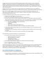
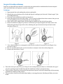
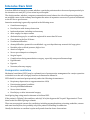
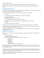
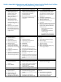
Package for Emergency Resuscitation and Intensive Care Unit For details refer to the WHO manual Surgical Care at the District Hospital and WHO Integrated Management for Emergency & Essential Surgical Care *IMEESC toolkit: http://www.who.int/surgery/en/index.html Anaesthesia and Oxygen Key Points: A reliable oxygen supply is essential for anaesthesia and for any seriously ill patients In many places, oxygen concentrators are the most suitable and economical way of providing oxygen, with backup cylinders in case of electricity failure Whatever your source of oxygen, you need an effective system for maintenance and spares Clinical staffs need to be trained to use oxygen safely, effectively and economically. A high concentration of oxygen is needed during and after anaesthesia, particularly if: – The patient is very young, old, sick, or anaemic – Using agents that cause cardio-respiratory depression, such as halothane. Air already contains 20.9% oxygen, so oxygen enrichment with a draw-over system is a very economical method of providing oxygen. Adding only 1 litre per minute may increase the oxygen concentration in the inspired gas to 35–40%. With oxygen enrichment at 5 litres per minute, a concentration of 80% may be achieved. Industrial-grade oxygen, such as that used for welding, is perfectly acceptable for the enrichment of a draw-over system and has been widely used for this purpose. Oxygen Sources In practice, there are two possible sources of oxygen for medical purposes: Cylinders: derived from liquid oxygen Concentrators: which separate oxygen from air. For remote hospitals that cannot obtain oxygen cylinders on a regular basis, there is a strong case for introducing concentrators. However, cylinders can be used to supply oxygen during power cuts and concentrators cannot. Without electricity, the flow of oxygen from a concentrator will stop within a few minutes. The ideal oxygen supply system is one based primarily on concentrators, but with a back-up supply from cylinders. Cylinder system Inexpensive to buy Expensive to operate Needs year-round supply of cylinders Training and maintenance needed Can store oxygen Concentrator system More expensive to buy Inexpensive to operate Needs only electricity Training and maintenance needed Cannot store oxygen: provides oxygen only when power supply is on Oxygen Cylinders Cylinders of oxygen are produced by a relatively expensive industrial process. An oxygen cylinder needs a special valve (regulator) to release the oxygen in a controlled way and a flow meter to control the flow. Without a flow meter, the use of oxygen from cylinders is very wasteful; without a regulator it is also extremely dangerous. Not all oxygen cylinders are the same; there are at least five different kinds of cylinder in use in different countries. A regulator will fit only one type of oxygen cylinder. Precise information on the type of oxygen cylinder in use should be obtained from the local oxygen supplier before ordering regulators. This should be confirmed by someone with technical knowledge who works in the hospital, such as an anaesthetist, chest physician or fully trained hospital technician. An international standard exists for the identification of oxygen cylinders, which specifies that they should be painted white. Unfortunately, the standard is widely ignored. Medical oxygen cylinders originating in the USA are normally green, while those originating in Commonwealth countries are usually black with white shoulders. Cylinders of industrial oxygen should also be identified clearly, but this is not always the case. Never use any cylinder to supply gas to a patient unless you are sure of its contents. Getting oxygen to patients requires more than simply having oxygen cylinders available. You must have in place an entire functioning system, comprising not only the apparatus for oxygen delivery, but also people who have been trained to operate it and a system for maintenance, repair and supply of spare parts. A complete system for using oxygen in cylinders requires: Reliable source of oxygen supply in cylinders Transport to get the cylinders to the hospital Procedures to ensure that the hospital orders the appropriate amount of oxygen Apparatus to deliver oxygen from the cylinder to the patient: – Suitable regulator – Flow meter – Oxygen delivery tubing – Humidifier – Tube to carry oxygen to the patient’s face – Nasal catheter (or mask) to deliver the oxygen to the patient’s airway Person with clinical training to give the correct amount of oxygen, in the correct manner, to the patients who need it Person with technical training to inspect the apparatus, maintain it in good condition and repair it when necessary Adequate budget to ensure the consistent availability of the oxygen supply. Safe use of oxygen cylinders The oxygen supply from a cylinder must be connected through a suitable pressure-reducing valve (regulator). For larger cylinders, this valve is incorporated into the cylinder’s pressure gauge; on a Boyle’s machine both the gauge and the pressure-reducing valve are part of the machine. Using oxygen from cylinders without a regulator is extremely dangerous. When connecting a cylinder to the anaesthetic apparatus, make sure that the connectors are free from dust or foreign bodies that might cause the valves to stick. 2 Never apply grease or oil, as it could catch fire in pure oxygen, especially at high pressure. Remember that an oxygen cylinder contains compressed oxygen in gaseous form and that the reading on the cylinder pressure gauge will therefore fall proportionately as the contents are used. A full oxygen cylinder normally has a pressure of around 13 400 kPa (132 atmospheres, 2000 p.s.i.). It should always be replaced if the internal pressure is less than 800 kPa (8 atmospheres, 120 p.s.i.) as failure is then imminent. Oxygen cylinders are dangerous objects. If they fall over, they may injure or even kill. Make sure that cylinders are safely stored and mounted. In storage, they should lie horizontally. In use, they should be securely fixed in the vertical position to a wall or be kept standing secured with a restraining strap or chain. Supplies, equipment and maintenance Compressed oxygen is expensive and using it may pose logistical and cost problems for small or remote hospitals. In the United Republic of Tanzania, for example, a recent survey showed that 75% of district hospitals had an oxygen supply for less than 25% of the year. A reliable system for cylinder oxygen depends on a good source of supply and reliable year-round transportation. In many countries, oxygen cylinders must be bought rather than rented and frequent losses of cylinders in transit impose additional costs. Fortunately, since oxygen is needed for a variety of industrial as well as medical applications, it is widely available. Because cylinders of “industrial” oxygen and of “medical” oxygen are produced by the same process (the fractional distillation of air), good-quality industrial oxygen is perfectly safe for medical use. It may also be easier to obtain and less expensive, since a price premium is often levied for “medical-grade” oxygen. However, if you obtain oxygen from an unorthodox source, you must check it for purity before use (a portable analyzer may be used). Efficient and economical use of oxygen – while still ensuring that the patient receives the maximum benefit – is important. If properly understood, oxygen supplies can be used quite economically. The oxygen concentration of air (21%) generally needs to be increased only to about 40% in order to bring great benefit to the majority of patients who need extra oxygen. Oxygen Concentrators Oxygen concentrators are suitable for use in all levels of hospital. They provide oxygen more cheaply than cylinders, as well as making oxygen available in hospitals where a regular supply of cylinders is difficult to obtain. Oxygen concentrators designed for use with individual patients normally give a flow rate of up to 4 litres per minute of near-pure oxygen at relatively low pressure. This oxygen can be used in exactly the same way as oxygen from a cylinder: As the supply for T-piece enrichment into a draw-over system For use with a nasal catheter, prongs or face mask to give postoperative or ward oxygen. If there is an electrical power failure, the oxygen flow from a concentrator will continue for about a minute only, so make sure you have a back-up system for use in such emergencies – either a generator to maintain electrical supply, or a cylinder of compressed oxygen. 3 Oxygen concentrators have been installed in many hospitals where cylinders are not consistently available. Concentrators ensure a more reliable and lower cost supply of oxygen than cylinders. An oxygen concentrator uses zeolite to separate oxygen from nitrogen in air. The oxygen produced by concentrators is at least 90% pure and can be used in the same way as oxygen from cylinders, with the same beneficial effects. Oxygen concentrators require much less energy than fractional distillation and have the additional advantage that oxygen is easily produced in the operating room or at the patient’s bedside, provided that there is an electricity supply (a small concentrator uses about 350 W). The purchase price of a concentrator is about half the cost of a year’s supply of oxygen from cylinders and running costs for electricity and spare parts are low. A complete system for oxygen delivery based on concentrators, requires: Manufacturer and supplier of concentrators Electricity in the hospital: either mains electricity or a generator System to ensure that a sufficient supply of major spare parts is purchased and stored centrally and an adequate supply of minor spare parts, such as air intake filters, is available at each hospital Apparatus to deliver oxygen from the concentrator to the patient, which includes: – Flow meter (included in every concentrator) – Oxygen delivery tubing – Humidifier – Tube to carry oxygen to the patient’s face – Nasal catheter (or mask) to deliver the oxygen to the patient’s airway Person with clinical training to give the correct amount of oxygen, in the correct manner, to the patients who need it Person with technical training to maintain the apparatus in good condition and to repair it when necessary Adequate budget to ensure the consistent availability of the oxygen supply. For successful use in a district hospital, a concentrator must: Be capable of functioning in adverse circumstances: – Ambient temperature up to 40 °C – Relative humidity up to 100% – Unstable mains voltage – Extremely dusty environment Be incapable of delivering an oxygen concentration of less than 70% oxygen Have a comprehensive service manual Have a supply of spare parts for two years’ use. A hospital planning to use oxygen concentrators should consider buying at least two. Remember that no piece of equipment will last for ever, especially if it is neglected. Hospitals need to plan for regular maintenance – usually after every 5000 hours of use. Servicing the machines is not complicated and can, if necessary, be carried out by the user after simple training. AND OTHER RISKS Care and maintenance of equipment The important principles of care and maintenance are: The anaesthetist working alone in a small hospital must understand and take responsibility for the upkeep of apparatus as well as for the care of patients 4 All equipment requires regular inspection, maintenance and repair to prevent it from rapidly deteriorating and becoming dangerous Make a detailed list or inventory of the equipment you have to enable you to identify any extra items needed As well as basic equipment, list spare parts, batteries and other consumables that will be needed and find out in advance how you can obtain them Try to estimate when new parts will be required and order spares well in advance, before the machine breaks down and leaves you in difficulty Ensure that all types of apparatus are kept in a clean and dust-free environment, away from extremes of temperature and covered when not in use Ensure that vaporizers are drained of anaesthetic if they are unlikely to be used for a week or more Put a cork or spigot in the end of any gas port or tubing during storage to prevent the entry of insects. Airway Management First priority is establishment or maintenance of airway patency. 1. Talk to the patient A patient who can speak clearly must have a clear airway. Airway obstruction by the tongue in the unconscious patient is often a problem. The unconscious patient may require assistance with airway and/or ventilation. If you suspect a head, neck or chest injury, protect the cervical spine during endotracheal intubation. 2. Give oxygen Give oxygen, if available, via self-inflating bag or mask. 3. Assess the airway Signs of airway obstruction include: Snoring or gurgling Stridor or abnormal breath sounds Agitation (hypoxia) Using the accessory muscles of ventilation/paradoxical chest movements Cyanosis. Be alert for foreign bodies. Intravenous sedation is absolutely contraindicated in this situation. 4. Consider the need for advanced airway management 5. Indications for advanced airway management techniques include: Persisting airway obstruction Penetrating neck trauma with hematoma (expanding) Apnea Hypoxia Severe head injury Chest trauma Maxillofacial injury. Airway obstruction requires urgent treatment. 5 Surgical Cricothyroidotomy Surgical cricothyroidotomy should be conducted in any patient where intubation has been attempted twice and failed and/or the patient cannot be ventilated. Technique 1. Hyperextend the neck, making the patient comfortable. 2. Identify the groove between the cricoid and thyroid cartilages just below the “Adam’s apple” (the protruding thyroid). 3. Clean the area and infiltrate with local anesthetic. 4. Incise through the skin vertically with a 1.5 cm cut and use blunt dissection to ensure that you can see the membrane between the thyroid and cricoid (Figure 1). 5. With a #22 or #23 scalpel blade, stab through the membrane into the hollow trachea. 6. Rotate the blade 90° (Figure 2), insert a curved artery forceps alongside the blade, remove the blade and open the forceps side to side, widening the space between the thyroid and cricoid cartilages (Figure 3). Figure 1 Figure 2 Figure 3 7. Pass a thin introducer or a nasogastric tube into the trachea if very small access (Figure 4) or proceed to step 9. 8. Run a 4–6 endotracheal tube over the introducer and pass it into the trachea (Figure 5). 9. Remove the introducer, if used. Figure 4 Figure 5 This tube can stay in place for up to 3 days. Do not attempt this procedure in a child under the age of 10 years; passing several needles through the membrane will give enough air entry. This procedure should be performed by an experienced person, with prior knowledge of the anatomy and medical condition of the patient. This procedure should not be undertaken lightly, as wrong placement, bleeding and delay can cause death. 6 Intensive Care Unit It is often difficult to know for certain whether a particular patient needs to be nursed postoperatively in the intensive care unit (ICU), if one exists in your hospital. The decision maker, whether a surgeon or anaesthetist, must balance the risk of the patient dying from an avoidable cause on the ordinary ward against the waste of expensive resources if a patient is admitted to the ICU for no good reason. Intensive monitoring is generally required in the following cases: Cranial neurosurgery Head injuries with airway obstruction Intubated patients, including tracheostomy After surgery for major trauma Abdominal surgery for a condition neglected for more than 24 hours Chest drain in the first 24 hours Ventilation difficulties Airway difficulties, potential or established: e.g. post-thyroidectomy removal of a large goitre Unstable pulse or blood pressure, high or low Anuria or oliguria Severe pre-eclampsia or eclampsia Surgical sepsis Complications during anaesthesia or surgery, especially unexpected haemorrhage Hypothermia Hypoxia Neonates, after any surgery. Postoperative ventilation Mechanical ventilation (IPPV) may be a planned part of postoperative management for a major operation or decided on at the end of surgery because circumstances demand it. IPPV should be continued postoperatively under the following circumstances: Respiratory depression or oxygen saturation <80% Deteriorating general condition Severely distended abdomen Severe chest trauma Head injury or after intracranial surgery. Avoid giving long acting muscle relaxants to facilitate IPPV. If the patient is “fighting” the ventilator, ask why? Is he/she hypercarbic? In pain? Hypertensive? Treat these needs first before giving a relaxant. There are non-surgical reasons for ventilation, including organophosphate poisoning, snakebite, tetanus and some head injuries, but probably only if the patient is breathing on admission. Usually the decision to ventilate is quite easily made from the above observations. 7 But, if in doubt, ventilate. With no ventilator, a patient in respiratory failure will rapidly die of hypoxia and hypercarbia. Many people die purely for lack of a short period of ventilation in the postoperative period or after trauma. Discharge from the ICU The decision to discharge the patient from the ICU very much depends on the quality of care to be found on the ward to which the patient is being transferred. The following conditions should be met before discharging the patient from ICU: Conscious Good airway, extubated and stable for several hours after extubation Breathing comfortably Stable blood pressure and urine output. Haemoglobin >6 g/dl or blood transfusion in progress Minimal nasogastric drainage and has bowel sounds, abdomen not distended Afebrile Looks better, sitting up, not confused. Pressure for beds to treat more urgent cases may mean that these guidelines have to be modified. If a patient dies after discharge from ICU, try to find out why the death took place and to learn from it, especially if it appears that the death was avoidable. Try to put a system in place where patients discharged from ICU are followed up for a week. Find out what happened to them. Equipment for the ICU The ICU does not necessarily need to have ventilators or other expensive machines. An ICU might be a ward where: Oxygen is available Drips are kept running overnight At least hourly measurements and observations are made of: – Blood pressure – Pulse rate – Urine output – Oxygenation – Conscious level – Other general observations of the patient. The monitoring of the patient all night long is the deciding factor in the success or failure of the ICU. Another important feature is whether staff take action when the measurements or observations show that something is wrong. The provision of one or more simple, reliable electric ventilators (not gas or oxygen dependent) will double the usefulness of a basic ICU. Small, portable mains/battery ventilators with integral compressors are available, although they are relatively expensive. The pulse oximeter should be the minimum standard of monitoring in every operating room where regular major surgery is carried out. 8 Guide to Anaesthetic Infrastructure and Supplies at Various Levels of Health Care Facilities Emergency and Essential Surgical Procedures Level 1 Small hospital/health center Rural hospital or health center with a small number of beds and a sparsely equipped operating room (OR) for minor procedures Provides emergency measurements in the treatment of 90-95% of trauma and obstetrics cases (excluding caesarean section) Referral of other patients (for example, obstructed labor, bowel obstruction) for further management at a higher level Procedures Normal delivery Uterine evacuation Circumcision Hydrocele reduction, incision and drainage Wound suturing Control of hemorrhage with pressure dressings Debridement and dressing of wounds Temporary reduction of fractures Cleaning or stabilization of open and closed fractures Chest drainage (possibly) Personnel Paramedical staff without formal anaesthesia training Nurse-midwife Drugs Ketamine 50 mg/ml injection, 10 ml Lidocaine 1% or 2% Diazepam 5 mg/ml injection, 2 ml injection Epinephrine (adrenaline) Level 2 District/provincial hospital District or provincial hospital with 100-300 beds and adequately equipped major and minor operating theaters Short term treatment of 95-99% of the major life-threatening conditions Procedures Same as Level 1 with the following additions: Caesarean section Laparotomy (usually not for bowel obstruction) Amputation Hernia repair Tubal ligation Closed fracture treatment and application of plaster of Paris Eye operations, including cataract extraction Removal of foreign bodies: e.g. in the airway Emergency ventilation and airway management for referred patients such as those with chest and head injuries Personnel One or two trained anaesthetists District medical officers, senior clinical officers, nurses, midwives Visiting specialists or resident surgeons and/or obstetricians/gynecologists Drugs Same as Level 1, but also: Thiopental 500 mg/1g powder Suxamethonium bromide 500 mg powder Atropine 0.5 mg injection Epinephrine (adrenaline) 1 mg Level 3 Referral hospital A referral hospital of 300–1000 or more beds with basic intensive care facilities. Treatment aims are the same as for Level 2, with the addition of: Ventilation in OR and ICU Prolonged endotracheal intubation Thoracic trauma care. Hemodynamic and inotropic treatment Basic ICU patient management and monitoring for up to 1 week : all types of cases, but with limited or no provision for: Multi-organ system failure Hemodialysis Complex neurological and cardiac surgery Prolonged respiratory failure Metabolic care or monitoring Procedures Same as Level 2 with the following additions: Facial and intracranial surgery Bowel surgery Pediatric and neonatal surgery Thoracic surgery Major eye surgery Major gynecological surgery, e.g. vesico-vaginal repair Personnel Clinical officers and specialists in anaesthesia and surgery Drugs Same as Level 2 with the following additions: Vecuronium 10 mg powder Pancuronium 4 mg injection Neostigmine 2.5 mg injection Trichloroethylene 500 ml 9 1 mg Atropine 0.6 mg/ml Equipment: capital outlay Adult and pediatric resuscitators Foot sucker Oxygen concentrator Equipment: disposable IV equipment Suction catheters Examination gloves injection Diazepam 10 mg injection Halothane 250 ml inhalation Lidocaine 5% heavy spinal solution 2 ml Bupivacaine 0.5% heavy or plain 4 ml Hydralazine 20 mg injection Frusemide (or Furosemide) 20 mg injection Dextrose 50% 20 ml injection Aminophylline 250 mg injection Ephedrine 30/50 mg ampoules Equipment: capital outlay Complete anaesthesia, resuscitation and airway management system consisting of: Oxygen source (concentrator or cylinder) Vaporizer(s) Hoses Valves Bellows or bag to inflate lungs Face masks Adult and pediatric resuscitator sets Pulse oximeter Laryngoscope Macintosh blades Foot sucker (electric) IV pressure infusor bag Magills forceps (adult and child), intubation stylet and/or bougie Equipment: disposable IV equipment (minimum fluids, normal saline, Ringer’s lactate and dextrose 5%) Suction catheters Examination gloves Sterile gloves Nasogastric tubes Oral airways Tracheal tubes Spinal needles Batteries inhalation Calcium chloride 10% 10 ml injection Potassium chloride 20% 10 ml injection for infusion Equipment: capital outlay Same as Level 2 with the following additions (one of each per OR or per ICU bed, except where stated): Pulse oximeter, spare probes, adult and pediatric* ECG (electrocardiogram) monitor* Anaesthesia ventilator, electric power source with manual override Infusion pumps (2 per bed) Pressure bag for IVI Electric sucker Defibrillator (one per OR / ICU) Automatic BP machine* Capnograph* Oxygen analyzer* Thermometer (temperature probe*) Electric warming blanket Electric overhead heater Infant incubator Laryngeal mask airways sizes 2, 3, 4 (3 sets per OR) Intubating bougies, adult and child (1 set per OR) * It is preferable to buy combined modalities all in one unit Equipment: disposable Same as Level 2 with the following additions: ECG dots Ventilator circuits Yankauer suckers Giving sets for IVI pumps Disposables for suction machines Disposables for capnography, oxygen analyzer, in accordance with manufacturers’ specifications: Sampling lines Water traps Connectors Filter-Fuel cells 10 WHO GENERIC ESSENTIAL EMERGENCY EQUIPMENT LIST This checklist of essential emergency equipment for resuscitation describes minimum requirements for emergency and essential surgical care at the first referral health facility Capital Outlays Quantity Date Checked Resuscitator bag valve and mask (adult) Resuscitator bag valve and mask (pediatric) Oxygen source (cylinder or concentrator) with mask and tubing Stethoscope Suction pump (manual or electric) with catheter Blood pressure measuring equipment Thermometer Scalpel with blades Retractor Scissors Oropharyngeal airway (adult size) Oropharyngeal airway (pediatric size) Forcep, artery Gloves (sterile) Gloves (examination) Needle holder Sterilizer Vaginal speculum Inventory list of equipment and supplies Best practice guidelines for emergency care Renewable Items Nasogastric tubes Light source (lamp & flash light) Intravenous fluid infusion set Intravenous cannulas/scalp vein infusion set Syringes with needles (disposable) Sharps disposal container Tourniquet Needles and sutures Splints for arm, leg Urinary catheters (Foleys disposable) Waste disposal container Face masks Eye protection Protective gowns/aprons Soap Supplementary equipment for use by skilled health professionals Magill Forceps (adult) Magill Forceps (pediatric) Endotracheal tubes (adult) Endotracheal tubes (pediatric) IV infusor bags Chest tube insertion equipment Laryngoscope handle Laryngoscope Macintosh blades (adult) with bulbs and batteries Laryngoscope Macintosh blades (pediatric) with bulbs and batteries Cricothyroidotomy set 11