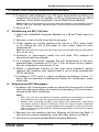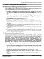
1
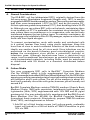
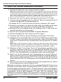
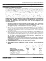
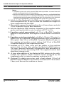
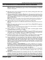
User Manual BacPAK™ Baculovirus Expression System User Manual United States/Canada 800.662.2566 Asia Pacific +1.650.919.7300 Europe +33.(0)1.3904.6880 Japan +81.(0)77.543.6116 Clontech Laboratories, Inc. A Takara Bio Company 1290 Terra Bella Ave. Mountain View, CA 94043 Technical Support (US) E-mail: [email protected] www.clontech.com Cat. No. 631402 PT1260-1 (PR862558) Published 5 June 2008 BacPAK™ Baculovirus Expression System User Manual Table of Contents I. Introduction 4 II. List of Components 6 III. Additional Materials Required 7 IV. Experimental Outline 8 V. Insect Cell Culture Guidelines 10 A. General Considerations 10 B. Culture Media 10 C. Establishing the Sf21 Cell Line 11 D. Subculturing Sf21 Monolayers 11 E. Suspension Cultures of Sf21 Cells 13 F. Storing Insect Cells in Liquid Nitrogen 14 VI. Plaque Assay Method 15 A. Practice Plaque Assay 15 B. Calculation of Virus Titer 17 C. Troubleshooting Plaque Assays 17 VII. Construction of a Recombinant Transfer Vector 18 A. Tailoring the Insert 18 B. Inserting the Target Gene into the Transfer Vector 18 VIII. Construction of a Recombinant Viral Expression Vector 19 A. Generating a Recombinant Virus 19 B. Troubleshooting Guide 21 C. Isolating Recombinant Viruses 22 IX. Virus Propagation and Evaluation 25 A. Preparation of Passage One Virus Stock 25 B. Evaluation of Recombinant Viruses 25 C. Processing and Storage of the Passage One Virus Stock 26 D. Amplification of Recombinant Viruses (Preparing Passage Two Virus Stock) E. Titration of Amplified Virus Stocks 26 27 X. Characterizing Recombinant Gene Expression 28 XI. Large-scale Target Protein Production Clontech Laboratories, Inc. www.clontech.com 2 29 Protocol No. PT1260-1 Version No. PR862558 BacPAK™ Baculovirus Expression System User Manual Table of Contents continued XII. References Appendix A: 30 Vector Maps & Multiple Cloning Site Sequences 31 List of Figures Figure 1. Figure 2. Figure 3. Figure 4. Figure 5. Figure 6. Figure 7. Target gene transfer to baculovirus expression vector Schematic diagram outlining BacPAK procedure Map of pBacPAK8 Transfer Vector Sequences in and around the pBacPAK8 MCS Map of pBacPAK9 Transfer Vector Sequences in and around the pBacPAK9 MCS Map of pBacPAK8-GUS Transfer Vector 5 9 31 31 32 32 33 List of Tables Table I. Guidelines for seeding densities Table II. Guidelines for preparing cells for analysis of gene production Protocol No. PT1260-1 www.clontech.com Version No. PR862558 13 28 Clontech Laboratories, Inc. 3 BacPAK™ Baculovirus Expression System User Manual I. Introduction Baculovirus gene expression is a popular method for producing large quantities of recombinant proteins in insect host cells. In most cases, posttranslational processing of eukaryotic proteins expressed in insect cells is similar to protein processing in mammalian cells. As a result, insect cellprocessed proteins have comparable biological activities and immunological reactivities to proteins expressed in mammalian cells. Protein yields from baculovirus systems are higher, and costs are significantly lower than in mammalian expression systems. The baculovirus expression system can express genes from bacteria, viruses, plants, and mammals at levels from 1–500 mg/liter; most proteins are expressed in the 10–100 mg/liter range, although making predictions is difficult. The baculovirus most commonly used to express foreign proteins is Autographa californica nuclear polyhedrosis virus (AcMNPV; Luckow, 1991; Vlak & Keus, 1990; Bishop & Possee, 1990; Miller, 1988; Luckow & Summers, 1988; O’Reilly et al., 1992). AcMNPV can be propagated in certain insect cell lines; the virus enters the cells and replication begins approximately 6 hours post-infection (h.p.i.). At approximately 20–48 h.p.i., transcription of nearly all genes ceases. The viral polyhedrin and p10 genes, however, are transcribed at high rates.The polyhedrin protein is essential for propagation of the virus in its natural habitat; however, in cell culture, polyhedrin is not needed, and its coding sequence can be replaced with a sequence for a target protein. Hence, the powerful polyhedrin promoter can drive high-level transcription of the insert, resulting in expression of a recombinant protein that can account for over 30% of total cellular protein. The large 134 kb-size of the AcMNPV genome (Ayres et al., 1994), makes direct manipulation of it difficult, so recombinant baculovirus expression vectors are constructed in two steps (Figure 1). First, a target gene is cloned into a modified polyhedrin locus contained in a relatively small transfer vector (<10 kb). The polyhedrin coding sequence has been deleted and replaced with a multiple cloning site (MCS). A target gene is inserted into this MCS, between the polyhedrin promoter and polyadenylation signals. Transfer vectors also contain a plasmid origin of replication and an antibiotic resistance gene for propagation in E. coli, but they are unable to replicate in insect cells. In the second step, the transfer vector and a viral expression vector are cotransfected into insect cells. Double recombination between viral sequences in the transfer vector and the corresponding sequences in the viral DNA transfers the target gene to the viral genome. The BacPAK™ Baculovirus Expression System uses BacPAK6, a specially engineered virus that facilitates construction and selection of recombinant expression vectors. BacPAK6 has an essential gene adjacent to the polyhedrin locus that provides selection for recombinant viruses (Kitts & Possee, 1993) (Figure 1). Sites for Bsu36 I, which does not cut wild-type AcMNPV DNA, were introduced into the genes flanking the polyhedrin expression locus Clontech Laboratories, Inc. www.clontech.com 4 Protocol No. PT1260-1 Version No. PR862558 BacPAK™ Baculovirus Expression System User Manual I. Introduction continued amp ori TARGET GENE r ESSENTIAL GENE ESSENTIAL GENE Transfer vector with insert Digested BacPAK6 viral DNA Recombination TARGET GENE ESSENTIAL GENE Recombinant baculovirus expression vector Polyhedrin promoter Figure 1. Transfer of a target gene to the baculovirus expression vector by forced recombination between a transfer vector and BacPAK6 viral DNA. of BacPAK6. Digesting BacPAK6 with Bsu36 I releases two fragments. The first carries part of a downstream gene, ORF1629, that is essential for viral replication (Possee et al., 1991). If the second large DNA fragment recircularizes by itself, the resulting viral DNA will lack an essential part of the genome and be unable to produce viable viruses. However, the transfer vector carries the missing ORF1629 sequence, and if the large fragment recombines with it, the resulting circular DNA will contain all the genes necessary for viral replication. This double recombination event restores the essential gene and transfers the target gene from the transfer vector to the viral genome. Cotransfections using Bsu36 I-digested BacPAK6 viral DNA produce recombinant viruses at frequencies approaching 100%. This User Manual contains directions for establishing insect cell cultures, as well as for isolating a recombinant baculovirus expression vector using the BacPAK system. More extensive protocols for using baculovirus expression systems are in the baculovirus laboratory manuals (O’Reilly et al., 1992; King & Possee, 1992; Richardson, 1995) Protocol No. PT1260-1 www.clontech.com Version No. PR862558 Clontech Laboratories, Inc. 5 BacPAK™ Baculovirus Expression System User Manual II. List of Components Store the following item at –180°C (liquid nitrogen) immediately upon receipt. • 2 x 10⁶ IPLB-Sf21 Insect Host Cells in TNM-FH/10% FBS/ 10% DMSO Store the following items at 4°C; do not freeze. The following components are sufficient for five transfections. • 25 µl BacPAK6 Viral DNA (Bsu36 I digest) • 25 µl Bacfectin For long-term storage of 6 months or longer, store the following reagents at –70°C. For storage less than 6 months, store at 4°C, protected from light. • 2 ml BacPAK6 Virus Stock For long-term storage, store the following reagents at –20°C. For storage less than 6 months, store at 4 °C. • 15 µg pBacPAK8 Transfer Vector (500 ng/µl) • 15 µg pBacPAK9 Transfer Vector (500 ng/µl) • 2.5 µg pBacPAK8-GUS Vector (100 ng/µl) • 20 µl Bac1 Primer (20 µM) • 20 µl Bac2 Primer (20 µM) Note: The following kit component is also available separately: • BacPAK6 DNA (Bsu36 I digest) Cat No. 631401 Clontech Laboratories, Inc. www.clontech.com 6 Protocol No. PT1260-1 Version No. PR862558 BacPAK™ Baculovirus Expression System User Manual III. Additional Materials Required The following materials are required but not supplied. • BacPAK Complete Medium Cat No. 631403. • • • • • • • • • You may also useTNM-FH insect cell medium (Grace’s medium supplemented with yeastolate and lactalbumin hydrolysate) with fetal bovine serum (cell culture grade; ask vendor for a lot tested with insect cells), and antibiotics. • • • • • • • • BacPAK Grace’s Basic Medium Cat No. 631404 CHROMA SPIN™ + TE-400 Columns Cat. No. 636076 Dimethylsulfoxide (DMSO) (cell culture grade) Neutral red stain (0.33%) Trypan blue dye (0.4%) X-GAL (25 mg/ml) (5-bromo-4-chloro- 3-indolyl-β-Dgalactopyranoside) in dimethylformamide (DMF). Store away from light at –20°C. X-GLUC (25 mg/ml) (5-bromo-4-chloro-3-indolyl-β-D-glucuronic acid) (Cat No. 631721) in DMSO. Store away from light at –20°C. RNase A (10 mg/ml) Store at –20°C Proteinase K (10 mg/ml; made fresh; Cat No. 635919) Store at –20°C SeaPlaque® Agarose (Cambrex Cat No. 50101) Sterile H2O 3 M NaOAc (pH 5.2) Lysis buffer 50mM Tris-HCl (pH 8.0) 10mM EDTA 5 %β-mercaptoethanol 0.4%Sodium dodecylsulfate Phosphate buffered saline (PBS) 140mM NaCl 27mM KCl 8mMNa2HPO4 1.5mM KH2PO4 (pH 7.3) TE buffer 10mM Tris-HCl (pH 8.0) 1mMEDTA Phenol:chloroform (50:50), equilibrated with 100 mM Tris-HCl (pH 8.0) Ethanol (100% and 70%) Protocol No. PT1260-1 www.clontech.com Version No. PR862558 Clontech Laboratories, Inc. 7 BacPAK™ Baculovirus Expression System User Manual IV. Experimental Outline Please refer to Figure 2 on the following page. • Obtain insect cell media and establish Sf21 cell line. This step will take 3–4 weeks (Section V.B–C). • Maintain working stocks of Sf21 cells (Sections V.D–E). • When the stock of cells has been passaged twice, freeze aliquots for long-term storage in liquid nitrogen. Aliquots of frozen cells provide a back-up in case the working stock dies or becomes contaminated. Frozen cells are also a source of fresh cells for replacing working stocks as they become old (Section V.F). • Practice assaying viral plaques using the BacPAK6 virus stock provided in the kit (Section VI.A). Isolating pure recombinant virus requires good viral plaques. Therefore, developing a good plaque assay technique before working with recombinant viruses is advisable. • Insert target gene into transfer vector (Section VII) and prepare plasmid DNA. • Produce a recombinant virus by cotransfecting Sf21 cells with BacPAK6 viral DNA and the transfer vector-target gene clone (Section VIII.A). • Perform plaque assays on the cotransfection supernatant to obtain individual viral plaques (Section VIII.C). • Test the putative recombinant viruses to confirm that they have incorporated the target gene and/or express the target protein (Section IX.A–B). • Amplify recombinant viruses to obtain working stocks (Section IX.C– D). • Titer amplified virus stock (Section IX.E). • Perform small-scale infections to characterize gene expression and to determine the optimum harvest time and infection ratio that will give maximum protein yield (Section X). • Scale-up: produce target protein in large quantities by infecting larger batches of insect cells (Section XI). Clontech Laboratories, Inc. www.clontech.com 8 Protocol No. PT1260-1 Version No. PR862558 BacPAK™ Baculovirus Expression System User Manual IV. Experimental Outline continued Prepare target vector (Section VII) • Insert target gene into transfer vector • Verify correct construct • Plasmid Preparation Establish Sf21 cells (Section V.C) Maintain working stocks of Sf21 cells Freeze cells for long-term storage (Sections V.D & V.E) (Section V.F?) Practice plaque assay (Section VI.A) Plasmid DNA BacPAK6 Viral DNA Cotransfect Sf21 cells with plasmid DNA and BacPAK6 viral DNA (Section VIII.A) Plaque assay of progeny viruses (Section VIII.C [& VI.A]) Pick several putative recombinant virus plaques & Confirm presence and/or expression of target gene (Sections IX.A & B) You may use theBacPAKTM Baculovirus Rapid Titer Kit (Cat. No. 631406) at steps IX.C & IX.E. Amplify recombinant virus (Sections IX.C & D) Titer amplified virus stock (Section IX.E) Characterize gene expression (Section X) Scale-up protein production (Section XI) Figure 2. Schematic diagram outlining BacPAK™ Baculovirus Expression Procedures. Protocol No. PT1260-1 www.clontech.com Version No. PR862558 Clontech Laboratories, Inc. 9 BacPAK™ Baculovirus Expression System User Manual V. Insect Cell Culture Guidelines A. General Considerations The IPLB-Sf21 cell line (abbreviated Sf21), originally derived from the fall army worm, Spodoptera frugiperda (Vaughn et al., 1977), is used to propagate AcMNPV-based expression vectors.These cells grow reasonably well from room temperature (~22°C) to 30°C and do not require CO2. At their optimum growth temperature (27°C), their doubling time is 20–24 hr. Although you can culture Sf21 cells on the bench, maintaining them in an incubator at 27°C is preferable for consistent virus infections. You may culture them as monolayers or in suspension; cells can be freely transferred between the two culture types. To maintain consistency, do not passage cells indefinitely. After 20–30 passages, replace a culture with fresh cells from liquid nitrogen. To prevent contamination, work with media and uninfected cells in a vertical laminar flow hood, using sterile technique. Keep this hood free of virus to avoid accidental infection of the stock cultures; ideally use another hood for all virus work. Virus infections can be performed on the bench following good microbiological practice, unless the recombinant virus carries a potentially harmful or infectious gene. Although baculoviruses have a restricted host range, treat recombinant baculoviruses as potential biohazards. All virus-contaminated materials, including fluids, must be autoclaved or disinfected with 5% bleach or a chemical disinfectant before disposal. B. Culture Media You may propagate Sf21 cells in BacPAK™ Complete Medium (Cat No. 631403), which is fully supplemented. You may also use serum-freemediasuchasBacPAKGrace’sBasicMedium(Cat.No.631404)for assaying or purifying secreted proteins. Insect cell medium does not contain pH indicators and is pale yellow. The pH of the medium is about 6.2, and it will gradually rise as the cells grow; however, pH will usually not exceed 6.4. BacPAK Complete Medium contains TNM-FH medium (Grace’s Basic Medium [Grace, 1962] with yeastolate, lactalbumin hydrolysate, and L-glutamine [Hink, 1970] supplemented with 10% FBS and 50 µg/ml gentamycin). You may substitute fully supplemented BacPAK Grace’s Basic Medium (Cat. No. 631404) for BacPAK Complete Medium throughout these protocols. Alternatively, prepare TNM-FH medium (Hink, 1970), and supplement as follows: 1.Add 50 ml of fetal bovine serum (cell culture-grade, preferably insect cell tested) to a 500-ml bottle of TNM-FH medium to give 10% v/v FBS. Clontech Laboratories, Inc. www.clontech.com 10 Protocol No. PT1260-1 Version No. PR862558 BacPAK™ Baculovirus Expression System User Manual V. Insect Cell Culture Guidelines continued 2.If desired, add antibiotics, e.g., 50 units of penicillin and 50 µg of streptomycin per ml of medium, or 50 µg of gentamycin per ml of medium, from a filter-sterilized, concentrated stock solution. Note: Antibiotic use is optional, but strongly recommended for cotransfections, plaque assays, and viral infections, because these cultures are prone to contamination. 3.Store at 4°C. C. Establishing the Sf21 Cell Line 1.Add 5 ml of BacPAK Complete Medium to a 25-cm2 flask; warm to 27°C. 2.Remove a vial of cells from liquid nitrogen. 3.Thaw rapidly by briefly dipping the vial in a 37°C water bath or by rolling the vial in the palm of your hand. Keep at room temperature. Note: Do not continue to warm the cells after they have thawed. Heating cells above 28°C will kill them. 4.Immerse or thoroughly swab the vial with 70% ethanol to decontaminate the outside. 5.In a laminar flow hood, transfer the cell suspension to the prewarmed flask. Incubate at 27°C for 1–3 hr to allow cells to attach. Do not incubate for more than 12 hr. 6.When a significant fraction of the cells have attached, gently remove the medium and replace with 5 ml of fresh prewarmed (27°C) medium. 7.Incubate at 27°C until a nearly confluent monolayer forms (~7 days). We recommend checking the flasks for confluency every other day. D. Subculturing Sf21 Monolayers 1.Examine cell monolayers under an inverted microscope to check that the cells are healthy and ready for passaging. The monolayer should be 80–90% confluent. Notes: • Sf21 cells are not susceptible to contact inhibition. If monolayers become overconfluent, the cells will start to float and divide in the media. • Healthy cells should be rounded, have distinct cell boundaries, and should not appear granular. Signs of unhealthy cells are a large number of floating cells before confluency, sausage-shaped cells stopped in mid-cell division, and cells with rough boundaries. Contaminated cultures will become cloudy within 24–48 hr. 2 Remove the old medium and any floating cells from the flask. If the cells are mainly detached, omit this step and go to Step 4. 3.Add 5 ml of prewarmed BacPAK Complete Medium. Protocol No. PT1260-1 www.clontech.com Version No. PR862558 Clontech Laboratories, Inc. 11 BacPAK™ Baculovirus Expression System User Manual V. Insect Cell Culture Guidelines continued 4.Gently dislodge the cells using a sterile scraper. Note: Many commercial scrapers are harsh and using them may result in significant numbers of dead cells. Nunc scrapers are acceptable, but the best ones are made by attaching a piece of silicon rubber tubing to a bent glass rod. Alternatively, wash cells using a stream of medium from a pipette. Sf21 cells attach less strongly to glass, and passaging them is easier if you use glass tissue culture flasks. 5 Disperse the cells by gently pipeting up and down 3–4 times. 6.Transfer the cell suspension to a 150-cm2 flask containing 30 ml of prewarmed BacPAK Complete Medium. 7.Incubate at 27°C until the cells are barely confluent (3–5 days). 8.Examine monolayers to check that the cells are healthy and ready for passaging. The monolayer should be 80–90% confluent. 9.Remove the old medium and any floating cells. If the cells are mainly detached, omit this step. 10.Add 10 ml of prewarmed BacPAK Complete Medium. 11.Gently dislodge the cells using a sterile scraper. 12.Disperse the cells by gently pipeting up and down 3–4 times. 13.Add 0.3 ml of cell suspension to 0.3 ml of 0.08% (w/v) trypan blue in PBS. Count cells with a hemocytometer; dead cells take up the blue stain. Determine the concentration and proportion of viable cells. Note: After careful harvesting, healthy monolayer cells from plastic flasks should have viabilities of 80–90%; monolayers harvested from glass flasks should have viabilities of >90%; and suspension cultures should have viabilities of >95%. 14.Remove all but 2 ml of the 10-ml cell suspension, and store it in a sterile container. Add 30 ml of prewarmed BacPAK Complete Medium to the remaining 2 ml of culture. Swirl to mix, and incubate at 27°C. For information on incubation times, see note below. 15.Add 2 ml of the reserved cell suspension to a second 150-cm2 flask containing 30 ml of prewarmed BacPAK Complete Medium. Swirl to mix, and incubate at 27°C. The cells from this flask will be frozen (Section V.F.) For information on incubation times, see note below. 16.Use a portion of the remaining reserved cell suspension to seed a 50-ml suspension culture (Section V.E). Maintain monolayer stocks of cells by repeating Steps 8–14. You must passage monolayers split 1:8 and grown at 27°C every 3–4 days; you must passage monolayers split 1:10 and grown at room temperature once a week. Depending upon your needs, you may split near-confluent monolayers at any ratio between 1:2 and 1:20. As needed, seed additional monolayer flasks and suspension cultures to provide cells for experiments. For additional information on seeding densities, see Table I. Clontech Laboratories, Inc. www.clontech.com 12 Protocol No. PT1260-1 Version No. PR862558 BacPAK™ Baculovirus Expression System User Manual V. Insect Cell Culture Guidelines continued table i. guidelines for seeding densities Size of vessel 25-cm2 flask 75-cm2 flask 150-cm2 flask Spinner/shake flasks Number of cells 106 1.0 x 3.0 x 106 6.0 x 10⁶ 2.0 x 10⁵/ml Volume of media 5 ml 10 ml 30 ml 50–500 ml E. Suspension Cultures of Sf21 Cells Suspension cultures, using either spinner or shake flasks, are easy to maintain and reproducibly give cells of a high viability (>95%) that are good for experimental work. Suspension cultures are particularly useful when large numbers of cells are needed. Growing cells in spinners requires a low-speed magnetic stir platform which can be placed inside a 27°C incubator. Note that some stir platforms generate too much heat to be used inside an incubator. Flat-bottomed pyrex flasks (100–1000 ml) containing a magnetic stir bar, and covered with a foil cap, can be used as spinner flasks. Alternatively, suspension cultures can be grown in shake flasks using an orbital shaker normally used for bacterial cultures. 1.Add an appropriate volume of prewarmed BacPAK Complete Medium to a sterile spinner or shake flask. Inoculate with cells to give a starting density of 4 x 10⁵ cells/ml. Note: Insect cells have a high oxygen demand; therefore, suspension cultures must have a high surface area to volume ratio or cell growth will be inhibited. The culture volume should be no more than two-fifths of the total volume of the flask; e.g., 40 ml of medium in a 100-ml flask, or 100 ml in a 250-ml flask. 2.Incubate cells at 27°C, and stir or shake at 50–100 rpm (use the minimum speed that will keep the cells in suspension). 3.Monitor the cell density daily until the culture reaches 2–3 x 10⁶ cells/ml (~4 days). Add 0.3 ml of cell suspension to 0.3 ml of 0.08% (w/v) trypan blue in PBS. Count the cells using a hemocytometer; viable cells exclude trypan blue, whereas dead cells take up the blue stain. 4.Use the cells to seed a fresh spinner/shake flask at a density of 1–2 x 10⁵ cells/ml. Alternatively, remove the excess cells and add fresh media to bring the density down to 1–2 x 10⁵ cells/ml. 5.Return to stirrer/shaker at 27°C, and monitor cell density daily, as above. Protocol No. PT1260-1 www.clontech.com Version No. PR862558 Clontech Laboratories, Inc. 13 BacPAK™ Baculovirus Expression System User Manual V. Insect Cell Culture Guidelines continued Notes: • Maintain one or more suspension cultures to provide cells for experimental work. Cells should only be used for virus infections and plaque assays if they are in the exponential phase of growth (usually 0.7–1 x 10⁶ cells/ml). Periodically (every 4–6 weeks) replace the working suspension cultures with fresh ones started from monolayer cells. • Take good care of your cells—the quality of virus plaques and the level of protein production are very dependent on the health of the host cells. F. Storing Insect Cells in Liquid Nitrogen Freezing aliquots of cells in liquid nitrogen provides a source of fresh cells to replace the working stocks when they become old. 1.Monitor the cells in a 150-cm2 flask to ensure that they are healthy and growing exponentially. When the monolayer reaches about 80% confluency, remove the old medium, add 5 ml of prewarmed BacPAK Complete Medium, gently scrape the cells, and disperse the cells by gently pipeting up and down. 2.Count the cells and ensure that they are at least 90% viable. 3.Adjust cell density to 4 x 10⁶ cells/ml with BacPAK Complete Medium. Chill the cells to 4°C. 4.Prepare an equal volume of BacPAK Complete Medium containing DMSO at 20% (v/v). Chill to 4°C. 5.Label cryogenic vials and put them on ice. 6.Add the BacPAK Complete Medium/DMSO to the cell suspension and mix. Keep on ice. 7.Place 1-ml aliquots of cells into each vial and cap tightly. 8.If available, place vials in a vapor-phase chamber of the liquid nitrogen container and freeze cells slowly overnight, before placing in the liquid phase. Alternatively, place vials at –20°C for 1–2 hr and then in a –70°C freezer overnight. Transfer to liquid nitrogen as rapidly as possible. 9.After a week or two, retrieve one vial and test the viability of the stored cells by following the protocol in Section V.C. Clontech Laboratories, Inc. www.clontech.com 14 Protocol No. PT1260-1 Version No. PR862558 BacPAK™ Baculovirus Expression System User Manual VI. Plaque Assay Method Plaque assays are designed to produce distinct viral plaques in a monolayer of host cells under conditions where each plaque is the result of a cell being infected by a single virus. Plaque assays can thus be used to isolate an individual recombinant virus from the pool of viruses generated by a cotransfection (SectionVIII.C). Plaque assays can also be used to determine the titer of a virus stock; however, titers can be obtained more quickly and easily using Clontech’s BacPAK Baculovirus Rapid Titer Kit (Cat. No. 631406). If plaque assays are to be used either to produce a pure recombinant virus clone or to titer virus stocks, it is advisable to develop good plaque assay technique by practicing using the virus stocks provided. A. Practice Plaque Assay BacPAK6 is a convenient virus for practice because it expresses β-galactosidase (Kitts & Possee, 1993) and produces plaques that can be stained blue with X-gal. 1.Remove an aliquot of exponentially growing Sf21 cells that have a viability of >95% and dilute with prewarmed BacPAK Complete Medium to make 18 ml of cell suspension at 7 x 10⁵ cells/ml. 2.Add 1.5 ml of the cell suspension to a 35-mm tissue culture dish, and rock to distribute evenly. Repeat for 9 more dishes. Each dish will receive approximately 1 x 10⁶ cells. Incubate the cultures at 27°C for 1–4 hr. Notes: • The correct cell density is critical to assay success. • To minimize problems with medium evaporation from the culture dishes during the incubation period, place the dishes in a plastic storage box that has a tight-fitting lid; place a folded, moist paper towel inside the box next to the dishes. • Seeding dishes with a volume of cell suspension less than 1.5 ml will result in an uneven distribution of cells over the dish. The volume added to each dish should be between 1.5 and 2.5 ml. 3.Make serial 1:10 dilutions of the BacPAK6 Virus Stock provided in the kit, in BacPAK Complete Medium to give final dilutions of 10-5 and 10-6. 4.Inspect the dishes to ensure that the cells have attached to form an even monolayer of about 70–80% confluency. Aspirate the medium from the cells using a sterile pasteur pipette or pipette tip. 5.Gently add 100 µl of the virus inoculum to the center of the dish, taking care not to displace any cells. Infect 4 dishes with the 10-6 and 4 with the 10-5 dilution of BacPAK6. Plate 100 µl of the dilution medium onto the remaining two dishes.These dishes will be useful for comparing with the infected dishes, and they provide a control that will reveal any contamination in the reagents. Protocol No. PT1260-1 www.clontech.com Version No. PR862558 Clontech Laboratories, Inc. 15 BacPAK™ Baculovirus Expression System User Manual VI. Plaque Assay Method continued 6.Incubate at room temperature for 1 hr on a level surface to allow the virus to infect the cells. 7.During this incubation, melt 10 ml of 2% agarose (2% SeaPlaque agarose in H2O, previously autoclaved) and cool to 37°C. Prewarm 10 ml of BacPAK Complete Medium to 37°C. 8.Add 24 µl of X-gal (25 mg/ml in DMF) per ml to the prewarmed BacPAK Complete medium (240 µl/10 ml, or final concentration 12 µl/ml medium when mixed with 2% agarose). 9.Remove the virus inoculum from the cells by tilting the dish and aspirating from the edge. Proceed immediately to step 10. 10.Add warm BacPAK Complete Medium to the agarose and mix; this makes a 1% agarose solution, which is used to overlay the infected cell monolayer to prevent virus progeny from spreading to other areas of the dish. Note: Water baths are a major source of microbial contamination; therefore, dry off the containers and flame the necks before mixing the agarose and medium. 11.Gently add 1.5 ml of the agarose overlay to each dish. Note: Allow the agarose to run down the side of the dish, taking care not to disturb the cells. 12.When the agarose overlay has set, add 1.5 ml of BacPAK Complete Medium to each dish. 13.Place dishes in a plastic storage box with a moist paper towel as described in the note to Step 2; incubate dishes at 27°C for 7 days. Note: Stain for virus plaques; half the dishes will be stained with neutral red only, and half with neutral red and X-gal. 14.Dilute neutral red to 0.03% with PBS (1 ml of 0.33% [w/v] neutral red stock + 10 ml of PBS). Add 1 ml of the 0.03% neutral red solution to each of the 10 dishes. Incubate at 27°C for 2–3 hr. 15.Aspirate off the stain, invert the dishes, and leave them in the dark at room temperature overnight to allow the plaques to clear and the blue color to fully develop. Notes: • Neutral red is taken up by healthy cells, but not by dead cells. Therefore, on the dishes stained with neutral red only, plaques will be clear circles about 0.5–3 mm in diameter, against a red or pink background. On the dishes stained with X-gal and neutral red, plaques will be blue against a red background. You should see blue foci in these dishes even if the plaques are small. Practice the plaque assay until you can see plaques with neutral red stain alone. • Neutral red is light-sensitive and will become grainy upon exposure to light. Clontech Laboratories, Inc. www.clontech.com 16 Protocol No. PT1260-1 Version No. PR862558 BacPAK™ Baculovirus Expression System User Manual VI. Plaque Assay Method continued B. Calculation of Virus Titer 1.Count the plaques on each dish that has a reasonable number of plaques (i.e., 10–30 per dish); from this count, calculate the average number of plaques per dish. 2.Since 0.1 ml of inoculum was applied to each dish, the titer of the virus stock (pfu/ml) is: (average plaques per dish) x 10 x (dilution factor)-1 3.Example calculation: 25 plaques x 10 x (10-5) -1 = 2.5 x 10⁷ pfu/ml C. Troubleshooting Plaque Assays To get good plaque formation, it is important to use cells which are in the exponential phase of growth and are >90% viable. The density at which the cells are seeded for the plaque assay is also critical. Problem Cause Cells are dead: Agarose overlay may have whole plate is been too hot. uniformly red. Virus inoculum may have been too high; resulting in complete lysis of the cells. Plaques are very Cells may have been seeded small, or invisible. too densely. Plaques are large, but diffuse. Cells may have been seeded too sparsely. Plaques appear There may have been some smeared. liquid under the agarose overlay. Solution Be sure to cool agarose to 37–42°C before use. Try higher dilutions. Seed dishes with fewer cells. Seed dishes with more cells. Be sure to aspirate all of the virus inoculum before adding the 1% agarose overlay. Cell monolayer Cells may have been disturbed Avoid touching the cell layer contains holes by addition of virus inoculum with pipettes and tips, or the agarose overlay. and make additions gently. Protocol No. PT1260-1 www.clontech.com Version No. PR862558 Clontech Laboratories, Inc. 17 BacPAK™ Baculovirus Expression System User Manual VII. Construction of a Recombinant Transfer Vector A.Tailoring the Insert 1.If using the vectors provided in the kit, the target gene must have its own ATG initiation codon, which should be the first ATG in the insert. 2.The coding sequence must end with a translation termination codon. 3.If you want the target protein to be secreted or directed to the membrane, the inserted gene should have the appropriate signal peptide and hydrophobic anchorage-encoding sequences. Many mammalian signals are recognized in insect cells. 4.The target gene should not contain introns; use cDNA. 5.The 5’-untranslated leader sequence should be as short as possible; remove leader regions with a high GC content or stable secondary structures if possible. 6.Transcription of the inserted gene is terminated by the polyhedrin polyadenylation signal in the transfer vector. B. Inserting the Target Gene into the Transfer Vector 1.Clone the insert into the appropriate site of pBacPAK8 or pBacPAK9 or other suitable vector. 2.Screen for transfer vectors having the insert in the correct orientation by PCR amplification using the Bac1 and Bac2 Primers, or by performing restriction digests of mini-prep DNA. 3.(Optional) Confirm the integrity of the junctions by sequencing with the Bac1 and Bac2 Primers. 4.Prepare plasmid DNA by CsCl isopycnic (density gradient) centrifugation or by alkaline lysis miniprep followed by purification with a CHROMA SPIN +TE-400 Column. You may also use a NucleoBond® Plasmid Purification Kit (Cat. Nos. 740571, 740571.100). Clontech Laboratories, Inc. www.clontech.com 18 Protocol No. PT1260-1 Version No. PR862558 BacPAK™ Baculovirus Expression System User Manual VIII.Construction of a Recombinant Viral Expression Vector A.Generating a Recombinant Virus Vector DNA (e.g., pBacPAK8 or pBacPAK9 containing the target gene) is transfected into Spodoptera frugiperda cells, along with Bsu36 I-digested BacPAK6 Viral DNA. In vivo homologous recombination between the plasmid and viral DNA rescues the viral DNA, and transfers the target gene to the viral genome (Kitts & Possee, 1993; Kitts, 1996). You may use pBacPAK8-GUS as a positive control for the cotransfection step. This transfer vector has the E. coli β-glucuronidase (GUS) gene cloned downstream of its polyhedrin promoter. Recombination of pBacPAK8-GUS with BacPAK6 DNA digest generates recombinant viruses that express β-glucuronidase. Expression of GUS can be detected by generation of a blue color from the chromogenic GUS substrate X-Gluc. 1.Remove an aliquot of exponentially growing Sf21 cells, and dilute with prewarmed (27°C) BacPAK Complete Medium to make a 6-ml cell suspension at a concentration of 7 x 10⁵ cells/ml. 2.Add 1.5 ml of cell suspension (approximately 1 x 10⁶ cells) to 2 or 3 35-mm tissue culture dishes and rock to distribute the cells. Place in a plastic storage box with a moist paper towel and incubate at 27°C for 1–2 hr. 3.Remove the medium from the cells and add 2 ml of BacPAK Grace’s Basic Medium. Swirl gently, remove the medium again and add 2 ml of BacPAK Grace’s Basic Medium. Incubate at room temperature for 10–30 min while the Bacfectin-DNA mixture is prepared, as described in the following steps. Note: A component in serum inhibits transfection; this washing step is necessary to replace normal medium with serum-free medium before adding the BacfectinDNA mixture to the cells. 4.Dilute the plasmid DNA to 100 ng/µl with TE buffer. 5.Make the following additions to two or three sterile microfuge tubes: (optional) Tube 1 Tube 2 Tube 3 Experiment (–) Control (+) Control Sterile H2O Plasmid DNA (100 ng/µl) pBacPAK8-GUS (100 ng/µl) BacPAK6 viral DNA (Bsu36 I digest) Final Volume Protocol No. PT1260-1 www.clontech.com Version No. PR862558 86 µl 5 µl — 5 µl 91 µl 5 µl — — 86 µl — 5 µl 5 µl 96 µl 96 µl 96 µl Clontech Laboratories, Inc. 19 BacPAK™ Baculovirus Expression System User Manual VIII.Construction of a Recombinant Vector continued Notes: • Transfecting the cells with plasmid DNA alone provides a control that will reveal any contamination in the reagents. • Baculovirus DNA is large and easily damaged by shearing, and BacPAK6 DNA will lose its infectivity if it is damaged. Therefore, the viral DNA should be handled with care throughout these procedures; for example, to mix solutions containing BacPAK6 DNA, gently flick the tube rather than vortexing it. 6.Add 4 µl of the Bacfectin to the DNAs and mix gently. Incubate at room temperature for 15 min to allow the transfection reagent to form complexes with the DNA. 7.Meanwhile, remove the medium from the cell monolayers and add 1.5 ml of BacPAK Grace’s Basic Medium. 8.Add the Bacfectin-DNA mixture dropwise to the medium while gently swirling the dish to mix. Incubate at 27°C for 5 hr. 9.If positive control was omitted: add 1.5 ml of BacPAK Complete Medium to experimental and negative control dishes. Incubate at 27°C. 10.If positive control was included: add 48 µl of X-gluc (25 mg/ml in DMSO) to 4 ml of BacPAK Complete Medium (final concentration 300 µg/ml). Add 1.5 ml of BacPAK Complete Medium/X-gluc to the negative and positive control dishes. Add 1.5 ml of BacPAK Complete Medium to the experimental dish. 11. Incubate at 27°C. Blue color will be visible in the positive control dish ~5 days after adding the Bacfectin-DNA mixture. The color indicates successful cotransfection and generation of recombinant viruses expressing GUS. The negative control dish should not change color. 12.~5 days after addition of the Bacfectin-DNA mixture to the cells, transfer the medium, which contains viruses produced by the transfected cells, to a sterile container and store at 4°C. 13.(Optional) To obtain more virus, add a fresh aliquot (1.5 ml) of BacPAK Complete Medium to each dish. Incubate at 27°C for another 2 days, and harvest the medium as above. Clontech Laboratories, Inc. www.clontech.com 20 Protocol No. PT1260-1 Version No. PR862558 BacPAK™ Baculovirus Expression System User Manual VIII.Construction of a Recombinant Vector continued B. Troubleshooting Guide Problem Cause Medium on transfected cells turns cloudy. Microbial contamination due to contaminated materials or poor sterile technique. Use freshly autoclaved H20, pipette tips, etc. Sterilize plasmid DNA by ethanol precipitation.To rescue,pass medium through a 0.2 µM sterile filter and plaque assay the filtrate.or you may add gentamycin pen/ strep to the medium. Few or no viral plaques recovered from cotransfection Poor plaque assay. Plaques on control plates will also be small or invisible. Practice assay. Make sure you use healthy cells. Dilutions were inappropriate for titer of virus. High virus titer will produce confluent plaques that may be mistaken for no plaques. Carefully compare with plates with medium only. Plate the cotransfection supernatant at higher dilutions, such as 10-4 & 10-5. Low transfection efficiency. Use undiluted cotransfection medium in plaque assay, OR use medium har vested from cotransfection after 4–5 days. Also, try a control cotransfection using pBacPAK8-GUS. Control transfection generates virus expressing GUS, but no virus produced in the experimental cotransfaction Control transfection does not generate virus expressing GUS (medium does not turn blue with X-Gluc). Solution Too much or too little plasmid Check DNA concentration DNA was used. of experimental transfer vector. Experimental transfer vector DNA contains impurities that inhibit transfection Clean your plasmid prep on a CHROMA-SPIN 400 column. Viral DNA damaged by shearing. Handle viral DNA gently; do not vortex. Transfection inhibited by serum or components in the media. Wash, soak, and wash cells in medium that is protein and serum-free. Viral DNA or Bacfectin damaged by freezing. Fresh batches of Bacfectin and BacPAK6 viral DNA. Protocol No. PT1260-1 www.clontech.com Version No. PR862558 Clontech Laboratories, Inc. 21 BacPAK™ Baculovirus Expression System User Manual VIII.Construction of a Recombinant Vector continued C. Isolating Recombinant Viruses Most viruses in the cotransfection supernatant will be recombinant; so you can use it as the primary stock of recombinant virus. This stock can be amplified, titered (Section IX), and used to express protein (Sections X & XI). However, it will contain a mixture of viruses, and its composition may change with repeated passage, resulting in altered expression. This short cut can be used to quickly produce a few batches of protein. If many batches of protein are to be produced, a clonal stock of virus should be produced to ensure consistency. You obtain a pure clone of a recombinant virus by diluting the cotransfection supernatant containing progeny viruses and doing a plaque assay to produce individual plaques. (Optional) The positive control cotransfection can be assayed to determine the yield and proportion of recombinant viruses expressing GUS. In this case, X-gluc is added to the overlay in a plaque assay of the control cotransfection supernatant (Steps 7 & 11). 1.Seed fourteen 35-mm dishes with 1 x 10⁶ Sf21 cells (in 1.5 ml of medium) and incubate at 27°C for 1–4 hr. 2.Make serial dilutions of the cotransfection supernatant (100 µl into 900 µl of BacPAK Complete Medium) to give final dilutions of 10-1, 10-2, and 10-3. 3.To provide a positive control, dilute the BacPAK6 virus stock provided so that 100 µl will produce 10–30 plaques on a dish (this will be a dilution between 10-5 and 10-6). 4.Inspect the dishes from Step 1 above to ensure that the cells have attached to form an even monolayer of about 70–80% confluency. Aspirate the medium from the cells using a sterile pasteur pipette or pipette tip. 5.Infect 4 dishes each with the 10-1, 10-2, and 10-3 dilutions of the cotransfection supernatant: gently add 100 µl of the appropriate virus inoculum to the center of the dish; take care not to displace any cells. For controls, infect one dish with 100 µl of the appropriate BacPAK6 virus dilution, and place 100 µl of the dilution medium on the remaining dish.These controls will be useful for comparison with the infected dishes; the negative control will reveal contamination in the reagents. 6.Incubate at room temperature for 1 hr on a level surface to allow the virus to infect the cells. 7.During this incubation, melt 12 ml of 2% SeaPlaque agarose (in H2O, previously autoclaved) and cool to 37°C. Prewarm 12 ml of BacPAK Complete Medium to 37°C. Clontech Laboratories, Inc. www.clontech.com 22 Protocol No. PT1260-1 Version No. PR862558 BacPAK™ Baculovirus Expression System User Manual III.Construction of a Recombinant Vector continued V Note: To assay for recombinant viruses in the control cotransfection, use BacPAK Complete Medium containing 300 µg/ml X-Gluc. (Final concentration in agarose overlay is 150 µg/ml.) 8.Remove the virus inoculum from the cells by tilting the dish and aspirating from the edge. 9.Add warm BacPAK Complete Medium to the agarose and mix; this makes a 1% agarose solution which is used to overlay the infected cell monolayer to prevent virus progeny from spreading to other areas of the dish. Note: Water baths are a major source of microbial contamination; therefore, dry off the containers and flame the necks before mixing the agarose and medium. 10.Gently add 1.5 ml of the agarose overlay to each dish. Note: Allow the agarose to run down the side of the dish, taking care not to disturb the cells. 11.When the agarose overlay has set, add 1.5 ml of BacPAK Complete Medium to each dish. Note: To assay for recombinant viruses in the control cotransfection, use BacPAK Complete Medium containing 150 µg/ml X-gluc. 12.Place dishes in a plastic storage box with a moist paper towel; incubate dishes at 27°C for 7 days. 13.Dilute neutral red to 0.03% with PBS (1.5 ml of 0.33% [w/v] neutral red stock + 15 ml of PBS). Add 1 ml of the 0.03% neutral red solution to each of the 14 dishes. Incubate at 27°C for 2–3 hr. 14.Aspirate off the stain, invert the dishes, and leave them in the dark at room temperature overnight to allow the plaques to clear. 15.Inspect dishes for viral plaques. Find dishes on which the diluted cotransfection supernatant produced only a few plaques and mark well-isolated ones by circling them with a pen on the outside bottom of the dish. Examine under a microscope to ensure that they contain virus-infected (unstained) cells and are not just holes in the cell monolayer. Positive control cotransfection: In the presence of X-gluc, recombinant viruses expressing GUS will give rise to blue plaques that vary in size. Small underdeveloped plaques may appear clear, but will often become blue on longer incubation. 16.Prepare a sterile microcentrifuge tube containing 0.5 ml of BacPAK Complete Medium for each well-isolated plaque that you have identified. 17.Pick the marked plaques by pushing the tip of a sterile Pasteur pipette through the agarose overlay into the plaque and gently sucking a plug of agarose into the pipette tip. Wash the agarose plug into the microcentrifuge tube. Vortex and store at 4°C overnight to Protocol No. PT1260-1 www.clontech.com Version No. PR862558 Clontech Laboratories, Inc. 23 BacPAK™ Baculovirus Expression System User Manual VIII.Construction of a Recombinant Vector continued allow viruses to diffuse out of the agarose.This is called a “plaquepick.” Note: Nearly all plaques produced from a BacPAK6 cotransfection are recombinant. However, we recommend picking up to 10 well-isolated plaques. Test three to four of these putative recombinant viruses for the target gene, and store the remainder at 4°C. Clontech Laboratories, Inc. www.clontech.com 24 Protocol No. PT1260-1 Version No. PR862558 BacPAK™ Baculovirus Expression System User Manual IX. Virus Propagation and Evaluation A. Preparation of Passage One Virus Stock Use plaque-picks to generate virus-infected cell proteins or DNA. This step also amplifies the virus in the plaque-pick. 1.Seed 35-mm dishes (one for each plaque-pick) with 5 x 10⁵ Sf21 cells in 1.5–2.5 ml of BacPAK Complete Medium. Incubate at 27°C for 1–6 hr. 2.Remove medium from cells. Gently add 100 µl of a plaque-pick near the center of the dish. As controls, plate 100 µl of medium and 100 µl of a 10-4 dilution of the BacPAK6 (parental) virus stock. 3.Incubate at room temperature for 1 hr. 4.Add 2 ml of BacPAK Complete Medium to each dish. 5.Incubate at 28°C for 3–4 days, until the cells look well-infected. Infected cells may be grainy, sausage-shaped, or have rough borders. 6.Transfer medium to a sterile centrifuge tube. Do not discard the dish; you will use the cells later. 7.Centrifuge medium at 1000 x g for 5 min to remove cells and debris. 8.The supernatant is the Passage One virus stock.Transfer it to a fresh sterile tube. Store at 4°C. See Section IX.C.1 for further processing of the Passage One virus stock. B. Evaluation of Recombinant Viruses Many screening methods can confirm that plaques picked from the cotransfection contain recombinant baculovirus. The probes available will dictate the method you choose. The preferred methods detect synthesis of the target protein; e.g. Western blotting, ELISA, or a biochemical assay for the expressed protein. If an antibody is not available, Southern blotting with a nucleic acid probe, or PCR with the Bac1 and Bac2 Primers (O’Reilly et al., 1992;Webb et al., 1991; Malitschek & Schartl, 1991; Sisk et al., 1992), can confirm that the target gene is in the viral genome. Detailed screening protocols will not be presented here, as they follow standard methods. 1.After removing medium from the virus-infected cell cultures (Step IV.A.6), add 1 ml of PBS to each dish and scrape cells into the buffer. 2.Pellet the cells in a microcentrifuge at 1,000 rpm for 1 min. 3.Remove supernatant and gently resuspend cells in 0.5 ml of PBS. 4.Repellet and discard supernatant. Analyze cell proteins or DNA as follows: Protocol No. PT1260-1 www.clontech.com Version No. PR862558 Clontech Laboratories, Inc. 25 BacPAK™ Baculovirus Expression System User Manual IX. Virus Propagation and Evaluation continued 5.Analysis of virus-infected cell proteins a. Resuspend the cell pellet in 50–100 µl of PBS. b. Add an appropriate volume of SDS-PAGE dissociation mix and boil for 5 min.You may run an aliquot of the denatured proteins on a standard polyacrylamide gel and analyze by Western blotting. 6.Analysis of virus-infected cell DNA a. Resuspend the cell pellet in 250 µl of TE buffer. b. Add 250 µl of lysis buffer, 12.5 µl of 10 mg/ml proteinase K, and 2 µl of 10 mg/ml RNase. Incubate at 37°C for 30 min. c. Add 500 µl of phenol:chloroform (50:50). Mix by inversion for 5 min. Spin in a microcentrifuge for 2 min to separate the phases. Move the aqueous layer to a fresh tube; repeat the extraction twice. d. Transfer the aqueous layer to a fresh tube; add 50 µl of 3 M sodium acetate and 1 ml of ethanol. Chill at –20°C for 10 min. Pellet the DNA in a microcentrifuge at room temperature for 5 min. Add 0.5 ml of 75% ethanol, vortex briefly and then repellet the DNA. Repeat the ethanol wash. Dry the pellet at room temperature for 30 min. e. Add 50 µl ofTE buffer to pellet, and soak at 4°C overnight. Gently resuspend DNA using a pipette tip. You may digest aliquots of this DNA with a restriction enzyme and analyze by Southern blotting. C. Processing and Storage of the Passage One Virus Stock 1.After confirming that the plaque comrpises recombinant virus containing the target gene, transfer 1 ml of recombinant virus Passage One stock to –70°C for long-term storage. Cryogenic agents are not required. 2.Determine the titer of the Passage One stock of recombinant virus (Section IX.E). You may use the BacPAK Baculovirus Rapid Titer Kit (Cat. No 631406) or a plaque assay. The titer should be in the range 1–5 x 10⁷ pfu/ml. D. Amplifying Recombinant Viruses (Preparing Passage Two Virus Stock) 1.Seed a 50-ml suspension culture with 1 x 10⁵ cells/ml and incubate at 27°C until the cell density reaches 4–5 x 10⁵ cells/ml (~2 days). 2.Calculate the volume of the Passage One virus stock that will contain 0.1 pfu for every cell in the suspension culture (Multiplicity of Infection, M.O.I., = 0.1), and add this volume to the suspension culture. Clontech Laboratories, Inc. www.clontech.com 26 Protocol No. PT1260-1 Version No. PR862558 BacPAK™ Baculovirus Expression System User Manual IX. Virus Propagation and Evaluation continued 3.Incubate at 27°C for 4–6 days until the cells are well-infected. 4.Centrifuge infected cells at 1,000 x g for 5 min to remove cells and debris. 5.Transfer the supernatant to a fresh sterile tube. This is the Passage Two virus stock. Freeze 5 x 5-ml aliquots at –70°C for long-term storage. Store the remainder at 4°C and use as the current working stock. 6.Determine the titer of the Passage Two stock (Section IX.E). The titer should be >5 x 10⁷ pfu/ml. 7.When the current working stock is depleted, thaw an aliquot of Passage Two stock, and generate a new working stock by infecting a 50–200-ml suspension culture (Steps 1–6 above). Note: Do not passage the virus repeatedly; baculoviruses can accumulate mutations. E. Titration of Amplified Virus Stocks You must obtain an accurate titer for a virus stock so that you can optimize subsequent infections to produce maximal yield of recombinant protein, or the highest titer of virus. The quickest and easiest method for determining titer is to use the BacPAK Baculovirus Rapid Titer Kit (Cat. No. 631406). With this kit, you can obtain a titer in as little as 48 hours. Alternatively, you may perform a plaque assay (Section VI.A). In this case, dilute amplified virus stock to 10-4, 10-5, and 10-6, and infect 2 or 3 dishes of cells for each dilution. Use 100 µl of virus dilution per plate. Plate out an appropriate dilution of BacPAK6 virus and 100 µl of medium as controls. Follow the procedure in Section VI.A, modifying volumes to match the total number of dishes being used. After the appropriate incubation time, count the plaques on the plates with reasonable numbers of plaques, and calculate the virus titer as explained in Section VI.B. Protocol No. PT1260-1 www.clontech.com Version No. PR862558 Clontech Laboratories, Inc. 27 BacPAK™ Baculovirus Expression System User Manual X. Characterizing Recombinant Gene Expression Before producing the target protein on a large scale, characterize gene expression from the recombinant virus, and determine the time course of protein production. You can infect cell monolayers of various sizes (Table II ) to obtain protein or RNA for analysis. Always include an infection with the parental BacPAK6 virus, which expresses β-galactosidase to high levels, and a mock-infected control. With these controls, you can find protein or RNA present in recombinant virus-infected cells but not in uninfected cells, or in cells infected with wild-type or parental viruses. table ii. guidelines for preparing cells for analysis of target gene production Dish/flask 35-mm dish 60-mm dish 150-mm dish 25-cm2 flask 75-cm2 flask 150-cm2 flask Number of cells to be seeded 2 hr overnight 1.5 1.0 x 10⁶ 2.5 2.0 x 10⁶ 15.0 10.0 x 10⁶ 2.0 1.5 x 10⁶ 6.0 4.0 x 10⁶ 12.0 8.0 x 10⁶ Virus Inoculum 0.1–0.5 ml 0.4–1.0 ml 2.0–6.0 ml 0.4–1.0 ml 0.1–3.0 ml Volume of Medium 1.5–2.0 ml 3.0–5.0 ml 20–30 ml 3.0–5.0 ml 10–15 ml 2.0–6.0 ml 20–30 ml When infecting cells for protein production, the object is to get all the cells infected synchronously. Therefore, a high M.O.I. (multiplicity of infection) is used. Initially an M.O.I. of 10 is recommended, but you may also want to try M.O.I.s of 5 and 20. Most proteins expressed from the polyhedrin promoter reach their maximum levels somewhere between 24 hr and 60 hr post-infection; the best time to harvest depends on the nature of the target protein. Clontech Laboratories, Inc. www.clontech.com 28 Protocol No. PT1260-1 Version No. PR862558 BacPAK™ Baculovirus Expression System User Manual XI.Large-scale Target Protein Production The preferred method for producing target protein is to infect cells grown in monolayer culture, since it is easier to achieve synchronous infections in monolayer cultures (than in suspension cultures). However, if you need more target protein than can practically be produced in a series of 150mm dishes or 150-cm2 flasks, you may want to use suspension cultures. To prepare suspension cultures for large-scale protein production, seed 100–500-ml suspension cultures with 2 x10⁵ Sf21 cells/ml, and infect them by adding the requisite volume of virus stock when they reach 1 x 10⁶ cells/ ml. To achieve maximal protein expression, use BacPAK Complete Medium (or other high-quality medium and fetal bovine serum) and log phase Sf21 cells that are at least 98% viable. Protocol No. PT1260-1 www.clontech.com Version No. PR862558 Clontech Laboratories, Inc. 29 BacPAK™ Baculovirus Expression System User Manual XII. References Ayres, M. D., Howard, S. C., Kuzio, J., Lopez-Ferber, M. & Possee, R. D. (1994) The complete DNA sequence of Autographa californica nuclear polyhedrosis virus. Virology 202:586–605. Bishop, D. H. L. & Possee, R. D. (1990) Baculovirus expression vectors. Advances GeneTechnol. 1:55–72. Grace, T. D. C. (1962) Establishment of four strains of cells from insect tissues grown in vitro. Nature 195:788–789. Hink, W. F. (1970) Established insect cell line from the cabbage looper, Trichoplusia ni. Nature 226:466–467. King, L. A. & Possee, R. D. (1992) The Baculovirus Expression System: A Laboratory Guide. (Chapman & Hall, NY). Kitts, P. A. (1996) Construction of baculovirus recombinants. Cytotechnology 20:111–123. Kitts, P. A. & Possee, R.D. (1993) A method for producing recombinant baculovirus expression vectors at high frequency. BioTechniques 14(5):810–817. Luckow,V. A. (1991) Cloning and expression of heterologous genes in insect cells with baculovirus vectors. in Recombinant DNA Technology & Applications., eds. Prokop, A., Bajpai, R. K. & Ho, C. S. (McGraw-Hill, Inc., NY), pp.97–152. Luckow, V. A. & Summers, M. D. (1988) Trends in the development of baculovirus expression vectors. Bio/Technology 6:47–55. Malitschek, B. & Schartl, M. (1991) Rapid identification of recombinant baculoviruses using PCR. BioTechniques 11:177–178. Miller, L. K. (1988) Baculoviruses as gene expression vectors. Ann. Rev. Microbiol. 42:177– 199. O’Reilly, D. R., Miller, L. K. & Luckow, V. A. (1992) Baculovirus Expression Vectors: A Laboratory Manual (W.H. Freeman & Co., NY). Possee, R. D. (1986) Cell-surface expression of influenza virus haemagglutinin in insect cells using a baculovirus vector. Virus Res. 5:43–59. Possee, R. D., Sun, T.-P., Howard, S. C., Ayres, M. D., Hill-Perkins, M. & Gearing, K. L. (1991) Nucleotide sequence of the Autographa californica nuclear polyhedrosis 9.4 kbp EcoR I-I and -R (polyhedrin gene) region. Virol. 185:229–241. Richardson, C. D. (1995) Baculovirus Expression Protocols. Methods in Molecular Biology, Volume 39 (Humana Press, NJ). Sisk, W. P., Bradley, J. D., Seivert, L. L., Vargas, R. A. & Horlick, R. A. (1992) An improved method for rapid screening of baculovirus recombinant plaques by PCR amplification. BioTechniques 13 (2):186. Vaughn, J. L., Goodwin, R. H.,Tompkins, G. J. & McCawley, P. (1977)The establishment of two cell lines from the insect Spodoptera frugiperda (Lepidoptera: Noctuidae) In Vitro 13:213–217. Vlak, J. M. & Keus, R. J. A. (1990) Baculovirus expression vector system for production of viral vaccines. in Viral Vaccines (Wiley-Liss, Inc., NY), pp. 91–128. Webb, A. C., Bradley, M. K., Phelan, S. A., Wu, J. Q. & Gehrke, L. (1991) Use of the polymerase chain reaction for screening and evaluation of recombinant baculovirus clones. BioTechniques 11:512–519. Clontech Laboratories, Inc. www.clontech.com 30 Protocol No. PT1260-1 Version No. PR862558 BacPAK™ Baculovirus Expression System User Manual Appendix A: Vector Maps and MCS Sequences AlwN I BstX I (4950) (314) Mlu I (546) ori 1250-bp AcMNPV pBacPAK8 Ampr Sca I PPolyhedrin 5.5 kb (3990) M13 ori EcoR V (1158) SnaB I poly A+ signal (1401) Hind III 1433-bp AcMNPV (1764) Dra III (3075) MCS BamH I (1253) Sse8387 I Pst I Stu I Xho I BstB I Xba I Bgl II Asp718I Kpn I Ecl136 II Sac I EcoR I Xma I Sma I Eag I Not I Pac I (1325) Figure 3. Map of pBacPAK8 Transfer Vector. 1151 • Bac1 Primer TGCTGATATC ATGGAGATAA TTAAAATGAT AACCATCTCG CAAATAAATA EcoRV 1201 • polyhedrin promoter +1 • AGTATTTTAC TGTTTTCGTA ACAGTTTTGT AATAAAAAAA CCTATAAATA 1251 • CGGATCCCTG CAGGCCTCGA GTTCGAATCT AGAAGATCTG GTACCGAGCT BamHI 1301 • BstBI StuI XhoI Sse8387I PstI XbaI stops BglII Asp718I KpnI Ecl136II SacI CGAATTCCCG GGCGGCCGCT TAATTAATTG ATCCGGGTTA TTAGTACATT EcoRI 1351 • XmaI SmaI EagI NotI PacI TATTAAGCGC TAGATTCTGT GCGTTGTTGA TTTACAGACA ATTGTTGTAC SnaBI GCG ATCTAAGACA CGCAACA 1401 • Bac2 Primer GTATTTTAAT AATTCATTAA ATTTATAATC Figure 4. Sequences in and around the pBacPAK8 multiple cloning sites. Protocol No. PT1260-1 www.clontech.com Version No. PR862558 Clontech Laboratories, Inc. 31 BacPAK™ Baculovirus Expression System User Manual Appendix A: Vector Maps and MCS Sequences continued AlwN I BstX I (4950) (314) Mlu I (546) ori 1250-bp AcMNPV Amp r pBacPAK9 Sca I 5.5 kb (3990) (1158) SnaB I poly A+ signal (1401) Hind III 1433-bp AcNPV M13 ori EcoR V PPolyhedrin (1764) Dra III (3075) MCS BamH I (1253) Xma I Sma I EcoR I Ecl136 II Sac I Asp718I Kpn I Bgl II Xba I BstB I Xho I Stu I Pst I Sse8387 I Eag I Not I Pac I (1325) Figure 5. Map of pBacPAK9 Transfer Vector. 1151 • Bac1 Primer TGCTGATATC ATGGAGATAA TTAAAATGAT AACCATCTCG CAAATAAATA EcoRV 1201 polyhedrin promoter • +1 • AGTATTTTAC TGTTTTCGTA ACAGTTTTGT AATAAAAAAA CCTATAAATA 1251 • CGGATCCCGG GAATTCGAGC TCGGTACCAG ATCTTCTAGA TTCGAACTCG BamHI 1301 • EcoRI XmaI SmaI Ecl136II SacI Asp718I KpnI BglII XbaI BstBI XhoI stops AGGCCTGCAG GGCGGCCGCT TAATTAATTG ATCCGGGTTA TTAGTACATT StuI 1351 • Sse8387I PstI EagI NotI PacI TATTAAGCGC TAGATTCTGT GCGTTGTTGA TTTACAGACA ATTGTTGTAC GCG ATCTAAGACA CGCAAC SnaBI 1401 • Bac2 Primer GTATTTTAAT AATTCATTAA ATTTATAATC Figure 6. Sequences in and around the pBacPAK9 multiple cloning sites. Clontech Laboratories, Inc. www.clontech.com 32 Protocol No. PT1260-1 Version No. PR862558 BacPAK™ Baculovirus Expression System User Manual Appendix A: Vector Maps and MCS Sequences continued ori BamH I Sse8387 I Pst I Stu I 1250-bp AcMNPV PPolyhedrin Ampr pBacPAK8-GUS 7.4 kb poly A signal M13 ori 1433-bp AcMNPV + G U S EcoR I Xma I Sma I Eag I Not I Pac I Figure 7. Map of pBacPAK8-GUS Transfer Vector. Protocol No. PT1260-1 www.clontech.com Version No. PR862558 Clontech Laboratories, Inc. 33 BacPAK™ Baculovirus Expression System User Manual Notice to Purchaser Clontech products are to be used for research purposes only. They may not be used for any other purpose, including, but not limited to, use in drugs, in vitro diagnostic purposes, therapeutics, or in humans. Clontech products may not be transferred to third parties, resold, modified for resale, or used to manufacture commercial products or to provide a service to third parties without written approval of Clontech Laboratories, Inc. SeaPlaque® is a registered trademark of FMC Corp. NucleoBond® is a registered trademark of Macherey-Nagel GmbH and Co. Clontech, the Clontech logo and all other trademarks are the property of Clontech Laboratories, Inc., unless noted otherwise. Clontech is aTakara Bio Company. ©2008 Clontech Laboratories, Inc. Clontech Laboratories, Inc. www.clontech.com 34 Protocol No. PT1260-1 Version No. PR862558