1
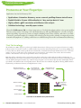
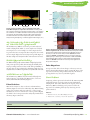
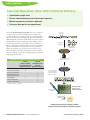
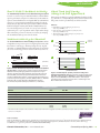
spring Volume XXIV, No. 2, 2009 Introducing A sensitive method for generating high-quality, full-length cDNA In This Issue MicroRNA Expression & Quantification Coexpress MicroRNAs with Fluorescent Proteins.. . . . . . . . . . . . . . . . . . . . . . . . . . . . . . . . . . . . . . . . . 2 Mir-X™ MicroRNA Quantification.. . . . . . . . . . . . . . . . . . . . . . . . . . . . . . . . . . . . . . . . . . . . . . . . . . . . . . . Cloning & Yeast One-Hybrid Technologies Synthesize cDNA the SMARTer™ Way. . . . . . . . . . . . . . . . . . . . . . . . . . . . . . . . . . . . . . . . . . . . . . . . . . . 4 6 Easily Identify and Characterize Protein-DNA Interactions with Our New Yeast One-Hybrid System.. . . . . . . . . . . . . . . . . . . . . . . . . . . . . . . . . . . . . . . . . . . . . . . 8 Antibody Microarray Analysis Proteome at Your Fingertips.. . . . . . . . . . . . . . . . . . . . . . . . . . . . . . . . . . . . . . . . . . . . . . . . . . . . . . . . . . . Adenoviral & Lentiviral Expression Systems 10 12 Live Cell Reporters, Now with Lentiviral Delivery.. . . . . . . . . . . . . . . . . . . . . . . . . . . . . . . . . . . . . . . 14 Concentrate Lentivirus… Effortlessly.. . . . . . . . . . . . . . . . . . . . . . . . . . . . . . . . . . . . . . . . . . . . . . . . . . . 16 Excellent Infection Rates with Lenti-X™ Systems and the RetroNectin Reagent.. . . . . . . . . 18 Obtain Adenoviral Titers in Less than 4 Hours.. . . . . . . . . . . . . . . . . . . . . . . . . . . . . . . . . . . . . . . . . . ® This issue and all prior issues are available online at www.clontech.com CLONTECHniques SMARTer™ PCR cDNA Synthesis New Products Coexpress MicroRNAs with Fluorescent Proteins These vectors coexpress your miRNA with either the mCherry or ZsGreen1 fluorescent protein marker • High-level miRNA and fluorescent protein coexpression • Simultaneously verify miRNA expression and transfection efficiency • Very bright fluorescent proteins Clontech offers two new vectors that provide high-level miRNA expression and allow you to verify it with fluorescent protein expression. The pmR-mCherry and pmR-ZsGreen1 Vectors couple your miRNA expression cassette to a bright red or green fluorescent reporter, for miRNA expression you can see and select. How Does It Work? Clone your miRNA sequence into the vector’s multiple cloning site (MCS) located in the 3' untranslated region of the fluorescent protein’s mRNA transcript. This enables both molecules to be expressed simultaneously from the vector’s strong CMV promoter. Each vector is equipped with the high-level CMV promoter, a selectable marker, and a fluorescent protein-miRNA expression cassette containing either mCherry or ZsGreen1—our two brightest Living Colors® Fluorescent Proteins (Figure 1). In short, you can clone and express your favorite miRNA, and then select, sort and/or visualize the cells in which it is expressed. Coding region for mCherry/ZsGreen1 5' AUG 3' untranslated region TAA mRNA translation AAAAAAAAAAAAA Cleavage of 3' UTR miRNA processing Target gene inhibition 5' Target gene mRNA AAAAAAAAAAAAA The pmR-mCherry and pmR-ZsGreen1 vectors will coexpress a fluorescent protein and an miRNA sequence that is embedded in the 3' UTR of the vector’s mRNA transcript. miRNA expression can be selected for and/or verified in transfected cells by monitoring red or green fluorescence. 2 Clontech Laboratories, Inc. • www.clontech.com Clontechniques Spring 2009 New Products miR NA Expression miR NA Quantification Once an miRNA sequence is cloned in the multiple cloning site of these vectors, miRNA expression can be delivered into any transfectable cell line. We used the pmR-ZsGreen1 and pmRmCherry vectors to express several different miRNAs in 293T cells (Figure 2). The miRNA sequences were amplified from human genomic DNA and then cloned into the vectors. The sequences included the indicated miRNA stem-loop along with ~300 bp of flanking DNA. Following transfection into separate cultures, samples of total RNA were prepared and treated with DNase prior to quantifying the expressed miRNAs using Clontech’s Mir-X™ miRNA qRT-PCR SYBR® Kit. Both vectors produced similarly high levels of expression for each miRNA, which were elevated to a range of values between 75- and >3000-fold over the vector-only controls. Clontech’s Mir-X miRNA qRT-PCR SYBR Kit has a diverse variety of applications, as it is able to detect and quantify multiple miRNAs, shRNAs, or mRNA targets in a single RNA sample. The complete, dual-function kit includes a fast and simple, one-step protocol for first-strand cDNA synthesis, as well as the reagents needed for the qPCR of your RNA target using SYBR Advantage® technology. From verifiable expression to accurate miRNA analysis, Clontech has the highly effective, state-of-the-art tools you need for investigating any miRNA network. 104 Primer miR-1 miR-9 miR-122a HSV TK polyA+ 103 mCherry/ ZsGreen1 pmR-mCherry/ ZsGreen1 Vector Kanr/Neor Fold increase in expression A PCMV IE pUC ori MCS 101 3' UTR SV40 polyA+ 102 PSV40 B pmR-mCherry 100 pmR-ZsGreen1 0 1 9 122a pmR-ZsGreen1 Figure 1. The pmR-mCherry and pmR-ZsGreen1 Vectors. Map of the vectors (Panel A). Cells transfected with the vectors exhibit red or green fluorescence (Panel B). 0 1 9 122a pmR-mCherry Figure 2. miRNA expression from pmR vectors. DNA sequences for the miR-1, miR-9, and miR-122 miRNAs were cloned into the pmRZsGreen1 and pmR-mCherry vectors, and the recombinant plasmids (1, 9, & 122a, respectively), as well as the parental vectors (0), were each transfected into separate cultures of 293T cells. After 48 hr, cells were harvested and the RNA was isolated for Mir-X miRNA qRT-PCR analysis using specific primers and the U6 snRNA as a normalization standard. Each primer was used with each RNA sample, but detected only the corresponding miRNA cognate. Ordering Information Product Size Cat. No. pmR-ZsGreen1 Vector 20 μg 632541 NEW! pmR-mCherry Vector 20 μg 632542 NEW! 200 rxns 600 rxns 638314 638316 NEW! Mir-X miRNA qRT-PCR SYBR Kit * *Includes a Mir-X miRNA First-Strand Synthesis Kit. Visit Visit sit our website for more details! click here… Notice to Purchaser Please see the Advantage ® and TITANIUM™ PCR Products, CMV Sequence, Fruit Fluorescent Protein Products, Hot Start Antibody, Living Colors ® Fluorescent Protein Products, Molecular Probes, Inc., and PCR licensing statements at www.clontech.com/licensing Clontech Laboratories, Inc. • www.clontech.com Clontechniques Spring 2009 3 New Products Mir-X™ MicroRNA Quantification Quickly and accurately quantify your miRNAs and their mRNA targets with this complete qRT-PCR kit. • Quantify any miRNA and its target from the same RNA sample • 2-kits-in-1: cDNA synthesis and qPCR • Simple, single-step cDNA synthesis reaction For the challenging task of unraveling microRNA (miRNA) expression profiles in tissues and cell lines, Clontech scientists have developed a sensitive, reliable, and easy-to-use miRNA quantification system. The Mir-X miRNA qRT-PCR SYBR® Kits are complete, dual-function systems for performing first-strand cDNA synthesis and quantitative PCR (qPCR) to precisely measure the level of your favorite miRNAs. The kits are available in economical, large-sized formats that provide 200 or 600 qPCR reactions. Simple and Sensitive A simple, single-step reaction uses an optimized mix of poly(A) polymerase and SMART™ MMLV Reverse Transcriptase to synthesize first-strand cDNA from your RNA sample. The cDNA is then specifically amplified and quantified by qPCR using your miRNA-specific primer and our SYBR Advantage® qPCR Premix. Multiple miRNA species, as well as the mRNA targets of the miRNAs, can be amplified from a single cDNA sample. The system is extremely sensitive and able to detect miRNAs down to 50 copies. First-strand cDNA synthesis from any RNA Add cDNA to miRNAspecific primers and SYBR Advantage Premix qPCR Data analysis Mir-X single-step cDNA synthesis 5' 3' Poly(A) polymerase 5' AAAAAAAAAAAAA-3' miRNA primer SYBR Advantage qPCR Oligo(dT) priming 5' AAAAAAAAAAAAA-3' NVTTTTTTTTTTTTT SYBR Advantage 5' 3' TTTTTTTTTTTTT 5' SMART MMLV RT 5' AAAAAAAAAAAAA-3' TTTTTTTTTTTTT 5' Mir-X miRNA qRT-PCR SYBR Kits use a single-step, single-tube reaction to produce first-strand cDNA, which is then specifically and quantitatively amplified using a miRNA-specific primer and SYBR Advantage qPCR chemistry. In the Mir-X cDNA synthesis reaction, RNAs are poly(A)-tailed using poly(A) polymerase, and then copied using a modified oligo(dT) primer and SMART MMLV Reverse Transcriptase. 4 Clontech Laboratories, Inc. • www.clontech.com Clontechniques Spring 2009 New Products Highly specific Detection A Let7a Let7b Let7c Let7d TGAGGTAGTAGGTTGTATAGTT TGAGGTAGTAGGTTGTGTGGTT TGAGGTAGTAGGTTGTATGGTT AGAGGTAGTAGGTTGCATAGTT Let7e Let7f Let7g Let7i TGAGGTAGGAGGTTGTATAGTT TGAGGTAGTAGATTGTATAGTT TGAGGTAGTAGTTTGTACAGTT TGAGGTAGTAGTTTGTGCTGTT B Percent of maximum 100 miRNA Let7a 80 Let7b 60 Let7c Let7d 40 Let7e 3 2 1 Let7i Let7b Let7c Let7d Let7e Primer Let7f Let7g Let7i Figure 1. Specific quantification of Let7 miRNA variants. Using miRNAspecific primers (Panel A), Mir-X qRT-PCR was able to specifically detect and quantify each member of a series of 8 synthetic Let7 variants that had been spiked into a background of yeast polyA+ RNA (Panel B). The primers detected each of their corresponding Let7 miRNA cognates, but did not detect the off-target variants in 63 of 64 possible combinations. Diverse Research Applications Since the Mir-X system is able to detect multiple miRNAs, shRNAs, or mRNA targets in a single RNA sample, it can be used for a variety of applications. In principle, any RNA that is, or can be, polyadenylated may be quantified using the Mir-X method. In mouse embryonic stem cells, we were able to monitor the alterations in expression for a panel of 12 miRNAs that respond to trichostatin A (TSA) treatment (Figure 2). Exposing aggregated mouse P19 cell clusters to retinoic acid (RA) causes them to acquire neural cell phenotypes, which are accompanied i f t7 b t7 Le Le a b t7 t7 Le Le a 99 99 m iR - 5p 5_ m iR - 3p m iR - 33 p 5_ Figure 2. Trichostatin A treatment alters miRNA expression in mouse ES cells. Mouse embryonic stem cells were harvested either prior to or after being treated with trichostatin A (TSA) for 18 hr. RNA was prepared from the cells, and was then analyzed by Mir-X miRNA qRT-PCR using primers specific for the 12 indicated miRNAs and for a p21 control mRNA known to be induced by TSA. 110 + RA RA – RA 90 70 50 30 10 –10 0 3 6 Days Let7g Let7a 33 m iR - p 5b _5 12 m iR - 24 5b _3 m iR - 12 21 m iR - _a m iR - p2 1 MicroRNA Let7f 20 0 4 0 Percent of maximum To demonstrate the specificity of Mir-X miRNA quantification, we used a series of 8 highly similar synthetic Let7 miRNA variants (Figure 1). We first spiked each of the Let7 miRNAs into separate samples of yeast polyA+ RNA and generated cDNA using the Mir-X singletube reaction. We then tested a panel of variant-specific primers with each cDNA sample to determine each primer’s ability to specifically and individually quantify the Let7 subtypes in the cDNA sample. Despite the high degrees of similarity among the variants and the primers (Figure 1, Panel A), Mir-X qPCR was highly specific in detecting each Let7 variant (Figure 1, Panel B). Fold induction 5 9 12 Figure 3. Induction of miR-9 in mouse P19 cells. P19 mouse embryonal carcinoma cells were plated on agarose-coated petri dishes and allowed to form embryoid bodies (EB) in the presence or absence of retinoic acid (RA). After five days of culture, EBs were dissociated with trypsin and replated on tissue culture dishes without RA. Cells were harvested on days 3–7 and 10–13, and the induction of miR-9 was followed and quantified using the Mir-X miRNA qRT-PCR SYBR Kit and primers specific for miR-9 and U6 (as a normalization control). by changes in the cellular miRNA pool. Using the Mir-X system, we tracked the abundance of one such miRNA, miR-9, which was induced by RA and continued to accumulate in these cells following a 5 day exposure to RA (Figure 3). Summary Mir-X miRNA qRT-PCR SYBR Kits are complete, dual-function qPCR systems that have the flexibility to monitor the level of your favorite miRNA or any other RNA species in your RNA sample. The single-tube cDNA synthesis is faster and far less complicated than other available methods, while the miRNA qPCR is very sensitive and extremely accurate. Ordering Information Product Size Cat. No. Mir-X miRNA qRT-PCR SYBR Kit* 200 rxns 600 rxns 638314 638316 NEW! Mir-X miRNA First-Strand Synthesis Kit 20 rxns 60 rxns 638313 638315 NEW! *Includes a Mir-X miRNA First-Strand Synthesis Kit. Visit Visit sit our website for more details! click here… Notice to Purchaser Please see the Advantage® and TITANIUM™ PCR Products, Hot Start Antibody, Molecular Probes, Inc., and PCR licensing statements at www.clontech.com/licensing Clontech Laboratories, Inc. • www.clontech.com Clontechniques Spring 2009 5 New Products Synthesize cDNA the SMARTer™ Way Get better results with SMARTer PCR cDNA Synthesis Kits • Generate high-quality cDNA from as little as 1–2 ng of total RNA • Preserve precious samples and maintain accurate gene representation • Higher specificity, lower background, and increased yield • Enrich for full-length cDNA • No adaptor ligation necessary • Amplify longer genes and rare transcripts SMART™ Technology Just Got SMARTer Clontech revolutionized cDNA synthesis with the invention of SMART (Switching Mechanism At the 5' end of RNA Transcript) technology. This unique technology utilizes the intrinsic terminal transferase and template-switching activity of Moloney Murine Leukemia Virus Reverse Transcriptase (MMLV RT) to accurately synthesize full-length cDNA in a single reverse transcription reaction (1), while incorporating adaptors on both ends of the cDNA. Incorporation of these universal primer binding sites in a single-step during first-strand cDNA synthesis eliminates the need for tedious second-strand synthesis and adaptor ligation, and facilitates a number of downstream applications, including RACE (Rapid Amplification of cDNA Ends), subtractive hybridization, and library construction. This simple and highly efficient cDNA synthesis method ensures higher specificity in amplifying your target cDNA, compared to conventional methods. Clontech has recently developed a new batch of kits featuring SMART technology with improved components—the SMARTer Kits for all your cDNA synthesis applications. These new SMARTer Kits include a modified SMARTer II A Oligonucleotide and SMARTScribe™ Reverse Transcriptase. The template-switching ability of SMARTScribe Reverse Transcriptase is enhanced when combined with the new SMARTer oligonucleotide; together these new components increase the likelihood of cloning your entire gene sequence, and result in high-quality, full-length cDNA, regardless of template size or abundance (2). Get High-Quality cDNA from Less R NA SMARTer cDNA Synthesis Kits are especially useful for researchers who have limited starting material, such as RNA derived from lasercapture microscopy samples, cells sorted by flow cytometry, or other extremely small samples. The SMARTer PCR cDNA Synthesis Kit allows first-strand cDNA synthesis from only 2 ng of total RNA (Figure 1, Panel A), much less starting material than is required 6 Clontech Laboratories, Inc. • www.clontech.com SMARTer cDNA Synthesis Starting material 5' poly A SMARTer oligo SMARTScribe MMLV RT PCR adaptor First-strand cDNA synthesis 5' Single step poly A dT Oligo(dT) primer with added sequence Second-strand cDNA synthesis LD-PCR amplification or primer extension dA Full-length ds cDNA dT Template for real-time PCR Probes for arrays Next-generation sequencing Subtractive hybridization Direct gene amplification RACE Library construction SMARTer cDNA synthesis occurs in a singlestep reverse transcription reaction. Following amplification, SMARTer cDNA can be used for a variety of downstream applications. Clontechniques Spring 2009 New Products A kb 10.0– M 18 PCR Cycles 21 24 27 B kb 10.0– 3.0– 2.0– 1.5– 3.0– 2.0– 1.5– 1.0– 1.0– 0.5– 0.5– M 18 PCR Cycles 21 24 27 Figure 1. Typical gel profile of ds cDNA synthesized with the SMARTer PCR cDNA Synthesis Kit (Panel A) and the SMARTer Pico PCR cDNA Synthesis Kit (Panel B), using the Control Human Placental Total RNA as a template. 2 ng (Panel A) or 1 ng (Panel B) of the Control Human Placental Total RNA was subjected to first-strand cDNA synthesis and purification, followed by PCR amplification as described in the user manual for each kit. 5 µl of each PCR product was electrophoresed on a 1.2% agarose/EtBr gel in 1X TAE buffer following the indicated number of PCR cycles. Lanes M: 1 kb DNA ladder size markers. The arrows indicate the strong band at 900 bp typically seen for human placental total RNA. Following reverse transcription, SMARTer cDNA can be used directly in 5'- and 3'-RACE PCR reactions, without an additional adaptor ligation step. A side-by-side test of the new SMARTer oligo and the SMART oligo included in the original kit revealed an increased overall yield of 5'-RACE PCR products with the SMARTer oligo (Figure 2). The SMARTer RACE Kit facilitates RACE PCR via the included Universal Primer Mix, which is also sold separately. Additionally, the kit includes random primers for researchers whose RNA lacks a poly(A) tail. References 1. Chenchik, A. et al. (1998) In Gene Cloning and Analysis by RT-PCR. Eds Siebert, P. & Larrick, J. (BioTechniques Books, MA) Ch 22. 2. Be SMART™ About First-Strand cDNA Synthesis (January 2009) Clontechniques XXIV(1):15–17. kb 10.0– for the original SMART PCR cDNA Synthesis Kit (Cat. No. 634902). Since extremely dilute RNA cannot be used in regular cDNA synthesis, we designed the new SMARTer Pico PCR cDNA Synthesis Kit to synthesize high-quality cDNA from even less starting material—as little as 1 ng of total RNA at a concentration as low as 20 pg/µl (Figure 1, Panel B). The SMARTer Pico Kit is the new and improved version of our original Super SMART PCR cDNA Synthesis Kit (Cat. No. 635000). The SMARTer Pico cDNA synthesis protocol makes it possible to use the entire volume of purified single-stranded cDNA for amplification, via increased reaction volumes and an additional column purification step. Both the SMARTer and SMARTer Pico protocols produce ds cDNA yields ranging from 1–2 µg. Amplify Longer Transcripts Clontech’s new SMARTer RACE cDNA Amplification Kit, an improved version of our original SMART RACE cDNA Amplification Kit (Cat. No. 634914), allows you to identify the complete sequence of your RNA transcript from a small region of known sequence within the transcript all the way to the 5'- or 3'-end of the RNA, starting with as little as 2 ng of total RNA. M 1 SMART II A 2 3 4 5 SMARTer II A 6 7 8 3.0– 2.0– 1.5– 1.0– 0.5– Figure 2. The SMARTer II A Oligo increases the overall yield of the 5'-RACE PCR products. Note the difference in intensity between the 2.6 kb 5'-RACE product in Lane 2 (produced by the SMART II A oligo) and that in Lane 6 (produced by the SMARTer II A oligo); both bands resulted from the amplification of only 2 ng of human placental total RNA template. In the assay shown, first-strand cDNA synthesis and 5'-RACE PCR amplification were performed as described in the user manual (PT4096-1). Lanes 1 & 5: No RNA 5'-RACE control. Lanes 2 & 6: Transferrin Receptor (TFR) 5'-RACE starting from 2 ng total RNA. Lanes 3 & 7: TFR 5'-RACE starting from 50 ng total RNA. Lanes 4 & 8: 5'-cDNA internal control using 2 ng total RNA as template. Lane M: 1 kb DNA Ladder. Ordering Information Product Size Cat. No. SMARTer™ PCR cDNA Synthesis Kit 10 rxns 20 rxns 634925 634926 NEW! SMARTer™ Pico PCR cDNA Synthesis Kit 10 rxns 634928 NEW! SMARTer™ RACE cDNA Amplification Kit 10 rxns 20 rxns 634923 634924 NEW! Universal Primer Mix 100 rxns 634922 NEW! Notice to Purchaser Visit Visit sit our website for more details! click here… Please see the SMART™ Amplification Products licensing statement at www.clontech.com/licensing Clontech Laboratories, Inc. • www.clontech.com Clontechniques Spring 2009 7 New Products Easily Identify and Characterize Protein-DNA Interactions with Our New Yeast One-Hybrid System • Highest performing yeast one-hybrid system • Aureobasidin A selection eliminates background • Complete system for easy construction and screening of cDNA libraries directly in yeast Clontech’s Matchmaker™ Gold Yeast One-Hybrid Library Screening System provides a simple and efficient method for identifying and characterizing novel protein-DNA interactions. The system uses SMART™ cDNA synthesis technology, which allows cDNA libraries to be created from any tissue source, starting with as little as 100 ng of total RNA. It also employs Aureobasidin A (AbA; 1) selection, which provides the most stringent yeast one-hybrid (Y1H) screening strategy available. The System In the Matchmaker Gold Yeast One-Hybrid Library Screening System, 1–3 copies of your target DNA sequence (i.e., the bait) are cloned into the reporter vector pAbAi. The resulting pBait-AbAi construct is then integrated into the genome of the Y1HGold yeast strain by homologous recombination to generate a bait-specific reporter strain. A cDNA library expressing potential DNA-binding proteins (i.e., prey) as fusions to the GAL4 transcription activation domain (AD) is constructed directly in the pBait-AbAi reporter strain. When a prey protein binds to the DNA target sequence (see figure below), transcription of the Aureobasidin A resistance gene (AbAr) is activated, allowing the cell to grow on medium containing the antibiotic Aureobasidin A (AbA). In library screens, the plasmids encoding the library-derived prey proteins can be rescued from the surviving yeast clones and subjected to further analysis. Library Proteins GAL4 AD Prey RNA Pol II Target DNA Element “Bait Sequence” 5' AbAr mRNA Screening for protein-DNA interactions with the Matchmaker Gold Yeast One-Hybrid System. 8 Clontech Laboratories, Inc. • www.clontech.com Clontechniques Spring 2009 New Products SMART Technology The cDNA inserts for the prey library are created by SMART cDNA synthesis, which results in the incorporation of known primer sequences at both ends of the cDNA. Consequently, SMART-generated cDNA: • is available for amplification by PCR—allowing the construction of libraries from nanogram amounts of starting RNA. • is flanked by sequences that are homologous to the cloning site of the linearized library vector, pGADT7-Rec—allowing homologous recombination between the cDNA and the pGADT7-Rec vector upon transformation into the bait-specific reporter strain (Figure 1). Get Screening Results Fast! With the Matchmaker Gold Yeast One-Hybrid Library Screening System, one-hybrid screening can be accomplished quickly and easily with the following steps: Step 1. Create a bait construct by cloning 1–3 copies of the target DNA-binding sequence into pAbAi. Step 2. Create a bait-specific reporter strain by transforming and integrating the linearized pBait-AbAi construct into the Y1HGold yeast strain and selecting on SD/–Ura medium, available in our Yeast Media Set 1 Plus. Step 3. Confirm the integration of the bait sequence by colony PCR using Matchmaker Insert Check PCR Mix 1. SMART cDNA x x pGADT7Rec Plate on SD/–Leu/ AbA Medium Step 4. Use SMART technology to synthesize cDNA that is flanked by sequences that are homologous to the ends of the linearized pGADT7-Rec vector. Step 5. Create and screen your Y1H library in a single step: In vivo recombination in Y1HGold[Bait] yeast Cotransform your bait-specific Y1HGold reporter strain with the SMART-generated cDNA and pGADT7Rec vector, and plate on SD/–Leu/+AbA. Figure 1. Use SMART technology and yeast biology to construct and screen your library. Your library is simultaneously constructed and screened directly in yeast. First, SMART cDNA synthesis technology is used to create a pool of cDNA that is flanked by sequences homologous to the ends of the linearized pGADT7-Rec vector. Next, the newly created Y1HGold-Bait reporter strain is transformed with the cDNA pool and pGADT7-Rec, which undergo homologous recombination within yeast. The yeast cells are then plated on SD/–Leu/+AbA to select for colonies that contain an active reporter (i.e., positive Y1H interactions). Step 6. Harvest the resulting colonies, which contain putative DNA-binding proteins, and analyze further (e.g., with the Matchmaker Insert Check PCR Mix 2 and the Easy Yeast Plasmid Isolation Kit). The Matchmaker Gold Yeast One-Hybrid Library Screening System is the most convenient and advanced Y1H screening tool available, allowing library screens to be accomplished in less time and with greater confidence than ever before. Aureobasidin A Selection Eliminates Background Matchmaker Gold Systems are unique because they use the AbAr gene as a novel reporter that confers resistance to AbA, a potent antifungal agent that is toxic to S. cerevisiae. Selecting for resistance to this highly stable depsipeptide makes Y1H library screening very straightforward (Figure 1), as AbA effectively kills yeast cells that are not expressing the AbAr reporter. Aureobasidin A and all of the required media are supplied in our Yeast Media Set 1 Plus. Reference 1. Takesako, K. et al. (1991) J. Antibiot. 44(9):919–924. Ordering Information Product Size Cat. No. Matchmaker Gold Yeast One-Hybrid Library Screening System 5 rxns 630491 NEW! Yeast Media Set 1 Plus each 630493 NEW! Matchmaker Insert Check PCR Mix 1 100 rxns 630496 NEW! Matchmaker Insert Check PCR Mix 2 100 rxns 630497 NEW! Easy Yeast Plasmid Isolation Kit 50 preps 630467 Notice to Purchaser Visit Visit sit our website for more details! click here… Please see the HotStart Antibody, PCR, PCR Polymerase, SMART™ Amplification Products, and Aureobasidin Resistance Gene licensing statements at www.clontech.com/licensing Clontech Laboratories, Inc. • www.clontech.com Clontechniques Spring 2009 9 Product Overview Proteome at Your Fingertips Applications for the Ab Microarray 500 • Applications—biomarker discovery, cancer research, profiling disease states & more • Rapid & flexible—Screen >500 antibodies in 1 day; analyze data in 1 hour • Highly reliable—≥80% correlation with Western blot analysis • Validated technology—over 40 papers to date Clontech’s Ab Microarray 500 is a robust, inexpensive tool for high-throughput analysis of proteomic profiles. This array can be used as a screening tool for biomarker discovery and target identification—as well as for rapid expression profiling of proteins in a variety of samples, including serum, tissues, cell lines, and cancer vs. normal or treated vs. untreated samples. For example, this technology has been used to identify overexpressed proteins in mantle cell lymphoma (1), yielding results that were validated using Western blot and immunohistochemistry. The Technology The Ab Microarray 500 is designed to perform rapid, reliable characterization of changes in protein expression between two samples. It allows scientists to focus their research on a smaller number of proteins that have biological relevance—by screening >500 proteins in one day. This array, which contains specific monoclonal antibodies, is incubated with Cy3™/Cy5™-labeled serum, tissue, or cell extract samples—in order to profile disease states, identify tumor-associated antigens, perform time course studies, discover biomarkers, and more. The antibodies on the array recognize human, mouse, and rat proteins. High data quality is achieved by using a Cy3/Cy5 dye swap detection technology together with an internally normalized ratio (2). This technology also has a built-in concentration step that allows for the detection of low-abundance proteins by accumulating antigens onto the antibody during the incubation step—thus increasing the signal. A one-hour data analysis procedure yields highly sensitive, reproducible, and reliable data that are biologically relevant. These data have been validated using Western blotting, ELISA, immunohistochemistry, and literature references (over 40 publications to date). Microarray with >500 pairs of unique monoclonal antibodies Array incubated with native protein extracts Native antigens bound to antibodies on array (cells, tissues, body fluids, etc.) ay 500 icroarr Ab M ay 500 icroarr Ab M Labeled analyte Western blot validation Capture antibody Array surface ELISA validation Immunohistochemistry validation Literature references Overview of the Ab Microarray 500 Protocol. 1 0 Clontech Laboratories, Inc. • www.clontech.com Clontechniques Spring 2009 Normalized intensity (log scale) Product Overview A 101 Tonsil MCL 1 MCL 2 MCL 3 CRIK 10 0 Hsp90 1 2 3 4 5 6 MCL samples Control MDM2 7 7 repeated Figure 1. Expression levels of the 13 overexpressed proteins in the MCL samples as compared to the control. The data points are colored in red/orange, indicating overexpression. The samples are numbered 1 to 7. MCL7 showed a reversed expression pattern compared to the other 6 MCL samples. MCL7 was tested twice and demonstrated a consistent expression pattern. Data were analyzed using GeneSpring™ software (Agilent Technologies, Inc.) B Data Reproducibility Six different histologically-confirmed MCL samples displayed the same protein expression profile, demonstrating the reliability of the array in providing consistent, accurate results. The reproducibility of the array data was also confirmed by the MCL7 sample that was analyzed twice (in 2 different experiments) and yielded the same result. Validation of Results The Ab Microarray 500 data from the mantle cell lymphoma analysis (Figure 1) was validated by Western blot (1; data not shown) and immunohistochemistry (Figure 2). False Positives Western blot analysis (1; data not shown) and immunohistochemistry (Figure 2) were used to confirm array data. Western blotting results were consistent with array results for 7 of the proteins identified, but not for 2 of them. One of the 2 proteins (Hsp90) also failed to show overexpression when analyzed by immunohistochemistry (Figure 2, Panel A). Tonsil MCL 1 MCL 2 Rb2 KU80 Ab Microarray 500 Analysis of Mantle Cell Lymphoma The Ab Microarray 500 was used in the proteomic analysis of mantle cell lymphoma (MCL; 1). Seven samples were analyzed and compared to a control sample. Six of the samples showed 13 proteins that were overexpressed when compared to the control, and one sample displayed an inverted expression profile (Figure 1). The latter sample was reanalyzed and the same results were observed. Paxillin Bcl-x Figure 2. Immunohistochemistry was used to analyze false positive and false negative results. Paraffin-embedded tissue biopsies available from the same patients were analyzed using antibodies present on the array in order to test for false positives (Panel A) and false negatives (Panel B). The Panel A results are consistent with the antibody array in detecting increased levels of CRIK and MDM2, and the Western blot in failing to demonstrate elevated Hsp90 levels. The Panel B results did not reveal any false negatives for the proteins analyzed. False Negatives Four proteins that did not show changes on the array were analyzed by immunohistochemistry (Figure 2, Panel B). None of the 4 proteins revealed any changes when analyzed by this method, proving that the array does not yield false negatives. Conclusion Comparing relative expression levels with the Ab Microarray 500 revealed the existence of 3 novel proteins associated with mantle cell lymphoma. The results demonstrate the capacity of the Ab Microarray 500 for identifying novel proteins as well as the importance of confirming antibody array data using an independent method. References 1. Ghobrial, I. M. et al. (2005) Blood 105(9):3722–3730. 2. Andersson, O. et al. (2005) J. Proteome Res. 4(3):758–767. Visit Visit sit our website for more details! click here… Ordering Information Product Size Cat. No. Ab Microarray 500 Slides 2 arrays 631790 Ab Microarray Express Buffer Kit each 631795 Clontech Laboratories, Inc. • www.clontech.com Clontechniques Spring 2009 1 1 New Products Obtain Adenoviral Titers in Less than 4 Hours Rapidly determine adenoviral titers with our new Adeno-X™ qPCR Titration Kit • Harvest, titrate, and infect in a single day • Measure titers from crude lysate or purified viral preps The Adeno-X qPCR Titration Kit provides an extremely fast, simple, and accurate method for titrating adenoviral stocks from all Ad5-based adenoviral vectors. The kit delivers results in just 4 hours, a vast improvement over standard titration methods (such as the plaque assay), which require up to 10 days to complete. Because qPCR titration is so fast, target cells can be infected with accurately titrated virus on the same day the virus is harvested. Obtain adenoviral lysate Purify viral DNA qPCR Flowchart of the procedures used for titrating adenoviral DNA with the Adeno-X qPCR Titration Kit. Table I: Comparison of Adeno-X qPCR Titration to Other Titration Methods Titration Method Sample Type Virus Purified Crude Adeno-X qPCR Titration Ratios qPCR (copies/ml) Fluor (IFU/ml) X-Gal (IFU/ml) qPCR/Fluor (copies/IFU) qPCR/X-Gal (copies/IFU) AdAcGFP1 5.62 x 10⁹ 3.03 x 10⁹ N/A 2 N/A AdAcGFP1 Prep A Prep B Prep C 1.44 x 10¹⁰ 1.46 x 10¹⁰ 1.38 x 10¹⁰ 1.90 x 10⁹ 2.26 x 10⁹ 2.73 x 10⁹ N/A N/A N/A 8 6 5 N/A N/A N/A Purified AdLacZ 1.01 x 10¹⁰ N/A 1.54 x 10⁹ N/A 7 Crude AdLacZ Prep A Prep B Prep C 1.33 x 10¹⁰ 1.24 x 10¹⁰ 1.21 x 10¹⁰ N/A N/A N/A 2.67 x 10⁹ 2.67 x 10⁹ 3.15 x 10⁹ N/A N/A N/A 5 5 4 a b a Adenoviral copy numbers were determined using the Adeno-X qPCR Titration Kit (Cat. No. 632252). b To determine fluorescence-based infectivity titers, adenoviral stocks were serially diluted (tenfold) and applied to HEK 293 cells. After 48 hr, fluorescent cells were scored with a fluorescence microscope. 1 2 Clontech Laboratories, Inc. • www.clontech.com Clontechniques Spring 2009 New Products The qPCR Titration Method The protocol combines qPCR with SYBR® Green chemistry, allowing you to determine the viral genome copy number in adenoviral preparations (i.e., crude lysates or purified stocks) from a calibrated DNA standard curve (Figure 1). The procedure is simple: viral DNA and control DNA (provided) are serially diluted and subjected to qPCR. The DNA copy number of each viral sample is then determined by comparing its Ct value to a standard curve generated by plotting the Ct values of the diluted control samples against their respective copy numbers (Figure 1). Titration assays demonstrating the consistency of this approach when used with either crude lysate or purified virus are shown in Figure 2. A B 24 22 20 Ct (dRn) Fluorescence (dRn) 100 10 1 – 18 16 14 12 10 10–2 8 0 5 10 15 20 Cycle no. 25 30 35 6 103 40 104 105 106 Initial quantity (copies) 107 108 Figure 1. The Adeno-X qPCR titration method exhibits a wide dynamic range. Adeno-X DNA Control Template was serially diluted to 108–103 copies per sample and analyzed with the Adeno-X qPCR Titration Kit. The amplification plots (Panel A) show a dynamic range of at least six orders of magnitude (each dilution is represented by a different colored plot). The standard curve (Panel B) demonstrates a strong linear correlation between the Ct and the DNA copy number (log scale), with R2 = 1.000 and a PCR efficiency of 96.2%. Once the genome copy number of your viral stock is determined, it can be correlated with the number of viral infectious units (IFU; determined independently) to establish a copy number/IFU relationship (Table I). Determination of the copy number/IFU relationship for a given prep allows you to normalize the amount of the prep used in each experiment, for consistent interassay results. Our method makes it possible to infect cells at a known multiplicity of infection (MOI), allowing you to produce results that are precise, consistent, and interpretable. The Adeno-X qPCR Titration Kit includes sufficient material for 200 qPCR reactions and allows multiple virus preparations to be titrated simultaneously. A B 100 Fluorescence (dRn) Fluorescence (dRn) 100 10–1 10–1 10–2 10–2 0 5 10 15 20 Cycle no. 25 30 35 0 40 5 10 15 20 Cycle no. 25 30 35 40 Figure 2. Titrate your adenovirus from crude lysate or purified viral particles. The Adeno-X qPCR Titration kit was used to titrate capsid-free viral DNA obtained from both crude lysate (Panel A) and purified viral particles (Panel B). In both cases, the capsid-free DNA was obtained with a virus purification kit (provided with the Adeno-X qPCR Titration Kit) and serially diluted (10X) prior to qPCR (each dilution is represented by a different colored plot). The titration procedure worked equally well regardless of the purity of the viral particles. Ordering Information Product Size Cat. No. Adeno-X qPCR Titration Kit 200 rxns 632252 Notice to Purchaser NEW! Visit Visit sit our website for more details! click here… Please see the Advantage ® and TITANIUM™ PCR Products, Hot Start Antibody, Molecular Probes, Inc., and PCR licensing statements at www.clontech.com/licensing Clontech Laboratories, Inc. • www.clontech.com Clontechniques Spring 2009 1 3 New Products Live Cell Reporters, Now with Lentiviral Delivery • Optimized for high titers • Choose chemiluminescent or fluorescent reporters • Measure promoter activity on demand • Get more data points per experiment Promoter of interest 5' LTR MCS Clontech’s Lenti-X™ Reporter Systems allow you to study your promoter of interest with a chemiluminescent or fluorescent on-demand reporter in virtually any cell type, including primary cultures, dividing and nondividing cells, stem cells, terminally differentiated cells, and neuronal cells. These are complete systems, which include our advanced 4th-generation packaging systems and lentivirus transfection system, plus your vector of choice (1; Figure 1). They deliver excellent signal-to-noise ratios by excluding reporter molecules that are expressed prior to the experiment (Figure 2) and allow you to monitor promoter activity at any time point and for any length of time that you choose. Because there is no cell lysis, you can observe multiple promoter activation cycles using the same cells. Ψ RRE MetLuc WPRE cPPT 3' LTR tTA Lentiphos HT transfection Lenti-X HT Packaging Mix 293T cells 1. Collect virus after 48 hr 2. Transduce host cells Table I: How Do I Choose My Reporter System? Lenti-X Lenti-X DD Fluorescent Ready-To-Glow Protein1 Secreted Luciferase Reporter System Reporter Systems Chemiluminescent detection (plate reader) Fluorescent detection (flow cytometry, fluorescence microscopy) Automatable License required for For-Profit organizations Promoter activation leads to expression of secreted luciferase protein in the medium Remove supernatant 1 AmCyan1, ZsGreen1, or tdTomato At desired time points, assay luciferase activity Add luciferase substrate Secreted luciferase LIGHT Product Detection by luminometer Flowchart of the Lenti-X Ready-To-Glow Secreted Luciferase Reporter System protocol. 1 4 Clontech Laboratories, Inc. • www.clontech.com Clontechniques Spring 2009 New Products The Lenti-X Ready-To-Glow™ Secreted Luciferase Reporter System is based on a secreted reporter, Metridia luciferase, which exhibits superior performance compared to traditional cytosolic luciferase reporters. Secreted luciferases are not degraded like cytosolic luciferase reporters, so you can monitor intermittent short bursts of promoter activity that you might have missed previously. In addition, Metridia luciferase has higher sensitivity than Renilla or firefly luciferases (2). To remove background, just “wash out” any reporter molecules synthesized prior to your experiment with a simple media change. Then activate the promoter and monitor its activity by sampling the accumulated stable reporter from the media. Measure Activity On Demand The Lenti-X DD Fluorescent Protein Reporter Systems combine a bright fluorescent protein reporter for high signal intensity with ProteoTuner™ technology to eliminate background (3). Liganddependent, on-demand stabilization of the fluorescent reporter allows you to start monitoring promoter activation whenever you choose. Save Time and Money Using Live Cell Reporters These systems are ideal for your studies with limited numbers of cells, e.g., stem cells, or to study multiple promoter activation cycles and/or time points, in order to produce many sets of data over time. References 1. High-Efficiency Lentiviral Packaging (October 2007) Clontechniques XXII(4): 1–2. 2. Ready-To-Glow™ Secreted Luciferase System (July 2006) Clontechniques XXI(2):12–13. 3. The Next Generation of Promoter Reporters (January 2009) Clontechniques XXIV(1)22–23. 160,000 A – Forskolin + Forskolin 120,000 RLU Don’t Miss Transient Activity 80,000 40,000 0 pLVX-CRE-MetLuc cPPT RRE /CTS 5' LTR Ψ WPRE 3' LTR 140 B – Forskolin 120 + Forskolin 100 B MCS pLVX-MetLuc MetLuc MCS DD FP RFU A 80 60 40 pLVX-DD-FP 20 Figure 1. Lenti-X chemiluminescent (Panel A) and fluorescent (Panel B) reporter vectors. Lenti-X vectors contain sequence elements that facilitate lentiviral packaging and/or boost expression of your reporter, including the LTRs, packaging signal (Ψ), Rev response element (RRE), and central polypurine tract/central termination sequence (cPPT/CTS) from HIV-1; and the woodchuck hepatitis virus post-transcriptional regulatory element (WPRE). MetLuc = Metridia luciferase. DD = ligand-dependent destabilization domain. FP = fluorescent protein (AmCyan1, ZsGreen1, or tdTomato). 0 pLVX-CRE-DD-ZsGreen1 Figure 2. Lenti-X Reporter Systems provide low background and high signal intensity. HEK 293 cells were transduced with pLVXCRE-MetLuc Reporter Vector (Panel A) or pLVX-CRE-DD-ZsGreen1 Reporter Vector (Panel B), treated with forskolin, and assayed according to the respective protocols. RLU = relative light units. RFU = relative fluorescence units. Ordering Information Product Size Cat. No. Lenti-X Ready-To-Glow Secreted Luciferase Reporter System each 631746 NEW! Lenti-X DD Cyan Reporter System each 631748 NEW! Lenti-X DD Green Reporter System each 631751 NEW! Lenti-X DD Red Reporter System each 631753 NEW! Lenti-X 293T Cell Line 1 ml 632180 Shield1* 60 μl 200 μl 500 μl 631037 631038 632189 * The number of reactions depends on the concentration of Shield1 used. At the maximum suggested concentration (1,000 nM), 60 μl = 30-plus reactions and 200 μl = 1,000-plus reactions in a six-well plate. Visit Visit sit our website for more details! click here… Notice to Purchaser Please see the BGH Poly A, CMV Sequence, cPPT Element, Lentiviral Expression Products, Living Colors® Fluorescent Protein Products, Metridia luciferase, ProteoTuner™ Protein Stabilization/Destabilization Products, Retroviral Packaging Systems, Tet-Based Expression Products, VSV-G Technology, and WPRE Technology licensing statements at www.clontech.com/licensing Clontech Laboratories, Inc. • www.clontech.com Clontechniques Spring 2009 1 5 New Products Concentrate Lentivirus… Effortlessly Increase your viral titer 100-fold with Lenti-X™ Concentrator—without ultracentrifugation • Simply mix and spin • Hassle-free and easily scaled up for large volumes • No ultracentrifugation required Need to concentrate your lentivirus preps, but don’t want the hassle of ultracentrifugation? Use Lenti-X Concentrator to increase your available titer up to 100-fold and infect your target cells at higher MOIs and in reduced volumes, without making more virus. Simple Protocol: Mix, Wait, Spin Lenti-X Concentrator provides a fast, simple, and highly efficient method for concentrating any lentiviral stock. In the simple protocol, you just mix your lentiviral supernatant with the Lenti-X Concentrator reagent, incubate for a short period, and spin the mixture in a standard centrifuge (see below). You’ll increase your vector titer by up to 100-fold in ~1 hr, and obtain excellent recoveries— with no ultracentrifugation. Lenti-X Concentrator works for all lentiviral supernatants, including those made from any of Clontech’s Lenti-X Systems, and the procedure can be scaled up or down to best suit your needs. Add Lenti-X Concentrator to clarified viral supernatant • Storeat–80°C • Titrate • Infect targets Incubate at 4°C for 30 min to overnight Centrifuge at 1,500 x g at 4°C for 45 min Collect pellet and resuspend in 1/10 to 1/100 original volume The Lenti-X Concentrator protocol. Add Lenti-X Concentrator reagent to clarified viral supernatant, incubate for 30 min to overnight at 4°C, and spin. That’s it. 1 6 Clontech Laboratories, Inc. • www.clontech.com Clontechniques Spring 2009 New Products Increase Titers by 100-fold Using the Lenti-X Concentrator protocol, we were able to increase the titer of a lentiviral supernatant from 107 to 109 IFU/ml, and recover 90% of the virus in 1/100 of the original volume (Figure 1). You can achieve similar results starting with any volume of supernatant. 3 ml 30 μl Titer (IFU/ml) 109 108 107 106 TotalIFU: Before After 3.8 x 10⁷ 3.4x10⁷ Scalable for Supernatants of Any Volume, Any Titer The Lenti-X Concentrator reagent is itself a 4X concentrate, so it can be added to any volume of supernatant containing any amount of virus or any starting titer. To illustrate, we diluted a sample of supernatant to 250 ml, then concentrated the virus and resuspended it in 2.5 ml, recovering >95% of the original virus (Figure 2). We also concentrated virus from serially diluted samples of lentiviral supernatant which were reduced to 1/100 of their original volumes using the Lenti-X Concentrator protocol (Figure 3). Regardless of the starting titer or volume, virtually all of the virus is recovered in the Lenti-X Concentrator pellets. Whether you need to reduce the volume of your viral supernatant, or increase its titer, Lenti-X Concentrator produces the results you need—quickly and simply, without the time-consuming hassles of ultracentrifugation. Figure 1. Efficient concentration with minimal loss. Lentiviral supernatant from a pLVX-ZsGreen1 vector was concentrated from 3 ml down to 30 µl using the Lenti-X Concentrator reagent, which reflected a 100-fold increase in viral titer. Measuring the total amount of virus contained in each sample indicated that the resuspended pellet captured 90% of the virus present in the original sample. Samples were titrated using HT1080 cells and analyzed by flow cytometry 48 hr post-transduction. Far Simpler Than Ultracentrifugation 2.5 ml Before After Titer (IFU/ml) 106 105 104 In a side-by-side comparison between Lenti-X Concentrator and ultracentrifugation, the advantages of Lenti-X are clearly evident (Table I). Lenti-X Concentrator is more flexible, faster, easier, and just as efficient as ultracentrifugation. 250 ml 107 Figure 2. Concentrate virus from large volumes. Lentiviral supernatant was diluted into 250 ml and then concentrated down to 2.5 ml using Lenti-X Concentrator. Titrations were performed using HT1080 cells and flow cytometry 48 hr post-transduction. 108 Feature Lenti-X Concentrator Ultracentrifugation Easily Scalable Yes No Specialized Equipment No Titer (IFU/ml) Table I: Lenti-X Concentrator vs. Ultracentrifugation Yes 106 105 104 103 Time Required ~1 hr 4 hr to overnight Ease-of-Use ++++ + Yield >90% >90% Before After 107 High Medium Starting Titer Low Figure 3. Concentrate virus from any starting titer. Tenfold serial dilutions of a high-titer lentiviral supernatant (high, medium, and low) were concentrated from a volume of 10 ml down to 100 µl using the Lenti-X Concentrator Reagent. Titrations were performed using HT1080 cells and flow cytometry 48 hr post-transduction. Visit Visit sit our website for more details! click here… Ordering Information Product Size Cat. No. Lenti-X Concentrator 100 ml 500 ml 631231 631232 Clontech Laboratories, Inc. • www.clontech.com NEW! Clontechniques Spring 2009 1 7 Product Overview Excellent Infection Rates with Lenti-X™ Systems and the RetroNectin® Reagent A simple protocol for difficult-to-transduce cells Why a superinfection? We have used our Lenti-X viral systems together with RetroNectin-coated dishes from Takara Bio to successfully transduce Jurkat T cells, which can transduce inefficiently. This double infection protocol increases the infection efficiency for Jurkat cells from <10% to >90% (1). To read the specifics of our experiment, please visit us on the web at www.clontech.com/jurkat Add viral supernatant to RetroNectin-coated dish Add cells Incubate or centrifuge, allowing lentivirus to bind Culture for 24 hr Harvest cells Remove supernatant and wash with PBS Repeat, adding cells to a second RetroNectin-coated dish Flowchart protocol for infecting Jurkat cells with Lenti-X vectors, using RetroNectin plates. Using this protocol drastically increases transduction efficiency (1). Visit Visit sit our website for more details! click here… Reference 1. Improve Viral Transductions with RetroNectin Reagent (October 2008) Clontechniques XXIII(4):7–8. Ordering Information Product Size Cat. No. Lenti-X Expression System each 632164 Lenti-X HT Packaging System 20 rxns 40 rxns 632160 632161 Lenti-X HT Ecotropic Packaging System 20 rxns 632199 Lenti-X 293T Cell Line 1 ml 632180 RetroNectin Reagent 0.5 mg 5 x 0.5 mg TAK T100A TAK T100B RetroNectin Precoated Dishes 10 dishes TAK T110A Notice to Purchaser For international orders of Takara products, please refer to the Takara Bio website (www.takara-bio.com) to locate a distributor in your area. RetroNectin® Reagent RetroNectin® is intended for research use only. Not for use in diagnostic or therapeutic procedures. For clinical grade CH-296, please contact TaKaRa Bio Inc. All trademarks are the property of their respective owners. A method to increase the efficiency of retrovirus mediated gene transfer (covered by the claims of U.S. Patent Nos. 5,686,278; 6,033,907; 7,083,979; and 6,670,177 and their foreign counterpart patent claims) is licensed to TAKARA BIO INC. exclusively and worldwide. Please see the BGH polyA, CMV Sequence, cPPT Element, Lentiviral Expression Products, Retroviral Packaging Systems, Tet-Based Expression Products, VSV-G Technology, and WPRE Technology licensing statements at www.clontech.com/licensing 1 8 Clontech Laboratories, Inc. • www.clontech.com Clontechniques Spring 2009 Clontechniques Managing Editor Sharon Fried Production Coordinator Martina Niou Clontech Laboratories, Inc. products are intended to be used for research purposes only. They are not to be used for drug or diagnostic purposes nor are they intended for human use. Products may not be resold, modified for resale, or used to manufacture commercial products without written approval of Clontech Laboratories, Inc. Clontechniques is published quarterly in Winter, Spring, Summer, and Fall by Clontech Laboratories, Inc., 1290 Terra Bella Avenue, Mountain View, CA 94043-1837, USA. Layout & Design Trademarks Jennifer Kolanek Cy3™ and Cy5™ are trademarks of GE Healthcare. GeneSpring™ is a trademark of Agilent Technologies, Inc. Contributing Editors Dominique DeBold Ilene Kaufman, Ph.D. Kurt Liittschwager, Ph.D. Kathleen Wunderlich RetroNectin® is a registered trademark of Takara Bio Inc. SYBR® is a registered trademark of Molecular Probes, Inc. Clontech Microsite Portal Visit our new Clontech Microsite Portal at www.clontech.com/microsites For an in-depth exploration of the following product lines: ProteoTuner PCR Fluorescent Proteins In-Fusion Ab Microarray 500 SMARTer cDNA Synthesis Clontech microsites offer valuable solutions for a variety of applications: • Learn how to rapidly cycle your protein’s concentration in order to understand its function at the ProteoTuner™ Microsite Upcoming Conferences Please visit us at these events to learn more about our products and technologies. NIH Research Festival October 8–9, 2009 Bethesda, MD Society for Neuroscience 2009 October 17–21, 2009 Chicago, IL http://www.sfn.org ASCB 49th Annual Meeting December 5–9, 2009 San Diego, CA http://www.ascb.org • Select the best fluorescent protein for your experiment, by color, application, or excitation/emission wavelength at the Fluorescent Proteins Microsite • Easily identify the best PCR enzyme for your experiment with our interactive selection guide at the PCR Microsite • Learn how to quickly and efficiently clone ANY insert into ANY location within ANY vector at the In-Fusion™ Microsite • Learn how to simultaneously profile changes in expression of >500 proteins in a single, one-day experiment at the Antibody Microarray 500 Microsite • Learn how to get high-quality cDNA from less RNA at the Get SMARTer™ Microsite Energy savings for this issue with environmentally friendly paper Total energy saved 1,954,575 BTUs Greenhouse gas reduction 255 lbs Wastewater reduction 1,172 gallons Solid waste reduction 130 lbs The paper used for this issue of Clontechniques is 70# Nature 10% Recycled Gloss Book. The inks used were soy-based. Regional Offices CORPORATE HEADQUARTERS ASIA PACIFIC EUROPE Japan Clontech Laboratories, Inc. Tel: +1.650.919.7300 e-mail: [email protected] Clontech Laboratories, Inc. Tel: +1.650.919.7300 e-mail: [email protected] Takara Bio Europe/Clontech Tel: +33.(0)1.3904.6880 e-mail: [email protected] Takara Bio Inc. Tel: +81.(0)77.543.6116 web: clontech.takara-bio.co.jp International Offices and Distributors AUSTRALIA Scientifix Pty. Ltd. Tel: +61.(0)3.8540.5900 Tel: 1800.007.900 toll free Fax: +61.(0)3.9548.7177 AUSTRIA Clontech Tel: 0800.296.141 Fax: +33.(0)1.3904.6870 Hanke Laboratory Products Tel: 01.798.53.1112 Fax: 01.798.53.1119 BALTIC STATES—Estonia, Latvia, Lithuania In vitro Eesti OU Tel: +372.630.6523 Fax: +372.630.6522 BELGIUM Westburg Tel: +31.33.495.0094 Fax: +31.33.495.1222 BRAZIL Sinapse Biotecnologia Tel: 55.11.6605.5759 Fax: 55.11.6605.5655 CANADA Clontech Laboratories, Inc. Tel: 800.662.2566 Fax: 800.424.1350 CHINA Takara Biomedical Biotechnology (Beijing) Co., Ltd. Tel: +86.(0)10.8072.0985 Tel: +86.(0)10.8072.0986 Fax: +86.(0)10.8072.0989 COLOMBIA LABB - Luis Alfredo Bastidas Barajas Tel: 57.1.6482566 Fax: 57.1.2583028 CZECH REPUBLIC I.T.A.-Intertact s.r.o. Tel: +420.224.810.196 Fax: +420.222.314.055 DENMARK Medinova Scientific A/S Tel: 39.56.20.00 Fax: 39.56.19.42 FINLAND In vitro Sweden AB Tel: 0800.11.46.27 Fax: +46.(0)8.306015 FRANCE Ozyme Tel: 01.34.60.24.24 Fax: 01.30.45.50.35 GERMANY Clontech Tel: 0800.1825178 Fax: +33.(0)1.3904.6870 GREECE BioSure Tel: +30.210.922.3246 Fax: +30.210.922.3252 HONG KONG Bio-Gene Technology Ltd. Tel: +852.2646.6101 Fax: +852.2686.8806 Hungary Central European Biosystems KFT. Tel: +36.23.502.195 Fax: +36.23.502.196 INDIA DSS Imagetech Pvt. Ltd. Tel: +91.(0)11.2695.0325 Fax: +91.(0)11.2695.9382 ISRAEL Danyel Biotech Ltd. Tel: 1.800.711.911 Tel: 08.936.6066 Fax: 08.936.6056 ITALY EuroClone S.p.A. Tel: 800.315.911 toll free Tel: 02.381.951 Fax: 02.3810.1465 JAPAN Takara Bio Inc. Tel: +81.(0)77.543.6116 Fax: +81.(0)77.543.9254 KOREA Takara Korea Biomedical Inc. Tel: +82.(0)2.2081.2525 Fax: +82.(0)2.2081.2500 LUXEMBURG Westburg Tel: +31.33.495.0094 Fax: +31.33.495.1222 MALAYSIA Interscience Sdn. Bhd. Tel: +60.(0)3.5740.9888 Fax: +60.(0)3.5740.9866 MEXICO Apco Inc. / Uniparts, S.A. Tel: 52.55.52.81.4718 Fax: 52.55.52.81.4722 NETHERLANDS Westburg Tel: +31.33.495.0094 Fax: +31.33.495.1222 NEW ZEALAND Norrie Biotech Tel: +64.(0)9.534.3559 Fax: +64.(0)9.534.0397 NORWAY AH diagnostics as Tel: 2323.3260 Fax: 2323.3270 Slovenia Medias International d.o.o Tel: +386.1.5202.300 Fax: +386.1.5202.495 Pakistan Global Marketing Service (GMS) Tel: +92.(0)51.484.1641 +92.(0)51.111.145.236 Fax: +92.(0)51.484.1861 +92.(0)51.484.2377 SOUTH AFRICA Southern Cross Biotechnology Tel: (27.21).6715.166 Fax: (27.21).6717.734 PHILIPPINES Diamed Enterprise Tel: +63.(0)49.536.0625 Fax: +63.(0)49.536.0625 Poland (North) DIAG-MED Tel: +48.22.8389.723 Fax: +48.22.8389.732 Poland (South) Immunogen sp. z o.o. Tel: +48.(32).249.40.20 Fax: +48.(32).241.31.84 PORTUGAL ENZIfarma S.A. Tel: 21.421.9330 Fax: 21.421.9339 REPUBLIC OF IRELAND & NORTHERN IRELAND Unitech LTD Tel: 01.4048300 Fax: 01.4048333 SINGAPORE Biomed Diagnostics Private Ltd. Tel: +65.6.298.4347 Fax: +65.6.298.4723 SLOVAKIA I.T.A.-Intertact s.r.o Tel: +420.224.810.196 Fax: +420.222.314.055 SPAIN Nucliber Tel: 915.062.940 Fax: 915.394.330 SWEDEN In vitro Sweden AB Tel: 08.306.010 Fax: 08.306.015 SWITZERLAND Clontech Tel: 0800.563.629 Fax: +33.(0)1.3904.6870 TAIWAN Unimed Healthcare Inc. Tel: +886.(0)2.2720.2215 Tel: +886.(0)2.2720.2216 Fax: +886.(0)22.723.3666 THAILAND ITS Thailand Co., Ltd. Tel: +66.(0)2.308.0611 Fax: +66.(0)2.308.0612 TURKEY TEZ Ileri Teknolojiler ve Tel: 90.312.266.2470 Fax: 90.312.266.2472 UNITED KINGDOM Clontech Tel: 0808.234.8063 Fax: +33.(0)1.3904.6870 Clontech Laboratories, Inc. A Takara Bio Company 1290 Terra Bella Ave. Mountain View, CA 94043 USA Clontech, Clontech logo, and all other trademarks are the property of Clontech Laboratories, Inc. unless noted otherwise. Clontech is a Takara Bio Company. No part of this publication may be reproduced, transmitted, transcribed, stored in retrieval systems, or translated into any language or computer language, in any form or by any means: electronic, mechanical, magnetic, optical, chemical, manual, or otherwise, without prior written permission from Clontech. ©2009 Clontech Laboratories, Inc. UD933047 IN (630873)