1
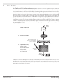
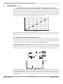
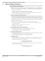
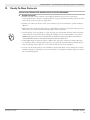
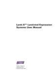
User Manual
Ready-To-Glow™ Secreted
Luciferase Reporter Systems
User Manual
United States/Canada
800.662.2566
Asia Pacific
+1.650.919.7300
Europe
+33.(0)1.3904.6880
Japan
+81.(0)77.543.6116
Clontech Laboratories, Inc.
A Takara Bio Company
1290 Terra Bella Ave.
Mountain View, CA 94043
Technical Support (US)
E-mail: [email protected]
www.clontech.com
PT3902-1 (PR922708)
Cat. Nos. 631729, 631735, 631742,
631744 & 631747
Published 10 April 2009
Ready-To-Glow™ Secreted Luciferase Reporter Systems User Manual
Table of Contents
I.
Introduction............................................................................................................................ 3
A.The Ready-To-Glow Reporter Assay..................................................................................................... 3
B.The Ready-To-Glow Reporter Vectors.................................................................................................. 4
C.Characteristics of the Secreted Metridia Luciferase Signal..................................................................... 5
D.The Ready-To-Glow Dual Secreted Reporter Assay.............................................................................. 6
II.
Additional Materials Required............................................................................................... 8
A.Microtiter Plates to Perform the Assay................................................................................................. 8
B.Vectors to Normalize Transfection Efficiency....................................................................................... 8
C.E. coli Competent Cells for Plasmid Propagation................................................................................. 8
D.Kits for Plasmid DNA Isolation........................................................................................................... 8
E. Sequencing Primers.............................................................................................................................. 8
III. Ready-To-Glow Protocols....................................................................................................... 9
A.Sample Preparation.............................................................................................................................. 9
B.Protocol for Non-Automated Metridia Luciferase Assays.................................................................... 10
C.Protocol for Automated Metridia Luciferase Assays............................................................................ 11
D.Chemiluminescent SEAP Reporter Assay (Dual Secreted Reporter System Only).............................. 12
IV. Troubleshooting Guide......................................................................................................... 13
V.
References............................................................................................................................. 14
Appendix A. Transfections & Experimental Controls................................................................. 15
A.Transfections...................................................................................................................................... 15
B.Proper Use of Controls...................................................................................................................... 15
Appendix B. Example Applications............................................................................................. 16
A.High-Throughput (HT) Applications................................................................................................ 16
B.Multiplexing Bioluminescent Secreted Reporters & Fluorescent Reporters........................................ 16
List of Figures
Figure 1. Flowchart of the Ready-To-Glow Secreted Luciferase Reporter Assay procedure........................ 3
Figure 2. High signal intensity and stability of secreted Metridia luciferase............................................... 5
Figure 3. Monitoring promoter activation using the secreted Metridia luciferase reporter......................... 5
Figure 4. Limit of detection of recombinant Metridia luciferase activity.................................................... 6
Figure 5. Monitoring the activities of two promoters using the Dual Secreted Reporter System................ 6
Figure 6. Similar secretion kinetics of Metridia secreted luciferase and SEAP............................................ 7
Figure 7. Use of secreted Metridia luciferase in an HTS application........................................................ 16
Figure 8. Multiplexing bioluminescent secreted reporters and fluorescent reporters................................ 17
List of Tables
Table I. Troubleshooting the Ready-To-Glow Secreted Luciferase Reporter Assay................................... 13
Table II. Troubleshooting the SEAP Assay (Dual Secreted Reporter System Only).................................. 14
Contact Us For Assistance
Customer Service/Ordering:
Technical Support:
Telephone: 800.662.2566 (toll-free)
Telephone: 800.662.2566 (toll-free)
Fax: 800.424.1350 (toll-free)
Fax: 650.424.1064
Web: www.clontech.com
Web: www.clontech.com
E-mail: [email protected]
E-mail: [email protected]
Protocol No. PT3902-1
www.clontech.com
Version No. PR922708
2
Clontech Laboratories, Inc.
A Takara Bio Company
Ready-To-Glow™ Secreted Luciferase Reporter Systems User Manual
I.
Introduction
A. The Ready-To-Glow Reporter Assay
The Ready-To-Glow Secreted Luciferase Reporter System is a versatile tool for the systematic analysis of eukaryotic
promoters and enhancers. The system uses secreted Metridia luciferase as a reporter molecule to monitor the activity of
promoters and enhancers, without the need for cell lysis, by sampling media supernatant. Each Ready-To-Glow system
includes a pMetLuc Reporter Vector, which contains either a specific promoter such as NFkB or CRE, or a multiple
cloning site where you can clone in the promoter sequence you are interested in. Transfect your cells of interest with
the Reporter Vector and culture until you are ready to begin your experiment. Then when you add an inducer, the
reporter molecule is expressed and secreted out of the cell into the supernatant. Simply add the luciferase substrate to
a sample of the media supernatant, and monitor pMetLuc-Reporter gene expression using a simple, sensitive, nonlysis and non-radioactive assay for secreted luciferase activity without the need for a cell lysis step (Figure 1). You can
perform the assay directly in the culture, or to transfer an aliquot of supernatant to another plate to perform the assay.
In either case, you will be left with live cells that can be used repeatedly, either for multiple time point assays or for
any other analysis on the cells.
1. Clone response element/
promoter of interest into
pMetLuc-Reporter vector
MCS
pMetLucReporter
Vector
Metridia
secreted
luciferase
2. Transfect host cell line
Promoter activation leads to
expression of secreted luciferase
protein in the medium
3. At desired time points,
assay luciferase activity
• Transfer media
sample to 96-well plate
• Add substrate
reaction buffer
• Assay luciferase activity
in a luminometer
Take sample of supernatant
Add luciferase
substrate
Secreted
luciferase
Product
LIGHT
Detection by
luminometer
Figure 1. Flowchart of the Ready-To-Glow Secreted Luciferase Reporter Assay procedure. The Ready-To-Glow Secreted Luciferase Reporter System combines the advantages of a live cell assay with the sensitivity of an enzyme-based system—all in a “one-step” reaction
which detects the activity of the secreted reporter enzyme in the supernatant of transfected cells without the need for cell lysis. This
flowchart describes the process for promoterless pMetLuc-Reporter Vectors. The procedure is identical for promoter-specific pMetLucReporter Vectors, except that Step 1 is eliminated.
Clontech Laboratories, Inc. www.clontech.com
A Takara Bio Company
Protocol No. PT3902–1
Version No. PR922708
3
Ready-To-Glow™ Secreted Luciferase Reporter Systems User Manual
I.
Introduction continued
The Metridia longa secreted luciferase gene was cloned from the marine copepod Metridia longa, and encodes a 24 kDa
protein containing a 17 amino acid N-terminal signal peptide for secretion (Markova et al., 2004). This secreted luciferase gene has been sequence-optimized by deleting possible cis-acting sites (splice sites), and increasing the overall GC
content to prolong mRNA half-life. It has also been human codon-optimized to enhance expression in cell lines.
The naturally secreted nature of Metridia luciferase provides several advantages for use as a transcription reporter:
• Cell lysis is eliminated.
• Gene expression kinetics studies can be simplified, by repeatedly collecting and assaying media from the same
culture (for multiple time points or multiple inducers).
• The same set of cultures can be used both for the secreted luciferase assay and for further investigations such
as DNA/RNA, protein, or cellular analysis because the transfected cells are not disturbed by measuring
luciferase activity in the media.
• Sample (culture media) collection can be automated by growing cultures and performing the assays in multiwell plates.
• The assay is more sensitive than fluorescence-based reporter assays or other cytosolic luciferase reporters.
• Guaranteed low background, because the medium can be removed and replaced at the start of the actual
promoter induction experiment.
• A broad dynamic range, because the reporter is secreted. Since there are no proteases in the culture media,
every reporter molecule that is expressed during the experiment contributes to the signal.
• The assay is suitable for high-throughput screening applications.
B. The Ready-To-Glow Reporter Vectors
Each Ready-To-Glow Reporter System includes one reporter vector and one control vector, along with sufficient
reagents to perform a minimum of 100 reactions.
The reporter vectors are:
• pMetLuc2-Reporter contains an MCS that allows promoter or promoter/enhancer elements to be inserted
upstream of the secreted Metridia luciferase (MetLuc) gene. Activation of the promoter/enhancer elements
of interest drives the expression of secreted Metridia luciferase, which is detected in the supernatant of the
transfected cells.
• pLVX-MetLuc Vector contains an MCS that allows promoter or promoter/enhancer elements to be inserted
upstream of the secreted Metridia luciferase (MetLuc) gene. Activation of the promoter/enhancer elements
of interest drives the expression of secreted Metridia luciferase, which is detected in the supernatant of the
transfected cells. Lentiviral delivery allows you to study your promoter of interest in virtually any cell type,
including primary cultures, dividing and nondividing cells, stem cells, terminally differentiated cells, and
neuronal cells
• pNFkB-MetLuc2-Reporter contains the NFkB promoter element upstream of the secreted Metridia luciferase (MetLuc) gene. It is designed to monitor NFkB signal transduction pathways. Adding TNF-α, IL-1,
or lymphokine receptor stimulants to the cell culture medium induces the binding of transcription factors to
the enhancer element and drives transcription of secreted Metridia luciferase.
• pCRE-MetLuc2-Reporter contains the CRE promoter element upstream of the secreted Metridia luciferase
(MetLuc) gene. It is designed to monitor the activation of cAMP binding protein (CREB) and cAMP-mediated signal transduction pathways. Adding cAMP or forskolin to the cell culture medium activates CREB or
ATF to bind CRE, and drives transcription of secreted Metridia luciferase.
Please visit our website at www.clontech.com/support/vectors.asp for more information and for vector maps.
Each reporter vector comes with a pMetLuc control vector, which is designed to function as a positive control. It
contains the secreted Metridia luciferase gene downstream of the constitutive immediate early promoter of the cytomegalovirus (pCMVIE), so cells transfected with this construct constitutively express and secrete Metridia luciferase.
Protocol No. PT3902-1
www.clontech.com
Version No. PR922708
4
Clontech Laboratories, Inc.
A Takara Bio Company
Ready-To-Glow™ Secreted Luciferase Reporter Systems User Manual
I.
Introduction continued
All of the pMetLuc vectors contain SV40 polyadenylation signals downstream of the MetLuc gene, which direct
proper processing of the 3’ end of the MetLuc mRNA. The vector backbone contains an SV40 origin of replication
in mammalian cells expressing the SV40 T antigen, a pUC origin of replication for propagation in E. coli, and an f1
origin for single-stranded DNA production. A neomycin-resistance cassette (Neor) allows stably transfected eukaryotic
cells to be selected using G418. This cassette consists of the SV40 early promoter, the neomycin/kanamycin resistance
gene of Tn5, and polyadenylation signals from the Herpes simplex virus thymidine kinase (HSV TK) gene. A bacterial
promoter (pKanr) upstream of the cassette allows kanamycin resistance in E. coli.
The pMetLuc2 vectors also include a synthetic transcription blocker (TB), composed of adjacent polyadenylation
and transcription pause sites located upstream of the pCMVIE promoter, which reduces background transcription
(Eggermont & Proudfoot, 1993).
C.
Characteristics of the Secreted Metridia Luciferase Signal
RLU
1. High Signal Intensity and Prolonged Stability of the Secreted Metridia Luciferase Signal
Compared to non-secreted luciferase reporters such as Renilla luciferase and firefly luciferase, secreted Metridia luciferase
exhibits a higher signal intensity and prolonged stability after substrate addition, making it easy to handle multiple
samples at the same time. The signal is stable between 20 and 45 minutes after substrate addition (Figure 2). Furthermore, the presence of 10% FBS in the media supernatant does not compromise signal intensity.
Firefly
Renilla
Metridia
Firefly neg
Renilla neg
Metridia neg
18 x 105
16 x 105
14 x 105
12 x 105
10 x 105
8 x 10⁵
6 x 10⁵
4 x 10⁵
2 x 10⁵
0
0
10
20
30
40
Time after addition of substrate (min)
50
Figure 2. High signal intensity and stability of secreted Metridia luciferase. CHO cells were plated into 96-well plates and transiently transfected with CMV-driven constructs encoding non-secreted firefly luciferase, non-secreted Renilla luciferase and sequence-optimized secreted Metridia luciferase. 24 hr after transfection, luciferase activity in equivalent samples was analyzed by addition of the recommended substrate. The signal was measured at different time points over a period of 45 min following the addition of substrate. neg=negative control.
Fold increase in reporter activity
[+TNF-α/–TNF-α (6 hr)]
In order to further examine the stability of the Metridia luciferase signal, we transiently transfected the pNFkB-MetLuc2-Reporter Vector into HeLa cells. 24 hours later, we induced the cells with TNF-α to activate the NFκB signaling
pathway. Six hours after that, we detected response element activation by assaying the cell supernatant for secreted
Metridia luciferase activity. Although signal intensity after substrate addition decreased with time, the calculated overall
fold induction was the same even 30 minutes after substrate addition (Figure 3; Haugwitz et al., 2008).
12
10
8
6
4
2
0
1
30
10
Time after addition of substrate (min)
Figure 3. Monitoring promoter activation using the secreted Metridia luciferase reporter. HeLa cells were transiently transfected with a
vector construct containing the NFkB response element driving the expression of sequence-optimized secreted Metridia luciferase. 24 hr
after transfection, the media was removed and replaced by media with or without TNF-α (100 ng/ml) to activate the NFkB signal transduction pathway. Six hr after addition of TNF-α, samples of the media were removed and analyzed for Metridia luciferase activity. The fold
induction was calculated for different time points following substrate addition.
Clontech Laboratories, Inc. www.clontech.com
A Takara Bio Company
Protocol No. PT3902–1
Version No. PR922708
5
Ready-To-Glow™ Secreted Luciferase Reporter Systems User Manual
I.
Introduction continued
2. High Sensitivity & Broad Linear Range of the Secreted Metridia Luciferase Signal
We determined the sensitivity (limit of detection) of Metridia luciferase in standard mammalian cell culture conditions
by spiking various amounts of purified protein into DMEM containing 10% FBS. Upon addition of the luciferase
substrate, we were able to detect levels as low as 40 fg of recombinant Metridia luciferase per ml (2 fg per well in a
96-well plate). This corresponds to approximately 46,000 molecules. We then determined the linear range of Metridia
luciferase activity by serial dilution of the recombinant protein, followed by substrate addition. Metridia luciferase
activity is linear over a range of at least 6 logs (Figure 4; Haugwitz et al., 2008).
108
107
Average RLU
106
105
104
10³
10²
100
10¹
10²
10³
Metridia luciferase (fg)
104
105
106
Figure 4. Limit of detection of recombinant Metridia luciferase activity. Various amounts of recombinant Metridia luciferase were spiked
into DMEM + 10% FBS in a 96-well plate. After addition of substrate, the plate was analyzed using a Molecular Devices® SpectraMax® L
plate reader.
D. The Ready-To-Glow Dual Secreted Reporter Assay
Multiplexing two different bioluminescent secreted reporters, using different substrates, provides even more information on the cell state without sacrificing the cells, and creates new advantages for experimental design and analysis. For
an extended discussion about the SEAP reporter system, please refer to PT3057-1, the Great EscAPe™ SEAP Reporter
System User Manual, which can be found at www.clontech.com/support/manuals.asp.
The Ready-To-Glow Dual Secreted Reporter Assay (No. 631734) uses two secreted reporters, Metridia luciferase and
Secreted Alkaline Phosphatase (SEAP), that can be detected and distinguished from each other by reporter-specific
substrates. When cells are cotransfected with both reporters and stimulated to express and secrete Metridia luciferase
or SEAP, or both, promoter-specific activity can be detected in a sample of culture media by adding the relevant
+TNF-α
ML
+ Substrate
LIGHT
SP
+ Substrate
LIGHT
NFκB-RE Metridia luciferase
?
?
+Forskolin
CRE
SEAP
Cell
Cell Supernatant
Fold increase in reporter activity
30
SEAP
signal
Secreted
Metridia
luciferase
signal
25
20
15
10
5
0
–TNF-α
+TNF-α
–Forskolin
+Forskolin
Figure 5. Monitoring the activities of two promoters using the Ready-To-Glow Dual Secreted Reporter System to investigate potential
cross-talk between signaling pathways in the same cell. HEK 293 cells cotransfected with pNFκB-TA-MetLuc and pCRE-SEAP constructs
were treated with fresh media alone, or with media containing either 1,000 ng/ml TNF-α or 10 μM forskolin. Media samples were collected
from the transfected cells 7 hr later, and each sample was tested in triplicate. No cross talk was observed between the cAMP and NFkB
signaling pathways.
Protocol No. PT3902-1
www.clontech.com
Version No. PR922708
6
Clontech Laboratories, Inc.
A Takara Bio Company
Ready-To-Glow™ Secreted Luciferase Reporter Systems User Manual
I.
Introduction continued
enzyme-specific substrate and measuring the resulting chemiluminescence. Because the reporters are secreted (and do
not require cell lysis), you will be left with live cells that can be used repeatedly, either for multiple time point assays
or for any other analysis on the cells.
Potential applications of the Ready-To-Glow Dual Secreted Reporter System include:
• Using one reporter as a transfection control for normalization purposes.
• Using one reporter as a positive, constitutive control to monitor the functionality of the secretory pathway.
• Monitoring two different signal transduction pathways simultaneously. This makes it possible to examine
potential cross-communication between intracellular signal transduction pathways. For example, we used
secreted Metridia luciferase and secreted alkaline phosphatase (SEAP) to determine whether the cAMP and
NFκB signal transduction pathways are interrelated (Figure 5).
1. The Ready-To-Glow Dual Secreted Reporter Vectors
The Ready-To-Glow Dual Secreted Reporter Vector Kit (Cat. No. 631735) includes four vectors (e.g., two vector sets).
It has been customized to work optimally with our Ready-To-Glow Dual Secreted Reporter Assay (Cat. No. 631734).
The reporter vectors are:
• pMetLuc2-Reporter contains an MCS that allows promoter or promoter/enhancer elements to be inserted
upstream of the secreted Metridia luciferase (MetLuc) gene. Activation of the promoter/enhancer elements
of interest drives the expression of secreted Metridia luciferase, which is detected in the supernatant of the
transfected cells.
• pSEAP2-Basic lacks eukaryotic promoter and enhancer sequences and has an MCS that allows putative
promoter DNA fragments to be inserted upstream of the SEAP gene. Enhancers can be cloned into either the
MCS or unique downstream sites. Activation of the promoter/enhancer elements of interest drives the expression of SEAP, which is detected in the supernatant of the transfected cells.
Each reporter vector comes with a control vector (pMetLuc2-Control and pSEAP2-Control, respectively), which
is designed to function as a positive control. The control vectors serve as positive controls for constitutive promoter
activity in the luciferase- and SEAP-based assays respectively.
RLU normalized %
2. Metridia Luciferase and SEAP Secretion Kinetics Are Comparable
The Metridia luciferase and SEAP reporters are secreted from stable or transiently transfected cells into the culture
medium, so you can sample repeatedly over time without sacrificing cells. Since the secretion kinetics of both reporters
are very similar (Figure 6), the amount of either reporter in the cell culture medium accurately reflects its promoter’s
activity.
120
100
80
60
40
20
0
pSEAP2Control
Vector
0
5
10
15
Time (hr)
20
25
pMetLucControl
Vector
Figure 6. Similar secretion kinetics of Metridia secreted luciferase and SEAP enable accurate comparisons of the relative timing of promoter activity. HeLa cells were plated into 6-well plates and transiently transfected with either the pMetLuc-Control vector or the pSEAPControl vector. Media samples from the transfected cells were collected from 3 wells at each time point by removing enough media to
run either the luciferase or the SEAP assay. Each sample was tested in triplicate, using a white bottom 96-well microtiter plate on a Turner
BioSystems Veritas® Luminometer.
For examples of how the Ready-To-Glow Secreted Reporter Systems can be applied to your promoter studies, please
see Appendix B.
Clontech Laboratories, Inc. www.clontech.com
A Takara Bio Company
Protocol No. PT3902–1
Version No. PR922708
7
Ready-To-Glow™ Secreted Luciferase Reporter Systems User Manual
II.
Additional Materials Required
A. Microtiter Plates to Perform the Assay
Bioluminescence detection of luciferase activity can be performed using either a tube or plate luminometer
when reactions are carried out in microcentrifuge tubes or in multiwell microtiter plates. Ready-To-Glow assays
can be carried out either in 0.5 ml microcentrifuge tubes or multiwell microtiter plates. The following type of
microtiter plate is recommended:
• Microlite™ 1 Luminescence Microtiter 96-well plates, Flat-Bottom
(VWR Scientific Products, Cat. No. 62403-124). These opaque white plates, which contain flat-bottom
wells, are recommended for bioluminescent assays and detection.
B.
Vectors to Normalize Transfection Efficiency
The Ready-To-Glow Dual Secreted Reporter Vector Kit (Cat. No. 631735) includes the pSEAP2-Basic and
-Control Vectors as well as the pMetLuc2-Reporter and -Control Vectors and the respective assays. The reporter
vectors can be used as transcription reporters, while the control vectors serve as positive controls for constitutive
promoter activity in the SEAP- (a secreted form of human alkaline phosphatase) and luciferase-based assays
respectively. For both vector sets, promoter activity can be directly correlated to the amount of reporter (SEAP
or luciferase) secreted in the medium.
Alternatively, any of the following vectors can be used to normalize transfection efficiency:
• Fluorescent protein vectors such as pAcGFP1-N1 Vector (Cat. No. 632469) or pDsRed-Express2-N1 Vector (Cat. No. 632537).
• pSEAP2-Control Vector (sold separately as Cat. No. 631717)
• pCMVβ Vector (Cat. No. 631719)
C.
E. coli Competent Cells for Plasmid Propagation
Either of the following competent E. coli strains can be used for plasmid propagation:
• Fusion-Blue™ Competent Cells (Cat. Nos. 636700 & 636758)
• Supercharge EZ10 Electrocompetent Cells (Cat. No. 636756)
D. Kits for Plasmid DNA Isolation
For rapid, high yield isolation of transfection grade plasmid DNA, we recommend using NucleoBond® Xtra Kits:
• NucleoBond Xtra Midi Plus Kits (Cat. Nos. 740412.50 & 740412.50)
• NucleoBond Xtra Maxi Plus Kits (Cat. Nos. 740416.10 & 740416.50)
E.
Sequencing Primers
• We recommend using the following primer (not available from Clontech) to sequence inserts cloned into
the pMetLuc2-Reporter Vector. This reverse primer can be used to sequence from the 5’ region of the
Metridia reporter gene region into the upstream MCS:
5’-CACGATGTCGATGTTGGGG-3’
(183–165 in pMetLuc2-Reporter Vector)
• We recommend the following primer (not available from Clontech) to sequence from the 5’ region of the SEAP
reporter gene region into the MCS:
5’-CCTCGGCTGCCTCGCGGTTCC-3’
(376–356 in pSEAP2-Control)
Protocol No. PT3902-1
www.clontech.com
Version No. PR922708
8
Clontech Laboratories, Inc.
A Takara Bio Company
Ready-To-Glow™ Secreted Luciferase Reporter Systems User Manual
III.
Ready-To-Glow Protocols
PLEASE READ THROUGH THE ENTIRE PROTOCOL BEFORE BEGINNING.
A. Sample Preparation
1. Transfect your cells of interest with the secreted Metridia luciferase reporter vector of your choice.
You can perform either a transient or stable transfection, using your transfection method of choice. For information about transfections, please see Appendix A.
2. Incubate your cells for the amount of time your transfection protocol recommends for optimal transfection
efficiency.
3. Remove the culture media and split the cells to a multiwell plate, following standard tissue culture protocols.
Alternatively, you can leave the cells in the culture plate for your experiment.
4. At the beginning of your experiment (i.e., when you begin your experimental treatment) remove the culture
media and replace it with fresh media containing the candidate promoter inducer/inhibitor of your choice.
Note: Changing the tissue culture media eliminates any background signal caused by the accumulation of
secreted Metridia luciferase in the supernatant before the start of the experiment.
Idea
5. Collect 50 µl samples of media supernatant at different time points after starting the treatment. How long
you should wait before collecting samples depends on your particular experiment. We recommend that you
begin by collecting media samples 2, 8, 12, and 24 hours after the start of the experiment.
6. You can assay the media samples for secreted luciferase activity immediately, or freeze them for later analysis.
If multiple samples are collected, they can be transferred into a 96-well plate for further analysis (See Section
II for recommended plates).
Clontech Laboratories, Inc. www.clontech.com
A Takara Bio Company
Protocol No. PT3902–1
Version No. PR922708
9
Ready-To-Glow™ Secreted Luciferase Reporter Systems User Manual
III.
Ready-To-Glow Protocols continued
B. Protocol for Non-Automated Metridia Luciferase Assays
1. Prepare the 10X Substrate Stock Solution.
Protocol
15 min.
Attention
Dissolve the Lyophilized Secreted Luciferase Substrate by adding Substrate Buffer:
Number of Reactions
Quantity of Substrate Buffer
100 rxns (Cat. No. 631726)
50 µl
500 rxns (Cat. No. 631727)
250 µl
1,000 rxns (Cat. No. 631728)
500 µl
DO NOT AGITATE or VORTEX the solution to dissolve the substrate. The substrate is sensitive to oxidation
in the presence of air bubbles caused by agitation. A 15 min incubation at room temperature (RT) can accelerate the reconstitution.
Store the 10X Substrate Stock Solution at –20° C. We do not recommend aliquoting the solution, as this may
induce oxidation.
2. Prepare the 1X Substrate/Reaction Buffer.
Dilute the 10X Substrate Stock Solution (Step III.B.1) 1:10 in Reaction Buffer. To calculate the total volume
of 1X Substrate/ Reaction Buffer needed in your experiment, multiply the number of samples in your experiment by a factor of 5.
For example, for 20 samples, you would prepare 20 x 5 = 100 μl of 1X Substrate/Reaction Buffer by diluting
10 μl of 10X Substrate Stock Solution in 90 μl of Reaction Buffer.
Attention
To ensure consistent results, prepare fresh 1X fresh Substrate/Reaction Buffer every time, without
introducing air bubbles. Slowly pipet the Substrate Stock Solution or use the tip to stir the mixture gently,
instead of using rigorous pipetting. NEVER VORTEX THE SUBSTRATE OR SUBSTRATE/REACTION BUFFER.
When the Substrate Stock is mixed with the colorless Reaction Buffer, the resulting 1X solution is light
yellow/orange in color. Allow the 1X Substrate/Reaction Buffer to remain at room temperature for 10 min
before use. It can remain at room temperature for up to 1 hr before use.
3. Transfer 50 μl of culture media from each sample (Step III.A.5) into one well of a 96 well plate (please see
Section II for a 96 well-plate recommendation).
te
No
Note: For statistical reasons, we recommend assaying triplicates of each sample.
4. Add 5 μl of 1X Substrate/Reaction Buffer (Step III.B.2) to each sample.
If you have many samples in a 96-well plate, we recommend using a multichannel pipette in order to reduce
the time between substrate addition and signal detection.
5. Transfer the plate to a luminometer and record light signals according to the manufacturer’s recommended
luminometer settings.
TIP: We recommend reading the plate(s) immediately, as well as 5 and 10 min after substrate addition. If the assay was not performed in a luminometer-compatible microtiter plate, transfer the entire solution from each well
to a suitable plate and place it in the instrument.
Protocol No. PT3902-1
www.clontech.com
Version No. PR922708
10
Clontech Laboratories, Inc.
A Takara Bio Company
Ready-To-Glow™ Secreted Luciferase Reporter Systems User Manual
III.
Ready-To-Glow Protocols continued
C. Protocol for Automated Metridia Luciferase Assays
1. Prepare the 100X Substrate Stock Solution:
Protocol
15 min.
Attention
Number of Reactions
Quantity of Substrate Buffer
1,000 rxns (Cat. No. 631739)
500 µl
5,000 rxns (Cat. No. 631740)
500 µl/ vial (2.5 ml in total, for 5 vials)
Note: DO NOT AGITATE or VORTEX the solution to dissolve the substrate. The substrate is sensitive to
oxidation in the presence of air bubbles caused by agitation. A 15 min incubation at room temperature (RT)
can accelerate the reconstitution.
Store the 100X Substrate Stock Solution at –20° C. We do not recommend aliquoting the solution, as this may
induce oxidation.
2. Prepare the 1X Substrate/Reaction Buffer:
Dilute the 100X Substrate Solution (Step III.C.1) 1:100 in Reaction Buffer. To calculate the total volume of
1X Substrate/ Reaction Buffer needed in your experiment, multiply the number of samples in your experiment
by a factor of 50.
For example, for 100 samples, you would prepare 100 x 50 = 5,000 μl of 1X Substrate/Reaction Buffer by
diluting 50 μl of 100X Substrate Stock Solution in 4,950 μl of Reaction Buffer.
Attention
To ensure consistent results, prepare fresh 1X fresh Substrate/Reaction Buffer every time, without
introducing air bubbles. Slowly pipet the Substrate Stock Solution or use the tip to stir the mixture gently,
instead of using rigorous pipetting. NEVER VORTEX THE SUBSTRATE OR SUBSTRATE/REACTION BUFFER.
When the Substrate Stock is mixed with the colorless Reaction Buffer, the resulting 1X solution is light
yellow/orange in color. Allow the 1X Substrate/Reaction Buffer to remain at room temperature for 10 min
before use. It can remain at room temperature for up to 1 hr before use.
3. Transfer 50 μl of culture media from each sample (Step III.A.5) into one well of a 96 well plate (please see
Section II for a 96 well-plate recommendation).
te
No
Note: For statistical reasons, we recommend assaying triplicates of each sample.
4. Add 50 μl of 1X Substrate/Reaction Buffer (Step III.C.2) to each sample, either by pipetting it into the wells
manually or via an automatic injector system connected to your plate reader.
Tip: Do not vortex the plate to mix the sample and substrate. If you have many samples, we recommend using
a multichannel pipette in order to reduce the time between substrate addition and signal detection.
5. Transfer the plate to a luminometer and record light signals according to the manufacturer’s recommended
luminometer settings.
TIP: We recommend reading the plate(s) immediately, as well as 5 and 10 min after adding the substrate.
Clontech Laboratories, Inc. www.clontech.com
A Takara Bio Company
Protocol No. PT3902–1
Version No. PR922708
11
Ready-To-Glow™ Secreted Luciferase Reporter Systems User Manual
III.
Ready-To-Glow Protocols continued
D. Chemiluminescent SEAP Reporter Assay (Dual Secreted Reporter System Only)
For transient transfection assays using the pSEAP2-Control vector (Sold as part of the Ready-To-Glow Dual Secreted
Reporter Vector Kit, Cat. No. 631735), the secreted form of human alkaline phosphatase (SEAP) is generally detected
in the medium 12–18 hours after transfection, with maximal levels detected between 48–72 hours. Optimal times
will vary depending on the cell type, cell density, and the particular experimental conditions. Each construct should
be transfected and assayed in triplicate.
1. Prepare Reagents and Samples for the SEAP Assay
a. Transfer 25 μl of cell culture medium from transfected cells or mock transfected cells (in triplicate) to a
96-well microtiter plate. If necessary, the plate can be frozen at –20ºC for future analysis.
ote
Note: We recommend Microlite™1 Luminescence Microtiter 96-well plates (VWR Scientific Products, Cat.
No. 62403-124).
N
b. Prepare 1X Dilution Buffer by diluting the 5X Dilution Buffer 1:5 with ddH2O.
2. Perform the SEAP Assay
You may need to dilute some samples in order to stay within the linear range of the assay. To determine the
linear range, assay a dilution series of your sample and a dilution series of recombinant human placental secreted
alkaline phosphatase before you assay your samples.
a. Allow the SEAP Substrate Solution to equilibrate to room temperature (22–25°C).
b. Add 75 μl of 1X Dilution Buffer to each sample (from Step 1.a.) in the 96-well microtiter plate.
c. Seal the plate with adhesive aluminum foil or a 96-well plate lid and incubate the diluted samples for 30
min at 65°C using a heat block or water bath.
d. Cool the samples on ice for 2–3 min, then equilibrate to room temperature.
e. Add 100 μl of SEAP Substrate Solution to each sample. Incubate for 10–60 min (30 min recommended)
at room temperature before reading.
f. Use a 96-well plate reader luminometer to detect and record the SEAP signal. Optimal readings will be
obtained 10–60 min after substrate addition.
Refer to your plate reader’s user manual for additional information regarding its performance and use.
Protocol No. PT3902-1
www.clontech.com
Version No. PR922708
12
Clontech Laboratories, Inc.
A Takara Bio Company
Ready-To-Glow™ Secreted Luciferase Reporter Systems User Manual
IV.
Troubleshooting Guide
Table I. Troubleshooting the Ready-To-Glow Secreted Luciferase Reporter Assay
Problem
Explanation
Solution
Little or no signal
from transfected
cells.
Number of transfected
cells is too low.
Ensure that the transfection efficiency has been optimized by
using the supplied control vector as an internal positive control
for luciferase expression.
Increase the number and/or density (or concentration) of cells
used in transfections.
Transfected cells are not
yet producing Metridia
luciferase.
If the signal from the positive control (media from cells transfected with the pMetLuc-Control Vector) is low, incubate the
transfected cells in media for a longer time before collecting
supernatant samples.
Increase the time of transfection before starting the experiment.
Plate reader requires
adjustment.
Refer to the instrument’s instructions for methods to increase the
sensitivity of light detection.
Signal quenches
rapidly (within a
few minutes of substrate addition).
Although the signal intensity decreases over time,
the overall fold induction
remains the same for ~30
min after substrate addition (Part I, Figure 3).
Continue with your experiment as usual.
Substrate stock
solution darkens
over time.
Possibly due to partial oxidation of the substrate.
Ensure that the substrate is working properly (using appropriate
positive and negative controls) before assaying experimental
samples.
High background
signal.
Decrease the volume of media assayed from experimental cultures. Alternatively, dilute your samples.
Signal exceeds the
linear range of the
assay.
Dilute your samples, using the same media that was used to
culture the cells.
If you are in doubt about the linear range of the assay, prepare
and assay a dilution series using the positive control (media
from cells transfected with the pMetLuc-Control Vector).
Clontech Laboratories, Inc. www.clontech.com
A Takara Bio Company
Protocol No. PT3902–1
Version No. PR922708
13
Ready-To-Glow™ Secreted Luciferase Reporter Systems User Manual
IV.
Troubleshooting Guide continued
Table II. Troubleshooting the SEAP Assay* (Dual Ready-To-Glow System Only)
Problem
Explanation
Solution
Little or no signal
from transfected
cells.
Number of transfected
cells is too low.
Ensure that the transfection efficiency has been optimized by using
pSEAP2-Control as an internal positive control for SEAP expression.
Increase the number and/or density (or concentration) of cells
used in transfections.
Increase the post-transfection interval prior to collecting media samples.
If background signals from negative controls (i.e., cells transfected
with pSEAP2-Basic) are low, increase the volume of media
assayed from experimental cultures from 25 µl to 50–75 µl (adjust
the 1X Dilution Buffer volume accordingly.)
Problem with assay
conditions.
Ensure that the assay conditions are correct and that the detection
method is working by assaying the positive control enzyme.
Ensure that the conditioned media does not contain an inhibitory
activity by adding 2 µl of Positive Control Placental Alkaline
Phosphatase to 25 µl of culture medium
Plate reader requires
adjustment.
High background
signal.
Refer to the instrument’s instructions for methods to increase the
sensitivity of light detection.
Ensure that the diluted media samples are heated for the full 30
min at 65°C as specified in Step III.D.2.c.
Decrease the volume of media assayed from experimental cultures.
Alternatively, dilute your samples.
Note: If diluting with fresh serum-containing media, please
ensure heat inactivation of any endogenous alkaline phosphatase
by incubation at 65°C for 30 min.
If possible, after transfection grow cells in media containing minimal serum. Serum levels >10% (v/v) may increase background.
Signal exceeds the
linear range of the
assay.
If in doubt about the linear range of the assay, prepare and assay
a dilution series using the Positive Control Placental Alkaline
Phosphatase.
Decrease the volume of media assayed from experimental
cultures. Alternatively, dilute your samples.
Note: If diluting with fresh serum-containing media, please ensure
heat inactivation of any endogenous alkaline phosphatase by
incubation at 65°C for 30 min.
Perform a time course after induction by collecting and assaying
media supernatant at different time points to find a suitable time
point, at which the signal falls in the linear range.
*For an extended discussion about the SEAP reporter system, please refer to PT3057-1, the Great EscAPe SEAP Reporter System User Manual, which can be
found at www.clontech.com/support/manuals.asp.
V.
References
Eggermont, J. & Proudfoot, N. (1993) Poly(A) signals and transcriptional pause sites combine to prevent interference
between RNA polymerase II promoters. EMBO J. 12(6):2539–2548.
Haas, M., et al. (1998) Effect of proteasome inhibitors on monocytic IkB-α and -β depletion, NFkB activation, and
cytokine production. J. Leukoc. Biol. 63(3): 395–404.
Markova, S. V. et al. (2004) Cloning and Expression of cDNA for a Luciferase from the Marine copepod Metridia
longa. J. Biol. Chem. 279(5):3212–3217.
Sambrook, J. & Russell, D. W. (2001) Molecular Cloning: A Laboratory Manual, Cold Springs Harbor Laboratory
Press (Cold Springs Harbor, NY).
Sun, L. & Carpenter, G. (1998) Epidermal growth factor activation of NFkB is mediated through IkB-α degradation
and intracellular free calcium. Oncogene 16(16): 2095–2102.
Protocol No. PT3902-1
www.clontech.com
Version No. PR922708
14
Clontech Laboratories, Inc.
A Takara Bio Company
Ready-To-Glow™ Secreted Luciferase Reporter Systems User Manual
Appendix A. Transfections & Experimental Controls
A. Transfections
1. Transfection Methods:
All Ready-To-Glow vectors can be transfected into mammalian cells by your standard transfection method of
choice. If you do not have an established transfection protocol or if you are working with a cell line for the first
time, we recommend comparing different protocols, using the pMetLuc2-Control vector and monitoring the
amount of secreted Metridia luciferase in the cell supernatant, in order to determine efficiency and choose an
appropriate protocol.
2. Transfection Considerations:
a. Perform each transfection in triplicate. If cells are sampled in multiwell plates, we recommend transfecting one master stock and then transferring cells from this master stock into the wells of a multiwell plate,
to avoid variations that may occur if cells in each well were transfected separately.
b. Normalize for transfection efficiency. When monitoring the effect of promoter and enhancer sequences
on gene expression, it is critical to include an internal control that will distinguish differences in the level
of transcription from variability in the efficiency of transfection (Sambrook & Russell, 2001 ). This is
easily done by cotransfecting a second plasmid that constitutively expresses another reporter that can be
clearly differentiated from secreted Metridia luciferase. The level of this second reporter can then be used
to normalize the levels of luciferase among different treatment groups and different transfections.
The Ready-To-Glow Dual Secreted Reporter Vector Kit (Cat. No. 631735) includes the pSEAP2-Basic
and -Control Vectors as well as the pMetLuc2-Reporter and -Control Vectors and the respective assays.
The reporter vectors can be used as transcription reporters, while the control vectors serve as positive controls for constitutive promoter activity in the SEAP- (a secreted form of human alkaline phosphatase) and
luciferase-based assays respectively. For both vector sets, promoter activity can be directly correlated to the
amount of reporter (SEAP or luciferase) secreted in the medium.
Other reporter proteins frequently used for normalization include SEAP (the pSEAP2-Control Vector, sold
separately as Cat. No. 631717), E. coli β-galactosidase (pCMVβ Vector, Cat. No. 631719), and fluorescent
protein vectors such as the pAcGFP1-N1 Vector (Cat. No. 632469) or the pCMV-DsRed-Express Vector
(Cat. No. 632416).
B.
Proper Use of Controls
1. Negative Controls:
A negative control is necessary to measure the background signal associated with the cell culture media.
To determine the background level, perform the assay using 50 µl samples of culture medium from mock‑transfected cells (without vector). The values obtained from such controls should be subtracted from experimental
results.
2. Positive Controls:
A positive control is necessary to confirm transfection and expression of exogenous DNA, and to verify that
your cells are secreting Metridia luciferase into the culture media.
To confirm the expression and secretion of functional luciferase in transfected cells, perform the assay using 50 µl of culture medium from cells transfected with the pMetLuc2‑Control Vector, which contains the
luciferase gene under transcriptional control of the constitutive CMV promoter. Cells transfected with this
plasmid should yield high activity within 12–36 hours (often even earlier) after transfection.
3. Normalizing Transfection Efficiencies:
It is critical to include an internal control, in order to distinguish differences in the level of transcription from
variability in the efficiency of transfection. See Section A.2.b above or Sambrook & Russell (2001) for more
information.
Clontech Laboratories, Inc. www.clontech.com
A Takara Bio Company
Protocol No. PT3902–1
Version No. PR922708
15
Ready-To-Glow™ Secreted Luciferase Reporter Systems User Manual
Appendix B. Example Applications
A. High-Throughput (HT) Applications
The Ready-To-Glow Secreted Luciferase System is a powerful tool for HT applications because it is easy to use, eliminates
cell lysis, and guarantees high sensitivity and a broad linear range. It has been used successfully in a 1,536-well format
to visualize the expression of secreted Metridia luciferase, even using a regular, low-sensitivity CCD camera (Figure 7).
In addition, the assay can be performed in the presence of cells by adding the substrate directly to the culture media.
Figure 7. Use of secreted Metridia luciferase in a High-Throughput Screening (HTS) application. This is a screen shot of a 1,536-well plate
containing stable CHO cells transfected with a forskolin-responsive Metridia luciferase gene (pASM-Lu164), taken with a CCD camera. 300
cells/well were plated in a 1,536-well microtiter plate. Cells were incubated with increasing concentrations of forskolin and the plate was
incubated for 4 hr at 37° C. Metridia substrate was added in increasing concentrations and the plate was visualized using a CCD camera
system (integration time: 60 sec). This pseudocolor image reflects luminescence intensity. Near white and gray regions are the brightest,
while black regions are the least bright. Courtesy of Bayer Health Care, Germany.
B. Multiplexing Bioluminescent Secreted Reporters & Fluorescent Reporters
Plate readers that can detect bioluminescent and fluorescent reporter activity at the same time, in the same well create
opportunities to develop new multiplex assays that are unlimited by the method of detection. When multiplexing a
secreted reporter and a fluorescent reporter, there is no requirement to sacrifice the cells—expanding the possibilities
even more.
We used this type of multi-mode, multiplexing approach to determine the role of the proteasomes in TNF-α-induced,
NFκB-dependent signaling. We used the Living Colors® Proteasome Sensor HEK 293 Cell Line to monitor proteasomal activity: if the proteasomes are active, only a very low level of green fluorescence is observed in the cytosol of
this cell line. However, if the function of the cellular proteasomes is compromised, there is a sharp rise in the fluorescent intensity in the cytosol of this stable cell line. At the same time, we used secreted Metridia luciferase to monitor
NFκB-based promoter activation.
Using these two reporters, we were able to determine the importance of proteasomal activity in the NFκB dependent,
TNF-α-induced signaling pathway. It has been shown previously that phosphorylation of IκB alone upon TNF-α
treatment is not sufficient to allow NFκB-induced promoter activation (Haas et al., 1998; Sun & Carpenter, 1998).
Consistent with these findings, our results show that phosphorylation of IκB must be followed by its proteasomal
degradation, in order to allow NFκB-based promoter activation (Figure 8; Haugwitz et al., 2008). This experiment
demonstrates the power of multiplexing bioluminescent and fluorescent reporters in a live cell assay: we were able to
detect two events simultaneously, from the same well of a 96-well plate, simply by collecting both fluorescent and
bioluminescent signals using a multi-mode plate reader.
Protocol No. PT3902-1
www.clontech.com
Version No. PR922708
16
Clontech Laboratories, Inc.
A Takara Bio Company
Ready-To-Glow™ Secreted Luciferase Reporter Systems User Manual
Appendix B. Example Applications continued
A
NFκB
IκB
1
P
No
ALLN
+ TNF-α
With
ALLN
P
IκB
NFκB
2
Active Proteasome
ALLN
IκB
NFκB
Inactive Proteasome
4
3
5
ZsProSensor-1
ZsProSensor-1
NFκB
NFκB Response Element
B
Metridia luciferase
NFκB Response Element
180
50,000
160
45,000
35,000
120
30,000
25,000
80
RLU
100
20,000
60
15,000
40
10,000
20
0
ZsProSensor-1
(RFU)
Metridia
Secreted
Luciferase
(RLU)
40,000
140
RFU
Metridia luciferase
5,000
−ALLN/−TNF-α
−ALLN/+TNF-α
+ALLN/−TNF-α
+ALLN/+TNF-α
0
Figure 8. Multiplexing bioluminescent secreted reporters and fluorescent reporters to determine the requirement for active proteasomes
in TNF-α-induced, NFkB-dependent signaling. Panel A. Inactive NFkB is sequestered in the cytoplasm by IkB. IkB must be phosphorylated
upon TNF-α induction (1) and degraded by the proteasome (2) in order for NFkB to translocate to the nucleus and initiate signaling (3).
Alternatively, when the proteasome is inhibited by the peptide ALLN (4), IkB is not degraded and NFkB cannot translocate (5). The status
of the proteasome (active or inactive) can be monitored based on ZsProSensor-1 levels. Panel B. NFkB activation by TNF-α requires proteasomal activity. High levels of Metridia luciferase signal were only observed in the absence of ALLN and the presence of TNF-α. When
the proteasomes were inactivated by ALLN (indicated by increasing levels of ZsProSensor-1 fluorescence), the NFkB signaling pathway
could not respond effectively to TNF-α stimulation.
Notice to Purchaser
Clontech products are to be used for research purposes only.They may not be used for any other purpose, including, but not limited to, use in
drugs, in vitro diagnostic purposes, therapeutics, or in humans. Clontech products may not be transferred to third parties, resold, modified for
resale, or used to manufacture commercial products or to provide a service to third parties without written approval of Clontech Laboratories, Inc.
CMV Sequence:
The CMV promoter is covered under U.S. Patent Nos. 5,168,062, and 5,385,839 assigned to the University of Iowa Research Foundation.
Metridia Luciferase:
Markova, S. V., Golz, S., Frank, L. A., Kalthof, B. & Vysotski, E. S. (2004) Cloning and expression of cDNA for a luciferase from the marine
copepod Metridia longa. A novel secreted bioluminescent reporter enzyme. J. Biol. Chem. 279(5):3212–3117.
Living Colors® Products AcGFP1, AmCyan, AsRed, mBanana, mCherry, DsRed, HcRed, mOrange, mPlum, mRaspberry, mStrawberry,
tdTomato, ZsGreen, ZsYellow and their variants:
Not-For-Profit Entities: Orders may be placed in the normal manner by contacting your local representative or Clontech Customer
Service at 650.919.7300. At its discretion, Clontech grants Not-For-Profit Entities a non-exclusive, personal, limited license to use this
product for non-commercial life science research use only. Such license specifically excludes the right to sell or otherwise transfer this
product, its components or derivatives thereof to third parties. No modifications to the protein coding sequence may be made without
express written permission from Clontech. Any other use of this product requires a license from Clontech. For license information,
please contact a licensing representative by phone at 650.919.7320 or by e-mail at [email protected].
For-Profit Entities wishing to use this product are required to obtain a license from Clontech. For license information, please contact a
licensing representative by phone at 650.919.7320 or by e-mail at [email protected].
Microlite™ is a trademark of Thermo Electron Corporation.
Molecular Devices® and SpectraMax® are registered trademarks of Molecular Devices, now a part of MDS Analytical Technologies.
NucleoBond® is a registered trademark of Macherey-Nagel GmbH & Co.
Veritas® is a registered trademark of Turner BioSystems.
Clontech, the Clontech logo and all other trademarks are the property of Clontech Laboratories, Inc., unless noted otherwise.
Clontech is a Takara Bio Company. ©2009 Clontech Laboratories, Inc.
Clontech Laboratories, Inc. www.clontech.com
A Takara Bio Company
Protocol No. PT3902–1
Version No. PR922708
17