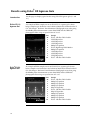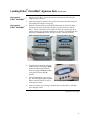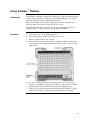
1
E-Gel® Technical Guide General information and protocols for using E-Gel® pre-cast agarose gels Version K December 12, 2008 25-0645 Corporate Headquarters Invitrogen Corporation 1600 Faraday Avenue Carlsbad, CA 92008 T: 1 760 603 7200 F: 1 760 602 6500 E: [email protected] For country-specific contact information visit our web site at www.invitrogen.com User Manual ii Table of Contents Table of Contents........................................................................................................................................ iii Warranty...................................................................................................................................................... vi General Information.................................................................................................................................. vii Accessory Products ..................................................................................................................................viii Introduction .................................................................................................................................1 E-Gel® Electrophoresis System ...................................................................................................................1 Gel Selection..................................................................................................................................................2 Low-Throughput E-Gel® Electrophoresis System....................................................................................4 Low-Throughput E-Gel® Well Formats.....................................................................................................5 E-Gel® iBase™ Power System.......................................................................................................................6 E-Gel® PowerBase™ ......................................................................................................................................8 E-Gel® Opener...............................................................................................................................................9 Gel Knife ......................................................................................................................................................11 Medium-Throughput E-Gel® Electrophoresis System ..........................................................................12 E-Gel® 48 Agarose Gels..............................................................................................................................13 High-Throughput E-Gel® Electrophoresis System.................................................................................14 E-Gel® 96 Agarose Gels..............................................................................................................................15 E- Base™ Power Supply..............................................................................................................................16 Nucleic Acid Gel Stains for E-Gel® Agarose Gels ..................................................................................18 SYBR Safe® DNA Gel Stain .......................................................................................................................19 Proprietary Fluorescent Nucleic Acid Gel Stain ....................................................................................21 Safe Imager™ Blue Light Transilluminators ............................................................................................22 Product Specifications................................................................................................................................27 Methods .....................................................................................................................................30 General Guidelines............................................................................................................................. 30 Electrophoresis of E-Gel® Agarose Gels.......................................................................................... 32 Sample Preparation for E-Gel® Agarose Gels .........................................................................................32 Loading Single Comb and Double Comb Gels.......................................................................................35 Running Single Comb and Double Comb Gels......................................................................................37 Results with E-Gel® Single Comb Gels ....................................................................................................40 Results with E-Gel® Double Comb Gels ..................................................................................................42 Troubleshooting..........................................................................................................................................43 Electrophoresis of E-Gel® with SYBR Safe® Gels ........................................................................... 44 Sample Preparation for E-Gel® with SYBR Safe® ...................................................................................44 Loading E-Gel® with SYBR Safe® Gels.....................................................................................................46 Running E-Gel® with SYBR Safe® Gels ....................................................................................................49 Visualizing E-Gel® with SYBR Safe® Agarose Gels................................................................................51 Results using E-Gel® with SYBR Safe® Agarose Gels ............................................................................52 Troubleshooting..........................................................................................................................................54 iii Electrophoresis of E-Gel® EX Agarose Gels.................................................................................... 56 Sample Preparation for E-Gel® EX Agarose Gels...................................................................................56 Preparing RNA Samples for E-Gel® EX Agarose Gels ..........................................................................58 Loading E-Gel® EX Agarose Gels .............................................................................................................59 Running E-Gel® EX Agarose Gels ............................................................................................................61 Visualizing E-Gel® EX Agarose Gels........................................................................................................63 Opening an E-Gel® EX Agarose Gel Cassette .........................................................................................64 Results using E-Gel® EX Agarose Gels ....................................................................................................66 Results using E-Gel® EX Agarose Gels for RNA Samples ....................................................................67 Troubleshooting..........................................................................................................................................68 DNA Purification Using E-Gel® CloneWell™ Agarose Gels............................................................. 70 Sample Preparation for E-Gel® CloneWell™ Agarose Gels ...................................................................70 Loading E-Gel® CloneWell™ Agarose Gels .............................................................................................72 Running E-Gel® CloneWell™ and Collecting DNA................................................................................75 Visualizing E-Gel® CloneWell™ Agarose Gels........................................................................................79 Results using E-Gel® CloneWell™ Agarose Gels ....................................................................................80 Troubleshooting..........................................................................................................................................81 DNA Purification Using E-Gel® SizeSelect™ Agarose Gels ............................................................ 83 Sample Preparation for E-Gel® SizeSelect™ Agarose Gels ....................................................................83 Run Time Estimation for SizeSelect™ Agarose Gels ..............................................................................85 Loading E-Gel® SizeSelect™ Agarose Gels...............................................................................................86 Running E-Gel® SizeSelect™ Agarose Gels..............................................................................................89 Visualizing E-Gel® SizeSelect™ Agarose Gels .........................................................................................92 Quantitation of DNA Isolated from E-Gel® SizeSelect™ Agarose Gels ...............................................93 Troubleshooting..........................................................................................................................................94 Electrophoresis of E-Gel® 48/96 Gels ...............................................................................................96 Sample Preparation for E-Gel® 48/96 Gels .............................................................................................96 Loading E-Gel® 48 and 96 Gels .................................................................................................................98 Running E-Gel® 48 and 96 Gels ..............................................................................................................103 E-Base™ Quick Reference Guide.............................................................................................................105 Using E-Holder™ Platform ......................................................................................................................107 Visualizing E-Gel® 96 with SYBR Safe® Agarose Gels.........................................................................108 Results with E-Gel® 48 Gels.....................................................................................................................109 Results with E-Gel® 96 Gels.....................................................................................................................112 Using E-Editor™ 2.02 Software................................................................................................................115 Troubleshooting........................................................................................................................................116 Appendix..................................................................................................................................118 Using E-Gel® 96 Mother/Daughter Base...............................................................................................118 E-Gel® 96 Mother/Daughter Base Quick Reference Guide ................................................................120 Using E-Gel® Base.....................................................................................................................................122 Downloading Firmware Upgrades for the E-Gel® iBase™ ..................................................................123 iv Parameters for E-Gel® iBase™ Programs................................................................................................124 Filter Selection Guide...............................................................................................................................125 Two-Step Loading of E-Gel® Agarose Gels...........................................................................................126 Explanation of Symbols and Warnings .................................................................................................128 Technical Support.....................................................................................................................................131 Purchaser Notification .............................................................................................................................132 v Warranty E-Gel® Equipment Warranty vi Invitrogen warrants that E-Gel® iBase™, E-Gel® Powerbase v.4, Mother E-Base™, Daughter E-Base™, and E-Holder™ will be free from defects in material and workmanship for a period of one (1) year from date of purchase. If a defect is present, Invitrogen will, at its option, repair, replace, or refund the purchase price of this product at no charge to you, provided it is returned during the warranty period. This warranty does not apply if the product has been damaged by accident, abuse, misuse or misapplication, or from ordinary wear and tear. This warranty shall be limited to the replacement of defective products. It is expressly agreed that this warranty will be in lieu of all warranties of fitness and in lieu of the warranty of merchantability. General Information Purpose of the Guide The E-Gel® Technical Guide contains information about the E-Gel® pre-cast agarose gels and is intended to supplement the Quick Reference Cards supplied with E-Gel® agarose gels. Details for sample preparation and electrophoresis conditions are included in this guide. To request the Quick Reference Card (QRC) or for additional information, contact Technical Support, (page 131) or download the appropriate QRC from our Website at www.invitrogen.com. Shipping and Storage All E-Gel® agarose gels are shipped at room temperature. Store E-Gel® pre-cast gels at room temperature. Do not allow the temperature to drop below 4°C or rise above 40°C. Gels are guaranteed to be stable for at least 2 to 6 months upon receipt. • • • Standard and Clear gels are stable for at least 6 months E-Gel® EX and E-Gel® SizeSelect™ are stable for at least 3 months E-Gel® with SYBR Safe are stable for at least 2 months. Please refer to the expiration date printed on the packaging of your E-Gel® agarose gel. All electrophoresis bases are shipped at room temperature. Store the E-Gel® Base, E-Gel® iBase™, E-Gel® PowerBase, and E-Base™ at room temperature. Avoid storing or using any electrophoresis bases at 4°C. • Some E-Gel® agarose gels contain ethidium bromide, a known mutagen. The concentration of ethidium bromide in each gel ranges from 0.1 to 0.3 μg/ml. All E-Gel® agarose gels contain 0.055% Proclin added as a preservative, apart from E-Gel® 48 4% gels, which contain 0.01% Thimerosal. Each gel is provided in a sealed package so you are protected from exposure. As a precaution, always wear gloves and protective clothing when handling the gel. • Dispose of used E-Gel® agarose gels containing ethidium bromide, E-Gel® EX, and E-Gel® SizeSelect™ agarose gels as hazardous waste. • Avoid overexposure of skin and eyes when using UV light. • Avoid overexposure of eyes when using intense blue light • Avoid touching the gel during electrophoresis. vii Accessory Products E-Gel® Agarose Gels The following E-Gel® agarose gels are available from Invitrogen. Ordering information is described below. Product Quantity Catalog no. ™ ® E-Gel CloneWell Gels E-Gel® CloneWell™ 0.8% SYBR Safe® Gels, 18 Pak 18 gels G6618-08 E-Gel® CloneWell™ 0.8% SYBR Safe® Gel and iBase™ Starter Kit 18 gels, E-Gel® iBase™ Power System, Safe Imager™ Blue Light Transilluminator, and E-Gel® High Range DNA Marker G6500ST G6500STEU G6500STUK 6 gels and E-Gel® PowerBase™ G6206-01 E-Gel® Single Comb Gels E-Gel® 1.2% with SYBR Safe® Starter Kit ® E-Gel 1.2% with SYBR Safe ® E-Gel® 2% with SYBR Safe® Starter Kit 18 gels G5218-01 6 gels and E-Gel® PowerBase™ G6206-02 E-Gel® 2% with SYBR Safe® 18 gels G5218-02 E-Gel® EX 1% Starter Kit 10 gels, E-Gel® iBase™ Power System, Safe Imager™ Blue Light Transilluminator, and E-Gel® 1 Kb Plus DNA Ladder G6511ST G6511STUK G6511STEU E-Gel® EX 1% 10 Pak 10 gels G4010-01 ® E-Gel EX 1% 20 Pak 20 gels G4020-01 E-Gel® EX 2% Starter Kit 10 gels, E-Gel® iBase™ Power System, Safe Imager™ Blue Light Transilluminator, and E-Gel® 1 Kb Plus DNA Ladder G6512ST G6512STUK G6512STEU E-Gel® EX 2% 10 Pak 10 gels G4010-02 E-Gel® EX 2% 20 Pak 20 gels G4020-02 ™ 10 gels G661002 E-Gel® SizeSelect 2% Starter Kit ™ 10 gels, E-Gel® iBase™ Power System, Safe Imager™ Blue Light Transilluminator, and 50 bp DNA Ladder G6612ST G6612STEU G6612STUK E-Gel® 0.8% with Ethidium Bromide Starter Pak 6 gels and E-Gel® PowerBase™ G6000-08 E-Gel® 0.8% with Ethidium Bromide 18 Pak 18 gels G5018-08 E-Gel® SizeSelect 2% 10 Pak ® ® E-Gel 1.2% with Ethidium Bromide Starter Pak 6 gels and E-Gel PowerBase ™ G6000-01 E-Gel® 1.2% with Ethidium Bromide 18 Pak 18 gels G5018-01 E-Gel® 2% with Ethidium Bromide Starter Pak 6 gels and E-Gel® PowerBase™ G6000-02 E-Gel® 2% with Ethidium Bromide 18 Pak 18 gels G5018-02 E-Gel® 4% with Ethidium Bromide 18 Pak 18 gels G5018-04 18 gels G6018-08 18 gels G6018-02 E-Gel® 48 1% with Ethidium Bromide gels 8 gels G8008-01 E-Gel® 48 2% with Ethidium Bromide gels 8 gels G8008-02 E-Gel® 48 4% with Ethidium Bromide gels 8 gels G8008-04 E-Gel® 96 2% with SYBR Safe® gels 8 gels G7208-02 E-Gel® 96 1% with Ethidium Bromide gels 8 gels G7008-01 E-Gel® 96 2% with Ethidium Bromide gels 8 gels G7008-02 ® E-Gel Double Comb Gels E-Gel® 0.8% double comb with Ethidium Bromide 18 Pak ® E-Gel 2% double comb with Ethidium Bromide 18 Pak E-Gel® 48 Gels ® E-Gel 96 Gels Continued on next page viii Accessory Products, Continued Electrophoresis Bases The following electrophoresis bases are available from Invitrogen for electrophoresis of E-Gel® agarose gels: • The E-Gel® iBase™ Power System (Invitrogen, Cat. nos. G6400, G6400EU, G6400UK) is used for electrophoresis of E-Gel® CloneWell™, E-Gel® EX, EGel® SizeSelect™, single comb, and double comb gels. • The E-Gel® PowerBase™ v.4 (available only in starter kits) is used for electrophoresis of E-Gel® single comb, and double comb gels. • The Mother E-Base™ (Invitrogen, Cat. no. EB-M03) is used for electrophoresis of one E-Gel® 48 or 96 gel. • The Daughter E-Base™ (Invitrogen, Cat. no. EB-D03) attaches to the Mother E-Base™ and is used for electrophoresis of two or more E-Gel® 48 or 96 gels. DNA Molecular Weight Markers A large variety of DNA molecular weight markers for use with E-Gel® agarose gels are available from Invitrogen. The recommended DNA marker for each gel type and ordering information is provided on pages 33, 45, 71, 57, 97. E-Gel® iBase™ USB Mini Cable E-Gel® iBase™ USB Mini Cable (Invitrogen, Cat. no. G6300) is used to download firmware upgrades for the E-Gel® iBase™ Power System from the Invitrogen website. E-Holder™ The E-Holder™ Platform is used to hold an E-Gel® 48 or 96 gel in place for robotic loading and is available from Invitrogen (Invitrogen, Cat. no. EH-03). The E-Holder™ is not a power supply unit, cannot be connected to an electrical outlet, and cannot be used to electrophorese E-Gel® agarose gels. Gel Knife The gel knife (Invitrogen, Cat. no. EI9010) is used to open E-Gel® EX cassettes for excision of DNA fragments or blotting. E-Gel® Opener The E-Gel® Opener is a device specifically designed to open E-Gel® single comb, and double comb, cassettes (excluding E-Gel® EX cassettes) for excision of DNA fragments or blotting. Ordering information is provided below. Product E-Gel® Opener E-Gel® Opener Replacement Blades E-Editor™ 2.02 Software Quantity 1 10 Catalog no. G5300-01 G5350-10 The E-Editor™ 2.02 software is available FREE of charge with the purchase of any E-Gel® 48 or 96 gels and related equipment. The software may be downloaded at www.invitrogen.com/egels. Continued on next page ix Accessory Products, Continued Safe Imager™ Transilluminators The E-Gel® Safe Imager™ Real-time Transilluminator and Safe Imager™ Blue Light Transilluminator are specifically designed for use with E-Gel® EX, E-Gel® SizeSelect™, and SYBR Safe® stained DNA gels. See pages 20–21 for viewing options. Product Quantity Catalog no. 1 G6500 E-Gel® Safe Imager™ Real-time Transilluminator (Device and Amber Filter) G6465 E-Gel® iBase™ and E-Gel® Safe Imager™ Combo Kit G6465EU 1 kit (US/EU/UK versions) G6465UK G6500ST E-Gel® Safe Imager™ Real-time Transilluminator G6500STEU 1 kit Starter Kit for Cloning (US/EU/UK versions) G6500STUK ™ 1 S37102 Safe Imager Blue Light Transilluminator Safe Imager™ Viewing Glasses 1 S37103 ™ 1 S37104 ™ 1 S37105 Safe Imager International Power Cord, replacement Safe Imager Amber Filter Unit, replacement Loading Buffers x Loading buffers are optional for E-Gel® agarose gels. The following loading buffers available from Invitrogen are suitable for use with E-Gel® agarose gels, but should be diluted 50–200 fold before use: Product Quantity Catalog no. 10X BlueJuice™ Gel Loading Buffer (10x) 3 × 1 ml 10816-015 TrackIt™ Cyan/Orange Loading Buffer 3 × 0.5 ml 10482-028 TrackIt™ Cyan/Yellow Loading Buffer 3 × 0.5 ml 10482-035 Introduction E-Gel® Electrophoresis System Introduction The E-Gel® agarose gel electrophoresis system is a complete bufferless system for agarose gel electrophoresis of DNA samples. The major components of the system are: • E-Gel® pre-cast agarose gels • Electrophoresis bases E-Gel® pre-cast agarose gels are self-contained gels that include electrodes packaged inside a dry, disposable, UV-transparent cassette. The E-Gel® agarose gels run in a specially designed device that is a base and power supply combined into one device (two bases are available for running E-Gels, the new iBase™ system and the original, economical E-Gel® Powerbase™). Advantages of E-Gel® Agarose Gels Throughput Capacity Using E-Gel® agarose gels for electrophoresis of DNA samples offer the following advantages: • Provides fast, safe, consistent, high-resolution electrophoresis • Eliminates the need to prepare agarose gels and buffers, and to stain gels • Compatible with most commercially available robotic systems for high-throughput agarose gel electrophoresis • Available in a variety of agarose percentages, well formats, and throughput capacities to suit your applications • Offered with a number of different DNA gel stains to accommodate your application • Includes E-Gel® CloneWell™ and E-Gel® SizeSelect™ gels, to accelerate and simplify DNA gel purification and improve cloning results Three categories of E-Gel® agarose electrophoresis systems are available from Invitrogen based on your throughput requirements. • Low-Throughput E-Gel® Electrophoresis System designed for electrophoresis of 8–16 DNA samples per gel. • Medium-Throughput E-Gel® Electrophoresis System designed for electrophoresis of 48 DNA samples per gel. This system is compatible for use with multichannel pipettors or automated liquid handling systems. • High-Throughput E-Gel® Electrophoresis System is designed for electrophoresis of 96 DNA samples per gel. This system is compatible for use with multichannel pipettors or automated liquid handling systems. 1 Gel Selection Choosing a Gel for Your Application To obtain the best results for your application, it is important to choose the correct agarose percentage and well format. The table below lists the various types of gel and resolution for each gel type. Gel Type No. Rows No. SampleLoading Wells Sample Volume Run Length % Agarose Resolution E-Gel® EX 1 10 sample 1 marker 20 μl 5.8 cm 1% 2% 100 bp–5 kb 50 bp–2 kb E-Gel® single comb with ethidium bromide 1 12 20 μl 5.8 cm 0.8% 1.2% 2% 4% 800 bp–10 kb 100 bp–5 kb 100 bp–2 kb 20 bp–500 bp E-Gel® with SYBR Safe® 1 12 20 μl 5.8 cm 1.2% 2% 100 bp–5 kb 100 bp–2 kb E-Gel® CloneWell™ 2* 8 sample 1 marker 20–25 μl 2.9 cm 0.8% 100 bp–6 kb E-Gel® SizeSelect™ 2* 8 sample 1 marker 20–25 μl 2.9 cm 2% 50 bp–2 kb E-Gel® double comb with ethidium bromide 2* 16 sample 2 marker 20 μl 10 μl 2.9 cm 0.8% 2% 1 kb–10 kb 100 bp–2 kb E-Gel® 48 Gel 2* 48 sample 4 marker 15 μl 15 μl 3.2 cm 1% 2% 4% 400 bp–10 kb 50 bp–3 kb 10 bp–400 bp 20 μl 20 μl *Wells compatible for loading with a multichannel pipettor. 1.6 cm 1% 2% 1 kb–10 kb 100 bp–2 kb E-Gel® 96 Gel 8* Apparatus Compatibility 96 sample 8 marker The table below lists the power systems compatible with the various types of E-Gel® agarose gels. E-Gel® iBase™ Power System* E-Gel® PowerBase™ v.4** Mother and Daughter E-Base™ Integrated Power Supply Y N N Y Y N Y N N E-Gel SizeSelect Y N N E-Gel® single comb and double comb with ethidium bromide Y Y N Gel Type E-Gel® CloneWell™ ® E-Gel with SYBR Safe ® ® E-Gel EX ® ™ N N Y E-Gel® 48/96 Gel ® ™ ® ™ *The E-Gel iBase Power System is compatible with the E-Gel Safe Imager Real-time Transilluminator. ** The E-Gel® PowerBase™ v.4 is compatible with the Safe Imager™ blue light transilluminator. Continued on next page 2 Gel Selection, Continued Advantages of E-Gel® EX Agarose Gels Advantages of E-Gel® with SYBR Safe® Agarose Gels E-Gel® EX pre-cast agarose gels are general use gels which contain a proprietary fluorescent nucleic acid stain with high sensitivity, allowing: • Detection down to 1 ng/band of DNA • Compatibility with blue light transillumination to dramatically reduce DNA damage • Separation of RNA • Easy opening of cassette with gel knife E-Gel® with SYBR Safe® contains SYBR Safe® DNA gel stain instead of ethidium bromide. Use E-Gel® with SYBR Safe® to: • • • Minimize your hazardous waste, since SYBR Safe® DNA gel stain is not classified as such under US Federal regulations. Protect yourself and your co-workers, because E-Gel® with SYBR Safe® eliminates the use of the strong mutagen ethidium bromide and reduces UV exposure. Maximize cloning efficiency, since E-Gel® with SYBR Safe® dramatically reduces DNA damage if using blue light transilluminators. For details on SYBR Safe® DNA gel stain, see page 18. Advantages of E-Gel® CloneWell™ Agarose Gels Advantages of E-Gel® SizeSelect™ Agarose Gels E-Gel® CloneWell™ agarose gels provide a simple method of DNA recovery, in which the purified DNA is removed directly from the gel with a pipette. In addition to the reasons stated above for E-Gel® With SYBR Safe®, use E-Gel® CloneWell™ 0.8% with SYBR Safe® for: • Fast and easy to purification of DNA fragments 100 bp –6,000 bp in size • Compatibility with blue light transillumination to dramatically reduce DNA damage E-Gel® SizeSelect™ 2% pre-cast agarose gels contain a proprietary fluorescent nucleic acid stain, allowing: • • • Detection to 1 ng/band of DNA Fast and easy to purification of DNA fragments 50 bp –2,000 bp in size Compatibility with blue light transillumination to dramatically reduce DNA damage 3 Low-Throughput E-Gel® Electrophoresis System Low-Throughput E-Gel® Electrophoresis System System Components The following components are available for low-throughput electrophoresis: • E-Gel® CloneWell™, E-Gel® with SYBR Safe®, E-Gel® EX, E-Gel® SizeSelect™, E-Gel® single comb, and E-Gel® double comb pre-cast agarose gels. Gels are available in a variety of percentages. Choose an appropriate gel based on your application (see table on page 2). • E-Gel® iBase™ Power System and E-Gel® PowerBase™ v.4. The E-Gel® iBase™ Power System and PowerBase™ v.4 are a base and a power supply in one device. These power systems connect directly to an electrical outlet using the adaptor supplied with the base (page 6). • E-Gel® Safe Imager™ Real-time Transilluminator. This transilluminator emits blue light, and is specifically designed for use with SYBR Safe® stained DNA gels run on the E-Gel® iBase™ Power System (page 22). • E-Gel® Opener. The E-Gel® Opener is an implement specifically designed to open any E-Gel® single comb, double comb, or E-Gel® with SYBR Safe® gel cassette (page 9). • Gel Knife. The Gel Knife is used to open E-Gel® EX cassettes (page 11). The Low-Throughput E-Gel® Electrophoresis System consists of the following components: • E-Gel® CloneWell™, E-Gel® EX, E-Gel® SizeSelect™, E-Gel® with SYBR Safe®, E-Gel® single comb, and E-Gel® double comb pre-cast agarose gels (next page) • E-Gel® iBase™ Power System or E-Gel® PowerBase™ v.4. The E-Gel® iBase™ Power System and E-Gel® PowerBase™ v.4 are a base and a power supply in one device. These power systems connect directly to an electrical outlet using the adaptor supplied with the base (page 6, 8) • E-Gel® Safe Imager™ Real-time Transilluminator, specifically designed for use with E-Gel® EX, E-Gel® SizeSelect™, and SYBR Safe® stained DNA gels run on E-Gel® iBase™ Power System (not suitable for viewing ethidium bromide stained gels) (page 23). • E-Gel® Opener (page 9) Note: The E-Gel® Base previously available from Invitrogen can be used for electrophoresis of E-Gel® with SYBR Safe®, E-Gel® single comb, and double comb agarose gels (page 122). Applications 4 E-Gel® agarose gels are suitable for analyzing or purifying: • PCR products • Restriction digests • RT-PCR reactions Low-Throughput E-Gel® Well Formats E-Gel® Single Comb and Double Comb Gels The E-Gel® single comb and double comb gels are bufferless gels containing electrodes embedded in the agarose matrix. Each gel contains an ion generating system (TAE buffer system), a pH balancing system, and ethidium bromide for DNA staining and is packaged inside an UV-transparent cassette. To create a patented bufferless system, each E-Gel® single comb and double comb cassette contains two ion exchange matrices (IEMs) that are in contact with the gel and electrodes. The IEMs supply a continuous flow of ions through out the gel resulting in a sustained electric field required for running the gel (see figure below). See page 27 for product specifications. 58 27 02 U .S .P .5 8 4 3 2 Upper IEM The upper IEM, near the cathode, contains acetate anions. ACUpper IEM OH- AC- EG el 1 A ga ro s e 6 Cathode (-) 7 (G P) 5 9 10 11 12 Running gel Tris+ Anode (+) Lower IEM The lower IEM, near the anode, contains Tris cations and ethidium bromide. Lower IEM Cu++ Tris+ Features of E-Gel® CloneWell™ and SizeSelect™ Agarose Gels E-Gel® CloneWell™ and E-Gel® SizeSelect™ pre-cast agarose gels provide a novel way to purify DNA bands, and offer the following advantages: • Saves time by not requiring additional gel purification steps after electrophoresis. • Simplifies DNA recovery, since purified DNA is removed directly from the well with a pipette. • Improves cloning results by minimizing UV-related DNA damage, leading to more colony forming units than other cloning methods. • Supplied as precast 0.8% E-Gel® CloneWell™ or 2% E-Gel® SizeSelect™ agarose gels in the familiar E-Gel® format, allowing fast, safe, consistent, and high-resolution separation of small and large DNA fragments. For details on E-Gel® CloneWell™ agarose gels, see page 70. For details on EGel® SizeSelect™ agarose gels, see page 83. 5 E-Gel® iBase™ Power System E-Gel® iBase™ Power System The E-Gel® iBase™ Power System (figure below) is an easy-to-use, automated device specifically designed to simplify electrophoresis of single comb or double comb E-Gel® agarose gels from Invitrogen. The E-Gel® iBase™ is a base and a power supply all in one device. The E-Gel® iBase™ Power System has an LCD display, which shows information about the program selected and running time. The display is located near the upper edge of the iBase™. Just below the display, the E-Gel® iBase™ Power System has four buttons (see image below): • A Go button, to start programs E-Gel® iBase™ Power System, top view • A Mode button, to toggle between programs, minutes, and seconds • An Up button (marked ▲), to select between programs on the display and increase running time • A Down button (marked ▼), to select between programs on the display and decrease running time A LED light is located in the middle of the four buttons, which indicates the status of the iBase™. The gel cassette is inserted into the two electrode connections at the lower half of the iBase™. At the back, the E-Gel® iBase™ Power System contains a USB port and a power inlet. The supplied power cord has a matching connector that inserts into the power inlet, and connects the E-Gel® iBase™ Power System to the electrical outlet. A separate, standalone power supply is not required to run the iBase™. The supplied USB cable can be connected to any internet ready computer to download firmware upgrades from the Invitrogen website (see page 123). E-Gel® iBase™ Power System, back view Continued on next page 6 E-Gel® iBase™ Power System, Continued iBase™ and Safe Imager™ Integrated System E-Gel® iBase™ Power System and E-Gel® Safe Imager™ Real-time Transilluminator form an integrated system for running and viewing SYBR® Safe stained E-gels®. The iBase™ fits neatly on the Real-time Transilluminator, and power is provided through a shared power cord/adapter (included with the E-Gel® iBase™ Power System). With the matching amber filter mounted on top of the iBase™ (included with the E-Gel® Safe Imager™ Real-time Transilluminator), you can follow the migration of DNA bands while they are running, or document your results at the end of the run directly. iBase™ and Safe Imager™ Integrated System E-Gel® iBase™ Power System Amber Filter E-Gel® Safe Imager™ Real-time Transilluminator 7 E-Gel® PowerBase™ E-Gel® PowerBase™ The E-Gel® PowerBase™ Version 4 (figure below) is an easy-to-use, automated device specifically designed to simplify electrophoresis of single comb or double comb E-Gel® agarose gels from Invitrogen. The E-Gel® PowerBase™ is a base and a power supply all in one device. The operation of the E-Gel® PowerBase™ v. 4 is controlled by two buttons on top of the base. The left button is for a double comb run and right button is for a single comb run (see the label on the unit). To select different electrophoresis runs for the PowerBase™, do one of the following (page 30 for details) • Press and release the button (run) OR • Press and hold the button (pre-run) electrical outlet light adaptor () pole buttons (+) pole Top E-Gel® Base Bottom The E-Gel® Base (see figure below) previously available from Invitrogen connects to a power supply and is used for electrophoresis of E-Gel® single comb, and double comb agarose gels (page 122 for details). Power Supply + - Black (-) () pole comb (wells underneath) Red (+) (+) pole Top 8 Bottom E-Gel® Opener Introduction Important Description of Opener The E-Gel® Opener is an easy-to-use device specifically designed to open any E-Gel® single comb, double comb, or E-Gel® with SYBR Safe® cassette for staining, excision of DNA fragments, or for blotting. Do not use the E-Gel® Opener to open the E-Gel® 48 or 96 cassettes. The E-Gel® 48 or 96 cassette cannot be opened. The E-Gel® Opener consists of an anodized aluminum platform housing two recessed steel blades, one which is stationary and one which is movable. The blades are brought into contact with the E-Gel® cassette by turning the large knob clockwise (see Figure 1). E-G Tigh te el ® Blade n Op e n RECOM Table edge • Before using the E-Gel® Opener for the first time, we recommend that you practice opening a few used E-Gels® to familiarize yourself with the process. Practice on E-Gels® that will not be used further for preparative purposes. • Electrophoresis must be complete before opening the E-Gel®. We recommend that you place the E-Gel® on the transilluminator and photograph the gel before proceeding further. If you plan to isolate DNA from the E-Gel®, we recommend that you open the gel and excise the gel fragment immediately after electrophoresis as bands will diffuse within 20 minutes. If you plan to blot the gel, keep your blotting apparatus ready before opening the gel. ION AT MEND Blade The blades on the E-Gel® Opener are extremely sharp. Do not insert your fingers into the area housing the blades! Pick up the E-Gel® Opener by holding the large knob only (see Figure 1 above). Exercise caution when handling and cleaning the E-Gel® Opener. Dispose of blades in a needle disposal container or a Sharps disposal box. Continued on next page 9 E-Gel® Opener, Continued Procedure for Opening an E-Gel® Single Comb and Double Comb Cassette The following section provides instructions to open an E-Gel® cassette. Before beginning, you should wear safety goggles and gloves. 1. Place the E-Gel® Opener on a flat surface, with the knob extending off the edge of the laboratory bench and facing the user. Set the E-Gel® Opener to its widest open position by turning the knob counterclockwise. 2. Insert the E-Gel® into the E-Gel® Opener so that two opposing sides of the gel cassette are aligned with the blades (see Figure 1). Position the E-Gel® such that the two sides fit into the grooves housing the blades. 3. Turn the knob steadily clockwise to bring the blades in contact with the E-Gel® cassette. As the knob is tightened, you will hear a series of pops. Continue to turn the knob until the resistance increases. Stop turning the knob as soon as the E-Gel® cassette begins to lift off the surface of the platform. Two sides of the E-Gel® will now be unsealed. Note: Once you observe the E-Gel® cassette begins to lift off the surface of the platform, do not continue to tighten the knob as you will damage the E-Gel®. 4. Unscrew the knob and remove the E-Gel®. The E-Gel® cassette fits snugly in the recessed groove, and you may have to carefully work the cassette from the housing. Turn the E-Gel® 90° and re-insert the gel cassette into the Opener so that the two remaining sealed sides can be opened. 5. Repeat Step 2 to open the remaining two sides of the E-Gel®. Stop turning the knob when you see the top of the E-Gel® cassette begins to lift off the gel. 6. Unscrew the knob and carefully remove the E-Gel® cassette. The 4 sides of the cassette should be unsealed. If not, repeat Steps 2–5 as necessary. Remove the E-Gel® and set the opened cassette on your bench. 7. If you plan to blot the gel, do not pick up the gel from the cassette. Lift off the top of the gel cassette. Place the blotting membrane on the gel and pick up the cassette with the gel and membrane. Flip the gel and membrane out of the cassette onto your gloved hand and then flip the gel and the membrane directly onto your wet blotting paper. If you plan to purify DNA from the gel, lift off the top of the gel cassette and excise the gel fragment. Transfer the gel slice to a microcentrifuge tube. 8. Cleaning and Storage 10 Discard E-Gel® agarose gels with ethidium bromide as hazardous waste. SYBR Safe® stain is not classified as hazardous waste under US Federal regulations, but contact your safety office for appropriate disposal methods. After use, clean the E-Gel® Opener with mild detergent and water to remove any excess agarose, ethidium bromide, and plastic from the platform. Use a squirt bottle and wipe the platform dry with a clean tissue. Do not insert your fingers into the area housing the blades, and do not immerse the E-Gel® Opener in water as the blades may rust. Store the E-Gel® Opener at room temperature. Gel Knife Introduction The Gel Knife is used to open the cassette for E-Gel® EX agarose gels. See page 64 for details on usage. Sharpened edge Cleaning and Storage Clean the Gel Knife with mild detergent and water after use, and store at room temperature. 11 Medium-Throughput E-Gel® Electrophoresis System MediumThroughput E-Gel® Electrophoresis System System Components The system consists of the following components: • E-Gel® 48 gels. Each E-Gel® 48 gel contains 48 sample lanes and 4 marker lanes and is designed for medium-throughput agarose electrophoresis of nucleic acids. • E-Base™ Electrophoresis Device. The E-Base™ is a base and a power supply all in one device and is an easy-to-use, pre-programmed device specifically designed for electrophoresis of E-Gel® 48 and 96 gels. • E-Editor™ 2.02 Software. The E-Editor™ 2.02 software allows you to quickly reconfigure digital images of E-Gel®48 or 96 gel results for analysis and documentation. The E-Editor™ 2.02 software can be downloaded for free from our Website at www.invitrogen.com/egels The Medium-Throughput E-Gel® Electrophoresis System is compatible for use with multichannel pipettors or automated liquid handling systems. The system consists of the following components: Applications • E-Gel® 48 gels (see below and next page) • Mother E-Base™ and Daughter E-Base™ (page 16) • E-Editor™ 2.02 Software (page 17) E-Gel® 48 agarose gels are suitable for analyzing multiple samples: • PCR products • Restriction digests • RT-PCR reactions • Primer dimers (4% E-Gel® 48 gels) • Library screenings • Diced dsRNA (4% E-Gel® 48 gels) • SNPs analysis Continued on next page 12 E-Gel® 48 Agarose Gels E-Gel® 48 Gels E-Gel® 48 gels are self-contained, pre-cast agarose gels that include agarose, a proprietary buffer system, ethidium bromide, and electrodes packaged inside a dry, disposable, UV-transparent cassette. Each E-Gel® 48 gel contains 48 sample lanes and 4 marker lanes and is designed for medium-throughput agarose electrophoresis of nucleic acids. This configuration provides a 3.2 cm run length. See page 27 for product specifications. The 4% E-Gel® 48 gels are prepared with high-resolution agarose to ensure quality resolution of DNA fragments below 400 bp (see next page for separation range). The wells of the E-Gel® 48 gel are compatible for loading with a multichannel pipettor. The lane numbers are labeled with fluorescent dye that transfers to the image and allows tracking of your samples during photo documentation of the gel. In addition, each E-Gel® 48 cassette is labeled with an individual barcode to facilitate identification of the gel using commercial barcode readers (page 98). Separation Range for E-Gel® 48 Gels The separation range for E-Gel® 48 gels is listed below: Sample Range bp Separation ® 1% E-Gel 48 400 bp–600 bp 50 bp 600 bp–1 kb 100 bp 1 kb–4 kb 500 bp 4 kb–10 kb 1 kb ® 2% E-Gel 48 100 bp–300 bp 25 bp 300 bp–700 bp 50 bp 700 bp–1200 bp 100 bp 1200 bp–2000 bp 200 bp 4% E-Gel® 48 5 bp–40 bp 5 bp 40 bp–80 bp 10 bp 80 bp–175 bp 20 bp 175 bp–300 bp 50 bp 300 bp–600 bp 100 bp 13 High-Throughput E-Gel® Electrophoresis System High-Throughput E-Gel® Electrophoresis System System Components The system consists of the following components: • E-Gel® 96 gels. Each E-Gel® 96 gel contains 96 sample lanes and 8 marker lanes in a patented, staggered-well format and is designed for highthroughput agarose electrophoresis of nucleic acids. • E-Base™ Electrophoresis Device. The E-Base™ is a base and a power supply all in one device and is an easy-to-use, pre-programmed device specifically designed for electrophoresis of E-Gel® 48 and 96 gels. • E-Holder™ Platform. The E-Holder™ Platform is designed to hold E-Gel® 96 gels during robotic loading. The E-Holder™ is used to load multiple gels on a robotic platform while other gels are running on the E-Base™. • E-Editor™ 2.02 Software. The E-Editor™ 2.02 software allows you to quickly reconfigure digital images of E-Gel®48 or 96 gel results for analysis and documentation. The E-Editor™ 2.02 software can be downloaded for free from our Website at www.invitrogen.com/egels The High-Throughput E-Gel® Electrophoresis System is compatible for use with multichannel pipettors or automated liquid handling systems. The system consists of the following components: Applications • E-Gel® 96 gels (see below and next page) • Mother E-Base™ and Daughter E-Base™ (page 16) • E-Holder™ Platform (page 17) • E-Editor™ 2.02 Software (page 17) E-Gel® 96 agarose gels are suitable for analyzing multiple samples: • PCR products • Restriction digests • RT-PCR reactions • Library screenings • SNPs analysis Continued on next page 14 E-Gel® 96 Agarose Gels E-Gel® 96 Gels E-Gel® 96 gels are self-contained, pre-cast agarose gels that include agarose, a proprietary buffer system, ethidium bromide or SYBR Safe® DNA stain, and electrodes packaged inside a dry, disposable, UV-transparent cassette. Each E-Gel® 96 gel contains 96 sample lanes and 8 marker lanes in a patented, staggered-well format (see figure on the next page). The wells of the E-Gel® 96 gel are compatible with the standard 96-well plate format for automated loading. See page 27 for product specifications. In addition, each E-Gel® 96 cassette is labeled with an individual barcode to facilitate identification of the gel using commercial barcode readers (page 98). The lane numbers are labeled with fluorescent dye that transfers to the image and allows tracking of your samples during photo documentation of the gel. During electrophoresis, samples migrate between the wells of the row below. For example, the bands of the lane B11 migrate between well C11 and C12 (see figure on the next page). This configuration provides a 1.6 cm run length, allowing resolution between 100 bp and 10 kb. The staggered well format of the gel cassette is compatible with automated liquid handling devices that use 8-, 12-, or 96-tip loaders. During sample loading, the samples will fall onto the slopes of the wells and be drawn into the wells by capillary force. A diagram of the E-Gel® 96 cassette is shown below. For details on the gel, see Diagram of ® E-Gel 96 Cassette previous page. 15 E- Base™ Power Supply E-Base™ Two types of bases are available from Invitrogen: The Mother E-Base™ (Invitrogen, Cat. no. EB-M03) has an electrical plug that can be connected directly to an electrical outlet and is used for electrophoresis of one E-Gel® 48, E-Gel® 96, E-PAGE™ 48, or E-PAGE™ 96 gels available from Invitrogen. The Daughter E-Base™ (Invitrogen, Cat. no. EB-D03) connects to the Mother EBase™, and together they can be used for the electrophoresis of two or more, EGel® 48, E-Gel® 96, E-PAGE™ 48, or E-PAGE™ 96 gels available from Invitrogen. Note: The Daughter E-Base™ does not have an electrical plug and cannot be used without a Mother E-Base™. See next page for a diagram of the bases. Mother E-Base™ Each Mother E-Base™ has a pwr/prg (power/program) button (right side) and a time button (left side) on the lower right side of the base. The lower left side of each Mother E-Base™ contains a light LED and a digital display. The gel cassette is inserted into the two electrode connections. The Mother E-Base™ is connected to an electrical outlet with the electrical plug. The E-Base™ is pre-programmed with 2 programs specific for each gel type as described below: Program EG EP EG EG EP Gel Type E-Gel® 96 E-PAGE™ 96 E-Gel® 48 (1% and 2%) E-Gel® 48 (4%) E-PAGE™ 48 Run Parameters Time: 12 minutes Time: 14 minutes Time: 20 minutes Time: 17 minutes Time: 23 minutes Mother E-Base™ to electrical outlet electrode (-) electrode (+) pwr/prg button time button digital display light LED Continued on next page 16 E- Base™ Power Supply, Continued Daughter E-Base™ The Daughter E-Base™ is similar to the Mother E-Base™ except the Daughter E-Base™ does not have an electrical cord and cannot be connected to an electrical outlet. The Daughter E-Base™ is connected to a Mother E-Base™ or to another Daughter E-Base™ (already connected to a Mother E-Base™). Once connected to a Mother E-Base™, each Daughter E-Base™ is designed to function independently of the Mother E-Base™ or other Daughter E-Bases™. Mother E-Base™/Daughter E-Base™ Mother ™ E-Base Daughter ™ E-Base E-Holder™ Platform The E-Holder™ Platform is designed to hold E-Gel® 96 gels during robotic loading. Use the E-Holder™ when you need to load multiple gels on a robotic platform while the other gels are running on the E-Base™. Note: The E-Holder™ is not a power supply unit, cannot be connected to an electrical outlet, and cannot be used to run gels. E-Editor™ 2.02 Software The E-Editor™ 2.02 software allows you to quickly reconfigure digital images of E-Gel®48 or 96 gel results for analysis and documentation. Capture an image of the gel and then, use the E-Editor™ 2.02 software to: • • • Align and arrange the lanes in the image Save the reconfigured image for further analysis Copy and paste selected lanes or the entire image into other applications for printing, saving, e-mailing, and/or publishing on the Web. The E-Editor™ 2.02 software can be downloaded for free at http://www.invitrogen.com/egels and following the instructions to download the software and user manual. 17 Nucleic Acid Gel Stains for E-Gel® Agarose Gels Available Nucleic Acid Gel Stains Advantages of SYBR Safe® DNA Gel Stain E-Gel® agarose gels come in four different formats for staining your DNA: • Regular E-Gel® agarose gels contain the standard DNA gel stain ethidium bromide. • E-Gel® with SYBR Safe® contains SYBR Safe® DNA gel stain, which is not classified as hazardous waste under US Federal regulations, and improves cloning efficiency when using blue light for imaging. • E-Gel® EX and E-Gel® SizeSelect™ agarose gels contain a proprietary fluorescent nucleic acid stain compatible with blue light visualization for increased nucleic acid detection sensitivity. SYBR Safe® DNA gel stain is a safer, more environmentally friendly alternative to ethidium bromide, and offers the following advantages: • SYBR Safe® DNA gel stain is not classified as hazardous waste under US Federal regulations and meets the requirements of the Clean Water Act and the National Pollutant Discharge Elimination System regulations. • SYBR Safe® DNA gel stain does not cause mutations, chromosomal aberrations, or transformations in appropriate mammalian test systems, in contrast to ethidium bromide which is a strong mutagen. • A single oral administration of SYBR Safe® DNA gel stain produces no signs of mortality or toxicity at a limit dose of 5000 mg/kg. • Visualizing E-Gel® with SYBR Safe® using blue light transilluminators dramatically reduces DNA damage that lowers cloning efficiency. For details on SYBR Safe® DNA gel stain, see page 18. Features of Proprietary Fluorescent Nucleic Acid Gel Stain 18 The proprietary fluorescent nucleic acid stain in E-Gel® EX and E-Gel® SizeSelect™ pre-cast agarose gels offer the following advantages: • Detection sensitivity to 1 ng/band of DNA. • Compatibility with blue light transillumination to reduce DNA damage that lowers cloning efficiency. SYBR Safe® DNA Gel Stain Introduction SYBR Safe® DNA gel stain has been specifically developed for reduced mutagenicity, making it safer than ethidium bromide for staining DNA in agarose gels. The detection sensitivity of E-Gel® with SYBR Safe® stain is similar to that of E-Gel® containing ethidium bromide. DNA bands stained with SYBR Safe® DNA gel stain can be detected using a standard UV transilluminator, a visible-light transilluminator or a laser-based scanner. Safety Features of SYBR Safe® • SYBR Safe® DNA gel stain is not classified as hazardous waste under US Federal regulations • SYBR Safe® stain meets the requirements of the Clean Water Act and the National Pollutant Discharge Elimination System regulations (though EGel® with SYBR Safe® generally does not generate liquid waste). • SYBR Safe® DNA gel stain does not induce transformations in primary cultures of Syrian hamster embryo (SHE) cells. In contrast, ethidium bromide tests positive in the SHE cell assay, consistent with its known activity as a strong mutagen. • SYBR Safe® stain does not cause mutations in mouse lymphoma cells at the TK locus, nor does it induce chromosomal aberrations in cultured human peripheral blood lymphocytes, with or without S9 metabolic activation. • Compared to ethidium bromide, SYBR Safe® DNA gel stain causes fewer mutations in the standard Ames test. Weakly positive results occurred in only four out of seven Salmonella strains and only with activation by a mammalian S9 fraction. • A single oral administration of SYBR Safe® DNA gel stain produces no signs of mortality or toxicity at a limit dose of 5000 mg/kg. View studies documenting the safety of SYBR Safe® in the SYBR Safe® White Paper document, available from http://probes.invitrogen.com/media/publications/494.pdf Disposal of SYBR Safe® SYBR Safe® DNA gel stain is not classified as hazardous waste, but as disposal regulations vary, please contact your safety office or local municipality for appropriate SYBR Safe disposal in your community. Continued on next page 19 SYBR Safe® DNA Gel Stain, Continued Excitation Emission 300 400 500 600 Wavelength (nm) Fluorescence emission Bound to nucleic acids, SYBR Safe® stain has fluorescence excitation maxima at 280 and 502 nm, and an emission maximum at 530 nm (see figure below). Fluorescence excitation Spectrum of SYBR Safe® Normalized fluorescence excitation and emission spectra of SYBR Safe® DNA gel stain, determined in the presence of DNA. 700 Visualization of SYBR Safe® Detect DNA bands stained with SYBR Safe® DNA gel stain using a blue light transilluminator, a standard UV transilluminator, or a laser-based scanner. For photographing gels, a special filter may be required; refer to page 51 for more information. Cloning Benefits of SYBR Safe® By using the SYBR Safe®-blue light for visualization, DNA damage is dramatically reduced, thus improving cloning efficiency. For more information, see the brochure (http://www.invitrogen.com/content/sfs/brochures/713_01825_011203_EGel_ bro.pdf), or the article on SYBR Safe® in Quest (Vol. 2 Issue 2, available July 2005; www.invitrogen.com/Quest), also available from Technical Support, page 131. 20 Proprietary Fluorescent Nucleic Acid Gel Stain Introduction A proprietary nucleic acid stain has been specifically developed for E-Gel® EX and E-Gel® SizeSelect™. This gel stain has high sensitivity, with detection down to 1 ng/band of DNA. In addition, this proprietary fluorescent nucleic acid stain can be viewed by blue light transilluminator, significantly reducing DNA damage that can reduce cloning efficiency. Disposal of E-Gel® EX and E-Gel® SizeSelect™ Agarose gels Dispose of E-Gel® EX and E-Gel® SizeSelect™ agarose gels as hazardous waste in the same manner as ethidium bromide containing gels. Contact your safety office or local municipality for appropriate disposal in your community. Spectrum of Proprietary Fluorescent Nucleic Acid Gel Stain When bound to nucleic acids, the proprietary nucleic acid stain in E-Gel® EX and E-Gel® SizeSelect™ agarose gels has fluorescence excitation maxima at 490 nm, and an emission maximum at 522 nm (see figure below). Normalized fluorescence excitation and emission spectra of proprietary DNA gel stain in E-Gel® EX and E-Gel® SizeSelect™ agarose gels, determined in the presence of DNA. Visualization of Proprietary Fluorescent Nucleic Acid Gel Stain Detect DNA bands stained with proprietary DNA gel stain using a blue light transilluminator, a standard UV transilluminator, or a laser-based scanner. For photographing gels, a special filter may be required; refer to page 51 for more information. Cloning Benefits of Proprietary Fluorescent Nucleic Acid Gel Stain Using a blue light transilluminator method dramatically reduces DNA damage. As a result, cloning efficiency can improve ten- to thousand-fold. For more information, see the brochure (http://www.invitrogen.com/content/sfs/brochures/713_01825_011203_EGel_ bro.pdf). 21 Safe Imager™ Blue Light Transilluminators Unlike UV transilluminators, the Safe Imager™ transilluminators do not produce UV light, which results in the following advantages: • The Safe Imager™ does not require UV protective equipment during use. • Blue light transillumination results in dramatically increased cloning efficiencies compared to UV transillumination. Instrument Specifications Caution Introduction Safe Imager™ Blue Light Transilluminator Viewing surface dimensions: 20 × 20 cm E-Gel® Safe Imager™ Real-time Transilluminator 6.2 × 7.7 cm Overall dimensions: 28 × 31 × 7 cm 20.0 x 11.0 x 4.3 cm Lamp life: 50,000 hours 50,000 hours Included accessories: Amber filter unit and viewing glasses for viewing results Amber filter unit and viewing glasses for viewing results. Emission maxima: 470 nm 480 nm Make sure to use the E-Gel® Safe Imager™ Amber filter unit or E-Gel® Safe Imager™ viewing glasses; they help to visualize the SYBR®-Safe stained DNA, and also prevent prolonged exposure of your eyes to the intense blue light. The Safe Imager™ Transilluminators are designed for viewing stained gels on the laboratory bench top. Safe Imager™ transilluminators are compatible with E-Gel® with SYBR® Safe gels, E-Gel® EX gels, E-Gel® CloneWell™ gels, and E-Gel® SizeSelect™ gels. Emission spectra for the Safe Imager™ Transilluminator Emission Advantages of Blue light Transillumination 350 400 450 500 550 Wavelength (nm) 600 Light from a LED source within the Safe Imager™ Blue Light Transilluminator passes through a blue filter producing a single-intensity signal at approximately 470 nm (see above), effective for the excitation of SYBR® DNAbinding dyes such as SYBR Safe® DNA gel stain, as well as many of our protein gel stains (such as SYPRO® Ruby, SYPRO® Orange, and Pro-Q® Diamond stains). Sensitivity obtained using this instrument is comparable to that obtained with a standard UV transilluminator. Continued on next page 22 Safe Imager™ Blue Light Transilluminators, Continued E-Gel® Safe Imager™ Real-time Transilluminator The E-Gel® Safe Imager™ Real-time Transilluminator is designed for viewing E-Gel® with SYBR® Safe, E-Gel® CloneWell™, E-Gel® EX and E-Gel® SizeSelect™ gels on the laboratory bench top for real time monitoring on the E-Gel® iBase™ Power System or for documentation purposes at the end of the run directly on the E-Gel® Safe Imager™. E-Gel® Safe Imager™ Real-time Transilluminator, top Light Source • A red ON/OFF button, located at the front • 30 seconds and 5 minutes automatic shut-off options LED Indicator light ON/OFF button USB port The E-Gel® Safe Imager™ Real-time Transilluminator has the following features: • An array of 12 LED sources behind a blue filter that emit high intensity blue light E-Gel® Safe Imager™ Real-time Transilluminator, back • A LED indicator light just behind the ON/OFF button, to indicate the status of the Safe Imager™. • A short electrical cord to connect to the iBase™ • USB port to enable future program updates Power inlet Attached short electrical cord E-Gel® Safe Imager™ Realtime Transilluminator Light from the array of 12 LED sources within the E-Gel® Safe Imager™ Realtime Transilluminators passes through a blue filter producing a single-intensity signal at approximately 480 nm, effective for the excitation of SYBR® Safe DNA gel stain, the proprietary stain in E-Gel® EX and E-Gel® SizeSelect™ gels, and many of our other nucleic acid and Emission spectrum for the E-Gel® Safe Imager™ Real-time Transilluminator protein stains such as SYBR® Gold, SYBR® Green I and II, SYPRO® Ruby, SYPRO® Orange, and Coomassie Fluor™ Orange. Unlike UV-transilluminators, the E-Gel® Safe Imager™ Real-time Transilluminator does not produce UV light and does not require UV-protective equipment during use. Blue light transillumination also results in dramatically increased cloning efficiencies compared to UV transillumination. The E-Gel® Safe Imager™ Real-time Transilluminator cannot be used for viewing ethidium bromide stained gels. Continued on next page 23 Safe Imager™ Blue Light Transilluminators, Continued Safety Information for the E-Gel® Safe Imager™ Real-time Transilluminator The E-Gel® Safe Imager™ Real-time Transilluminator is an electrical device. • • • Never touch the power cord or outlet with wet hands. Do not use this device in damp areas or while standing on damp floors. Do not attempt to open the E-Gel® Safe Imager™ Real-time Transilluminator. The E-Gel® Safe Imager™ Real-time Transilluminator should be used with the power cord supplied your starter kit, or with the E-Gel® iBase™ Power System. This power cord has a universal transformer compatible with 90 V to 220 V. Only these power cords should be used to power the device. Attach the power cord to the E-Gel® Safe Imager™ Real-time Transilluminator at the back of the device. Plug the other end of the power cord into a properly grounded electrical outlet, ensuring the correct plug adaptor is attached. Always disconnect the E-Gel® Safe Imager™ Real-time Transilluminator from electrical outlet before cleaning device. The E-Gel® Safe Imager™ Real-time Transilluminator does not produce UV-light, however, it does utilize an intense blue light for viewing gels. It should be noted that published literature has identified blue light as a possible risk factor for macular degeneration, however, no clinical studies have been published. Therefore, the E-Gel® Safe Imager™ amber filter unit or E-Gel® Safe Imager™ viewing glasses provided with this device should always be used to protect your eyes while viewing gels. Note: The amber filter unit is NOT a safety screen for UV emission, and will NOT protect your eyes when viewing gels on UV transilluminators. And although the viewing glasses do block UV light, they are not designed for use as UV safety glasses. Do not leave the E-Gel® Safe Imager™ Real-time Transilluminator switched on for extended periods of time. After viewing and documenting the gel or sample, always switch the unit off. Viewing Gels with the E-Gel® Safe Imager™ Real-time Transilluminator 1. Place the Amber filter unit on top of the sample as shown below, or use the viewing glasses when excising bands from DNA gels. Switch the E-Gel® Safe Imager™ Real-time Transilluminator on using the ON/OFF button in one of these ways: • To turn on the light for 30 seconds press and release the ON/OFF button. The LED indicator light will be a flashing green throughout the run. • To turn on the light for 5 minutes press and hold the ON/OFF button for a few seconds. The LED indicator light will turn a steady green followed by a flashing green the last 30 seconds of the run. Any SYBR®-Safe stained DNA present should be immediately visible after light is on and amber filter unit or viewing glasses are in position. 2. 3. To turn off the light, press and release the ON/OFF button. The LED indicator light will turn red. Note: A flashing red LED indicates an error. Wait until the LED turns a steady red before turning on the device again. If the LED does not turn red after the run, disconnect the Safe Imager™ and try again after a few minutes. If this problem persists, contact the Invitrogen Technical Service (page 131). Continued on next page 24 Safe Imager™ Blue Light Transilluminators, Continued Safety Information Safe Imager™ Blue Light Transilluminator The Safe Imager™ Blue Light Transilluminator is an electrical device. Never touch the power cord or outlet with wet hands. Do not use this device in damp areas or while standing on damp floors. The Safe Imager™ Blue Light Transilluminator is supplied with an international power cord. This power cord has a universal transformer (compatible with either 110 V or 220 V electrical outlets) and a selection of plug adaptors so that it may be used with any electricity supply. Use only the power cord supplied with the Safe Imager™ transilluminator to power the device. Attach the supplied power cord to the Safe Imager™ transilluminator at the back of the device. Plug the other end of the power cord into a properly grounded electrical outlet, ensuring the correct plug adaptor is attached. Always disconnect the Safe Imager™ transilluminator from the electrical outlet before cleaning the device. The Safe Imager™ blue light transilluminator does not produce UV-light. However, the intense blue light emitted by the Safe Imager™ may promote macular degeneration upon prolonged exposure, especially in those prone to such problems (e.g. people with fair complexion and blue eyes, nutritional or endocrine defects, or those who are aging). Use the Safe Imager™ amber filter unit or Safe Imager™ viewing glasses provided with this device to protect your eyes. The amber filter unit and viewing glasses are for viewing stained gels using the Safe Imager™ blue light transilluminator. The amber filter unit is NOT a safety screen for UV emission, and will NOT protect your eyes when viewing gels on UV transilluminators. Although the viewing glasses do block UV light, they are not designed for use as UV safety glasses. Do not leave the Safe Imager™ switched on for extended periods of time. After viewing and documenting the gel or sample, always switch the unit off. Do not attempt to open the Safe Imager™. Operating the Safe 1. Ensure that the Safe Imager™ Blue Light Transilluminator is placed on a level bench and there is enough air circulation around the unit to prevent Imager™ Blue overheating. Plug the power cord into electrical outlet. Light 2. Before handling your gel or sample, ensure that the personal safety Transilluminator 3. 4. equipment you are using is appropriate for the hazards posed by the chemicals that may be present. Place the gel or sample onto the surface of the Safe Imager™ transilluminator. Place the amber filter unit on top of the sample or stained gel. If you are using a gel that is larger than the viewing area you may rest the amber filter unit directly on top of the gel, or forgo the amber filter unit and rely solely on the viewing glasses. The viewing glasses are also useful when excising bands from DNA gels, as they allow the bands to be visualized while leaving the gel surface unobstructed. Make sure to use either the Safe Imager™ amber filter unit or Safe Imager™ viewing glasses; they not only help to visualize the SYBR®-stained DNA, but also prevent prolonged exposure of your eyes to the intense blue light. Switch the Safe Imager™ transilluminator ON using the ON/OFF switch located at the front of the instrument. Any SYBR®-stained DNA present (in solution or in gel bands) should be immediately visible after the light is on and the amber filter unit or viewing glasses are in position. Continued on next page 25 Safe Imager™ Blue Light Transilluminators, Continued Imaging Cleaning and Maintenance 26 • To document your results you may use any standard imaging device. Due to the small footprint, the Safe Imager™ transilluminators may fit inside the cabinet of your current gel documentation system. In many cases, satisfactory results are obtained by placing the amber filter unit on top of the gel and photographing/imaging using standard procedures. • Your CCD documentation systems may already include an appropriate filter for imaging the gel (see page 51 for filter guidelines and contact the manufacturer for filter specifications). You may use this filter in place of the amber filter unit. • The Safe Imager™ transilluminators have a very slim design compared to UV transilluminators; the distance between the camera and the gel may have to be adjusted. • After viewing or documenting the results, switch the Safe Imager™ transilluminator off. Clean the Safe Imager™ transilluminators with a dry cloth, or with water and mild soap. Ethanol may also be used. Avoid damaging or scratching the glass surface of the Safe Imager™ transilluminator with abrasive cleaners, sharp instruments, or harsh solvents. Before cleaning the instrument, disconnect it from the electrical outlet. Product Specifications E-Gel® Single Comb and Double Comb Gel Specifications E-Gel® 48 Gel Specifications The E-Gel® cassette is 8 cm × 10 cm and 0.6 cm thick. The thickness of the E-Gel® gel is 3 mm and the volume of the gel is 20 ml. • Single comb gel—Each well is 4.1 mm wide and the space between wells is 1 mm. The running distance is 5.8 cm. Each gel contains 12 lanes. • Double comb gel—The sample well is 4.6 mm wide and the marker well is 2.8 mm wide. The running distance from each comb is 2.9 cm. Each gel contains two rows of 8 sample wells and 2 marker wells (M). The wells of the double comb gel are compatible for loading with a multichannel pipettor. Each E-Gel® 48 gel contains 48 sample wells and 4 marker wells (M). Cassette Size: 13.5 cm (l) × 10.8 cm (w) × 0.67 cm (thick) Gel Thickness: 3.7 mm Gel Volume: 50 ml Gel Percentage: 1%, 2%, and 4% Well Depth: 3 mm Dimensions of the Well: 3.6 mm (l) × 2.2 mm (w) Running Distance: (one well to the next) 32 mm Space between Well Center: 4.5 mm ® The wells of the E-Gel 48 gel are compatible for loading with a multichannel pipettor. E-Gel® 96 Gel Specifications Each E-Gel® 96 gel contains 96 sample wells and 8 marker wells (M). Cassette Size: 13.5 cm (l) × 10.8 cm (w) × 0.67 cm (thick) Gel Thickness: 3.7 mm Gel Volume: 50 ml Gel Percentage: 1% and 2% Well Depth: 3 mm Well Opening: 3.8 mm × 1.8 mm Well Bottom: 3.3 mm × 1.1 mm Running Distance: (one well to the next) 16 mm Space between Wells: 9 mm ® The wells of the E-Gel 96 cassette are compatible with a multichannel pipettor or 8, 12, or 96-tip robotic loading devices. Continued on next page 27 Product Specifications, Continued E-Base™ Specifications The specifications for Mother E-Base™ and Daughter E-Base™ are listed below. Dimensions: 14.6 cm x 15 cm × 5.3 cm Weight: Mother E-Base™- 370 g Daughter E-Base™- 271 g Safety: Double Insulation, UL listed, and CE certified Temperature: Ambient 15°C to 40°C Built-in Features: Digital time display (00–99 minutes), alarm, light LED The SBS (Society for Biomolecules Screening) standard 96-well plate format of the E-Base™ fits on most robotic platforms allowing the loading and electrophoresis of gels on the E-Base™ directly on the robot. E-Gel® iBase™ Specifications E-Gel® Safe Imager™ Real-time Transilluminator Specifications The specifications for E-Gel® iBase ™ are listed below. Dimensions: 18.4 cm × 11 cm × 5.75 cm Weight: 500 g Safety: UL listed and CE certified Temperature: Ambient 15°C to 40°C Built-in Features: Alarm, light LED, LCD Display The specifications for the E-Gel® Safe Imager™ Real-time Transilluminator are listed below. Viewing surface dimensions: 62 × 77 mm Case dimensions: 200 × 110 x 43 mm Amber filter dimensions: 121 x 138 x 31 mm 243 g Weight of Safe Imager™: Weight of Filter: 55 g Electrical Requirements: 48 VDC, 0.8 A max Temperature: Ambient 5˚ C to 40˚ C Built in Features: LED light LED life: 50,000 hours LED Specifications: Array of 12 high power LEDs emitting at 480 ± 5 nm. The LEDs used radiate less than 10 Lumens each at 200 mA. Included accessories: Amber filter unit and viewing glasses for viewing results. Adapter Specifications Use only the UL Listed adapter supplied with the starter kit, or with the E-Gel® iBase™ Power System. Input: 100–240 VAC, 50/60Hz, 1A Output: 48 VDC, 0.8 A. Continued on next page 28 Product Specifications, Continued E-Gel® PowerBase™ v.4 Specifications The specifications for E-Gel® PowerBase™ V.4 are listed below. Dimensions: 12.5 cm x 13 cm x 13.5 cm Weight: 1.19 lbs (540 g) with adaptor Safety: UL listed and CE certified Temperature: Ambient 15°C to 40°C Built-in Features: Alarm, light LED E-Gel® iBase™ Adaptor Specifications The E-Gel® iBase™ is designed for use with an adaptor included with the iBase™. Use only the UL Listed, original adapter supplied. Input: 100–240 VAC, 50/60Hz, 1A Output: 48 VDC, 0.8 A. E-Gel® PowerBase™ Adaptor Specifications The E-Gel® PowerBase™ v.4 is designed for use with an adaptor included with the PowerBase™. Use only UL Listed Class 2 Direct Plug-in Adaptor included with the PowerBase™. Input and Output supplied by the adaptor are shown in the table below. Country E-Gel® Base Specifications Input Output US and Canada 110–120 V AC, 60 Hz 12 V DC, 880 mA Europe 220–240 V AC, 50 Hz 12 V DC, 880 mA The specifications for E-Gel® Base are listed below: Dimensions: 12.5 x 13 x 3.5 cm Weight: 3.18 oz. (90 g) Temperature: Ambient 15°C to 40°C 29 Methods General Guidelines Introduction For optimal results, follow these general guidelines for preparing your DNA sample. For specific details related to running each type of E-Gel® agarose gel, refer to the section for that particular gel type. Materials Needed • • • DNA sample Loading buffer (optional) Molecular weight markers General Guidelines • • • • Run gels stored at room temperature Keep samples uniform and load deionized water into empty wells Load gel within 15 minutes of opening the pouch Run gel within 1 minute of loading samples Loading E-Gel® Agarose Gels DNA samples are loaded in E-Gel® agarose gels using a One-Step Loading or Two-Step Loading method. The One-Step Loading method is the standard method for loading E-Gel® agarose gels. The Two-Step Loading method is an optional method that is only necessary if the One-Step Loading method produces fuzzy or indistinct bands, or the gel has been removed from its plastic pouch for an extended period of time (see Appendix, page 126 for details). Loading Buffer Loading buffer is optional. Samples can be loaded directly into the wells, if no loading buffer is used. If you are using loading buffer, mix the required amount of DNA with the loading buffer. We recommend using a loading buffer with the following formulation in its final concentration: E-Gel® agarose gels E-Gel® CloneWell™ and SizeSelect™ gels • • • • • • 10 mM Tris-HCl, pH 7.5 1 mM EDTA 0.005% bromophenol blue 0.005% xylene cyanol FF 10 mM Tris-HCl, pH 7.5 1 mM EDTA If using 10X BlueJuice™ Gel Loading Buffer or TrackIt™ Loading Buffer from Invitrogen (page viii), dilute this buffer 50- to 200-fold to obtain optimal results with E-Gel® agarose gels. Continued on next page 30 General Guidelines, Continued DNA Ladders DNA ladders can be used to estimate the size of fragments, and to track the progress of a run. Suggested ladders are listed in the description for running each type of gel. E-Gel® 1 Kb Plus Ladder The E-Gel® 1Kb Plus DNA Ladder is recommended for use with E-Gel® EX and E-Gel® SizeSelect™ gels, as well as other E-Gel® precast gels. TrackIt™ Ladders If using TrackIt™ DNA ladders for molecular weight estimation, do not use more than 2 μl in a total load volume of 20 μl. TrackIt™ DNA ladders are not recommended for use with E-Gel® EX or E-Gel® SizeSelect™ agarose gels. High Salt Samples Important: Samples containing ≥50 mM NaCl, 100 mM KCl, 10 mM acetate ions, or 10 mM EDTA (i.e. certain restriction enzyme and PCR buffers) will cause loss of resolution on E-Gel® agarose gels. To obtain the best results, dilute samples which contain high salt levels 2- to 20-fold. 1. Take the volume listed below for the type of sample you wish to dilute: Source Sample Volume Restriction Digest* (fragment size >1 kb) 1 μl Restriction Digest* (fragment size <1 kb) 5–10 μl PCR** 1–5 μl * Digest of 500 ng–1 μg DNA in 20 μl ** PCR reaction size of 50 μl 2. Dilute samples as described below for the type of gel you are using: Gel Type ® Dilution ™ E-Gel CloneWell agarose gel E-Gel® single comb gel E-Gel® double comb gel E-Gel® with SYBR Safe® E-Gel® EX gel E-Gel® SizeSelect™ gel E-Gel® 96 gel E-Gel® 48 gel Dilute samples with loading buffer, deionized water, or TE to a final volume of 20–25 μl Dilute samples with loading buffer, deionized water, or TE to a final volume of 20 μl Dilute samples with loading buffer, deionized water, or TE to a final volume of 15 μl 31 Electrophoresis of E-Gel® Agarose Gels Sample Preparation for E-Gel® Agarose Gels Introduction For optimal results, follow the guidelines for preparing your DNA sample as described in this section. Materials Needed • DNA sample • Loading buffer (optional) • Molecular weight markers (page 33) Amount of DNA Use 20–100 ng DNA per band for samples containing one unique band, or up to 500 ng per lane for samples containing multiple bands. If you are unsure how much to use, test a range of concentrations to determine the optimal concentration for your particular sample. Excess DNA will cause poor resolution. Total Sample Volume The recommended total sample volume for each gel type is listed in the table below. Note: For best results, keep all sample volumes uniform. If you do not have enough samples to load all the wells of the gel, load an identical volume of deionized water into any empty wells. Gel Type E-Gel® single comb gel ® E-Gel double comb gel Preparing Samples Total Sample Volume 20 μl 20 μl Prepare your samples by adding deionized water to the required amount of DNA to bring the total sample volume to 20 μl. For samples that are in a high-salt buffer, refer to page 31. Loading Buffer Loading buffer is optional. See page 30 for more details. One-Step Loading Method Load samples in the appropriate sample volume directly into the wells. The E-Gel® agarose gel should be loaded within 15 minutes of opening the pouch, and run the gel within 1 minute of loading samples. Continued on next page 32 Sample Preparation for E-Gel®, Continued DNA Molecular Weight Markers We recommend using the following DNA molecular weight markers for different types of E-Gel® agarose gels to obtain good resolution. Note: Supercoiled DNA molecular weight markers may produce a slightly fuzzy pattern when run on E-Gel® agarose gels containing ethidium bromide. Product Markers Catalog no. Amount Used ® Single comb E-Gel gels 0.8% E-Gel® 1 Kb Plus DNA Ladder ® E-Gel High Range DNA Marker 12352-019 1 Kb Plus DNA Ladder 10787-018 500 bp DNA Ladder 10594-018 High DNA Mass Ladder 10496-016 ™ TrackIt 1 Kb Plus DNA Ladder 1.2% 10488-090 ® E-Gel High Range DNA Marker 12352-019 100 bp DNA Ladder 15628-019 1 Kb Plus DNA Ladder 10787-018 High DNA Mass Ladder 10496-016 E-Gel 1 Kb Plus DNA Ladder ™ 10488-058 ™ 10488-085 TrackIt 1 Kb Plus DNA Ladder ® 10488-090 ® E-Gel Low Range Quantitative DNA Marker 12373-031 25 bp DNA Ladder 10597-011 50 bp DNA Ladder 10416-014 100 bp DNA Ladder 15628-019 Low DNA Mass Ladder 10068-013 E-Gel 1 Kb Plus DNA Ladder ™ 10488-022 ™ 10488-043 ™ TrackIt 10 bp DNA Ladder 10488-019 TrackIt™ 25 bp DNA Ladder 10488-022 TrackIt™ 50 bp DNA Ladder 10488-043 TrackIt 25 bp DNA Ladder TrackIt 50 bp DNA Ladder 4% ™ Load 100–250 ng markers in a volume of 20 μl. 10488-085 ® TrackIt 100 bp DNA Ladder 2% 10488-090 TrackIt 1 Kb Plus DNA Ladder 10488-085 10 bp DNA Ladder 10821-014 25 bp DNA Ladder 10597-011 50 bp DNA Ladder 10416-014 Continued on next page 33 Sample Preparation for E-Gel®, Continued Product Markers Catalog no. Amount Used ® Double comb E-Gel gels 0.8% E-Gel® 1 Kb Plus DNA Ladder ® 2% E-Gel High Range DNA Marker 12352-019 Low DNA Mass Ladder 10068-013 High DNA Mass Ladder 10496-016 E-Gel® 1 Kb Plus DNA Ladder 10488-090 ® E-Gel Low Range Quantitative DNA Marker 12373-031 ™ 10488-043 ™ 10488-085 TrackIt 50 bp DNA Ladder TrackIt 1 Kb Plus DNA Ladder 34 10488-090 Load 100–250 ng markers in a volume of 10 μl in marker well. Loading Single Comb and Double Comb Gels Introduction After you have prepared your samples, you are ready to proceed with electrophoresis. Instructions are provided below to load and run E-Gel® single comb, and double comb gels using the E-Gel® iBase™ Power System or E-Gel® PowerBase™ v.4 for electrophoresis. For details on using the E-Gel® agarose gels with the E-Gel® Base, see page 122. Pre-running E-Gel® You must first pre-run the E-Gel® agarose gels for 2 minutes with the comb in place before loading your samples to ensure proper resolution of your bands. Each E-Gel® cassette is supplied individually wrapped and ready for use. To set up and use an E-Gel® agarose gel using an E-Gel® iBase™ Power System, follow the instructions below. If you are using a PowerBase™ v.4., see next page. Pre-running Using iBase™ Power System 1. Attach the power cord of the E-Gel® iBase™ to the power inlet and then to the electrical outlet. Use only properly grounded AC outlets and cords. 2. Open the package and remove the gel. Do not remove the comb until you start loading the samples (page 36). 3. Slide the cassette into the two electrode connections on the E-Gel® iBase™ Power System. Press on the left side of the cassette to secure it into the E-Gel® iBase™. The two electrodes on the right side of the gel cassette must be in contact with the two electrode connections on the base. The LED light will illuminate a steady red to show that the cassette is correctly inserted. Slide cassette into electrodes Press left side to secure 4. Toggle between program, minutes, and seconds by pressing the Mode button until the program blinks. 5. Select the program PRE-RUN 2 minutes using the Up/Down (▲\▼) buttons to change the program. 6. Press the Go button to pre-run the gel. The LED light changes to green light to indicate that the cassette is in the pre-run mode. 7. After two minutes pre-run stops automatically as indicated by a red light and a beeping sound. Continued on next page 35 Loading Single Comb and Double Comb Gels, Continued Pre-running Using PowerBase™ v.4 1. Plug the PowerBase™ v.4 into an electrical outlet using the adaptor plug. 2. Open the package containing the gel and insert the gel (with the comb in place) into the apparatus right edge first. Press firmly at the top and bottom to seat the gel in the base. You should hear a snap when it is in place. The Invitrogen logo should be located at the bottom of the base, close to the positive pole. See the diagram below. A steady, red light will illuminate when the E-Gel® gel is correctly inserted (Ready Mode). electrical outlet light adaptor () pole buttons (+) pole 4. Method of Loading Samples Top Bottom 3. Press and hold either button until the red light turns to a flashing green light. This indicates that the 2-minute pre-run has started. 5. At the end of the pre-run, current will automatically shut off. The flashing green light will change to a flashing red light and the PowerBase™ will beep rapidly. 6. Press and release either button to stop the beeping (you will hear only one beep). The light will change from a flashing red light to a steady red light. The E-Gel® agarose gels are designed for loading samples manually or using a multichannel pipettor. We recommend the following methods of sample loading based on the gel type: Gel Type Manual ® Manual or multichannel pipettor E-Gel single comb E-Gel double comb One-Step Loading Method 36 Method of Loading ® All wells in the gel must contain sample or water. Avoid introducing bubbles while loading, as bubbles will cause bands to distort. 1. Remove the comb from the E-Gel® gel using both hands to lift the comb gently by rolling the comb slowly towards you. Be careful to pull the comb straight up from both sides. Do not bend the comb. Remove any excess fluid using a pipette. 2. Load samples in 20 μl volume into the wells. Load 20 μl of water into any remaining empty wells. 3. Load 100–250 ng of the appropriate molecular weight markers (page 33). Running Single Comb and Double Comb Gels Introduction After you have loaded your samples, you are ready to proceed with electrophoresis. Instructions are provided below to run E-Gel® agarose gels using the E-Gel® iBase™ Power System or E-Gel® PowerBase™ v.4. For details on using the E-Gel® agarose gels with the E-Gel® Base, see page 122. Electrophoresis Using iBase™ Power System 1. Toggle between program, minutes, and seconds on the E-Gel® iBase™ by pressing the Mode button until the program blinks. Use the Up/Down (▲\▼) buttons to select the proper program: Gel Type E-Gel® (0.8%, 1.2%, 2%) Program Default Run Time Maximal Run Time RUN E-Gel 26 minutes 40 minutes ® RUN E-Gel 4% 30 minutes 40 minutes ® RUN E-Gel DC 13 minutes 20 minutes E-Gel 4% E-Gel double comb (0.8%, 2%) Note: For 0.8%, 1.2% and 2% E-Gels, the E-Gel® iBase™ Power System has the option of speed-runs if you want quick results. See below for more information. Speed Runs Using iBase™ 2. If you want to change the run time, press the Mode button until the minutes or seconds blink and change the values using the Up/Down buttons (up to the maximal run time indicated in the table). 3. Take out the comb and load your samples. Be sure to load molecular weight markers and add water to any empty wells. 4. To start electrophoresis, press the Go button; a green light illuminates to show that the run is in progress. The LCD displays the count down time while the run is in progress. 5. The run stops automatically when the programmed time has elapsed. The E-Gel® iBase™ signals the end of the run with a flashing red light and rapid beeping for 30 seconds followed by a single beep every minute. The LCD displays “Run Complete Press Go”. 6. Press and release the Go button to stop the beeping. The light turns to a steady red light and the LCD display shows the last selected time and program. 7. Remove the E-Gel® cassette from the E-Gel® iBase™. You are now ready to proceed to imaging or any other application with the gel. The E-Gel® iBASE™ is pre-programmed with a program for quick runs to get a “yes/no” result. The program SPEED E-Gel utilizes high power and is suitable for 0.8%, 1.2% and 2% E-Gels only. This program is limited to 7 minutes, where the bands migrate less than half the length of the gel. A run exceeding 7 minutes, under these conditions results in a defective run. This mode is not compatible with E-Gel 4% gels. Continued on next page 37 Running Single Comb and Double Comb Gels, Continued Interrupting a Run Using iBase™ You can interrupt an electrophoresis run on the E-Gel® iBase™ at any time by pressing and releasing the Go button to stop the current. The stopped current is indicated by a flashing red light and the digital display flashes to indicate that the run was interrupted. The display also shows “Press GO to Run, Hold Go to Reset”. You can remove the gel from the E-Gel® iBase™ to check the progress of the run. Then: Running in Reverse Direction Using iBase™ • To continue the run from the point at which it was stopped, reinsert the gel and press and release the Go button. The light changes to steady green and the LCD display shows the count down time. It is also possible to change the remaining run time (but not the program) as described on the previous page before continuing the run. • To cancel the rest of the interrupted run, press and hold the Go button for a few seconds. The LCD display will reset and the base will return to Ready Mode. If desired, you can then select a new program or run time as described on the previous page and rerun the gel. The E-Gel® iBase™ is pre-programmed with a program to run E-Gel® gels in a reverse direction. This is particularly useful for isolating fragments using E-Gel® CloneWell™ and E-Gel® SizeSelect™ agarose gels. 1. Toggle between program, minutes, and seconds by pressing the Mode button (M) until the program blinks. 2. Select the REVERSE E-Gel Program using the Up/Down (▲\▼) buttons to change the program. 3. If you want to change the run time, press the Mode button until the minutes or seconds blink and change the values using the Up/Down buttons (the maximal run time for reverse running is 3 minutes). 4. To start electrophoresis press the Go button, a green light will illuminate to show that the run is in progress. The LCD display will show the count down time while the run is in progress. 5. The iBase™ will signal the end of the run with a flashing red light and rapid beeping for 30 seconds followed by a single beep every minute, while the LCD display will read “Run Complete Press GO”. 6. Press and release the Go button to stop the beeping. The light turns to a steady red light and the LCD display shows the last selected time and program. 7. Remove the E-Gel® cassette from the E-Gel® iBase™. You are now ready to proceed to imaging or any other application with the gel. Continued on next page 38 Running Single Comb and Double Comb Gels, Continued Electrophoresis Using PowerBase™ v.4 1. On the E-Gel® PowerBase™ v.4, choose between a 30-minute run for singlecomb gels and a 15-minute run for double-comb gels. For the 30-minute run, press and release the 30-min button to start the 30-minute electrophoresis run. The light will change to a steady green light. For the 15-minute run, press and release the 15-min button. A steady blue light appears to indicate the beginning of the 15-minute run. Note: The actual running time of the E-Gel® gel may vary between 15– 17 minutes for double-comb gels and 30–33 minutes for single-comb gels. 2. Current through the E-Gel® gel automatically shuts off at the end of each run. The E-Gel® PowerBase™ v.4 signals the end of the run with a flashing red light and rapid beeping. 3. Press and release either button to stop the beeping. The light will turn to a steady red light. 4. At the end of the run, remove the gel cassette from the power unit and analyze your results using a UV transilluminator. E-Gel® agarose gels can only be used once. Do not re-use them. Opening the E-Gel® Cassette To open the E-Gel® cassette for staining, excision of DNA fragments, or for blotting, see page 9 for details. 39 Results with E-Gel® Single Comb Gels Introduction Results obtained using single comb or double comb E-Gel® gels are shown below and on the following pages. All gels were photographed using a Kodak EDAS120 system. You can also use a mini transilluminator to view the bands. Note: You may vary the amount of markers loaded to improve photography of the gel. 0.8% Single Comb Gel Results obtained using a 0.8% E-Gel® gel are shown below using 20 μl per lane. Digestion of pUC18 with Pst I (lane 5) linearizes the plasmid (2.7 kb). Digestion of pcDNA™3.1 (5.4 kb) with Nco I (lane 3) yields 3 fragments (735 bp, 1.4 kb, and 3.3 kb). Lane 1 2 3 4 5 6 7 8 9 10 11 12 1.2% Single Comb Gel Sample High DNA Mass Ladder (200 ng) 500 bp DNA Ladder (620 ng) pcDNA™3.1/Nco I cut (150 ng) pcDNA™3.1 uncut (120 ng) pUC18/Pst I (60 ng) 1 Kb Plus DNA Ladder (300 ng) 1 Kb Plus DNA Ladder (300 ng) 3 kb PCR fragment 4 kb PCR fragment 5 kb PCR fragment 500 bp DNA Ladder (620 ng) High DNA Mass Ladder (200 ng) Results obtained using a 1.2% E-Gel® gel are shown below using 20 μl per lane. Digestion of pUC18 and pcDNA™3.1 are as described above. Lane 1 2 3 4 5 6 7 8 9 10 11 12 Sample High DNA Mass Ladder (200 ng) 250 bp DNA Ladder (400 ng) pcDNA™3.1/Nco I cut (150 ng) pcDNA™3.1 uncut (120 ng) pUC18/Pst I (60 ng) 1 Kb Plus DNA Ladder (300 ng) 1 Kb Plus DNA Ladder (300 ng) 1 kb PCR fragment 2 kb PCR fragment 3 kb PCR fragment 250 bp DNA Ladder (400 ng) High DNA Mass Ladder (200 ng) Continued on next page 40 Results with E-Gel® Single Comb Gels, Continued 2% Single Comb Gel Results obtained using a 2% agarose gel are shown below using 20 μl per lane. Digestion of pUC18 and pcDNA™3.1 are as described on the previous page. Lane Sample 1 Low DNA Mass Ladder (250 ng) 2 100 bp DNA Ladder (300 ng) 3 pcDNA™3.1/Nco I cut (150 ng) 4 pcDNA™3.1 uncut (120 ng) 5 pUC18/Pst I (60 ng) 6 50 bp DNA Ladder (300 ng) 7 50 bp DNA Ladder (300 ng) 8 450 bp PCR fragment 9 500 bp PCR fragment 10 1 kb PCR fragment 11 100 bp DNA Ladder (300 ng) 12 Low DNA Mass Ladder (250 ng) 4% Single Comb Gel Results obtained using a 4% agarose gel are shown below using 20 μl per lane. Lane Sample 1 50 bp DNA Ladder (300 ng) 2 50 bp DNA Ladder (300 ng) 3 450 bp PCR fragment 4 500 bp PCR fragment 5 25 bp DNA Ladder (400 ng) 6 25 bp DNA Ladder (400 ng) 7 10 bp DNA Ladder (740 ng) 8 10 bp DNA Ladder (740 ng) 9 450 bp PCR fragment 10 500 bp PCR fragment 11 50 bp DNA Ladder (300 ng) 12 50 bp DNA Ladder (300 ng) Continued on next page 41 Results with E-Gel® Double Comb Gels Introduction Results obtained using double comb E-Gel® gels are shown below. All gels were photographed using a Kodak EDAS120 system. You can also use a mini transilluminator to view the bands. Note: You may vary the amount of markers loaded to improve photography of the gel. 0.8% Double Comb Gel Results obtained using a 0.8% double comb E-Gel® gel are shown below (10 μl loaded in M lanes; 20 μl loaded in sample lanes). Digestion of pUC18 and pcDNA™3.1 are as described for the 0.8% single comb gel (page 40). Lane Sample 1 High DNA Mass Ladder (200 ng) 2 pcDNA3.1/Nco I cut (150 ng) 3 pcDNA3.1 uncut (120 ng) 4 pUC18/Pst I (60 ng) M Low DNA Mass Ladder (125 ng) 5 1 kb PCR fragment 6 3 kb PCR fragment 7 5 kb PCR fragment 8 High DNA Mass Ladder (200 ng) Lanes 9–16 contain samples as described for Lanes 1–8. 2% Double Comb Gel Results obtained using a 2% double comb E-Gel® gel are shown below (10 μl loaded in M lanes; 20 μl loaded in sample lanes). Digestion of pUC18 and pcDNA™3.1 are as described for the 0.8% single comb gel (page 40). Lane Sample 1 1 Kb Plus DNA Ladder (300 ng) 2 pcDNA™3.1/Nco I cut (150 ng) 3 pcDNA™3.1 uncut (120 ng) 4 pUC18/Pst I (60 ng) M Low DNA Mass Ladder (125 ng) 5 500 kb PCR fragment 6 1 kb PCR fragment 7 2 kb PCR fragment 8 1 Kb Plus DNA Ladder (300 ng) Lanes 9–16 contain samples as described for Lanes 1–8. We have adjusted the brightness and contrast to improve the reproduction quality of the E-Gel® gel images in this manual. 42 Troubleshooting Troubleshooting Problem No current The table below provides solutions to some problems that you may encounter with E-Gel® single comb and double comb gels. Cause Solution Copper contacts in the base are damaged Make sure the copper contacts in the base are intact. Expired or defective gel cassette Use fresh gel cassette. Use properly stored gels before the specified expiration date. E-Gel® cassette is not Remove cassette and reinsert; a steady red light inserted properly into a illuminates on the base when the cassette is correctly base inserted and power is on. Poor resolution or smearing of bands Incorrect adaptor used Use only UL Listed Class 2 Direct Plug-in Adaptor included with the E-Gel® iBase™ and PowerBase™. Sample is overloaded Load 20–100 ng of sample DNA per band. Less DNA is required since E-Gel® agarose gels are thinner. High salt concentration Dilute your high-salt samples as described on page 31 Very low volume of sample loaded or sample was not loaded properly Sample leaking from the wells Failure Mode indicated by continuous rapid beeping and “Cassette Missing Hold Go to Reset” or a steady red light Gel was not electrophoresed immediately after sample loading Avoid introducing bubbles while loading the samples. Bubbles will cause band distortion. Load the recommended sample volume based on the gel type and loading method. For proper band separation, we recommend keeping sample volumes uniform. Load deionized water or TE into any empty wells. For best results, run the gel within 15 minutes of sample loading. If you cannot run the gel immediately after sample loading, use the Two-Step Loading method (page 126). Expired gel used Use properly stored gels before the expiration date. Longer electrophoresis run time or high current during the run Longer run times cause an increase in the current, resulting in poor band migration or melted gel. Do not run the gel longer than recommended time for each gel type. Sample is overloaded Load the recommended sample volume per well. Use the Two-Step Loading method(page 126). Wells damaged during comb removal Remove the comb gently without damaging the wells. Defective cassette Disconnect the base and replace gel cassette with a fresh gel cassette. Press and release the power button or Go button to return to Ready Mode. Cold cassette or improper operating conditions Use a cassette stored at room temperature. Avoid storing gel cassettes at 4°C. Use E-Gel® iBase™, PowerBase™, and EGel® Base at room temperature (20–25°C). 43 Electrophoresis of E-Gel® with SYBR Safe® Gels Sample Preparation for E-Gel® with SYBR Safe® Introduction E-Gel® with SYBR Safe® agarose gels contain the safer and environmentally friendly SYBR Safe® DNA gel stain, enabling visualization of bands with a blue light transilluminator, thus minimizing DNA damage. For optimal results, follow the guidelines for preparing your DNA sample as described in this section. Note: For instructions to run E-Gel® 96 with SYBR Safe® gels, refer to the chapter Electrophoresis of E-Gel® 48/96 Gels (page 96). Materials Needed • DNA sample • Loading buffer (optional) • Molecular weight markers (page 45) Amount of DNA Use 20–100 ng DNA per band for samples containing one unique band, or up to 500 ng per lane (E-Gel® 1.2% with SYBR Safe®) or 700 ng per lane (E-Gel® 2% with SYBR Safe®) of samples containing multiple bands. If you are unsure how much to use, test a range of concentrations to determine the optimal concentration for your particular sample. Excess DNA will cause poor resolution. Total Sample Volume The recommended total sample volume for E-Gel® with SYBR Safe® is 20 μl. Preparing Samples Note: For best results, keep all sample volumes uniform. If you do not have enough samples to load all the wells of the gel, load an identical volume of deionized water into any empty wells. Prepare your samples by adding deionized water to the required amount of DNA to bring the total sample volume to 20 μl. For samples that are in a high-salt buffer, refer to page 31. Loading Buffer Loading buffer is optional. See page 30 for more details. One-Step Loading Method Load samples in the appropriate sample volume directly into the wells. The E-Gel® agarose gel should be loaded within 15 minutes of opening the pouch, and run the gel within 1 minute of loading samples. Continued on next page 44 Sample Preparation for E-Gel® with SYBR Safe®, Continued DNA Molecular Weight Markers We recommend using the following DNA molecular weight markers for different types of E-Gel® agarose gels to obtain good resolution. Note: Supercoiled DNA molecular weight markers may produce a slightly fuzzy pattern when run on E-Gel® with SYBR Safe® agarose gels. Product ® E-Gel 1.2% with SYBR Safe® E-Gel® 2% with SYBR Safe® Markers Catalog no. ® 10488-090 ® E-Gel High Range DNA Marker 12352-019 100 bp DNA Ladder 15628-019 1 Kb Plus DNA Ladder 10787-018 High DNA Mass Ladder 10496-016 E-Gel® 1 Kb Plus DNA Ladder 10488-090 E-Gel® Low Range Quantitative DNA Marker 12373-031 25 bp DNA Ladder 10597-011 50 bp DNA Ladder 10416-014 100 bp DNA Ladder 15628-019 Low DNA Mass Ladder 10068-013 E-Gel 1 Kb Plus DNA Ladder Amount Used Load 500–750 ng markers in a volume of 20 μl. 45 Loading E-Gel® with SYBR Safe® Gels Introduction After you have prepared your samples, you are ready to proceed with electrophoresis. Instructions are provided below to load E-Gel® with SYBR Safe® (available as single comb gels) using the E-Gel® iBase™ or PowerBase™ v.4. For details on using the E-Gel® agarose gels with the E-Gel® Base, see page 122. Pre-running E-Gel® You must first pre-run the E-Gel® with SYBR Safe® agarose gel for 2 minutes with the comb in place before loading your samples to ensure proper resolution with SYBR Safe® of your DNA fragments. Each E-Gel® cassette is supplied individually wrapped and ready for use. To set up and use an E-Gel® with SYBR Safe® gel using an iBase™ Power System, follow the instructions below. If you are using a PowerBase™ v.4., see next page. Installing iBase™ Power System Alone If using only the iBase™ Power System, attach the power cord of the iBase™ to the power inlet and then to the electrical outlet. Use only properly grounded AC outlets and cords. Installing iBase™ and Safe Imager™ If using the iBase™ Power System and Safe Imager™ Real-time Transilluminator: 1. 2. 3. Place the iBase™ directly onto the E-Gel® Safe Imager™ Real-time Transilluminator so that the legs of the iBase fit directly into the grooves of the Safe Imager™ as shown in the image below. Plug the short electrical cord of the E-Gel® Safe Imager™ Real-time Transilluminator (a) into the power inlet of the iBase™ (b). Plug the connecting end of the power cord with the transformer into the back inlet of the Safe Imager™ (c) and connect the power cord to the electrical socket. b a c To power outlet Continued on next page 46 Loading E-Gel® with SYBR Safe® Gels, Continued Pre-running Using iBase™ Power System 1. Open the package and remove the gel. Do not remove the comb until you start loading the samples (page 48). 2. Slide the cassette into the two electrode connections on the E-Gel® iBase™ Power System. Press on the left side of the cassette to secure it into the iBase™. The two electrodes on the right side of the gel cassette must be in contact with the two electrode connections on the base. The LED light will illuminate a steady red to show that the cassette is correctly inserted. Slide cassette into electrodes Press left side to secure 3. Toggle between program, minutes, and seconds by pressing the Mode button until the program blinks. 4. Select the program PRE-RUN 2 minutes using the Up/Down (▲\▼) buttons to change the program. 5. Press the Go button to pre-run the gel. The LED light changes to green light to indicate that the cassette is in the pre-run mode. 6. After two minutes pre-run stops automatically as indicated by a red light and a beeping sound. Continued on next page 47 Loading E-Gel® with SYBR Safe® Gels, Continued Pre-running Using PowerBase™ v.4 1. Plug the PowerBase™ v.4 into an electrical outlet using the adaptor plug. 2. Open the package containing the gel and insert the gel (with the comb in place) into the apparatus right edge first. Press firmly at the top and bottom to seat the gel in the base. You should hear a snap when it is in place. The Invitrogen logo should be located at the bottom of the base, close to the positive pole. See the diagram below. A steady, red light illuminates when the E-Gel® gel is correctly inserted (Ready Mode). electrical outlet light adaptor () pole buttons (+) pole Top Bottom 3. Press and hold either button until the red light turns to a flashing green light. This indicates that the 2-minute pre-run has started. 4. At the end of the pre-run, the current automatically shuts off. The flashing green light changes to a flashing red light and the PowerBase™ beeps rapidly. 5. Press and release either button to stop the beeping (you will hear only one beep). The light changes from a flashing red light to a steady red light. Method of Loading Samples The E-Gel® with SYBR Safe® agarose gels are designed for manual loading of samples. Loading E-Gel® All wells in the gel must contain sample or water. Avoid introducing bubbles while loading, as bubbles will cause bands to distort. 48 1. Remove the comb from the E-Gel® with SYBR Safe® gel using both hands to lift the comb gently by rolling the comb slowly towards you. Be careful to pull the comb straight up from both sides. Do not bend the comb. Remove any excess fluid using a pipette. 2. Load 20 μl of sample per sample well (page 44 for sample preparation). 3. Load 20 μl (500–700 ng) of the appropriate molecular weight markers (page 45). 4. Load 20 μl of water into any remaining empty wells. Running E-Gel® with SYBR Safe® Gels Introduction After you have loaded your samples, you are ready to proceed with electrophoresis. Instructions are provided below to run E-Gel® with SYBR Safe® (which come as single comb gels) using the E-Gel® iBase™ Power System or E-Gel® PowerBase™ v.4. For details on using the E-Gel® agarose gels with the E-Gel® Base, see page 122. Electrophoresis Using iBase™ Power System Speed Runs Using iBase™ 1. Toggle between program, minutes, and seconds on the iBase™ by pressing the Mode button until the program blinks. Select the program RUN E-Gel using the Up/Down (▲\▼) buttons to change the program. Note: The iBase™ Power System has the option of speed-runs if you want quick results. See below for more information. 2. Default run time for RUN E-Gel is 26 minutes. If you want to change the run time, press the Mode button until the minutes or seconds blink and change the values using the Up/Down buttons (up to the maximal run time of 40 minutes). 3. Remove the comb and load your samples. Be sure to load molecular weight markers and add water to any empty wells. 4. To start electrophoresis, press the Go button; a green light illuminates to show that the run is in progress. The LCD displays the count down time while the run is in progress. 5. The run will stop automatically when the programmed time has elapsed. The iBase™ signals the end of the run with a flashing red light and rapid beeping for 30 seconds followed by a single beep every minute. The LCD displays “Run Complete Press Go”. 6. Press and release the Go button to stop the beeping. The light turns to a steady red light and the LCD display shows the last selected time and program. 7. Remove the E-Gel® cassette from the iBase™. You are now ready to proceed to imaging or any other application with the gel. The iBASE™ is pre-programmed with a program for quick runs to get a “yes/no” result. The program SPEED E-Gel utilizes high power and is suitable for 1.2% and 2% E-Gels with SYBR Safe®. This program is limited to 7 minutes, where the bands migrate less than half the length of the gel. A run exceeding 7 minutes, under these conditions results in a defective run. Continued on next page 49 Running E-Gel® with SYBR Safe® Gels, Continued Interrupting a Run Using iBase™ You can interrupt an electrophoresis run on the iBase™at any time by pressing and releasing the Go button to stop the current. The stopped current is indicated by a flashing red light and the digital display flashes to indicate that the run was interrupted. The display also shows “Press GO to Run, Hold Go to Reset”. You can remove the gel from the iBase™ to check the progress of the run. Then: Electrophoresis Using PowerBase™ v.4 • To continue the run from the point at which it was stopped, reinsert the gel and press and release the Go button. The light changes to steady green and the LCD display shows the count down time. It is also possible to change the remaining run time (but not the program) as described on the previous page before continuing the run. • To cancel the rest of the interrupted run, press and hold the Go button for a few seconds. The LCD display will reset and the base will return to Ready Mode. If desired, you can then select a new program or run time as described on the previous page and rerun the gel. 1. Choose the 30-minute run on the PowerBase™ v.4 for E-Gel® with SYBR Safe® gels. Press and release the 30-min button to start the electrophoresis run. The light will change to a steady green light. Note: The actual running time may vary between 30–33 minutes. 2. Current through the E-Gel® gel automatically shuts off at the end of each run. The E-Gel® PowerBase™ v.4 signals the end of the run with a flashing red light and rapid beeping. 3. Press and release either button to stop the beeping. The light will turn to a steady red light. 4. At the end of the run, remove the gel cassette from the power unit and analyze your results as described in the next sections. • E-Gel® agarose gels can only be used once. Do not re-use them. • E-Gel® with SYBR Safe® gels already contain the SYBR Safe® DNA gel stain, and therefore do not have to be stained after the electrophoresis run. Opening the E-Gel® with SYBR Safe® Cassette To open the E-Gel® with SYBR Safe® cassette for excision of DNA fragments, or for blotting, see page 9 for details. Disposal of E-Gel® with SYBR Safe® SYBR Safe® DNA gel stain shows no or very low mutagenic activity when tested by an independent, licensed testing laboratory, and this stain is not classified as hazardous waste under US Federal regulations. As disposal regulations vary, please contact your safety office or local municipality for appropriate SYBR Safe disposal in your community. 50 Visualizing E-Gel® with SYBR Safe® Agarose Gels Introduction Bound to nucleic acids, SYBR Safe® DNA gel stain has fluorescence excitation maxima at 280 and 502 nm, and an emission maximum at 530 nm. Use a blue light or UV light transilluminator to view the gel; a filter is required to photograph the gel (your standard ethidium bromide filter may not be appropriate; see below). Viewing E-Gel® with SYBR Safe® View E-Gel® with SYBR Safe® using these instruments: • Blue light transilluminator: The E-Gel® Safe Imager™ Real-time Transilluminator and Safe Imager™ Blue Light Transilluminator (Invitrogen, Cat. nos G6500 and S37102) are designed specifically for use with SYBR Safe® stained DNA gels. Refer to page 22 for instructions on using the E-Gel® Safe Imager™ Real-time Transilluminator or Safe Imager™ Blue Light Transilluminator. Blue light transilluminators available from other manufacturers are also compatible for use with E-Gel® with SYBR Safe®. • Standard 300 nm UV transilluminator • Imaging systems such as laser based scanners equipped with an excitation source in the UV range or between 470–530 nm Note: If you plan to excise bands for cloning, use a blue light transilluminator to visualize your DNA. UV light sources can lead to reduced cloning efficiencies. Using a blue light transilluminator will also minimize your personal UV exposure. Imaging E-Gel® with SYBR Safe® Photograph E-Gel® with SYBR Safe® using a CCD camera or a laser-based scanner. Required Filters For photographing gels, refer to page 125 to determine the optimal filter sets to use, or contact the instrument manufacturer for advice. Important Exposure Time and Gain Setting Do not use ethidium bromide filters that block light above 500 nm for photographing E-Gel® with SYBR Safe®. While yielding similar sensitivities to ethidium bromide, SYBR Safe® is somewhat dimmer yet with a lower background than ethidium bromide. As a result a slightly longer exposure time, or higher gain setting may be necessary. 51 Results using E-Gel® with SYBR Safe® Agarose Gels Introduction On this page, we display typical results using E-Gel® with SYBR Safe® agarose gels (1.2% and 2%). On the next page, examples of the same E-Gel® 2% with SYBR Safe® recorded with different imaging methods are shown. E-Gel® 1.2% with SYBR Safe® An example of DNA samples run on an E-Gel® 1.2% with SYBR Safe® is shown below. Samples were loaded in a total volume of 20 μl and visualized on a standard 312 nm UV transilluminator. Photographs were taken using the MiniBis photo documentation system from DNR, and the SYBR Safe® photographic filter using an exposure time of 1.8 sec. ® E-Gel with SYBR Safe™ E-Gel® 2% with SYBR Safe® Lane 1 2 3 4 5 6 7 8 9 10 11 12 Sample High DNA Mass Ladder (130 ng) E-Gel® High Range DNA Marker (200 ng) 1 kb PCR product (100 ng) 3 kb PCR product (200 ng) 9 kb PCR product (200 ng) 1 Kb plus DNA Ladder (500 ng) 1 Kb plus DNA Ladder (500 ng) pBR322 EcoR I cut (100 ng) pBR322 uncut (100 ng) pUC19 EcoR I cut (50 ng) E-Gel® High Range DNA Marker (200 ng) High DNA Mass Ladder (130 ng) An example of DNA samples run on an E-Gel® 2% with SYBR Safe® is shown below. Samples were loaded in a total volume of 20 μl and visualized on a standard 312 nm UV transilluminator. Photographs were taken using the MiniBis photo documentation system from DNR, and the SYBR Safe® photographic filter using an exposure time of 1.8 sec. ® E-Gel with SYBR Safe™ Lane 1 2 3 4 5 6 7 8 9 10 11 12 Sample Low DNA Mass Ladder (470 ng) E-Gel® Low Range DNA Marker (350 ng) 240 bp PCR product (500 ng) 317 bp PCR product (700 ng) 1 kb PCR product (100 ng) 100 bp DNA Ladder (900 ng) 50 bp DNA Ladder (700 ng) 100 bp DNA Ladder (900 ng) pUC19 EcoR I cut (50 ng) pUC19 uncut (50 ng) E-Gel® Low Range DNA Marker (350 ng) Low DNA Mass Ladder (470 ng) Continued on next page 52 Results using E-Gel® with SYBR Safe® Agarose Gels, Continued Examples Using Different Imaging Methods DNA samples run on an E-Gel® 2% with SYBR Safe® are shown below, recorded using different imaging methods. Samples were loaded in a total volume of 20 μl and visualized using the indicated transilluminator and filter. Photographs were taken using the MiniBis photo documentation system from DNR. Standard 312 nm UV transilluminator Long pass ethidium bromide filter ® E-Gel with SYBR Safe® Lane 1 2 3 4 5 6 7 8 9 10 11 12 Standard 312 nm UV transilluminator SYBR Safe® photographic filter ® E-Gel with SYBR Safe® Blue light transilluminator SYBR Safe® photographic filter ® E-Gel with SYBR Safe® Sample Low DNA Mass Ladder (470 ng) E-Gel® Low Range DNA Marker (350 ng) 240 bp PCR product (500 ng) 317 bp PCR product (700 ng) 1 kb PCR product (100 ng) 100 bp DNA Ladder (900 ng) 50 bp DNA Ladder (700 ng) 100 bp DNA Ladder (900 ng) pUC19 EcoR I cut (50 ng) pUC19 uncut (50 ng) E-Gel® Low Range DNA Marker (350 ng) Low DNA Mass Ladder (470 ng) Continued on next page 53 Troubleshooting Troubleshooting Problem No current The table below provides solutions to some problems that you may encounter with E-Gel® with SYBR Safe® agarose gels. Cause Copper contacts in the base are damaged due to improper use Make sure the copper contacts in the base are intact. Expired or defective gel cassette Use fresh gel cassette. Use properly stored gels before the specified expiration date. E-Gel® with SYBR Safe® cassette is not inserted properly into a base Remove cassette and reinsert; a steady red light illuminates on the base when the cassette is correctly inserted and power is on. Incorrect adaptor used Use only UL Listed Class 2 Direct Plug-in Adaptor included with the E-Gel® iBase™ and PowerBase™. Poor resolution Sample is overloaded or smearing of High salt concentration bands Aberrant pre-run step Melted gel Solution Do not load more than 200 ng of sample DNA per band. Dilute your high-salt samples as described on page 31. Be sure to pre-run the gel but do not exceed 2 minutes. Very low volume of sample loaded or sample was not loaded properly Avoid introducing bubbles while loading the samples. Bubbles will cause band distortion. Load the recommended sample volume based on the gel type and loading method. For proper band separation, we recommend keeping sample volumes uniform. Load deionized water or TE into any empty wells. Gel was not electrophoresed immediately after sample loading For best results, run the gel within 15 minutes of sample loading. If you cannot run the gel immediately after sample loading, use the Two-Step Loading method (page 126). Expired gel used Use properly stored gels before the expiration date. Longer electrophoresis run time or high current during the run Longer run times cause an increase in the current, resulting in poor band migration or a melted gel. Do not run the gel longer than recommended time for each gel type. Increased current due to longer run times Do not run the gel longer than 40 minutes. Sample leaking Sample is overloaded from the wells Wells damaged during comb removal Load the recommended sample volume per well. Use the Two-Step Loading method (page 126). Remove the comb gently without damaging the wells. Continued on next page 54 Troubleshooting, Continued Problem Cause Solution Failure Mode indicated by continuous rapid beeping and “Cassette Missing Hold Go to Reset” or a steady red light Defective cassette Disconnect the base and replace gel cassette with a fresh gel cassette. Press and release the power button or Go button for 2 seconds to return to Ready Mode. Cold cassette or improper operating conditions Use a cassette stored at room temperature. Avoid storing gel cassettes at 4°C. Use E-Gel® iBase™, E-Gel® Base, and E-Gel® PowerBase™ at room temperature (20–25°C). Speckles visible Dust fluorescing in same wavelength as SYBR-Safe™ Make sure gel is clean before imaging. High background, suboptimal, or no image No filters or wrong filter set. Refer to page 123 to determine the optimal filter sets to use, or contact the instrument manufacturer for advice. Photographic settings not optimal. Optimize settings of your system for E-Gel® with SYBR Safe® empirically. You may need to increase the exposure time or gain setting. Stripes visible on image No IR coating on camera when using an UV system. Use IR blocking filter or emission filter with IR coating. Low cloning efficiency Used a UV light source to visualize DNA Use a blue light transilluminator, such as the Safe Imager™ Blue Light Transilluminator or E-Gel® Safe Imager™ Real-time Transilluminator (see page x). 55 Electrophoresis of E-Gel® EX Agarose Gels Sample Preparation for E-Gel® EX Agarose Gels Introduction E-Gel® EX agarose gels are pre-cast 1% and 2% gels in the E-Gel® format with 11 wells and a novel, openable format. The gels contain a proprietary fluorescent nucleic acid stain (excitation: 490 nm, emission: 520 nm) that can be viewed with blue light, which minimizes DNA damage and allows detection down to 1 ng/band of DNA. For optimal results using E-Gel® EX agarose gels, follow the guidelines for preparing your DNA sample as described in this section. Materials Needed • DNA sample • Loading buffer with diluted tracking dye (optional) • Molecular weight markers (page 57) Amount of DNA Total Sample Volume For optimal results, refer to the following tables when determining the amount of sample to run on an E-Gel® EX agarose gel. If you are unsure how much to use, test a range of concentrations to determine the optimal concentration for your particular sample. Excess DNA will cause poor resolution. E-Gel® EX 1% agarose gel Single DNA Multiple DNA Band Bands 1–100 ng 1–50 ng/band Optimal Sample Amount 3–25 ng Maximum Sample Amount 250 ng E-Gel® EX 2% agarose gel Single DNA Multiple DNA Band Bands 1–300 ng 1–100 ng/band Optimal Sample Amount 5–150 ng Maximum Sample Amount 500 ng The recommended total sample volume for E-Gel® EX is 20 μl. Note: For best results, keep all sample volumes uniform. If you do not have enough samples to load all the wells of the gel, load an identical volume of deionized water into any empty wells. The proprietary fluorescent nucleic acid stain in E-Gel® EX agarose gels is more sensitive than ethidium bromide. For cloning, be sure to load enough DNA for your application, as quantities of DNA that are sub-optimal for cloning can still produce a strong signal. Preparing Samples Prepare your samples by adding deionized water or TE to the required amount of DNA to bring the total sample volume to 20 μl. For samples that are in a high-salt buffer, refer to page 31. Continued on next page 56 Sample Preparation for E-Gel® EX Agarose Gels, Continued Loading Buffer Loading buffer is optional. See page 30 for more details. Loading Method Load samples in the appropriate sample volume directly into the wells. The E-Gel® agarose gel should be loaded within 15 minutes of opening the pouch, and run the gel within 1 minute of loading samples. DNA Molecular Weight Markers We recommend using the following DNA molecular weight markers for different types of E-Gel® EX agarose gels to obtain good resolution. Note: Supercoiled DNA molecular weight markers may produce a slightly fuzzy pattern when run on E-Gel® EX agarose gels. Product ® E-Gel EX 1% agarose ® E-Gel EX 2% agarose Markers Catalog no. ® 10488-090 ® E-Gel High Range DNA Marker 12352-019 High DNA Mass Ladder 10496-016 E-Gel 1 Kb Plus DNA Ladder ® 10488-090 ® E-Gel Low Range Quantitative DNA Marker 12373-031 Low DNA Mass Ladder 10068-013 E-Gel 1 Kb Plus DNA Ladder Amount Used Load 100–250 ng markers in a volume of 20 μl 57 Preparing RNA Samples for E-Gel® EX Agarose Gels Introduction E-Gel® EX agarose gels can be used to run RNA samples. RNA can be run under denaturing or non-denaturing conditions. Use non-denaturing conditions only when checking for RNA quality, where accurately determining size is not critical. Non- Denaturing Conditions 1. 2. 3. Denaturing Agents The only denaturing agent that is compatible with the E-Gel® EX system is Formamide, 50–95%. Lower concentrations are also acceptable. Denaturing Conditions There are two methods for denaturing your RNA sample to run on an E-Gel® EX agarose gel. Mix RNA sample with RNase-free water such that the final volume is 20 μl. Do not heat. Load the entire sample onto the E-Gel® EX. Run RNA using the E-Gel® EX program (program 7) for 10 minutes. Method 1: 1. Mix RNA (250 ng–2 μg) sample with formamide (to 50–95%) such that the final volume is 20 μl. 2. Heat samples at 65°C for 5 minutes to denature RNA. 3. Place samples on ice immediately after heating. 4. Load entire sample onto E-Gel® EX. 5. Run RNA using the E-Gel® EX program (program 7) for 10 minutes. Method 2: 1. Mix RNA (250 ng–2 μg) sample with RNAse-free water or loading buffer such that the final volume is 20 μl. 2. Heat samples at 65°C for 5 minutes to denature RNA. Important Using other denaturing agents like Glyoxal, Formaldehyde, or Urea results in very poor separation and band morphology on E-Gel® EX. It is not recommended to run samples that were loaded with RNA loading buffer on the same gel as samples that are loaded with water. Continued on next page 58 Loading E-Gel® EX Agarose Gels Introduction After you have prepared your samples, you are ready to proceed with electrophoresis. Instructions are provided below to load E-Gel® EX agarose gels (which come as single comb format) using the E-Gel® iBase™. Installing iBase™ Power System Alone If using the iBase™ Power System without an E-Gel® Safe Imager™: Connect the cord with the transformer to the power inlet of the iBase™, and plug the other end of the power cord into an electrical outlet. Use only properly grounded AC outlets and cords. Installing iBase™ and Safe Imager™ If using the iBase™ Power System and Safe Imager™ Real-time Transilluminator: 1. 2. 3. Place the iBase™ directly onto the E-Gel® Safe Imager™ Real-time Transilluminator so that the legs of the iBase fit directly into the grooves of the Safe Imager™ as shown in the image below. Plug the short electrical cord of the E-Gel® Safe Imager™ Real-time Transilluminator (a) into the power inlet of the iBase™ (b). Plug the connecting end of the power cord with the transformer into the back inlet of the Safe Imager™ (c) and connect the power cord to the electrical socket. b a c To power outlet Continued on next page 59 Loading E-Gel® EX Agarose Gels, Continued Inserting the E-Gel® EX Agarose Gel 1. Open the package and remove the gel. Gently remove the comb from the EGel® EX agarose gel using both hands to lift the comb gently by rolling the comb slowly towards you. Be careful to pull the comb straight up from both sides. Do not bend the comb. Remove any excess fluid using a pipette. 2. Slide the cassette into the two electrode connections on the E-Gel® iBase™ Power System. Press firmly at the top and bottom of the left side of the cassette to secure it into the iBase™. The two electrodes on the right side of the gel cassette must be in contact with the two electrode connections on the base. The LED light will illuminate a steady red to show that the cassette is correctly inserted. Slide cassette into electrodes Press left side to secure Important: Do not pre-run E-Gel® EX agarose gels. Method of Loading Samples The E-Gel® EX agarose gels are designed for manual loading of samples. Loading E-Gel® All wells in the gel must be loaded with either sample or water. Avoid introducing bubbles while loading, as bubbles will cause bands to distort. 1. Load 20 μl of sample per sample well (page 56 for sample preparation). 2. Load 20 μl (100–250 ng) of the appropriate molecular weight markers (page 57). 3. Load 20 μl of water into any remaining empty wells. Continued on next page 60 Running E-Gel® EX Agarose Gels Introduction After you have loaded your samples, you are ready to proceed with electrophoresis. Instructions are provided below to run E-Gel® EX agarose gels using the E-Gel® iBase™ Power System. Electrophoresis Using iBase™ Power System 1. Toggle between program, minutes, and seconds on the iBase™ by pressing the Mode button until the program blinks. Select the program E-Gel® EX (program 7) using the Up/Down (▲\▼) buttons to change the program. 2. Default run time for E-Gel® EX is 10 minutes. If you want to change the run time, press the Mode button until the minutes or seconds blink and change the values using the Up/Down buttons (up to the maximal run time of 20 minutes). 3. To start electrophoresis, press the Go button; a green light illuminates to show that the run is in progress. The LCD displays the count down time while the run is in progress. 4. The run will stop automatically when the programmed time has elapsed. The iBase™ signals the end of the run with a flashing red light and rapid beeping for 30 seconds followed by a single beep every minute. The LCD displays “Run Complete Press Go”. 5. Press and release the Go button to stop the beeping. The light turns to a steady red light and the LCD display shows the last selected time and program. 6. Remove the E-Gel® EX cassette from the iBase™. You are now ready to proceed to imaging or any other application with the gel. Continued on next page 61 Running E-Gel® EX Agarose Gels, Continued Interrupting a Run Using iBase™ You can interrupt an electrophoresis run on the iBase™at any time by pressing and releasing the Go button to stop the current. The stopped current is indicated by a flashing red light and the digital display flashes to indicate that the run was interrupted. The display also shows “Press GO to Run, Hold Go to Reset”. You can remove the gel from the iBase™ to check the progress of the run. Then: • To continue the run from the point at which it was stopped, reinsert the gel and press and release the Go button. The light changes to steady green and the LCD display shows the count down time. It is also possible to change the remaining run time (but not the program) as described on the previous page before continuing the run. • To cancel the rest of the interrupted run, press and hold the Go button for a few seconds. The LCD display will reset and the base will return to Ready Mode. If desired, you can then select a new program or run time as described on the previous page and rerun the gel. • E-Gel® EX agarose gels can only be used once. Do not re-use them. • E-Gel® EX agarose gels already contain a proprietary DNA gel stain, and therefore do not have to be stained after the electrophoresis run. Continued on next page 62 Visualizing E-Gel® EX Agarose Gels Introduction Bound to nucleic acids, the proprietary DNA gel stain in E-Gel® EX agarose gels has fluorescence excitation maxima at 490 nm, and an emission maximum at 522 nm. Use a blue light or UV light transilluminator to view the gel; a filter is required to photograph the gel (your standard ethidium bromide filter may not be appropriate; see below). Viewing E-Gel® EX Agarose Gels View E-Gel® EX agarose using these instruments: • Blue light transilluminator. The E-Gel® Safe Imager™ Real-time Transilluminator and Safe Imager™ Blue Light Transilluminator (Invitrogen, Cat. nos G6500 and S37102) are compatible for use with E-Gel® EX agarose gels. Refer to the next section for instructions on using the E-Gel® Safe Imager™ Real-time Transilluminator or Safe Imager™ Blue Light Transilluminator. See page 22 for details. Blue light transilluminators available from other manufacturers are also compatible for use with E-Gel® EX agarose gels. • Standard 300 nm UV transilluminator • Imaging systems such as laser based scanners equipped with an excitation source in the UV range or between 470–530 nm Note: If you plan to excise bands for cloning, use a blue light transilluminator to visualize your DNA. UV light sources can lead to reduced cloning efficiencies. Using a blue light transilluminator will also minimize your personal UV exposure. Imaging E-Gel® EX Agarose Gels Photograph E-Gel® EX agarose gels using a CCD camera or a laser-based scanner. Required Filters For photographing gels, refer to page 125 to determine the optimal filter sets to use, or contact the instrument manufacturer for advice. Important Exposure Time and Gain Setting Do not use ethidium bromide filters that block light above 500 nm for photographing E-Gel® EX agarose gels. E-Gel® EX agarose gels have greater sensitivity than ethidium bromide stained gels. As a result a shorter exposure time, or lower gain setting may be necessary. 63 Opening an E-Gel® EX Agarose Gel Cassette Introduction Procedure The E-Gel® EX agarose gel has a novel openable format that allows the cassette to be opened with a Gel Knife (Invitrogen, Cat. no. EI9010) for excision of DNA fragments or for blotting. The following section provides instructions to open an E-Gel® EX cassette. Before beginning, put on safety goggles and gloves. 1. Place the E-Gel® EX cassette on a flat surface, with the wells facing upward. 2. Insert the sharp edge of the gel knife (a) into the groove around the edge of the cassette edge (b) and lever the knife up and down (c). c b a 3. Work around the perimeter of the entire cassette and repeat this action for every edge. Continued on next page 64 Opening an E-Gel® EX Agarose Gel Cassette, continued Disposal of E-Gel® EX Agarose Gels 4. The 4 sides of the cassette should be unsealed. Lift off the top of the gel cassette. 5. If you plan to transfer DNA from the gel by blotting, only the main running gel is required. Remove the upper and lower IEM (see page 5) and well areas with the Gel Knife. 6. If you plan to purify DNA from the gel, excise the gel fragment. Transfer the gel slice to a microcentrifuge tube. E-Gel® EX agarose gels should be disposed of as hazardous waste in the same manner as ethidium bromide containing gels. Contact your safety office or local municipality for appropriate disposal in your community. 65 Results using E-Gel® EX Agarose Gels Introduction On this page we display typical results using E-Gel® EX agarose gels (1% and 2%). E-Gel® EX 1% Agarose Gel An example of DNA samples run on an E-Gel® EX 1% agarose gel is shown below. Samples were loaded in a total volume of 20 μl and visualized on the EGel® Safe Imager™ Real-time Transilluminator. Photographs were taken using the MiniBis photo documentation system from DNR, and the SYBR Safe® photographic filter using an exposure time of 1.8 sec. ® E-Gel EX Agarose Gel E-Gel® EX 2% Agarose Gel Lane M 1 2 3 4 5 6 7 8 9 10 Sample E-Gel® 1 Kb Plus DNA Ladder 5 kb PCR product 2 kb PCR product 1 kb PCR product 500 bp PCR product E-Gel® High Range DNA Marker 500 bp PCR product 1 kb PCR product 2 kb PCR product 5 kb PCR product E-Gel® 1 Kb Plus DNA Ladder An example of DNA samples run on an E-Gel® EX 2% agarose gel is shown below. Samples were loaded in a total volume of 20 μl and visualized on the EGel® Safe Imager™ Real-time Transilluminator. Photographs were taken using the MiniBis photo documentation system from DNR, and the SYBR Safe® photographic filter using an exposure time of 1.8 sec. ® E-Gel EX Agarose Gel Lane M 1 2 3 4 5 6 7 8 9 10 Sample E-Gel® 1 Kb Plus DNA Ladder 500 bp PCR product 200 bp PCR product 100 bp PCR product 50 bp PCR product E-Gel® 1 Kb Plus DNA Ladder 50 bp PCR product 100 bp PCR product 200 bp PCR product 500 bp PCR product E-Gel® 1 Kb Plus DNA Ladder Continued on next page 66 Results using E-Gel® EX Agarose Gels for RNA Samples E-Gel® EX 1% Agarose Gel An example of mouse brain RNA samples and the Invitrogen 0.1–2 Kb RNA Ladder run on an E-Gel® EX 1% agarose gel is shown below. Samples were loaded in a total volume of 20 μl and visualized on the E-Gel® Safe Imager™ Realtime Transilluminator. Photographs were taken using the MiniBis photo documentation system from DNR, and the SYBR Safe® photographic filter using an exposure time of 1.8 sec. ® E-Gel EX Agarose Gel E-Gel® EX 2% Agarose Gel Lane M 1 2 3 4 5 6 7 8 9 10 Sample RNA Ladder, native RNA Ladder, native + heat RNA Ladder, native + heat Total RNA, native Total RNA, native Total RNA + 65°C, 5 minutes Total RNA + 65°C, 5 minutes Total RNA + formamide + 65°C, 5 minutes Total RNA + formamide + 65°C, 5 minutes RNA Ladder, native + heat RNA Ladder, native An example of mouse brain RNA samples and the Invitrogen 0.1–2 Kb RNA Ladder run on an E-Gel® EX 2% agarose gel is shown below. Samples were loaded in a total volume of 20 μl and visualized on the E-Gel® Safe Imager™ Realtime Transilluminator. Photographs were taken using the MiniBis photo documentation system from DNR, and the SYBR Safe® photographic filter using an exposure time of 1.8 sec. ® E-Gel EX Agarose Gel Lane M 1 2 3 4 5 6 7 8 9 10 Sample RNA Ladder, native RNA Ladder, native + heat RNA Ladder, native + heat Total RNA, native Total RNA, native Total RNA + 65°C, 5 minutes Total RNA + 65°C, 5 minutes Total RNA + formamide + 65°C, 5 minutes Total RNA + formamide + 65°C, 5 minutes RNA Ladder, native + heat Ladder + formamide + 65°C, 5 minutes Continued on next page 67 Troubleshooting Troubleshooting Problem No current The table below provides solutions to some problems that you may encounter with E-Gel® EX agarose gels. Cause Copper contacts in the base are damaged due to improper use Make sure the copper contacts in the base are intact. Expired or defective gel cassette Use fresh gel cassette. Use properly stored gels before the specified expiration date. E-Gel® EX cassette is not inserted properly into a base Remove cassette and reinsert; a steady red light illuminates on the base when the cassette is correctly inserted and power is on. Incorrect adaptor used Use only UL Listed Class 2 Direct Plug-in Adaptor included with the E-Gel® iBase™. Poor resolution Sample is overloaded or smearing of bands High salt concentration Melted gel Solution Do not load more than the recommended amount of DNA sample per band (see page 56). Dilute your high-salt samples as described on page 31. Aberrant pre-run step Do not pre-run E-Gel® EX agarose gels. Very low volume of sample loaded or sample was not loaded properly Avoid introducing bubbles while loading the samples. Bubbles will cause band distortion. Load the recommended sample volume based on the gel type and loading method. For proper band separation, we recommend keeping sample volumes uniform. Load deionized water or TE into any empty wells. Gel was not electrophoresed immediately after sample loading For best results, run the gel within 1 minute of sample loading. Expired gel used Use properly stored gels before the expiration date. Longer electrophoresis run time or high current during the run Longer run times cause an increase in the current, resulting in poor band migration or a melted gel. Do not run the gel longer than recommended time for each gel type. Increased current due to longer run times Do not run the gel longer than 15 minutes. Sample leaking Sample is overloaded from the wells Wells damaged during comb removal Load the recommended sample volume per well. Use the Two-Step Loading method(page 126). Remove the comb gently without damaging the wells. Continued on next page 68 Troubleshooting, Continued Problem Cause Solution RNA sample cannot be seen Inhibition of visualization by heat and denaturing agent Wait 10–15 minutes for gel to cool before visualization. Failure Mode indicated by continuous rapid beeping and “Cassette Missing Hold Go to Reset” or a steady red light Defective cassette Disconnect the base and replace gel cassette with a fresh gel cassette. Press and release the power button or Go button for 2 seconds to return to Ready Mode. Cold cassette or improper operating conditions Use a cassette stored at room temperature. Avoid storing gel cassettes at 4°C. Use E-Gel® iBase™ at room temperature (20–25°C). Speckles visible Dust fluorescing in same wavelength as SYBR-Safe™ Make sure gel is clean before imaging. High background, suboptimal, or no image No filters or wrong filter set. Refer to page 123 to determine the optimal filter sets to use, or contact the instrument manufacturer for advice. Photographic settings not optimal. Optimize settings of your system for E-Gel® EX agarose gels empirically. You may need to increase the exposure time or gain setting. Stripes visible on image No IR coating on camera when using an UV system. Use IR blocking filter or emission filter with IR coating. Low cloning efficiency Used a UV light source to visualize DNA Use a blue light transilluminator, such as the Safe Imager™ Blue Light Transilluminator (Invitrogen, Cat. no. S37102). 69 DNA Purification Using E-Gel® CloneWell™ Agarose Gels Sample Preparation for E-Gel® CloneWell™ Agarose Gels Introduction E-Gel® CloneWell™ pre-cast 0.8 % agarose gels provide a novel way to purify DNA bands with no purification necessary for downstream applications such as cloning. E-Gel® CloneWell™ gels contain the safer and environmentally friendly SYBR Safe® DNA gel stain, enabling visualization of bands with a blue light transilluminator, thus minimizing DNA damage. For optimal results, follow the guidelines for preparing your DNA sample as described in this section. Materials Needed • DNA sample • Loading buffer (optional) • Molecular weight markers (page 71) Amount of DNA For best results, use 50–200 ng DNA per band (though 20–400 ng per band is acceptable). Load up to 500–700 ng of samples containing multiple bands per well. If you are unsure how much to use, test a range of concentrations to determine the optimal concentration for your particular sample. Excess DNA will cause poor resolution. Total Sample Volume Load 20–25 μl total sample volume per well. Lower volumes may result in poor band shape. Excess sample may cause well to well contamination. Load 5–10 μl DNA molecular weight marker into the small middle well. Note: For best results, keep all sample volumes uniform. If you do not have enough samples to load all wells of the gel, load an equal volume of deionized water into any empty wells. Preparing Samples Loading Buffer Prepare your samples as described below: 1. Prepare the samples by adding deionized water to the required amount of DNA to bring the total sample volume to 20–25 μl. 2. Prepare the DNA molecular weight marker by adding deionized water to the required amount of DNA to bring the total sample volume to 5–10 μl. Instead of water, you may use a loading buffer to prepare samples or DNA molecular weight marker. See page 30 for more details. Do not use a tracking dye to avoid masking the bands. For samples that are in a high-salt buffer, refer to page 31. Loading Method Load samples in the appropriate sample volume directly into the wells. The E-Gel® agarose gel should be loaded within 15 minutes of opening the pouch, and run the gel within 1 minute of loading samples. Continued on next page 70 Sample Preparation for E-Gel® CloneWell™ Agarose Gels, Continued DNA Molecular Weight Markers We recommend using the following DNA molecular weight markers for E-Gel® CloneWell™ 0.8% with SYBR Safe® to obtain good resolution. Note: Supercoiled DNA molecular weight markers may produce a slightly fuzzy pattern when run on E-Gel® CloneWell™ 0.8% with SYBR Safe® agarose gels. Markers ® Catalog no. E-Gel 1 Kb Plus DNA Ladder 10488-090 E-Gel® High Range DNA Marker 12352-019 1 Kb Plus DNA Ladder 10787-018 High DNA Mass Ladder 10496-016 Amount Used Load 500–700 ng markers in a volume of 5– 10 μl. 71 Loading E-Gel® CloneWell™ Agarose Gels Introduction Important After you have prepared your samples, you are ready to proceed with electrophoresis. Instructions are provided below to load an E-Gel® CloneWell™ agarose gel. Before loading the gel, make sure you have a blue light transilluminator set up for viewing the bands. See page 79 for instructions. You must first pre-run the E-Gel® CloneWell™ agarose gel for 2 minutes with Pre-running ® ™ the comb in place before loading your samples to ensure proper resolution of E-Gel CloneWell your DNA fragments. Each E-Gel® cassette is supplied individually wrapped and ready for use. To set up and use an E-Gel® CloneWell™ agarose gel using an iBase™ Power System, follow the instructions below. Installing iBase™ Power System Alone If using only the iBase™ Power System, attach the power cord of the iBase™ to the power inlet and then to the electrical outlet. Use only properly grounded AC outlets and cords. Installing iBase™ and Safe Imager™ If using the iBase™ Power System and Safe Imager™ Real-time Transilluminator: 1. 2. 3. Place the iBase™ directly onto the E-Gel® Safe Imager™ Real-time Transilluminator so that the legs of the iBase fit directly into the grooves of the Safe Imager™ as shown in the image below. Plug the short electrical cord of the E-Gel® Safe Imager™ Real-time Transilluminator (a) into the power inlet of the iBase™ (b). Plug the connecting end of the power cord with the transformer into the back inlet of the Safe Imager™ (c) and connect the power cord to the electrical socket. b a c To power outlet Continued on next page 72 Loading E-Gel® CloneWell™ Agarose Gels, Continued 1. Plug the E-Gel® iBase™ Power System into an electrical outlet using the Pre-running ® ™ adaptor plug on the base. E-Gel CloneWell 2. Open the package and remove the gel. Do not remove the comb until you start loading the samples (next page). 3. Slide the cassette into the two electrode connections on the E-Gel® iBase™ Pre-running ® ™ Power System. Press on the left side of the cassette to secure it into the E-Gel CloneWell iBase™. The two electrodes on the right side of the gel cassette must be in contact with the two electrode connections on the base. The LED light will illuminate a steady red to show that the cassette is correctly inserted. Slide cassette into electrodes 4. Toggle between program, minutes, and seconds by pressing the Mode button until the program blinks. Select the program PRE-RUN 2 min using the Up/Down (▲\▼) buttons. 5. Press the Go button to pre-run the gel. The LED light changes to green light to indicate that the cassette is in the pre-run mode. 6. Press left side to secure Run the program PRE-RUN 2 min After two minutes pre-run stops automatically as indicated by a red light and a beeping sound. Continued on next page 73 Loading E-Gel® CloneWell™ Agarose Gels, Continued Loading Samples After you have pre-run the gel, load the samples within 15 minutes as described below. All wells in the gel must contain sample or water. Avoid introducing bubbles while loading, as bubbles will cause bands to distort. 1. 2. 3. Remove the combs from the top and bottom row of the E-Gel® CloneWell™ Agarose Gel using both hands to lift the comb gently by rolling the comb slowly towards you. Be careful to pull the comb straight up from both sides. Do not bend the comb. Remove any excess fluid using a pipette. Load 20–25 μl prepared sample per sample well in the top row (see page 70 for sample preparation). Load 5–10 μl of the appropriate molecular weight marker (page 71) in the small middle well. 4. Load 25 μl of water into any remaining empty wells of the top row. 5. Load 25–30 μl of water into the wells of the bottom row. Load Samples and Molecular Weight Marker in the top row and water in the bottom row. 74 Running E-Gel® CloneWell™ and Collecting DNA Introduction After you have loaded your samples, you are ready to proceed with electrophoresis and retrieve your DNA. This consists of three steps: 1. Run your fragments to reach the reference line just above the bottom row. 2. Run the fragments from the reference line into the bottom well, while monitoring the progress constantly 3. Retrieve your fragment from the bottom well. Instructions are provided below to run an E-Gel® CloneWell™ agarose gel using the E-Gel® iBase™ Power System. Monitoring the E-Gel® CloneWell™ The progress of the E-Gel® CloneWell™ agarose gel needs to be monitored during the run with a blue light transilluminator, such as the Safe Imager™ Blue Light Transilluminator or E-Gel® Safe Imager™ Real-time Transilluminator (see page x). Refer to page 79 for instructions on using the Safe Imager™ Blue Light Transilluminator and E-Gel® Safe Imager™ Real-time Transilluminator. Blue light transilluminators available from other manufacturers are also compatible for use with E-Gel® CloneWell™. Note: Do not use a UV transilluminator, since UV light sources could lead to reduced cloning efficiencies. Estimated Run Time to Reference Line Refer to the Run Time table below to estimate run times of your fragments to the Reference Line. Same bands in different wells may migrate differently; DNA fragment sizes, amounts and salt content may also slightly affect the migration rates. The run times indicated are estimates; monitor your gel occasionally during the run. Band Size Run time to Reference Line 200 bp 14–18 minutes 400 bp 15–19 minutes 800 bp 17–21 minutes 1000 bp 19–23 minutes 2000 bp 21–25 minutes 3000 bp 24–28 minutes 4000 bp 28–32 minutes 6000 bp 32–36 minutes Continued on next page 75 Running E-Gel® CloneWell™ and Collecting DNA, Continued Electrophoresis of Bands to Reference Line 1. If you haven’t already done so, place the E-Gel® iBase™ Power System over a blue light transilluminator. Use the orange cover or orange goggles for viewing the bands. For instructions using the Safe Imager™ Transilluminator, see page 22. 2. Toggle between program, minutes, and seconds by pressing the Mode button until the program blinks. Select the program Run CloneWell™ using the Up/Down (▲\▼) buttons. 3. Place the iBase™ and CloneWell™ on the Safe Imager™ to allow for monitoring during the run. Toggle between program, minutes, and seconds by pressing the Mode button until the minutes blink. Enter the estimated run time to the Reference line (see previous page) using the Up/Down (▲\▼) buttons. 4. Press the Go button on the iBase™ to run your band of interest to reach the printed reference line just above the bottom row of wells. The red light turns to a green light indicating the start of the run. Run the program Run CloneWell™ with the estimated run time. 5. Monitor your gel occasionally during the run. If your band of interest reaches the reference line, press the Go button to stop the run. Continue with the next section. 6. At the end of the run, the iBase™ stops after the entered run time and displays a flashing red light and beeps rapidly. If your band did not reach the reference line, run the gel for a few more minutes until the band reaches the line Run the gel until the band of interest reaches the Reference Line Continued on next page 76 Running E-Gel® CloneWell™ and Collecting DNA, Continued Electrophoresis of Bands from Reference Line to Collection Well 1. 2. Once the band reaches the reference line, refill the second row again with sterile water until the well is full (some pre-filled water is lost during the run). Note: If more concentrated DNA is desired, do not completely fill the bottom well. This will result in the retrieved DNA being more concentrated. Press the Go button to run the gel for the time listed in the table below until the band enters the collection well. During this period of time, monitor the run over a Safe Imager™. At the end of this run, you may see the band of your interest migrating into the well. Note: We recommend monitoring the run in a darkened room for optimal results. Small DNA amounts and low molecular weight bands may be difficult to view inside the well. Band Size Retrieving DNA From Reference Line to Collection Well 200 bp 1–2 minutes 400 bp 1–2 minutes 800 bp 1–2 minutes 1000 bp 1–2 minutes 2000 bp 1.5–2.5 minutes 3000 bp 1.5–2.5 minutes 4000 bp 2–3 minutes 6000 bp 2–3 minutes 1. Collect DNA from the well using a pipette. Proceed to your application using the collected DNA without any further purification. 2. You may continue to collect more DNA bands from the same well (be sure to fill more water into the second row well) or from other wells. 3. If your band of interest overruns the collection well and re-enters the gel, use the REVERSE E-Gel program of the iBase™ Power System to run the band backwards into the collection well (see next page) Retrieve DNA from bottom row Continued on next page 77 Running E-Gel® CloneWell™ and Collecting DNA, Continued Running in Reverse Direction 78 The E-Gel® iBase™ Power System is pre-programmed with a program to run EGel® agarose gels in a reverse direction. 1. Toggle between program, minutes, and seconds by pressing the Mode button until the program blinks. 2. Select the REVERSE E-Gel Program using the Up/Down (▲\▼) buttons to change the program. 3. If you want to change the run time, press the Mode button until the minutes or seconds blink and change the values using the Up/Down buttons (the maximal run time for reverse running is 3 minutes). 4. To start electrophoresis press the Go button, a green light will illuminate to show that the run is in progress. The LCD display will show the count down time while the run is in progress. 5. During this period of time, monitor the run over a Safe Imager™. 6. When you see the band of your interest migrating into the well, press the Go button to stop the run, and collect the DNA from the well using a pipette. • E-Gel® agarose gels can only be used once. Do not re-use them. • E-Gel® CloneWell™ gels already contain the SYBR Safe® DNA gel stain, and therefore do not have to be stained. Visualizing E-Gel® CloneWell™ Agarose Gels Introduction Viewing E-Gel® CloneWell™ Agarose Gels Bound to nucleic acids, SYBR Safe® DNA gel stain has fluorescence excitation maxima at 280 and 502 nm, and an emission maximum at 530 nm. To minimize DNA damage, use a blue light transilluminator to view the gel; a filter is required to photograph the gel (your standard ethidium bromide filter may not be appropriate; see below). View E-Gel® CloneWell™ agarose gels using a Blue light transilluminator. The E-Gel® Safe Imager™ Real-time Trans-illuminator and Safe Imager™ Blue Light Transilluminator (see page x) are designed specifically for use with SYBR Safe® stained DNA gels. Refer to page 22 for instructions on using the E-Gel® Safe Imager™ Real-time Transilluminator or Safe Imager™ Blue Light Transilluminator. Blue light transilluminators available from other manufacturers are also compatible for use with E-Gel® with SYBR Safe®. Note: UV light sources can lead to reduced cloning efficiencies. Imaging E-Gel® CloneWell™ Agarose Gels Photograph E-Gel® with SYBR Safe® using a CCD camera or a laser-based scanner. Required Filters For photographing gels, refer to page 125 to determine the optimal filter sets to use, or contact the instrument manufacturer for advice. Important Exposure Time and Gain Setting Do not use ethidium bromide filters that block light above 500 nm for photographing E-Gel® with SYBR Safe®. While yielding similar sensitivities to ethidium bromide, SYBR Safe® is somewhat dimmer yet with a lower background than ethidium bromide. As a result a slightly longer exposure time, or higher gain setting may be necessary. 79 Results using E-Gel® CloneWell™ Agarose Gels Introduction On this page, we display typical results using E-Gel® CloneWell™ Agarose Gels. Example of E-Gel® CloneWell™ An example of DNA samples run on an E-Gel® CloneWell™ is shown below. Bands that have entered the Collection Well completely are indicated by arrows. The gel was visualized on a Safe Imager™ Transilluminator. CloneWell™ Cloning E-Gel® CloneWell™ was tested in cloning experiments using restriction based cloning, Topo® cloning and Gateway® cloning. In all cases, colony counts were several-fold higher using E-Gel® CloneWell™ compared to ethidium bromide gels. Exact numbers may vary depending on the experiment, but you should get many more colonies when using E-Gel® CloneWell™ for your fragment isolation. 80 Troubleshooting Troubleshooting Problem No current The table below provides solutions to some problems that you may encounter with E-Gel® CloneWell™ agarose gels. Cause Solution Copper contacts in the base are damaged due to improper use Make sure the copper contacts in the base are intact. Expired or defective gel cassette Use fresh gel cassette. Use properly stored gels before the specified expiration date. E-Gel® CloneWell™ cassette is Remove cassette and reinsert; a steady red light not inserted properly into a illuminates on the base when the cassette is correctly base inserted and power is on. Incorrect adaptor used Poor resolution Sample is overloaded or smearing of High salt concentration bands Aberrant pre-run step Melted gel Use only UL Listed Class 2 Direct Plug-in Adaptor included with the E-Gel® iBase™ Power System. Do not load more than 200 ng of sample DNA per band. Dilute your high-salt samples as described on page 31. Be sure to pre-run the gel but do not exceed 2 minutes. Very low volume of sample loaded or sample was not loaded properly Avoid introducing bubbles while loading the samples. Bubbles will cause band distortion. Load the recommended sample volume based on the gel type and loading method. For proper band separation, we recommend keeping sample volumes uniform. Load deionized water or TE into any empty wells. Gel was not electrophoresed immediately after sample loading For best results, run the gel within 15 minutes of sample loading. Expired gel used Use properly stored gels before the expiration date. Longer electrophoresis run time or high current during the run Longer run times cause an increase in the current, resulting in poor band migration or a melted gel. Do not run the gel longer than recommended time for each gel type. Increased current due to longer run times Do not run the gel longer than 40 minutes. Sample leaking Sample is overloaded from the wells Wells damaged during comb removal Load the recommended sample volume per well. Remove the comb gently without damaging the wells. Continued on next page 81 Troubleshooting, Continued Problem Cause Solution Failure Mode Defective cassette indicated by a flashing red light and Cold cassette or improper continuous operating conditions rapid beeping Disconnect the base and replace gel cassette with a fresh gel cassette. Press and release the power button to return to Ready Mode. Speckles visible Dust fluorescing in same wavelength as SYBR-Safe™ Make sure gel is clean before imaging. High background, suboptimal, or no image No filters or wrong filter set. Refer to page 123 to determine the optimal filter sets to use, or contact the instrument manufacturer for advice. Photographic settings not optimal. Optimize settings of your system for E-Gel® CloneWell™ empirically. You may need to increase the exposure time or gain setting. Stripes visible on image No IR coating on camera. Use IR blocking filter or emission filter with IR coating. Low cloning efficiency Used a UV light source to visualize DNA Use a blue light transilluminator, such as the Safe Imager™ Blue Light Transilluminator (Invitrogen, Cat. no. S37102). Use a cassette stored at room temperature. Avoid storing gel cassettes at 4°C. Use E-Gel® iBase™ at room temperature (20–25°C). Band of Run time too long. interest below collection well Use the REVERSE program of the iBase™ Power System to run the band backwards into the collection well (see the iBase™ Power System manual) Low volume for collection Missed refilling water Refill the second row with sterile water until the well is full prior to running your band of interest into the collection well. Low yield Band is too big Collect DNA from the well in two or more fractions. Be sure to load the recommended DNA amount. 82 DNA Purification Using E-Gel® SizeSelect™ Agarose Gels Sample Preparation for E-Gel® SizeSelect™ Agarose Gels Introduction E-Gel® SizeSelect™ 2% pre-cast agarose gels feature two rows of wells; a top row for loading samples, and a bottom row to retrieve your DNA bands of interest. The gels contain a proprietary fluorescent nucleic acid stain that is visualized by blue light transilluminator (excitation/emission at 490/522 nm) and allows detection down to 1.5 ng/band of DNA. The recovered DNA has been shown to be compatible with nick translation and amplification steps without further purification. For optimal results, follow the guidelines for preparing your DNA sample described in this section. Materials Needed • DNA sample • Loading buffer with diluted tracking dye (optional) • Molecular weight markers (page 57) Amount of DNA For optimal results, refer to the following tables when determining the amount of sample to run on an E-Gel® SizeSelect™ agarose gel. If you are unsure how much to use, test a range of concentrations to determine the optimal concentration for your particular sample. Excess DNA will cause poor resolution. E-Gel® SizeSelect 2% agarose gel Single DNA Multiple DNA Band Bands 1–300 ng 1–100 ng/band Total Sample Volume Optimal Sample Amount 5–150 ng Maximum Sample Amount 500 ng The recommended total sample volume for E-Gel® SizeSelect™ is 20–25 μl. Load 5–10 μl DNA molecular weight marker into the small middle well (lane M). Note: For best results, keep all sample volumes uniform. If you do not have enough samples to load all the wells of the gel, load an identical volume of deionized water into any empty wells. Preparing Samples Prepare your samples as described below: 1. 2. Prepare the samples by adding deionized water to the required amount of DNA to bring the total sample volume to 20–25 μl. Prepare the DNA molecular weight marker by adding deionized water to the required amount of DNA to bring the total sample volume to 5–10 μl. Continued on next page 83 Sample Preparation for E-Gel® SizeSelect™ Agarose, Continued Loading Buffer Instead of water, you may use a loading buffer to prepare samples or DNA molecular weight marker. See page 30 for more details. Tracking dye can be used, but must be very dilute to avoid masking the bands. Loading Method Load samples in the appropriate sample volume directly into the wells. The E-Gel® agarose gel should be loaded within 15 minutes of opening the pouch, and run the gel within 1 minute of loading samples. The proprietary fluorescent nucleic acid stain in E-Gel® SizeSelect™ agarose gels is more sensitive than ethidium bromide. For downstream applications such as cloning or sequencing, be sure to load enough DNA for your application, as quantities of DNA that are sub-optimal for your purposes can still produce a strong signal. DNA Molecular Weight Markers We recommend using the following DNA molecular weight markers for different types of E-Gel® SizeSelect™ agarose gels to obtain good resolution. Note: Supercoiled DNA molecular weight markers may produce a slightly fuzzy pattern when run on E-Gel® SizeSelect™ agarose gels. Product ® E-Gel SizeSelect™ 2% agarose 84 Markers ® Catalog no. E-Gel 1 Kb Plus DNA Ladder 10488-090 25 bp DNA Ladder 10597-011 50 bp DNA Ladder 10416-014 100 bp DNA Ladder 15628-019 Low DNA Mass Ladder 10068-013 Amount Used Load 100–250 ng markers in a volume of 5–10 μl. Run Time Estimation for SizeSelect™ Agarose Gels Introduction Refer to the Run Time Table below to estimate the run time for your DNA fragment to reach the reference line, and then from the reference line to reach the collection well. Be sure to monitor your gel during the run. If the amount of DNA is low, the band may not be visible. Viewing the gel in a darkened room may improve visualization. The Invitrogen 50 bp ladder (see page ix) can be run as a size reference marker. Collect the band of interest when the two bands in the 50 bp ladder that bracket the band of interest in size are just beginning to enter (for the larger marker) and exit (for the smaller marker) the collection well in the marker lane. Run Time Estimation Band Size Run Time to Reference Line Time from Reference Line to Collection Well 50 bp 8.5−10 minutes 0.5−1 minute 100 bp 9−10.5 minutes 0.5−1 minute 150 bp 10−11.5 minutes 0.5−1 minute 200 bp 11−12.5 minutes 0.5−1.5 minute 300 bp 12−14 minutes 0.5−1.5 minute 400 bp 13−15 minutes 0.5−1.5 minute 500 bp 14.5−16.5 minutes 0.5−1.5 minute 650 bp 16−18 minutes 1−1.5 minute 800 bp 17.5−19.5 minutes 1−2 minute 1000 bp 18.5−20.5 minutes 1−2 minute The run times indicated are estimates. Some bands in different wells may migrate differently, as DNA fragment size, DNA quantity, and salt content may slightly affect migration rates. 85 Loading E-Gel® SizeSelect™ Agarose Gels Introduction After you have prepared your samples, you are ready to proceed with electrophoresis. Instructions are provided below to load E-Gel® SizeSelect™ agarose gels using the E-Gel® iBase™. Installing iBase™ Power System Alone If using the iBase™ Power System without an E-Gel® Safe Imager™: Connect the cord with the transformer to the power inlet of the iBase™, and plug the other end of the power cord into an electrical outlet. Use only properly grounded AC outlets and cords. Installing iBase™ and Safe Imager™ If using the iBase™ Power System and Safe Imager™ Real-time Transilluminator: 1. 2. 3. Place the iBase™ directly onto the E-Gel® Safe Imager™ Real-time Transilluminator so that the legs of the iBase fit directly into the grooves of the Safe Imager™ as shown in the image below. Plug the short electrical cord of the E-Gel® Safe Imager™ Real-time Transilluminator (a) into the power inlet of the iBase™ (b). Plug the connecting end of the power cord with the transformer into the back inlet of the Safe Imager™ (c) and connect the power cord to the electrical socket. b a c To power outlet Continued on next page 86 Loading E-Gel® SizeSelect™ Agarose Gels, Continued 4. Open the package and remove the gel. Gently remove the combs from the Inserting the ® ™ upper and lower wells of the E-Gel® SizeSelect™ agarose gel using both E-Gel SizeSelect hands to lift the comb gently by rolling the comb slowly towards you. Be Agarose Gel careful to pull the comb straight up from both sides. Do not bend the comb. Remove any excess fluid using a pipette. 7. Slide the cassette into the two electrode connections on the E-Gel® iBase™ Power System. Press firmly at the left edge of the cassette to secure it into the iBase™. The two electrodes on the right side of the gel cassette must be in contact with the two electrode connections on the base. The LED light will illuminate a steady red to show that the cassette is correctly inserted. Slide cassette into electrodes Press left side to secure Important: Do not pre-run E-Gel® SizeSelect™ agarose gels. Continued on next page 87 Loading E-Gel® SizeSelect™ Agarose Gels, Continued Loading E-Gel® SizeSelect™ Agarose Gels 88 All wells in the gel must be loaded with either sample or water. Avoid introducing bubbles while loading, as bubbles will cause bands to distort. Important: Do not pre-run E-Gel® SizeSelect™ agarose gel. 1. Load 20−25 μl of sample into each well of the upper row (page 83 for sample preparation). 2. Load 5−10 μl (100–250 ng) of the appropriately diluted molecular weight markers (page 84) into the middle well (lane M). 3. Load 25 μl of deionized water into any remaining empty wells of the upper row. 4. Load 25 μl of deionized water into all of the wells in the lower row (collection wells). Load 5−10 μl of deionized water into lane M of the lower row. Running E-Gel® SizeSelect™ Agarose Gels Introduction After you have loaded your samples, you are ready to proceed with electrophoresis. Instructions are provided below to run E-Gel® SizeSelect™ agarose gels using the EGel® iBase™ Power System. Electrophoresis Using iBase™ Power System 1. Place the amber filter over the E-Gel® iBase™. 2. Toggle between program, minutes, and seconds on the iBase™ by pressing the Mode button until the program blinks. Select the program Run SizeSelect™ 2% (program 8) using the Up/Down (▲\▼) buttons to change the program. Note: The iBase™ Power System has the option of speed-runs if you want quick results. See below for more information. 3. Set time to Run Time to Reference Line as listed in the Run Time Estimation Table for the appropriate band size. Default run time for E-Gel® SizeSelect™ is 8 minutes. If you want to change the run time, press the Mode button until the minutes or seconds blink and change the values using the Up/Down buttons (up to the maximal run time of 20 minutes). Continued on next page 89 Running E-Gel® SizeSelect™ Agarose Gels, Continued Electrophoresis Using iBase™ Power System, continued 4. The end of the run is signaled with a flashing red light and rapid beeping. If the band has not reached the reference line, run the gel longer until the band reaches the line. Press and release the Go button to stop the beeping. The light turns to a steady red light and the LCD display shows the last selected time and program. 5. When the band reaches the reference line, refill the collection wells to 25 μl with sterile water. The refill volume may vary between wells. Do not overfill. 6. Enter the appropriate time listed under Run Time from Reference Line to Collection Well from the Run Time Estimation Table for your band. Press Go to run the gel. Monitor the run carefully. As the run ends, the band of interest may be seen migrating into the collection well. 7. Collect DNA from the wells using a pipette without piercing the bottom of the well. Proceed to your application using the collected DNA. If the band of interest has overshot the collection well, use the “Reverse E-Gel program” to run the band back into the collection well. Refer to the E-Gel® Technical Guide for instructions. Remove the E-Gel® SizeSelect™ cassette from the iBase™. You are now ready to proceed to imaging or any other application with the gel. 8. Additional DNA bands can be collected from the same well(s). Be sure to refill the collection wells with more water, as water is lost during the run. Continued on next page 90 Running E-Gel® SizeSelect™ Agarose Gels, Continued Interrupting a Run Using iBase™ You can interrupt an electrophoresis run on the iBase™at any time by pressing and releasing the Go button to stop the current. The stopped current is indicated by a flashing red light and the digital display flashes to indicate that the run was interrupted. The display also shows “Press GO to Run, Hold Go to Reset”. You can remove the gel from the iBase™ to check the progress of the run. Then: Running in Reverse Direction Using iBase™ • To continue the run from the point at which it was stopped, reinsert the gel and press and release the Go button. The light changes to steady green and the LCD display shows the count down time. It is also possible to change the remaining run time (but not the program) as described on the previous page before continuing the run. • To cancel the rest of the interrupted run, press and hold the Go button for a few seconds. The LCD display will reset and the base will return to Ready Mode. If desired, you can then select a new program or run time as described on the previous page and rerun the gel. The iBase™ is pre-programmed with a program to run E-Gel® agarose gels in the reverse direction. This is particularly useful for isolating fragments using E-Gel® CloneWell™ and SizeSelect™ agarose gels. 1. Toggle between program, minutes, and seconds by pressing the Mode button (M) until the program blinks. 2. Select the REVERSE E-Gel Program using the Up/Down (▲\▼) buttons to change the program. 3. If you want to change the run time, press the Mode button until the minutes or seconds blink and change the values using the Up/Down buttons (the maximal run time for reverse running is 3 minutes). 4. To start electrophoresis press the Go button, a green light will illuminate to show that the run is in progress. The LCD display will show the count down time while the run is in progress. 5. The iBase™ will signal the end of the run with a flashing red light and rapid beeping for 30 seconds followed by a single beep every minute, while the LCD display will read “Run Complete Press GO”. 6. Press and release the Go button to stop the beeping. The light turns to a steady red light and the LCD display shows the last selected time and program. 7. Remove the E-Gel® cassette from the iBase™. You are now ready to proceed to imaging or any other application with the gel. • E-Gel® SizeSelect™ agarose gels can only be used once. Do not re-use them. • E-Gel® SizeSelect™ agarose gels already contain a proprietary DNA gel stain, and therefore do not have to be stained after the electrophoresis run. Continued on next page 91 Visualizing E-Gel® SizeSelect™ Agarose Gels Introduction The proprietary DNA gel stain in E-Gel® SizeSelect™ agarose gels has an excitation maxima at 490 nm, and an emission maximum at 522 nm when bound to nucleic acid. Use a blue light or UV light transilluminator to view the gel; a filter is required to photograph the gel (see below for details). Viewing E-Gel® SizeSelect™ Agarose Gels View E-Gel® SizeSelect™ agarose using these instruments: • Blue light transilluminator. The E-Gel® Safe Imager™ Real-time Transilluminator and Safe Imager™ Blue Light Transilluminator (Invitrogen, Cat. nos G6500 and S37102) are compatible for use with E-Gel® SizeSelect™ agarose gels. Refer to the next section for instructions on using the E-Gel® Safe Imager™ Real-time Transilluminator or Safe Imager™ Blue Light Transilluminator. See page 22 for details. Blue light transilluminators available from other manufacturers are also compatible for use with E-Gel® SizeSelect™ agarose gels. • Standard 300 nm UV transilluminator • Imaging systems such as laser based scanners equipped with an excitation source in the UV range or between 470–530 nm View SizeSelect™ agarose gels using amber filter or amber viewing goggles For imaging with a laser based scanner, verify the system has an excitation source compatible with the proprietary dye. • • Note: If you plan to excise bands for cloning, use a blue light transilluminator to visualize your DNA. UV light sources in combination can lead to reduced cloning efficiencies. Using a blue light transilluminator will also minimize your personal UV exposure. Imaging E-Gel® SizeSelect™ Agarose Gels Photograph E-Gel® SizeSelect™ agarose gels using a CCD camera or a laserbased scanner. Required Filters For photographing gels, refer to page 125 to determine the optimal filter sets to use, or contact the instrument manufacturer for advice. Important Exposure Time and Gain Setting 92 Do not use ethidium bromide filters that block light above 500 nm for photographing E-Gel® SizeSelect™ agarose gels. E-Gel® SizeSelect™ agarose gels have greater sensitivity than ethidium bromide stained gels. As a result a shorter exposure time, or lower gain setting may be necessary. Quantitation of DNA Isolated from E-Gel® SizeSelect™ Agarose Gels Quantitation Size and quantity of recovered DNA can be assessed by gel electrophoresis. For accurate quantitation of DNA recovered from SizeSelect™ gels for applications such as next generation sequencing libraries, we recommend performing qPCR. For fluorometric quantitation we recommend using the Qubit® fluorometer (Invitrogen, Cat. no. Q32857) with the Quant-iT™ dsDNA HS Assay Kit™ (Invitrogen, Cat. nos Q32851 or Q32854) from Invitrogen. An accurate working range can accommodate up to 40 ng of DNA in the final Qubit reaction mixture. For spectrophotometric quantitation of recovered DNA, we recommend buffer exchange using the Invitrogen PureLink™ PCR Micro kit (Invitrogen, Cat. nos K310010 or K310050). 93 Troubleshooting Troubleshooting Problem No current The table below provides solutions to some problems that you may encounter with E-Gel® SizeSelect™ agarose gels. Cause Solution Copper contacts in the base are damaged due to improper use Make sure the copper contacts in the base are intact. Expired or defective gel cassette Use fresh gel cassette. Use properly stored gels before the specified expiration date. E-Gel® SizeSelect™ cassette is Remove cassette and reinsert; a steady red light not inserted properly into a illuminates on the base when the cassette is correctly base inserted and power is on. Incorrect adaptor used Poor resolution Sample is overloaded smeared bands, poor High salt concentration migration Aberrant pre-run step Melted gel Use only UL Listed Class 2 Direct Plug-in Adaptor included with the E-Gel® iBase™. Do not load more than the recommended amount of DNA sample per band (see page 56). Dilute your high-salt samples as described on page 31. Do not pre-run E-Gel® SizeSelect™ agarose gels. Very low volume of sample loaded or sample was not loaded properly Avoid introducing bubbles while loading the samples. Bubbles will cause band distortion. Load the recommended sample volume based on the gel type and loading method. For proper band separation, we recommend keeping sample volumes uniform. Load deionized water or TE into any empty wells. Gel was not electrophoresed immediately after sample loading For best results, run the gel within 1 minute of sample loading. Expired gel used Use properly stored gels before the expiration date. Longer electrophoresis run time or high current during the run Longer run times cause an increase in the current, resulting in poor band migration or a melted gel. Do not run the gel longer than recommended time for each gel type. Increased current due to longer run times Do not run the gel longer than 25 minutes. Sample leaking Sample is overloaded from the wells Wells damaged during comb removal Load the recommended sample volume per well. Use the Two-Step Loading method (page 126). Remove the comb gently without damaging the wells. Continued on next page 94 Troubleshooting, Continued Problem Cause Solution High background, suboptimal, or no image No filters or wrong filter set. Refer to page 123 to determine the optimal filter sets to use, or contact the instrument manufacturer for advice. Photographic settings not optimal. Optimize settings of your system for E-Gel® SizeSelect™ agarose gels empirically. You may need to increase the exposure time or gain setting. Stripes visible on image No IR coating on camera when using an UV system. Use IR blocking filter or emission filter with IR coating. Band of interest below collection well Low volume in collection well Low yield, bands smeared Low yield, bands not visible Run time too long Use Reverse program as described in the E-Gel® Technical Guide to run band back into collection well. Well not refilled prior to collection Fill the second row of wells with sterile water prior to running your band of interest into the wells. Excess quantity of DNA Collect DNA from the well in two or more fractions (refill with water after each collection). Load the recommended amount of DNA. Load the recommended amount of DNA. View gel in darkened room or use 50 bp ladder as reference marker. Refer to Run Time Table to determine when to collect the sample. Low quantity of DNA 95 Electrophoresis of E-Gel® 48/96 Gels Sample Preparation for E-Gel® 48/96 Gels Introduction E-Gel® 48 and E-Gel® 96 gels are designed for medium- and high-throughput electrophoresis of DNA fragments. For optimal results, follow the guidelines for preparing your DNA sample as described in this section. Materials Needed • DNA sample • Loading buffer (optional) • Molecular weight markers (page 97) Amount of DNA Use 20–100 ng DNA per band for samples containing one unique band, or up to 500 ng per lane for samples containing multiple bands. If you are unsure how much to use, test a range of concentrations to determine the optimal concentration for your particular sample. Excess DNA will cause poor resolution. Total Sample Volume The recommended total sample volume for each gel type is listed in the table below. Note: For best results, keep all sample volumes uniform. If you do not have enough samples to load all wells of the gel, load an equal volume of buffer containing the same salt concentration as samples into any empty wells. Gel Type E-Gel 48 gel 15 μl ® 20 μl E-Gel 96 gel Preparing Samples Prepare your samples based on the loading method used as described below: E-Gel® 48 gel: Add deionized water to the required amount of DNA to bring the total sample volume to 15 μl. Loading Buffer Total Sample Volume ® E-Gel® 96 gel: 20 μl Add deionized water to the required amount of DNA to bring the total sample volume to 20 μl. Loading buffer is optional. See page 30 for more details. Continued on next page 96 Sample Preparation for E-Gel® 48/96 Gels, Continued DNA Molecular Weight Markers We recommend using the following DNA molecular weight markers for different types of E-Gel® agarose gels to obtain good resolution. Note: Supercoiled DNA molecular weight markers may produce a slightly fuzzy pattern when run on E-Gel® agarose gels containing ethidium bromide. Product Markers Catalog no. Amount Used ® E-Gel 48 gels 1% 1 Kb Plus DNA Ladder 10787-018 TrackIt™ 1 Kb Plus DNA Ladder 10488-085 500 bp DNA Ladder 10594-018 ® E-Gel High Range DNA Marker 2% 4% 10488-043 ™ TrackIt 100 bp DNA Ladder 10488-058 50 bp DNA Ladder 10416-014 100 bp DNA Ladder 15628-019 Low DNA Mass Ladder 10068-013 E-Gel® Low Range Quantitative DNA Marker TrackIt™ 25 bp DNA Ladder 12373-031 10488-022 TrackIt™ 50 bp DNA Ladder 10488-043 25 bp DNA Ladder 10597-011 50 bp DNA Ladder 10416-014 TrackIt 50 bp DNA Ladder ® E-Gel Low Range Quantitative DNA Ladder E-Gel® 96 gels E-Gel® 96 High Range DNA Marker 1% 2% 12352-019 ™ E-Gel® Low Range Quantitative DNA Ladder Load 100–250 ng of markers in a volume of 15 μl in marker well. Use a buffer containing the same salt concentration as your samples. 12373-031 12352-019 12373-031 Load 100–250 ng of markers in a volume of 20 μl in marker well. Use a buffer containing the same salt concentration as your samples. Continued on next page 97 Loading E-Gel® 48 and 96 Gels Introduction This section describes the procedure for loading and running samples on E-Gel® 48 and E-Gel® 96 gels using the Mother E-Base™ and Daughter E-Base™. The Mother E-Base™ and Daughter E-Base™ are designed to fit most robotic platforms allowing you to load and run E-Gel® 48 and 96 Gels directly on the robot. If you need to load multiple gels on a robotic platform while other gels are running on the E-Base™, use an E-Holder™ Platform (page 107 for details). • The E-Gel® 48 and 96 gels can only be used once. Do not re-use. • The E-Gel® 48 and 96 gels are compatible with the E-Gel® 96 mother base and daughter base available previously from Invitrogen. For instructions on using the gels with E-Gel® 96 mother base and daughter base, see page 118. Using the Barcode Each E-Gel® 48 and 96 gel is labeled with an individual barcode (with a number). The barcode facilitates identification of each gel cassette during the electrophoresis of multiple gels. Each E-Gel® 48 and 96 gel contains an EAN 39 type of barcode, which is recognized by the majority of commercially available robotic barcode readers. Refer to the manufacturer’s instructions to set up the barcode reader. Note: When capturing an image of the E-Gel® 48 or 96 gel, note that the barcode label is easily overexposed. To ensure that the barcode label is distinct and readable in the image, experiment with different shutter settings for your particular camera. 98 Loading E-Gel® 48 and 96 Gels, Continued Aligning the Robotic Loading Assembly The wells of the E-Gel® 96 gel are staggered to provide maximum run length (see Figure 1, below). For proper loading of samples, it is important to program your robotic loading system to set the A1 tip of the 8, 12 or 96-tip robotic head over the E-Gel® 96 gel cassette as described below. Set the position of the first tip, approximately 1 mm above the slope of the A1 well (see Figure 2, below). This will ensure that the remaining tips are aligned above the slopes of the remaining wells. Refer to the manufacturer’s manual of your robot to program this setting. After programming the setting, load your samples. During loading, the samples will fall onto the slopes of the wells and be drawn into the wells by capillary force. Figure 1 1 2 3 4 5 6 7 8 9 10 11 12 M A B C D E F G H Figure 2 1 A B 2 Well Well 1 mm Well Well Continued on next page 99 Loading E-Gel® 48 and 96 Gels, Continued • For optimal results, load each E-Gel® 48 and 96 gel within 15 minutes of removing the gel from the plastic pouch and run the gel within 15 minutes of loading. If a gel has been out of its plastic pouch for more than 15 minutes, you must use the Two-Step Loading method described on page 126. • Do not pre-run E-Gel® 48 and 96 gels. • Store and run E-Gel® agarose gels at room temperature. Important Selecting Program on E-Base™ The recommended program for E-Gel® is EG, and the run time for E-Gel® 48 1% and 2% gels is 20 minutes, E-Gel® 48 4% is 17 minutes, and E-Gel® 96 gels is 12 minutes. Alternatively, E-Gel® 96 gels can be run using the EP program with a 6 minute run time, though in some cases this may result in a slight reduction of run quality. You will need to select an appropriate program on the base prior to inserting a gel into the base. Note: If you had previously set the E-Base™ to the desired program or set the time, the last used program or time is displayed. 1. Plug the Mother E-Base™ into an electrical outlet using the plug on the base. If using Daughter E-Base™, connect the Daughter E-Base™ to a Mother E-Base™ or to another Daughter E-Base™ connected to a Mother E-Base™. 2. The display will show EP (default) or last program used (EG or EP). 3. Select the appropriate program based on the gel: Program EG EP EG EG Gel Type E-Gel® 96 E- Gel® 96 E-Gel® 48 (1% and 2%) E-Gel® 48 (4%) Run Parameters Time: 12 minutes Time: 6 minutes Time: 20 minutes Time: 17 minutes The default time is 12 minutes. Change the time manually by pressing and holding the time button until the run time appropriate for your type of gel is displayed. Continued on next page 100 Loading E-Gel® 48 and 96 Gels, Continued Setting the Time The initial default time setting on an E-Base™ for program EG is 12 minutes. Follow instructions below to increase or decrease the time setting, if desired. Do not run an E-Gel® 96 gel for more than 20 minutes or E-Gel® 48 gel for more than 30 minutes. To increase or decrease the default run time when no cassette is inserted on the base, use the following steps: 1. Connect the Mother E-Base™ to an electrical outlet. If you are using a Daughter E-Base™, connect the Daughter E-Base™ to the Mother E-Base™ and then connect the Mother E-Base™ to an electrical outlet. 2. Press and release the time button located on the lower right corner of the base to view the time setting. 3. Press and hold the time button to increase the time continuously. 4. When you reach the desired default time, release the time button. If the time button is not released, the time setting will increase until it reaches 00. To begin cycling through the numbers again, starting from 00, press the time button again. Note: To increase the run time when a cassette is inserted, press and release the time button to increase the time setting by 1-minute intervals or press and hold the time button to increase the time continuously. To increase the run time while a run is in progress, see next page. To manually interrupt or stop a run, see page 104. Inserting Gel in the E-Base™ Each E-Gel® 48 and 96 gel is supplied individually wrapped and ready for use. Use short, rigid tips for robotic loading. To load your samples on the E-Holder™, refer to page 107 for detailed instructions. 1. Open the package and remove the gel. 2. Remove the plastic comb from the gel. 3. Slide the gel into the two electrode connections on the Mother E-Base™ or Daughter E-Base™. The two copper electrodes on the right side of the gel cassette must be in contact with the two electrode connections on the base, as shown below. 4. When the gel is properly inserted into the base, a fan in the base will begin to run and a red light will illuminate at the lower left corner of the base. The digital display will show the appropriate time for a selected program or last time setting (Ready Mode). Note: If you accidentally inserted a gel into the base before selecting program EG, remove the gel, select program EG, and then reinsert the gel in the base. Continued on next page 101 Loading E-Gel® 48 and 96 Gels, Continued Method of Loading Samples We recommend the following methods of sample loading based on the gel type: Gel Type ® One-Step Loading Method Method of Loading E-Gel 48 Manual, multichannel pipettor (load samples into alternate wells of the gel followed by a second round of loading into the remaining wells), or robotic loading devices (8- or 12-tip) E-Gel® 96 Manual, multichannel pipettor, or robotic loading devices (8-, 12-, or 96-tip) Load DNA samples into the gel as described below (see page 96 for sample preparation). Avoid introducing bubbles while loading, as they will cause the bands to distort. The gel should be loaded within 15 minutes of removal from its plastic pouch. Load prepared samples into each well • For E-Gel® 48 Gels Load 15 μl of prepared sample into sample wells. Load 15 μl of sample buffer containing the same salt concentration as your sample into any remaining empty wells. • For E-Gel® 96 Gels Load 20 μl of sample into each well. Load 20 μl of sample buffer containing the same salt concentration as your sample into any remaining empty wells. Load the appropriate DNA markers (page 97) in the marker (M) wells of an E-Gel® 48 and 96 gels. 102 Running E-Gel® 48 and 96 Gels Using E-Base™ Instructions for running E-Gel® 48 and E-Gel® 96 gels in a Mother E-Base™ or Daughter E-Base™ are provided below. Note: It is not necessary to have a gel in the Mother E-Base™ if you are using a Daughter E-Base™. However, the Mother E-Base™ must be plugged into an electrical outlet. 1. To begin electrophoresis, press and release the pwr/prg button located on the lower right corner of the Mother E-Base™. The red light will change to a green light and the digital display will show the count down time while the run is in progress. If you are using a Daughter E-Base™, press and release the pwr/prg button located on the lower right corner of the Daughter E-Base™. To add to the run time while the run is in progress, press the time button to select the desired time and then release the time button. To interrupt or stop a run in progress, see next page. 2. The Mother E-Base™ or Daughter E-Base™ will signal the end of the run with a flashing red light and rapid beeping for 2 minutes followed by a single beep every minute. At the end of the run, the digital display will show the original time setting (not any time change that was made during the electrophoresis). The digital display will also show the elapsed time (up to 19 minutes with a negative sign) since the end of the run. 3. Press and release the pwr/prg button to stop the beeping. The light will turn to a steady red and the digital display will show the last time setting. 4. Remove the gel cassette from the Mother E-Base™ or Daughter E-Base™. You are now ready to capture an image of the gel. Note: The bands in the gel will diffuse within 20–40 minutes. Continued on next page 103 Running E-Gel® 48 and 96 Gels, Continued We recommend that you disconnect the Mother E-Base™ from the electrical outlet when not in use for a prolonged period of time. Interrupting an Electrophoresis Run You can interrupt an electrophoresis run at any time by pressing and releasing the pwr/prg button to stop the current. The stopped current is indicated by a steady red light and the digital display will flash to indicate that the run was interrupted. You can remove the gel from the E-Base™ to check the progress of the run. Then: • To continue the run from the point at which it was stopped, reinsert the gel and press and release the pwr/prg button. The light changes to steady green and the digital display shows the count down time. • To cancel the rest of the interrupted run, press and hold the pwr/prg button for a few seconds. The digital display will reset and the base will return to Ready Mode. If desired, you can then program a new run time as described on page 101 and rerun the gel. In case of an external power failure (loss of electricity or the electrical cord is accidentally removed from the outlet), the run will continue when the power resumes. The Mother E-Base™ or Daughter E-Base™ will signal the end of the run as described on the previous page, except the light will be an alternating red/green to indicate that an external power failure had occurred during the run. Maintaining E-Base™ 104 The surfaces of the Mother E-Base™ and Daughter E-Base™ should be kept free of contaminants. To clean, disconnect bases from power source and wipe clean with a dry cloth. Do not attempt to open the Mother E-Base™ or Daughter E-Base™. To honor the warranty, bases should only be opened and serviced by Invitrogen. E-Base™ Quick Reference Guide Introduction A quick reference guide for operating the Mother E-Base™ and Daughter E-Base™ is provided below. Operating modes and electrophoresis runs are described. Mode Action Sound Light Digital Display Without gel cassette -EP, last program used (EP or EG) With gel cassette in -last time setting Default time setting (12 minutes for EG, 14 minutes for EP, or last time setting) Count down time Base plugged in Mother E-Base™ connected to an electrical outlet 1 beep No light if a cassette is not inserted, or red light if a cassette is inserted Ready (with no current flowing through gel) Gel cassette inserted into a base -- Steady red Run Press and release the pwr/prg button Automatic -- Steady green Continuous beeping for 2 minutes followed by a single beep every minute Continuous beeping for 2 minutes followed by a single beep every minute -- Flashing red until the time button is pressed Negative time display (00 to –19 minutes) Alternating red and green Negative time display (00 to –19 minutes) With gel cassette in steady red Without gel cassette - no light Flashing time display End of run Run ends after an external power failure Automatic Pause (manually end the run) Press and release the pwr/prg button during the run Continued on next page 105 E-Base™ Quick Reference Guide, Continued Mode Action Sound Return to Ready mode after an automatic stop Restart after a manual stop Return to Ready mode after a manual stop Press and release the pwr/prg button -- Steady red Press and release the pwr/prg button Press and hold the pwr/prg button -- Steady green Count down time -- With gel cassette in – steady red Without gel cassette – no light Press and hold pwr/prg button for 2 seconds and remove gel from the base -- Continuous loud beeping With gel cassette in – last time setting Without gel cassette - last program setting Flashing “ER” -- With gel cassette in - Press and release the time button With and without gel cassette - Press and hold the time button -- With gel cassette – steady red EP, last program used (EP or EG) Time increases by 1 minute increments -- Press and release the pwr/prg button when no cassette is inserted into the E-Base™ to select the desired program 1 beep With gel cassette in – steady red Without gel cassette – no light No light Failure No cassette Time setting Program setting 106 -- Light Digital Display Last time setting Time increases continuously and automatically stops at 00 Selected program EP or EG Using E-Holder™ Platform Introduction The E-Holder™ Platform is designed to hold E-Gel® 48 and96 gels during robotic loading. Use the E-Holder™ when you need to load multiple gels on a robotic platform while other gels are running on the E-Base™ Note: The E-Holder™ is not a power supply unit, cannot be connected to an electrical outlet, and cannot be used to run E-Gel® 96 gels. To obtain the best results, run E-Gel® 48 or96 gels on the Mother E-Base™ or Daughter E-Base™ within 15 minutes after loading on E-Holder™. Procedure 1. Place the E-Holder™ on the robotic platform. 2. Open the package and remove the E-Gel® 48 or 96 gel. 3. Remove comb from the E-Gel® cassette. 4. Place the E-Gel® cassette in the E-Holder™. Align the bottom left end of the cassette in the lower left alignment corner of the E-Holder™ as shown in the figure below. 5. Set up your robotic system to load samples into the E-Gel® 48 or 96 gel placed on an E-Holder™. Program your robotic system to load the samples approximately 5 minutes before the previous electrophoresis run is complete. This will ensure that the loaded gel from the E-Holder™ will be placed onto an E-Base™ within the recommended time of 15 minutes. 107 Visualizing E-Gel® 96 with SYBR Safe® Agarose Gels Introduction Bound to nucleic acids, SYBR Safe® DNA gel stain has fluorescence excitation maxima at 280 and 502 nm, and an emission maximum at 530 nm. Use a blue light or UV light transilluminator to view the gel; a filter is required to photograph the gel (your standard ethidium bromide filter may not be appropriate; see below). Viewing E-Gel® with SYBR Safe® View E-Gel® with SYBR Safe® using these instruments: • Blue light transilluminator. The Safe Imager™ Blue Light Transilluminator (Invitrogen, Cat. no. S37102) is designed specifically for use with SYBR Safe® stained DNA gels. Refer page 22 for instructions on using the Safe Imager™ Blue Light Transilluminator. Blue light transilluminators available from other manufacturers are also compatible for use with E-Gel® with SYBR Safe®. • Standard 300 nm UV transilluminator • Imaging systems such as laser based scanners equipped with an excitation source in the UV range or between 470–530 nm Note: If you plan to excise bands for cloning, use a blue light transilluminator to visualize your DNA. UV light sources in combination with SYBR Safe® stain could lead to reduced cloning efficiencies. Using a blue light transilluminator will also minimize your personal UV exposure. Imaging E-Gel® with SYBR Safe® Photograph E-Gel® with SYBR Safe® using a CCD camera or a laser-based scanner. Required Filters For photographing gels, refer to page 125 to determine the optimal filter sets to use, or contact the instrument manufacturer for advice. Important Exposure Time and Gain Setting 108 Do not use ethidium bromide filters that block light above 500 nm for photographing E-Gel® with SYBR Safe®. While yielding similar sensitivities to ethidium bromide, SYBR Safe® is somewhat dimmer yet with a lower background than ethidium bromide. As a result a slightly longer exposure time, or higher gain setting may be necessary. Results with E-Gel® 48 Gels 1% E-Gel® 48 Gel Results obtained using a 1% E-Gel® 48 gel is shown in the figure below. The gel was electrophoresed for 23 minutes using the standard conditions described in this manual and imaged using a KODAK EDAS290 system. You can use a mini transilluminator to view the bands. You may vary the amount of markers loaded on the gel to improve gel imaging. The gel contains following samples: Lane Sample 1, 24, M (lower left, lower right) High Mass DNA Ladder (4 μl/well) 2, 3, 22, 23, 32, 33, 36, 37, 40, 41 PCR product, 317 bp (100 ng/well) 4, 5, 20, 21, 30, 31,42, 43 PCR product, 1 kb (100 ng/well) 6, 7, 18, 19, 28, 29, 44, 45 PCR product, 3 kb (100 ng/well) 8, 9, 12, 13, 16, 17, 26, 27, 46, 47 PCR product, 9 kb (100 ng/well) 10, 11, 14, 15, 34, 35, 38, 39 E-Gel® High Range DNA Ladder (10 μl/well) 25, 48, M (upper right, upper left) 1 Kb Plus DNA Ladder (0.5 μg/well) Continued on next page 109 Results with E-Gel® 48 Gels, Continued 2% E-Gel® 48 Gel Results obtained using a 2% E-Gel® 48 gel is shown in the figure below. The gel was electrophoresed for 20 minutes using the standard conditions described in this manual and imaged using a KODAK EDAS290 system. You can use a mini transilluminator to view the bands. You may vary the amount of markers loaded on the gel to improve gel imaging. The gel contains following samples: 110 Lane Sample 1, 24, M (lower left, lower right) 100 bp DNA Ladder (0.4 μg/well) 2, 3, 22, 23, 34, 35, 38, 39 PCR product, 150 bp (100 ng/well) 4, 5, 8, 9, 20, 21, 40, 41 PCR product, 240 bp (100 ng/well) 6, 7, 17, 18, 30, 31, 41, 42 PCR product, 317 bp (100 ng/well) 8, 9, 16, 17, 28, 29, 44, 45 PCR product, 1 kb (100 ng/well) 10, 11, 14, 15, 26, 27, 45, 47 PCR product, 3 kb (100 ng/well) 12, 13, 36, 37 E-Gel® 96 Low Range Quantitative Ladder (10 μl/well) 25, 48, M (upper left, upper right) 50 bp DNA Ladder (0.4 μg/well) Results with E-Gel® 48 Gels, Continued 4% E-Gel® 48 Gel Results obtained using a 4% E-Gel® 48 gel is shown in the figure below. The gel was electrophoresed for 20 minutes using the standard conditions described in this manual and imaged using a KODAK EDAS290 system. You can use a mini transilluminator to view the bands. You may vary the amount of markers loaded on the gel to improve gel imaging. The gel contains following samples: Lane 46, 47, M (upper left) 1, 20, 21, 38, 39, 48, M (lower right) 24, 25, M (lower left) 26, 27, M (upper right) 8, 13, 14, 34, 35 2, 3 6, 7 4, 5 22 23 9, 10, 44, 45 11, 12, 36, 37 15, 28, 29 16, 30, 31 17, 32, 33 18, 40, 41 19, 42, 43 Sample 10 bp DNA Ladder (1 μg/well) 25 bp DNA Ladder (0.5 μg/well) 50 bp DNA Ladder (0.5 μg/well) 100 bp DNA Ladder (0.5 μg/well) E-Gel® Low Range Quantitative DNA Ladder (10 μl/well) Synthetic 21-mer siRNA (short interfering RNA, 100 ng/well) dsRNA diced (cut) with Dicer enzyme (100 ng/well) Undiced dsRNA (100 ng/well) ssDNA, 60 mer (200 ng/well) ssDNA (lane 22) annealed to form dsDNA, 60 bp (100 ng/well) PCR product Hinf I cut PCR product Aat II cut PCR product, 40 bp (100 ng/well) PCR product, 72 bp (100 ng/well) PCR product, 150 bp (100 ng/well) PCR product, 240 bp (100 ng/well) PCR product, 317 bp (100 ng/well) 111 Results with E-Gel® 96 Gels 2% E-Gel® 96 with SYBR Safe® Results obtained using a 2% E-Gel® 96 with SYBR Safe® gel are shown in the figure below. The gel was electrophoresed for 12 minutes using the standard conditions described in this manual and imaged on a Safe Imager™ Blue Light Transilluminator. You can vary the amount of markers loaded to improve gel imaging. Note: The wells of the E-Gel® 96 gel are staggered. DNA bands migrate between adjacent wells in the row below. For example, the bands of lane A2 will migrate between wells B1 and B2. The box highlights a lane The gel contains the following samples: 112 Lane 1, 2, 11,12: Sample 25 ng 10 kb fragment (Fermentas, Cat. no. SM1751) 3, 4, 9, 10: 25 ng 300 ng fragment (Fermentas, Cat. no. SM1621) 5, 6, 7, 8: 25 ng of pUC18 (Fermentas Cat. no. SD0051, 2.68 kb) cut with EcoR I (Invitrogen, Cat. no. 15202-013) M 8 ul of Low Range Quantitative marker (Invitrogen, Cat. no. 12373-031 Results with E-Gel® 96 Gels, Continued 1% E-Gel® 96 Gel Results obtained using a 1% E-Gel® 96 gel are shown in the figure below. The gel was electrophoresed for 12 minutes using the standard conditions described in this manual and imaged using a KODAK EDAS120 system. You can use a mini transilluminator to view the bands. You can vary the amount of markers loaded to improve gel imaging. Note: The wells of the E-Gel® 96 gel are staggered. DNA bands migrate between adjacent wells in the row below. For example, the bands of lane A2 will migrate between wells B1 and B2. The box highlights a lane The gel contains the following samples: Lane 1, 2, 3 Sample 1 kb PCR product (100 ng) 4, 5, 6, 10, 11, 12 3 kb PCR product (100 ng) 7, 8, 9 9 kb PCR product (100 ng) M E-Gel® High Range DNA Marker 113 Results with E-Gel® 96 Gels, Continued 2% E-Gel® 96 Gel Results obtained using a 2% E-Gel® 96 gel are shown in the figure below. The gel was electrophoresed for 12 minutes using the standard conditions described in this manual and imaged using a KODAK EDAS120 system. You can use a mini transilluminator to view the bands. You can vary the amount of markers loaded to improve gel imaging. The gel contains the following samples: 114 Lane 1, 2, 3, 10, 11, 12 Sample 125 bp PCR product (100 ng) 4, 5, 6 240 bp PCR product (100 ng) 7, 8, 9 1 kb PCR product (100 ng) M E-Gel® Low Range Quantitative DNA Ladder Using E-Editor™ 2.02 Software Introduction The E-Editor™ 2.02 software for Windows® allows you to reconfigure digital images of E-Gel® 48 and E-Gel® 96 gels for analysis and documentation. The staggered lanes in an E-Gel® 96 gels are difficult to compare and analyze by standard 1-D gel analysis programs such as Bio-Rad’s Quantity One, Phoretix 1D, or Kodak 1D software. E-Editor™ 2.02 software reconfigures the wells of an E-Gel® 48 and E-Gel® 96 gel into a side-by-side format for easy comparison and analysis. You can reconfigure gels that were scanned in the original gel cassette, or gels that were removed from the cassette. You can also group the images of multiple gels loaded from a 384-well microtiter plate into a single image with a layout corresponding to that of the original plate. Capture an image of the gel as described below and then, use the E-Editor™ 2.02 software to: • Align and arrange the lanes in the image • Save the reconfigured image for further analysis • Copy and paste selected lanes or the entire reconfigured image into other applications for printing, saving, e-mailing, and/or publishing on the Web Imaging the Gel Use an appropriate gel documentation system to capture a digital image of the gel. When imaging, the gel should be properly aligned (i.e., not at an angle) and gel features should be clear and distinct. Proceed to Downloading Software. Downloading Software E-Editor™ 2.02 software can be downloaded for free from the Invitrogen website. Go to www.invitrogen.com/egels and follow the instructions to download the software and user manual. 115 Troubleshooting Troubleshooting The table below provides some solutions to possible problems you might encounter during the electrophoresis of E-Gel® 48 and 96 agarose gels. To troubleshoot problems with single and double comb E-Gel®, see page 43. Problem No current Poor resolution or smearing of bands Cause Solution Daughter E-Base™ used without Mother E-Base™ Do not use the Daughter E-Base™ without a Mother E-Base™. The Daughter E-Base™ does not have an electrical plug to connect to an electrical outlet. Copper contacts in the base are damaged due to improper use Make sure the copper contacts in the base are intact. Expired or defective gel cassette used Use fresh gel cassette. Use properly stored gels before the specified expiration date. Gel cassette is not inserted properly into a base Remove cassette and reinsert; a red light illuminates at the lower left corner of the base, a fan in the base begins to run, and digital display indicates time for a selected program or last time setting (Ready Mode) when the gel is properly inserted into the base. Sample is overloaded Do not load more than 20–100 ng of sample DNA per band. Less DNA is required since E-Gel® agarose gels are thinner. High salt concentration Dilute your high-salt samples as described on page 31. Very low volumes of sample loaded or sample was not loaded properly Avoid introducing bubbles while loading the samples. Bubbles will cause band distortion. Load the recommended sample volume based on the gel type and loading method. For proper band separation, we recommend keeping sample volumes uniform. Load an equal volume of sample buffer containing the same salt concentration as your sample into the empty wells. Gel was not electrophoresed immediately after sample loading For best results, run the gel within 15 minutes of sample loading. If you cannot run the gel immediately after sample loading, use the Two-Step Loading method (page 126). If you are using the E-Holder™, program your robotic system to load the gel 5 minutes before the end of the previous gel’s run. A1 tip not aligned Be sure to align the A1 tip properly prior to loading your samples on an E-Gel® 96 gel (page 99). Expired gel used Use properly stored gels before the specified expiration date. Longer electrophoresis run time or high current during the run Longer run times cause an increase in the current, resulting in poor band migration. Do not run the gel longer than the recommended time for each gel type. Continued on next page 116 Troubleshooting, Continued Problem Cause Solution Uneven run on E-Gel® 48 gels Differential salt concentration in adjacent lanes Be sure to load 15 μl of sample buffer containing the same salt concentration as the sample into any remaining empty wells. Keep all sample volumes uniform. Slanted bands in marker lanes on E-Gel® 48 gels Differential salt concentrations Prepare the marker in a buffer containing the same in adjacent lanes salt concentration as the samples. Sample leaking from the wells Sample is overloaded Be sure to load the recommended volume of sample per well. Use the Two-Step Loading method(page 126). Wells damaged during comb removal Be sure to remove the comb gently without damaging the wells. Over-run the gel Accidentally selected a or need more time different program to run gel Select EG if you are using E-Gel® 96 gels. For E-Gel® 48 gels, select EG and then manually change the time to 20 minutes. If you accidentally selected a different program and are at the beginning of the run, stop the run and select the desired program. If you are well into the run, check the gel to see where the loading dye is running. Estimate the amount of time remaining and then manually stop the run. Failure Mode indicated by a steady red and continuous rapid beeping, and flashing “ER” on an E-Base™ Defective cassette Disconnect the base and replace gel cassette with a fresh gel cassette. Press and release the power button to return to Ready Mode. Cold cassette Use a room temperature cassette stored at room temperature. Avoid storing gel cassettes at 4°C. Improper operating conditions Use the E-Base™ at room temperature (20–25°C). Dust fluorescing in same wavelength as SYBR-Safe™ Make sure gel is clean before imaging. Speckles visible (SYBR Safe® gel) High background, No filters or wrong filter set. suboptimal, or no image (SYBR Safe® gel) Photographic settings not optimal. Refer to page 123 to determine the optimal filter sets to use, or contact the instrument manufacturer for advice. Stripes visible on image (SYBR Safe® gel) Use IR blocking filter or emission filter with IR coating. No IR coating on camera when using an UV system. Optimize settings of your system for E-Gel® with SYBR Safe® empirically. You may need to increase the exposure time or gain setting. 117 Appendix Using E-Gel® 96 Mother/Daughter Base Introduction Instructions are provided below to perform electrophoresis of E-Gel® 48 and 96 gels with E-Gel® 96 mother base and daughter base previously available from Invitrogen. For instructions on using the Mother E-Base™ and Daughter E-Base™, see page 103. Note: The E-Gel® 96 mother base and daughter base are designed differently than Mother and Daughter E-Base™. Using E-Gel® 96 Mother Base and Daughter Base The recommended run time for E-Gel® 48 gels is 20 minutes and E-Gel® 96 gels is 12 minutes. 1. Connect the electrical plug from the E-Gel® 96 mother base to an appropriate electrical outlet (110 V or 220 V). If a gel cassette is not inserted, the light on the mother base is not illuminated. If you are using an E-Gel® 96 daughter base, connect the daughter base to a mother base. 2. A red light at the lower left corner of the mother base and daughter base will illuminate when the E-Gel® 48 or 96 cassette is correctly inserted and the digital display will show the default time (e.g., 12 minutes) or the last programmed time. 3. To begin electrophoresis, press and release the power button located on the lower right corner of the mother base (see figure below) and daughter base. The red light will change to a green light while the run is in progress. While the run is in progress, you can add to the run time by pressing the time button. To interrupt or stop a run in progress, see next page. 4. The mother base will signal the end of the run with a flashing red light and rapid beeping for 2 minutes followed by a single beep every minute. The digital display will show the elapsed time (up to 19 minutes with a negative sign) since the end of the run. 5. Press and release the power button to stop the beeping. The light will turn to a steady red and the digital display will show the last time setting. 6. Remove the gel cassette from the mother base and daughter base. You are now ready to capture an image of the gel. Note: The bands in the gel will diffuse within 20 minutes. Continued on next page 118 Using E-Gel® 96 Mother/Daughter Base, Continued Interrupting an Electrophoresis Run You can interrupt an electrophoresis run at any time by pressing and releasing the power button to stop the current. The stopped current is indicated by a steady red light, and the digital display will flash to indicate that the run has been interrupted. You can remove the gel from the mother or daughter base to check the progress of the run. Then: • To continue the run from the point at which it was stopped, reinsert the gel and press and release the power button. • To cancel the rest of the interrupted run, press and hold the power button for a few seconds. The digital display will reset and the base will return to “ready” mode. If desired, you can then program a new run time. In case of a power failure, the run will continue when the power resumes. The mother or daughter base will signal the end of the run as described on the previous page, except the light will be an alternating red/green and ‘ER’ is displayed in the digital display to indicate that a power failure has occurred during the run. 119 E-Gel® 96 Mother/Daughter Base Quick Reference Guide Introduction A quick reference guide for operating the E-Gel® 96 mother and daughter base is provided below. Operating modes and electrophoresis runs are described below. Mode Action Sound Light Digital Display Base plugged in Mother base connected to an electrical outlet 1 beep No light if a cassette is not inserted, or red light if a cassette is inserted On Ready (with no current flowing through gel) E-Gel® 96 cassette inserted into a base -- Steady red Default time setting (12 minutes) or last time setting Run Press and release the power button -- Steady green Count down time End of run Automatic Continuous beeping for 2 minutes followed by a single beep every minute Flashing red until the time button is pressed Negative time display (00 to –19 minutes) Run ends after a power failure during the run Automatic Continuous beeping for 2 minutes followed by a single beep every minute Alternating red and green Negative time display (00 to –19 minutes) Pause (manually end the run) Press and release the power button during the run -- With gel cassette in — steady red Without gel cassette — no light Flashing time display Return to Ready mode after an automatic stop Press and release the power button -- Steady red Last time setting Continued on next page 120 E-Gel® 96 Mother/Daughter Base Quick Reference Guide, Continued Mode Action Sound Restart after a manual stop Press and release the power button -- Steady green Count down time Return to Ready mode after a manual stop Press and hold the power button -- With gel cassette in – steady red Without gel cassette – no light Last time setting Failure Remove the gel cassette from the base Steady red Flashing “ER” No cassette Time setting -- Rapid beeping Light Digital Display -- -- Last time setting Press and release the time button -- -- Time increases by 1 minute increments Press and hold the time button -- -- Time increases continuously and automatically stops at 00 121 Using E-Gel® Base Introduction Instructions are provided below to perform electrophoresis of E-Gel® with SYBR Safe®, E-Gel® single comb gels, and double comb gels with the E-Gel® Base previously available from Invitrogen. For instructions on using the E-Gel® PowerBase™, see page 35. Note: You will need a power supply for electrophoresis with an E-Gel® Base. Pre-run with an E-Gel® Base You must first pre-run the E-Gel® agarose gel for 2 minutes with the comb in place before loading your samples to ensure proper resolution of your DNA fragments. Each E-Gel® cassette is supplied individually wrapped and ready for use. To set up and use an E-Gel® gel, follow the instructions below: 1. Open the package containing the gel and insert the gel (with the comb in place) into the apparatus right edge first. 2. Press firmly at the top and bottom to seat the gel in the base. You should hear a snap when it is in place. The Invitrogen logo should be located at the bottom of the base, close to the positive pole. See the diagram below. Power Supply + - Black (-) () pole comb (wells underneath) Red (+) (+) pole Top Running E-Gel® on an E-Gel® Base 122 Bottom 3. Connect electrical leads from the base unit to the power supply. 4. Pre-run the gel (with the comb in place) for 1–2 minutes at 60–70 V (or 40–50 mA). Do not exceed 2 minutes. Turn off the power supply. 5. Gently remove the comb from the gel. 6. Load the samples in the wells of the E-Gel® as described on page 32. Proceed to running the gel, below. 1. Run the gel at 60–70 volts (constant voltage) or 40–50 mA (constant current) for 30 minutes for single comb gels or 15 minutes for double comb gels. Do not run longer than 45 minutes (single comb gels) or 25 minutes (double comb gels). Longer run times will damage the gel. Do not allow the current to exceed 60 mA. Turn down the voltage to decrease current. 2. At the end of the run, remove the gel cassette from the power unit and analyze your results on a UV transilluminator. Downloading Firmware Upgrades for the E-Gel® iBase™ Introduction Instructions are provided below to upgrade the firmware on the E-Gel® iBase™ Power System. iBase™ Updater Firmware upgrade requires installation of the iBase™ Updater program. 1. Download the iBase Updater file (iBaseUpdater.zip) from www.invitrogen.com/ibase. 2. Extract the iBaseUpdater.exe file from the zip folder. 3. Double-click the iBaseUpdater.exe file and follow the instructions to install the program. To launch the iBase™ Updater, click on Start > All Programs > Invitrogen > Updaters and select iBase™ Updater. Firmware Update 1. Disconnect the electrical plug of the iBase™ from the electrical outlet. 2. Make sure the USB cable is not connected. 3. Press and hold the Go button (red button). 4. Continue holding the Go button and insert the power plug into the electrical outlet. Then connect the cable to the iBase™ unit. 5. Release the Go button and connect the iBase™ to the computer with a USB A to B cable (A into the computer, B end into the iBase™). The computer should now begin to search for the iBase™. This step may take several minutes. A B 6. The program indicates that it is searching for the iBase™. 7. The program indicates that the iBase™ has been found. 8. Press Next to begin the iBase Firmware Update. Do not disconnect or use device until iBase™ Update is complete. 9. Once the update is complete, the program will indicate that the update was successful. 10. Disconnect the USB cable from the iBase™ device. 11. The iBase™ is now updated. Troubleshooting In case a message "The Update Failed". Retry the program and if the problem persists contact Technical Support (see page 131) for further assistance. 123 Parameters for E-Gel® iBase™ Programs Introduction The E-Gel® iBase™ Power System contains a number of different programs to run different types of E-Gel® agarose gels. Refer to the table below for the run parameters, default time, and maximum allowable time for each program. Program Number 0 1 2 3 4 5 6* 7 Gel Types --- Program name Default Run Time (min.) Maximal Run Time (min.) PRE-RUN 2 2 ® E-Gel 0.8-2.0% 26 40 ® E-Gel 4% 30 40 13 20 E-Gel (0.8%, 1.2%, 2%) E-Gel 4% ® E-Gel double comb (0.8%, 2%) E-Gel DC ® ™ CloneWell 0.8% 12 60 ® ™ REVERSE E-Gel 2 3 SPEED E-Gel 7 7 E-Gel EX 10 20 E-Gel CloneWell 0.8% E-Gel CloneWell Reverse run ® E-Gel (0.8%, 1.2%, 2%) ® E-Gel EX 1%, 2% ® ™ ™ 8 E-Gel SizeSelect 2% SizeSelect 2% 8 20 ® ® ™ ® *This mode is not compatible with E-Gel 4%, E-Gel EX, E-Gel CloneWell , or E-Gel SizeSelect™ gels. 124 Filter Selection Guide Filter Selection Guide Use the filter recommended with your instrument below to photograph E-Gel® with SYBR Safe®, E-Gel® EX, or E-Gel® SizeSelect™ agarose gels. We have shown the most popular instruments; other instruments with an excitation source in the UV range or between 470–530 nm may also be used with the proper filter. Contact your instrument manufacturer for advice. Instrument (Manufacturer) AlphaImager (Alpha Innotech) Excitation Source 302 nm Emission Filter SYB-500 AlphaImager HP (Alpha Innotech) 302 nm SYB-500 AlphaDigiDoc RT (Alpha Innotech) UV transilluminator Shroud, Camera Stand (Alpha Innotech) UV transilluminator SYB-100 DE500 or DE400 light cabinet 2.17” diameter (Alpha Innotech) UV transilluminator SYB-500 DE500 or DE400 light cabinet 2” diameter (Alpha Innotech) UV transilluminator SYB-400 VersaDoc Imaging Systems (Bio-Rad) Broadband UV 520LP Molecular Imager FX Systems (Bio-Rad) 488 nm 530 nm BP Gel Doc Systems (Bio-Rad) 302 nm 520DF30 (#170-8074) Typhoon 9400/9410 (GE Healthcare) 488 nm 520 BP 40 Typhoon 9200/9210/8600/8610 (GE Healthcare) 488 nm 526 SP FluorImager (GE Healthcare) 488 nm 530 DF 30 Storm (GE Healthcare) Blue (fluorescence mode) VDS-CL (GE Healthcare) Transmission UV Low Ultracam/Gel Imager (Ultra-Lum) UV Yellow Filter (#990-0804-07) Omega Systems (Ultra-Lum) UV 520 nm Polaroid Camera (Polaroid) UV SYBR Safe® Photographic Filter (S27100) FOTO/Analyst Express/Investigator/Plus/Luminary (FOTODYNE) UV Fluorescent Green (#60-2034) FOTO/Analyst Minivisionary (FOTODYNE) UV Fluorescent Green (#62-4289) FOTO/Analyst Apprentice (FOTODYNE) UV Fluorescent Green (#62-2535) FOTO/Analyst Luminary (FOTODYNE) UV Fluorescent Green (#60-2056) FCR-10 (Polaroid) UV #3-4218 FUJI FLA-3000 (FUJI Film) 473 nm 520LP BioDocIt/AC1/EC3/BioSpectrum (UVP) 302 nm SYBR® Green (#38-0219-01) or SYBR® Gold (#38-0221-01) Gel Logic (Kodak) UV 535 nm WB50 Syngene Instruments (Syngene) UV 500–600 nm Shortpass filter 125 Two-Step Loading of E-Gel® Agarose Gels Introduction For optimal results, follow the guidelines for preparing your DNA sample as described in this section. Recommended Volumes The recommended total sample volume for each gel type is listed in the table below. Note: For best results, keep all sample volumes uniform. If you do not have enough samples to load all wells of the gel, load an equal volume of deionized water (all E-Gel® gels) or buffer containing the same salt concentration as samples (E-Gel® 48/96 gels) into any empty wells. Total Volume Gel Type Second Step 10 μl 10 μl ® 10 μl 10 μl ® 10 μl 10 μl ® E-Gel 48 gel 5 μl 10 μl E-Gel® 96 gel 10 μl 10 μl 10 μl 10 μl 10 μl 10 μl E-Gel® single comb gel E-Gel double comb gel E-Gel EX agarose gel ® E-Gel SizeSelect™ agarose gel ® E-Gel with SYBR Safe Loading Buffer First Step ® Loading buffer is required for the Two-Step Loading method for E-Gel® agarose gels. Mix the required amount of DNA with the loading buffer (see below). The total volume of the DNA sample and loading buffer should not exceed the volume listed for the second step (see table above). We recommend using a loading buffer with the following formulation in its final concentration: E-Gel® agarose gels E-Gel® CloneWell™, EX, and SizeSelect™ gels • • • • • • 10% glycerol (or 6% Ficoll 400) 10 mM Tris-HCl, pH 7.5 1 mM EDTA 0.005% bromophenol blue 0.005% xylene cyanol FF 10% glycerol (or 6% Ficoll 400) If using 10X BlueJuice™ Gel Loading Buffer or TrackIt™ Loading Buffer from Invitrogen (page viii), dilute this buffer 50- to 200-fold and add 10% glycerol. Continued on next page 126 Two-Step Loading of E-Gel® Agarose Gels, Continued Two-Step Loading Method All wells in the gel must be loaded with either sample or water. Avoid introducing bubbles while loading, as bubbles will cause bands to distort. 1. 2. 3. 4. High Salt Samples Load deionized water into each well (include wells for sample, molecular weight marker and empty wells). See table on page 126 for volume to load in the first step. Do not premix with sample. Load 10 μl of sample with loading buffer per sample well. Load 10 μl of the appropriate molecular weight markers with loading buffer into the marker well (page 57). Load 10 μl of water (loading buffer may be added) into any remaining empty wells. Dilute samples with glycerol loading buffer (next page), glycerol in deionized water, or glycerol in TE buffer to obtain a final glycerol concentration of 10% in a final sample volume of 10 μl (see page 31 for details). 127 Explanation of Symbols and Warnings E-Base™ E189045 The Mother E-Base™ and Daughter E-Base™ comply with the Underwriters Laboratories Inc. regulation and the European Community Safety requirements. Operation of the E-Gel® bases is subject to the following conditions: • Indoor use. • Altitude below 2,000 meters. • Temperature range: 5° to 40° C. • Maximum relative humidity: 80%. • Installation categories (over voltage categories) • Mains supply voltage fluctuations not to exceed 10% of the nominal voltage (100–240V, 50/60Hz, 1500 mA). • The Mother E-Base™ has been tested with up to 3 Daughter E-Bases™ connected at one time. • Mains plug is a disconnect device and must be easily accessible. • Do not attempt to open E-Base™ devices. To honor the warranty, E-Base™ can only be opened and serviced by Invitrogen. • The protection provided by the equipment may be impaired if the equipment is used in a manner not specified by Invitrogen. II; Pollution degree 2 Ethrog Biotechnologies Ltd., an Invitrogen company, is the manufacturer and owner of the UL file. For more information, contact: Ethrog Biotechnologies Ltd. Ness-Ziona Science Park Bldg 14, P.O. Box 444 Ness-Ziona, Israel 74103 Caution The Caution symbol denotes a risk of safety hazard. Refer to accompanying documentation. WEEE (Waste Electrical and Electronic Equipment) symbol WEEE Double Insulation Class II product Continued on next page 128 Explanation of Symbols and Warnings, Continued E-Gel® iBase™ Power System E189045 The E-Gel® iBase™ Power System complies with the Underwriters Laboratories Inc. regulation and the European Community Safety requirements. Operation of the E-Gel® iBase™ Power System is subject to the following conditions: • Indoor use. • Altitude below 2,000 meters. • Temperature range: 5° to 40° C. • Maximum relative humidity: 80%. • Installation categories (over voltage categories) II; Pollution degree 2 • Mains plug is a disconnect device and must be easily accessible. • Do not attempt to open the iBase Device. To honor the warranty, iBase™ device can only be opened and serviced by Invitrogen. • The protection provided by the equipment may be impaired if the equipment is used in a manner not specified by Invitrogen. • The device must be connected to a mains socket outlet with protective earthing connections. • Ventilation requirements: no special requirements The E-Gel® iBase™ Power System complies with part 15 of the FCC rules. Operation of the device is subject to the following two conditions: • The device may not cause harmful interference • The device must accept any interference received, including interference that may cause undesired operation. Ethrog Biotechnologies Ltd., an Invitrogen company, is the manufacturer and owner of the UL file. For more information, contact: Ethrog Biotechnologies Ltd. Ness-Ziona Science Park Bldg 22, P.O. Box 4035 Ness-Ziona, Israel 74103 For more information, contact Technical Support (see page 131). E-Gel® PowerBase™ The E-Gel® PowerBase™ complies with the Underwriters Laboratories Inc. regulation and is listed under file no. E189045 in the US and Canada. This device complies with part 15 of the FCC Rules. Operation is subject to the following two conditions: • • This device may not cause harmful interference. This device must accept any interference received, including interference that may cause undesired operation. Continued on next page 129 Explanation of Symbols and Warnings, Continued E-Gel® iBase™ E189045 EN60825-1 The E-Gel® iBase™ Power System and E-Gel® Safe Imager™ Real-time Transilluminator comply with the Underwriters Laboratories Inc. regulation and the European Community Safety requirements. Operation of the E-Gel® iBase™ Power System and E-Gel® Safe Imager™ Real-time Transilluminator are subject to the following conditions: • Indoor use. • Altitude below 2,000 meters. • Temperature range: 5° to 40° C. • Maximum relative humidity: 80%. • Installation categories (over voltage categories) II; Pollution degree 2 • Mains plug is a disconnect device and must be easily accessible. • Do not attempt to open the iBase or Safe Imager™ device. To honor the warranty, iBase™ and Safe Imager™ device can only be opened and serviced by Invitrogen. • The protection provided by the equipment may be impaired if the equipment is used in a manner not specified by Invitrogen. • The device must be connected to a mains socket outlet with protective earthing connections. • Ventilation requirements: no special requirements The E-Gel® iBase™ Power System and E-Gel® Safe Imager™ Real-time Transilluminator comply with part 15 of the FCC rules. Operation of the devices are subject to the following conditions: • The device may not cause harmful interference • The device must accept any interference received, including interference that may cause undesired operation. Ethrog Biotechnologies Ltd., an Invitrogen company, is the manufacturer and owner of the UL file. For more information, contact Technical Service (page 131) or Ethrog: Ethrog Biotechnologies Ltd. Ness-Ziona Science Park Bldg 22, P.O. Box 4035 Ness-Ziona, Israel 74103 Caution The Caution symbol denotes a risk of safety hazard. Refer to accompanying documentation. The E-Gel® Safe Imager™ Real-time Transilluminator is classified as a Class 1 LED product, which is indicated by the symbol to the left. A yellow label is affixed to the side of the E-Gel® Safe Imager™ Amber filter saying: “Caution – Class 2 LED radiation when open, do not stare into the beam.” 130 Technical Support Web Resources Contact Us Visit the Invitrogen Website at www.invitrogen.com for: • Technical resources, including manuals, vector maps and sequences, application notes, MSDSs, FAQs, formulations, citations, handbooks, etc. • Complete technical support contact information • Access to the Invitrogen Online Catalog • Additional product information and special offers For more information or technical assistance, call, write, fax, or email. Additional international offices are listed on our Web page (www.invitrogen.com). Corporate Headquarters: Invitrogen Corporation 5791 Van Allen Way Carlsbad, CA 92008 USA Tel: 1 760 603 7200 Tel (Toll Free): 1 800 955 6288 Fax: 1 760 602 6500 [email protected] Japanese Headquarters: Invitrogen Japan LOOP-X Bldg. 6F 3-9-15, Kaigan Minato-ku, Tokyo 108-0022 Tel: 81 3 5730 6509 Fax: 81 3 5730 6519 [email protected] European Headquarters: Invitrogen Ltd Inchinnan Business Park 3 Fountain Drive Paisley PA4 9RF, UK Tel: +44 (0) 141 814 6100 Fax: +44 (0) 141 814 6117 [email protected] MSDS MSDSs (Material Safety Data Sheets) are available at www.invitrogen.com/msds Product Qualification The Certificate of Analysis provides detailed quality control and product qualification information for each product. Certificates of Analysis are available on our website. Go to www.invitrogen.com/support and search for the Certificate of Analysis by product lot number, which is printed on the box. Limited Warranty Invitrogen is committed to providing our customers with high-quality goods and services. Our goal is to ensure that every customer is 100% satisfied with our products and our service. If you should have any questions or concerns about an Invitrogen product or service, contact our Technical Service Representatives. Invitrogen warrants that all of its products will perform according to specifications stated on the certificate of analysis. The company will replace, free of charge, any product that does not meet those specifications. This warranty limits Invitrogen Corporation’s liability only to the cost of the product. No warranty is granted for products beyond their listed expiration date. No warranty is applicable unless all product components are stored in accordance with instructions. Invitrogen reserves the right to select the method(s) used to analyze a product unless Invitrogen agrees to a specified method in writing prior to acceptance of the order. Invitrogen makes every effort to ensure the accuracy of its publications, but realizes that the occasional typographical or other error is inevitable. Therefore Invitrogen makes no warranty of any kind regarding the contents of any publications or documentation. If you discover an error in any of our publications, please report it to our Technical Service Representatives. Invitrogen assumes no responsibility or liability for any special, incidental, indirect or consequential loss or damage whatsoever. The above limited warranty is sole and exclusive. No other warranty is made, whether expressed or implied, including any warranty of merchantability or fitness for a particular purpose. 131 Purchaser Notification Limited Use Label License No. 5: Invitrogen Technology The purchase of this product conveys to the buyer the non-transferable right to use the purchased amount of the product and components of the product in research conducted by the buyer (whether the buyer is an academic or for-profit entity). The buyer cannot sell or otherwise transfer (a) this product (b) its components or (c) materials made using this product or its components to a third party or otherwise use this product or its components or materials made using this product or its components for Commercial Purposes. The buyer may transfer information or materials made through the use of this product to a scientific collaborator, provided that such transfer is not for any Commercial Purpose, and that such collaborator agrees in writing (a) not to transfer such materials to any third party, and (b) to use such transferred materials and/or information solely for research and not for Commercial Purposes. Commercial Purposes means any activity by a party for consideration and may include, but is not limited to: (1) use of the product or its components in manufacturing; (2) use of the product or its components to provide a service, information, or data; (3) use of the product or its components for therapeutic, diagnostic or prophylactic purposes; or (4) resale of the product or its components, whether or not such product or its components are resold for use in research. Invitrogen Corporation will not assert a claim against the buyer of infringement of patents owned or controlled by Invitrogen Corporation which cover this product based upon the manufacture, use or sale of a therapeutic, clinical diagnostic, vaccine or prophylactic product developed in research by the buyer in which this product or its components was employed, provided that neither this product nor any of its components was used in the manufacture of such product. If the purchaser is not willing to accept the limitations of this limited use statement, Invitrogen is willing to accept return of the product with a full refund. For information on purchasing a license to this product for purposes other than research, contact Licensing Department, Invitrogen Corporation, 5791 Van Allen Way, Carlsbad, California 92008. Phone (760) 603-7200. Fax (760) 6026500. Email: [email protected] Continued on next page 132 Purchaser Notification, Continued Limited Use Label License No. 223: Labeling and Detection Technology The manufacture, use, sale or import of this product may be subject to one or more pending US patent applications and corresponding foreign equivalents owned by Invitrogen Corp. The purchase of this product conveys to the buyer the non-transferable right to use the purchased amount of the product and components of the product in research conducted by the buyer (whether the buyer is an academic or for-pro? t entity). The buyer cannot sell or otherwise transfer (a) this product (b) its components or (c) materials made using this product or its components to a third party or otherwise use this product or its components or materials made using this product or its components for Commercial Purposes. The buyer may transfer information or materials made through the use of this product to a scientific collaborator, provided that such transfer is not for any Commercial Purpose, and that such collaborator agrees in writing (a) to not transfer such materials to any third party, and (b) to use such transferred materials and/or information solely for research and not for Commercial Purposes. Commercial Purposes means any activity by a party for consideration and may include, but is not limited to: (1) use of the product or its components in manufacturing; (2) use of the product or its components to provide a service, information, or data; (3) use of the product or its components for therapeutic, diagnostic or prophylactic purposes; or (4) resale of the product or its components, whether or not such product or its components are resold for use in research. Invitrogen Corporation will not assert a claim against the buyer of infringement of the above patents based upon the manufacture, use, or sale of a therapeutic, clinical diagnostic, vaccine or prophylactic product developed in research by the buyer in which this product or its components was employed, provided that neither this product nor any of its components was used in the manufacture of such product. If the purchaser is not willing to accept the limitations of this limited use statement, Invitrogen is willing to accept return of the product with a full refund For information on purchasing a license to this product for purposes other than research, contact Molecular Probes, Inc., Business Development, 29851 Willow Creek Road, Eugene, OR 97402. Tel: (541)465-8300. Fax: (541)335-0504. Limited Use Label License No. 188: SYBR Safe® Nucleic Acid Stain This product is provided under an agreement only for the use in staining nucleic acids in gels. ©2004–2008 Invitrogen Corporation. All rights reserved. For research use only. Not intended for any animal or human therapeutic or diagnostic use. Windows® is a registered trademark of Microsoft Corporation. 133 Corporate Headquarters Invitrogen Corporation 5791 Van Allen Way Carlsbad, CA 92008 T: 1 760 603 7200 F: 1 760 602 6500 E: [email protected] For country-specific contact information, visit our web site at www.invitrogen.com User Manual