1
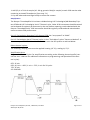
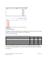
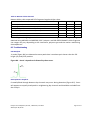
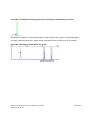
Devyser Thrombophilia
Art. No.: 8-A035
For in-vitro Diagnostic Use
Instructions for Use
Devyser Thrombophilia, CE-IVD, 7-A030-EN, v.4-2014
© Devyser AB, 2014
Page 1 of 27
Table of Contents
Table of Contents
1. Introduction to Devyser Thrombophilia
Intended Use
Included in the Kit
Test Procedure
Background
Principle of the Procedure
2. Warnings and Precautions
3. Symbols used on Labels
4. Required Material
4.1 Included in the kit
Configuration
Components
4.2 Required but not Provided
Reagent Preparation
DNA Extraction
Amplification
Detection
Size Standard
4.3 Dye-Set Calibration
5. Storage and Handling Requirements
6. Sample Requirements
Clinical Samples
DNA Extraction and Measurement
Procedure and Storage
7. Instructions for Use
7.1 Workflow Devyser Thrombophilia
7.2 Sample Preparation and PCR Amplification
Sample Preparation
Addition of Sample
Amplification
7.3 Detection
Sample Preparation
Sample Preparation for Capillary Electrophoresis
Instrument Preparation
Run Modules
8. Data Analysis
Background to data analysis
8.1 The Devyser Thrombophilia kit
Devyser Thrombophilia, CE-IVD, 7-A030-EN, v.4-2014
© Devyser AB, 2014
2
4
4
4
4
4
5
6
7
8
8
8
8
8
8
8
8
8
8
9
10
11
11
11
11
12
12
12
12
12
13
14
14
14
14
14
16
16
16
Page 2 of 27
Normal alleles
Mutant alleles
8.2 Data interpretation
Normal (non mutated) alleles
Heterozygous mutation
Homozygous mutation
Peak cutoff signals (rfu)
Sizing of DNA fragments
Table 1. Normal alleles detected
Table 2. Mutant alleles detected
8.3 Troubleshooting
PCR Artefacts
Electrophoretic Artefacts
9. Performance Characteristics
Sensitivity
Specificity
Reproducibility
Within run reproducibility
Between run reproducibility
Clinical Evaluation
Cross Reactivity
10. Procedural Limitations
11. Notice to Purchaser
12. References
13. Contact Information
Devyser Thrombophilia, CE-IVD, 7-A030-EN, v.4-2014
© Devyser AB, 2014
16
16
16
16
16
16
16
18
18
19
19
19
19
21
21
21
21
21
21
22
23
24
25
26
27
Page 3 of 27
1. Introduction to Devyser Thrombophilia
Intended Use
The Devyser Thrombophilia kit is an in vitro diagnostic product for qualitative detection of
genetic variants that may be associated with thrombophilia. The test can distinguish between
individuals who are heterozygote and homozygote for all tested genetic variants.
The following alleles are detected:
- Factor V Leiden (normal and mutated)
- Factor V R2 (normal and mutated)
- Prothrombin/FII 20210 (normal and mutated)
- MTHFR 677 (normal and mutated)
- MTHFR 1298 (normal and mutated)
- PAI-1/Serpin 1 (4G and 5G)
Included in the Kit
Analysis of the different alles is performed using one single amplification mix (Thromb Mix).
Test Procedure
DNA extraction: The Devyser Thrombophilia kit has been validated using QIAamp DNA Blood
Mini Kit (Qiagen, cat.# 51104) for extraction of DNA from human whole blood.
Amplification: The Devyser Thrombophilia kit has been validated using Life Technologies/ABI
GeneAmp® System 9700 and Life Technologies Veriti® Thermal cycler.
Detection: Life Technologies/ABI Genetic Analyzers (ABI 310, 3100, 3130, 3500, 3730) that support detection of Devyser Dye-Set DEV-5.
Background
Thrombophilia is caused by abnormalities in blood coagulation that increases the risk of thrombosis (1, 2). These abnormalities are frequently seen in patients who have an episode of thrombosis that was not provoked by other causes (3). A significant proportion of the population has a
detectable abnormality, but most of these only develop thrombosis in the presence of an additional risk factor (2).
Congenital thrombophilia is frequently caused by mutations in genes coding for coagulation factors such as factor V Leiden and prothrombin (1, 4, 5).
Common conditions associated with thrombophilia are deep vein thrombosis (DVT) and pulmonary embolism (PE), collectively referred to as venous thromboembolism (VTE). Thrombophilia has also been linked to recurrent miscarriage (6, 7).
Devyser Thrombophilia, CE-IVD, 7-A030-EN, v.4-2014
© Devyser AB, 2014
Page 4 of 27
Principle of the Procedure
The Devyser Thrombophilia kit is based on multiplex allele specific PCR amplification for detection of normal, (non-mutated) and mutated alleles in the following loci:
- Factor V Leiden
- Factor V R2
- Prothrombin/FII 20210
- MTHFR 677
- MTHFR 1298
- PAI-1/Serpin 1 4G/5G
Allele specific PCR amplification generates fluorescently labelled fragments that are analysed
by capillary electrophoresis (CE) on a Genetic Analyzer instrument. Amplified fragments are
identified based on size and fluorescent labels.
Devyser Thrombophilia, CE-IVD, 7-A030-EN, v.4-2014
© Devyser AB, 2014
Page 5 of 27
2. Warnings and Precautions
A.
Devyser Thrombophilia has been validated using a total PCR reaction volume of 12,5 µL. Changing the reaction volume will compromise the kit performance.
B.
Avoid microbial contamination of ragents when removing aliquots from reagent vials. The use
of sterile disposable aerosol barrier pipette tips is recommended.
C.
Do not pool reagents from different lots or from different vials of the same lot.
D.
Do not use a kit after its expiry date.
E.
Do not use opened or damaged kit reagent vials.
F.
Work flow in the laboratory should proceed in a unidirectional manner, beginning in the reagent preparation area and moving to the DNA extraction area and then to the amplification
area and finally to the detection area. Pre-amplification activities should begin with reagent
preparation and proceed to DNA extraction. Reagent preparation activities and DNA extraction
activities should be performed in separate areas. Supplies and equipment should be dedicated
to each activity and not used for other activities or moved between areas. Gloves should be
worn in each area and should be changed before leaving that area. Equipment and supplies
used for reagent preparation should not be used for DNA extraction activities or for pipetting or
processing amplified DNA or other sources of target DNA. Amplification and detection supplies
and equipment should remain in the amplification and detection area at all times.
G.
Handling of kit components and samples, their use, storage and disposal should be in accordance with the procedures defined by national biohazard safety guidelines or regulations.
H.
Wear powder free disposable gloves, laboratory coats and eye protection when handling specimens and kit reagents. Wash hands thoroughly after handling specimens and kit reagents.
Devyser Thrombophilia, CE-IVD, 7-A030-EN, v.4-2014
© Devyser AB, 2014
Page 6 of 27
3. Symbols used on Labels
Lot or batch number
Expiry date
Number of tests
Store below temperature shown
Catalogue number
Manufacturer
In vitro diagnostic device
Devyser Thrombophilia, CE-IVD, 7-A030-EN, v.4-2014
© Devyser AB, 2014
Page 7 of 27
4. Required Material
4.1 Included in the kit
Configuration
The Devyser Thrombophilia test kit contains reagents for analysis of 48 samples.
Components Cap Colour
Orange
Turquoise
Tube Colour
Clear
Amber
Label
PCR Activator
Thromb Mix
Art.Nr.
4-A018
4-A213
Kit Content
1
1
4.2 Required but not Provided
Reagent Preparation
· Consumables for the Thermal Cycler
· Micropipette/dispenser with aerosol barrier tips or displacement tips
· Disposable protective gloves (powder free)
DNA Extraction
· Reagents and equipment according to manufacturer's instructions for use
· Micropipette/multipipette with aerosol barrier tips
Amplification
· Thermal Cycler: Life Technologies/ABI GeneAmp® PCR System 9700 and Life Technologies Veriti® Thermal cycler. For use of alternative thermal cyclers the following ramping rates must be
applied: heating 1,6 ˚C/s, cooling 1,6 ˚C/s
· Micropipette/dispenser with aerosol barrier tips or displacement tips
Detection
· Life Technologies/ABI Genetic Analyzer (ABI 310, 3100, 3130, 3500, 3730)
· Performance optimized polymers: POP-4™ or POP-7™
· Hi-Di™ Formamide, Genetic Analysis Grade
· 1x Genetic Analyzer Buffer
· Micropipette/multipipette/dispenser with aerosol barrier tips or displacement tips
Size Standard
560 SIZER ORANGE (Devyser cat.# 8-A402) or GeneScan™ 600 LIZ® Size Standard (Life Technologies cat.# 4366589)
Devyser Thrombophilia, CE-IVD, 7-A030-EN, v.4-2014
© Devyser AB, 2014
Page 8 of 27
4.3 Dye-Set Calibration
ABI 3100, 3130, 3730:
Use DEV-5 Dye-Set MultiCap kit (Devyser cat.# 8-A401) in the “Any5Dye” Dye-Set.
ABI 3500:
Use DEV-5 Dye-Set MultiCap kit (Devyser cat.# 8-A401) and generate the DEV-5 Dye-Set.
ABI 310 Matrix file generation:
Use DEV-5 Dye-Set SingleCap kit (Devyser cat.# 8-A400). Run with module file “GS STR POP4 (1
mL) G5.md5”
Detailed instructions for Dye-Set calibration may be downloaded from the download section at:
http://www.devyser.com/qf-pcr-accessories/dev-5-calibrators.
Devyser Thrombophilia, CE-IVD, 7-A030-EN, v.4-2014
© Devyser AB, 2014
Page 9 of 27
5. Storage and Handling Requirements
A.
Store all components below -18 °C.
B.
The activated reaction mix (prepared by addition of Thromb Mix to the PCR Activator tube)
may be stored at +2 to +8 °C for at least 7 days and at below -18 °C for at least 90 days. Avoid
repeated freeze-thawing.
C.
Dispose of unused reagents and waste in accordance with country, federal, state and local regulations.
D.
Do not mix reagents from different kit lot numbers.
Devyser Thrombophilia, CE-IVD, 7-A030-EN, v.4-2014
© Devyser AB, 2014
Page 10 of 27
6. Sample Requirements
Clinical Samples
The Devyser Thrombophilia kit is for use with human genomic DNA extracted from whole
blood.
DNA Extraction and Measurement
Preparation of DNA from whole blood samples
Results are consistently obtained with DNA extracted from human whole blood using QIAamp
DNA Blood Mini Kit (Qiagen, cat.# 51104). Follow the protocol starting with 200 μL fresh whole
blood and elute in 200 μL elution buffer. It is normally not necessary to determine the concentration of the purified DNA sample if this procedure is followed. The purified DNA can be
used directly for PCR without any further dilution.
It is recommended that alternative DNA extraction methods and sample materials are thoroughly evaluated with the Devyser Thrombophilia kit prior to the results being used for diagnostic use. For the recommended PCR conditions and analysis settings (see sections 7.2- 7.3),
results are consistently obtained at DNA concentrations between 25 and 150 ng/PCR reaction
(10 - 60 ng genomic DNA/µL sample).
All DNA concentrations referred to in this handbook were determined using Qubit® dsDNA HS
Assay Kit (Life Technologies, cat.# Q32851). The DNA concentration determined in a sample
using Qubit® dsDNA HS Assay Kit may differ from the DNA concentration determined by other
methods.
Procedure and Storage
According to manufacturer’s instructions for use.
Devyser Thrombophilia, CE-IVD, 7-A030-EN, v.4-2014
© Devyser AB, 2014
Page 11 of 27
7. Instructions for Use
7.1 Workflow Devyser Thrombophilia
Each Devyser Thrombophilia kit (art.# 8-A035) contains reagents for 48 samples. The activated
reaction mix should be prepared before preparing the DNA samples if the complete process is
performed in one day. The opposite order is advisable only if the DNA samples are prepared
the day before amplification or earlier.
The activated reaction mix is prepared by adding the Thromb mix to the PCR Activator. It is recommended that the activated reaction mix is dispensed into appropriate PCR reaction tubes
after preparation. Before dispensing ensure that the activated reaction mix is properly mixed
(see below). Dispense in 10 µL aliquots and store at below – 18 °C.
Devyser Thrombophilia has been validated using a total PCR reaction volume of 12,5 µL. Changing the reaction volume will compromise the kit performance.
Ensure that the Thromb mix is completely thawed before use.
1. Centrifuge each tube briefly to collect the content. Do not vortex the tubes at this step.
2. Add 500 µL from the Thromb mix to the PCR Activator.
3. Carefully mix by pipetting several times from the bottom of each tube.
4. Vortex the activated reaction mix tube and centrifuge briefly to collect the content.
5. Add 10 µL of the activated reaction mix to separate PCR reaction tubes.
6. Cap the reaction tubes and centrifuge briefly to collect the contents.
7. Continue to step 7.2.
The activated reaction mix is stable at +2-8 °C for at least 7 days and at below -18 °C for at
least 90 days. Avoid repeated freeze-thawing.
7.2 Sample Preparation and PCR Amplification
Sample Preparation
It is recommended that alternative DNA extraction methods and sample materials are thoroughly evaluated with the Devyser Thrombophilia kit prior to the results being used for diagnostic use. For the recommended PCR conditions and analysis settings (see below), results are
consistently obtained at DNA concentrations between 25 and 150 ng/PCR reaction (10- 60 ng
genomic DNA/µL sample). See also section 6.
Addition of Sample
Samples and controls should be added in a dedicated area separated from reagent preparation,
amplification and detection areas.
Devyser Thrombophilia, CE-IVD, 7-A030-EN, v.4-2014
© Devyser AB, 2014
Page 12 of 27
1. Add 2,5 µL of clinical sample (10 - 60 ng genomic DNA/µL sample) to each PCR reaction tube
containing activated Thrombo mix (from step 7.1)
2. Cap the tubes and centrifuge briefly to collect the content.
Amplification
The Devyser Thrombophilia kit has been validated using Life Technologies/ABI GeneAmp® System 9700 and Life Technologies Veriti® Thermal cycler. Other PCR instruments should be tested
and evaluated for optimal performance by the user before reporting results obtained with the
Devyser Thrombophilia kit. PCR instruments should be regularly calibrated and maintained to
ensure accurate PCR performance.
For Life Technologies GeneAmp® PCR System 9700: Set “ramp speed” to “MAX”.
For Life Technologies Veriti® Thermal cycler: In the "Tools Menu" select "Convert a Method". In
the next step select "9700 Max Mode" and then enter the PCR profile as outlined below.
Other thermal cyclers:
The following ramping rates must be applied: heating 1,6 ˚C/s, cooling 1,6 ˚C/s.
Amplification Area:
Program the Thermal Cycler for amplification according to the following thermal profile (consult the User´s Manual for additional information on programming and operation of the thermal cycler):
95°C 15 min
94°C; 0,5 min -> 62°C; 1 min -> 72°C; 1 min for 25 cycles
72°C 15 min
4°C FOREVER
Devyser Thrombophilia, CE-IVD, 7-A030-EN, v.4-2014
© Devyser AB, 2014
Page 13 of 27
1. Set reaction volume to 13 µL.
2. Set the appropriate ramping rates (heating 1,6 ˚C/s, cooling 1,6 ˚C/).
3. Start the amplification (duration approximately 2,5 hrs).
4. Following amplification, remove the tubes containing completed PCR amplification reaction
from the thermal cycler and place into a suitable holder. Centrifuge briefly to collect the content. Remove the caps carefully to avoid aerosol contamination. Do not bring amplified material into the pre-amplification areas. Amplified material should be restricted to amplification
and detection areas.
7.3 Detection
Sample Preparation
Refer to the respective Life Technologies/ABI Genetic Analyzers User Manual for instructions on
maintenance and handling. Prior to running the Devyser Thrombophilia kit, the instrument must
be spectrally calibrated to support detection of the Dye-Set DEV-5. See section 4.3 for details.
Sample Preparation for Capillary Electrophoresis
1. Prepare a loading cocktail by combining and mixing 2 µL of the size standard (e.g. 560 SIZER
ORANGE) with 100 µL Hi-DiTM Formamide (sufficient mix for 6 wells/tubes).
2. Vortex for 15 seconds.
3. Dispense 15 µL of the loading cocktail into the required number of wells of a microwell plate
or into individual tubes to be placed on the Genetic Analyzer.
4. Add 1,5 µL of the sample PCR product to the corresponding well/tube containing loading cocktail.
5. Seal the plate/tubes.
Instrument Preparation
Create a sample sheet using the data collection software with the following settings:
l
l
l
Sample ID
Dye-Set: Any5Dye/DEV-5
Recommended run Module: See below for different polymers and instruments
Run Modules
The amount of PCR product injected into the capillaries can be adjusted by increasing/decreasing the injection time and/or injection voltage.
Devyser Thrombophilia, CE-IVD, 7-A030-EN, v.4-2014
© Devyser AB, 2014
Page 14 of 27
ABI 310(Run with module file “GS STR POP4 (1 mL) G5.md5”)
Run Parameters
POP-4
Capillary length
47 cm
Run temperature
60 ˚C
Injection voltage
10 kV
Injection time
5s
Run voltage
15 kV
Run time
30 min
ABI 3100/3130
Run Parameters
Capillary length
Run temperature
Injection voltage
Injection time
Run voltage
Run time
POP-4/POP-7
36 cm
60 ˚C
1,5 kV
20 s
15 kV
1500 s
ABI 3500
Run Parameters
Capillary length
Run temperature
Injection voltage
Injection time
Run voltage
Run time
POP-7
50 cm
60 ˚C
1,6 kV
15 s
19,5 kV
1500 s
ABI 3730
Run Parameters
Capillary length
Run temperature
Injection voltage
Injection time
Run voltage
Run time
POP-7
36 cm
60 ˚C
1,6 kV
15 s
15 kV
1500 s
Devyser Thrombophilia, CE-IVD, 7-A030-EN, v.4-2014
© Devyser AB, 2014
Page 15 of 27
8. Data Analysis
Background to data analysis
An individual has two copies (alleles) of each of the investigated loci and is considered being
homozygous for a given DNA sequence when both alleles have the same sequence. An individual is considered being heterozygous for a given DNA sequence when the two alleles differ in
sequence.
8.1 The Devyser Thrombophilia kit
The Devyser Thrombophilia kit is used for the qualitative analysis of mutations in the loci outlined in tables 1 and 2. Analysis of the different alleles is performed using one single amplification mix (Thromb Mix). All detected normal and mutant alleles are listed in tables 1 and 2.
Normal alleles
PCR fragments representing normal DNA sequences are detected in the blue channel as outlined in table 1 below.
Mutant alleles
PCR fragments representing mutant DNA sequences are detected in the green channel as outlined in table 2 below.
8.2 Data interpretation
Normal (non mutated) alleles
The sample is to be considered normal for a certain locus if a normal allele fragment is
detected while the corresponding mutant allele fragment is absent (Figure 8.1).
Heterozygous mutation
The sample is to be considered heterozygously mutated in a certain locus if both a normal
allele fragment and the corresponding mutation allele fragment are present (Figure 8.2).
Homozygous mutation
The sample is to be considered homozygously mutated in a certain locus if the normal allele
fragment is absent, while the corresponding mutant allele fragment is present (Figure 8.3).
Peak cutoff signals (rfu)
It is recommended that an instrument specific cutoff range is determined. Do not to use lower
cutoff values than 200 rfu (ABI 310, 3100 and 3130) and 500 rfu (ABI 3730, 3500). Do not analyse samples where peak signals are saturated.
Devyser Thrombophilia, CE-IVD, 7-A030-EN, v.4-2014
© Devyser AB, 2014
Page 16 of 27
Figure 8.1. Homozygous wildtype MTHFR 1298 (1298A)
Figure 8.2. Heterozygous MTHFR 1298 (1298A/1298A>C)
Devyser Thrombophilia, CE-IVD, 7-A030-EN, v.4-2014
© Devyser AB, 2014
Page 17 of 27
Figure 8.3. Homozygous mutated MTHFR 1298 (1298A>C)
Sizing of DNA fragments
PCR fragments obtained using the Devyser Thrombophilia kit should be sized with the 560 SIZER
ORANGE using a fragment analysis software (e.g. GeneMapper).
Table 1. Normal alleles detected
Normal alleles, their expected PCR fragment length and dye colour.
Gene
Methyltetrahydrofolate Reductase (MTHFR)
Plasminogen Activator Inhibitor 1 (PAI-I/SERPIN 1 )
Methyltetrahydrofolate Reductase (MTHFR)
Factor II (Prothrombin)
Factor V (R2)
Factor V (Leiden)
Allele
1298A
5G
677C
20210G
4070A
1691G
Size (bp)*
215
233
295
312
381
437
Colour
Blue
Blue
Blue
Blue
Blue
Blue
*Allele fragment lengths given in this table are based on average observed fragment lengths
obtained using ABI3130 and ABI3500, POP-7 polymer and 560 SIZER ORANGE. Allele fragment
size ranges may vary depending on the instrument, polymer type and size marker used during
electrophoresis.
Devyser Thrombophilia, CE-IVD, 7-A030-EN, v.4-2014
© Devyser AB, 2014
Page 18 of 27
Table 2. Mutant alleles detected
Mutant alleles, their expected PCR fragment length and dye colour.
Gene
Factor V (Leiden)
Plasminogen Activator Inhibitor 1 (PAI-I/SERPIN 1 )
Methyltetrahydrofolate Reductase (MTHFR)
Methyltetrahydrofolate Reductase (MTHFR)
Factor V (R2)
Factor II (Prothrombin)
Allele Size (bp)*
1691G>A
125
4G
134
677C>T
151
1298A>C
182
4070A>G
388
20210G>A
426
Colour
Green
Green
Green
Green
Green
Green
*Allele fragment lengths given in this table are based on average observed fragment lengths
obtained using ABI3130 and ABI3500, POP-7 polymer and 560 SIZER ORANGE. Allele fragment
size ranges may vary depending on the instrument, polymer type and size marker used during
electrophoresis.
8.3 Troubleshooting
PCR Artefacts
-A peaks (figure 8.4) are detected as extra peaks that is one base pair shorter than the full
length (+A peak) PCR product.
Figure 8.4. -A and +A peaks as indicated by the arrows.
Electrophoretic Artefacts
Crosstalk/bleed through between dye channels may occur during detection (Figure 8.5). Crosstalk appears as equally sized peaks in neighbouring dye channels and should be excluded from
the analysis.
Devyser Thrombophilia, CE-IVD, 7-A030-EN, v.4-2014
© Devyser AB, 2014
Page 19 of 27
Figure 8.5. Crosstalk peak (from green to blue channel) as indicated by the arrow.
Dye blobs may appear in the sample analysis range (figure 8.6). In general, dye blobs appear
as broad, undefined peaks of a single colour and tend to occur relatively early in the data.
Figure 8.6. Dye blob as indicated by the arrow
Devyser Thrombophilia, CE-IVD, 7-A030-EN, v.4-2014
© Devyser AB, 2014
Page 20 of 27
9. Performance Characteristics
Sensitivity
Definition: The proportion (%) of subjects with a well defined genetic condition and whose test
values are positive within the defined decision limit.
Experimental design:
One hundred (100) DNA samples previously characterised to carry at least one of the listed
mutations (chapter 8, table 2) were tested using Devyser Thrombophilia. All results obtained
using Devyser Thrombophilia correlated with the previously obtained results.
Result: >99% sensitivitya
Specificity
Definition: The proportion (%) of subjects who do not have a well defined genetic condition and
whose test results are negative or within the defined decision limit.
Experimental design:
One hundred one (101) DNA samples previously characterised to carry at least one copy of all
the listed normal alleles (chapter 8, table 1) were tested using Devyser Thrombophilia.All
results obtained using Devyser Thrombophilia correlated with the previously obtained results.
Result: > 99% specificity
Reproducibility
Within run reproducibility
Definition: The degree (%) of agreement between measurements conducted on replicate specimens
of the same measured where the measurements are carried out under unchanged conditions.
Experimental design: Forty-eight (48) replicates of a normal male sample was amplified in one
run using identical reagent batches, operator, PCR- and capillary electrophoresis instruments.
All 48 replicates gave the expected results.
Result: >98 % within run reproducibility
Between run reproducibility
Definition: The degree (%) of agreement between measurements conducted on replicate specimens of the same measurand where the measurements are carried out under changed
Devyser Thrombophilia, CE-IVD, 7-A030-EN, v.4-2014
© Devyser AB, 2014
Page 21 of 27
conditions.
Experimental design: One hundred and nine (109) replicates of a normal male sample were
amplified on 3 different occasions using different reagent batches, operators, PCR- and capillary electrophoresis instruments. All 109 replicates gave the expected results.
Result: >99 % between run reproducibility
Clinical Evaluation
One hundred and three (103) DNA samples previously characterised for at least one of the
parameters analysed by Devyser Thrombophilia were tested blind. All samples were successfully analysed. Results are summarised in table 1 below.
Table 1. Results from the clinical evaluation of 103 previously characterised clinical samples
Locus
Factor V G1691 (Leiden)
wt/wt
wt/mut
mut/mut
Plasminogen Activator Inhibitor 1 (PAI-I/SERPIN 1 )
wt/wt
wt/mut
mut/mut
Methyltetrahydrofolate Reductase (MTHFR) C677
wt/wt
wt/mut
mut/mut
Methyltetrahydrofolate Reductase (MTHFR) A1298
wt/wt
wt/mut
mut/mut
wt/wt
wt/mut
mut/mut
Factor V H1299 (R2)
wt/wt
wt/mut
mut/mut
Devyser Thrombophilia, CE-IVD, 7-A030-EN, v.4-2014
© Devyser AB, 2014
Number of samples
Correlation (%)
81
14
4
100
100
100
16
22
21
100
100
100
31
49
14
100
100
100
42
43
10
100
100
100
85
8
4
100
100
100
77
12
6
100
100
100
Page 22 of 27
Cross Reactivity
No cross reactivities have been observed.
Devyser Thrombophilia, CE-IVD, 7-A030-EN, v.4-2014
© Devyser AB, 2014
Page 23 of 27
10. Procedural Limitations
A.
Use of this product should be limited to personnel trained in the techniques of PCR and capillary
electrophoresis.
B.
The Devyser Thrombophilia kit has been validated using Life Technologies/ABI Thermal Cycler
GeneAmp® 9700 and Life Technologies Veriti® Thermal cycler. It is recommended that alternative thermocycler instruments are thoroughly evaluated with the Devyser Thrombophilia kit
prior to the results being used for diagnostic use.
C.
The Devyser Thrombophilia kit has been validated using QIAamp DNA Blood Mini Kit for extraction of DNA from human whole blood. Performance with other matrices and DNA extraction
kits has not been validated and may result in false negative or false positive results.
D.
Devyser Thrombophilia kit should be used only for the detection of specific mutations according
to the instructions for use. Many other mutations are possible that may not be detected using
Devyser Thrombophilia. The assay has not been validated for diagnosis of Thrombophilia.
Results obtained with Devyser Thrombophilia kit can only be directly applied to the tissue or specific sample material tested.
E.
Only the following mutations are tested: Factor V G1691A, Factor V H1299R, Prothrombin
G20210A, PAI-1/SERPIN1 4G/5G, MTHFR C677T and MTHFR A1298C.
F.
Diagnostic errors can occur due to rare sequence variations.
Devyser Thrombophilia, CE-IVD, 7-A030-EN, v.4-2014
© Devyser AB, 2014
Page 24 of 27
11. Notice to Purchaser
Results from Devyser Thrombophilia should be interpreted with consideration of the overall picture obtained from clinical and laboratory findings. Devyser AB will not accept responsibility for
any clinical decisions taken.
LIZ®, Veriti® and GeneAmp® are registered trademarks of Life Technologies Corporation. GeneScanTM, POP-4TM, POP-7TM and Hi-DiTM are trademarks of Life Technologies Corporation.
Purchase of this product does not provide a license to perform PCR under patents owned by any
third party.
Devyser Thrombophilia, CE-IVD, 7-A030-EN, v.4-2014
© Devyser AB, 2014
Page 25 of 27
12. References
1. Mitchell RS, Kumar V, Abbas AK, Fausto N (2007). "Chapter 4".Robbins Basic Pathology
(Eighth ed.). Philadelphia: Saunders. ISBN 1-4160-2973-7.
2. Heit JA (2007)."Thrombophilia: common questions on laboratory assessment and management". Hematology Am. Soc. Hematol. Educ. Program 2007 (1): 127–35.doi:10.1182/asheducation-2007.1.127. PMID 18024620.
3. Kyrle PA, Rosendaal FR, Eichinger S (December 2010). "Risk assessment for recurrent venous
thrombosis". Lancet 376 (9757): 2032–9. doi:10.1016/S0140-6736(10)60962-2. PMID 21131039.
4. Rosendaal FR (2005). "Venous thrombosis: the role of genes, environment, and behavior".
Hematology Am. Soc. Hematol. Educ. Program 2005(1): 1–12. doi:10.1182/asheducation2005.1.1. PMID 16304352.
5. Crowther MA, Kelton JG (2003). "Congenital thrombophilic states associated with venous
thrombosis: a qualitative overview and proposed classification system". Ann. Intern. Med. 138
(2): 128–34. doi:10.7326/0003-4819-138-2-200301210-00014. PMID 12529095. Lay summary.
6. Scarvelis D, Wells PS (October 2006). "Diagnosis and treatment of deep-vein thrombosis".
CMAJ 175 (9): 1087–92. doi:10.1503/cmaj.060366. PMC 1609160. PMID 17060659.
7. Rai R, Regan L (August 2006). "Recurrent miscarriage". Lancet 368 (9535): 601–11.
doi:10.1016/S0140-6736(06)69204-0. PMID 16905025.
Devyser Thrombophilia, CE-IVD, 7-A030-EN, v.4-2014
© Devyser AB, 2014
Page 26 of 27
13. Contact Information
Devyser AB
Instrumentvägen 19
SE-126 53 Hägersten
SWEDEN
Phone: +46-8-562 15 850
Homepage: www.devyser.com
Technical Support
Phone: +46-8-562 15 850
E-mail: [email protected]
Devyser Thrombophilia, CE-IVD, 7-A030-EN, v.4-2014
© Devyser AB, 2014
Page 27 of 27