1
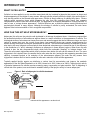
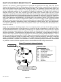
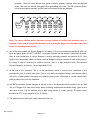
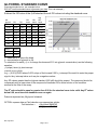
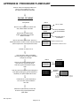
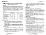
Cellulite Treatment Screening Kit Human Adipocyte Lipolysis Assay Free Glycerol Detection Cat# LIP-10 INSTRUCTION MANUAL ZBM0017.05 STORAGE CONDITIONS Human Adipocytes All orders are delivered via Federal Express Priority courier at room temperature. All orders must be processed immediately upon arrival. NOTE: Domestic customers: Assay must be performed 5-7 days AFTER receipt. International customers: Assay must be performed 3-5 days AFTER receipt Glycerol Reagent A & Buffers: 4°C Use reconstituted Glycerol Reagent A within 7 days. Glycerol Standard & Controls: -20°C Assay plate A (96-well) cultured human adipocytes: 37°C humidified incubator Long-term storage: Remove the glycerol reagent A and buffers from the box and place at 4°C, store the rest of the kit at -20°C. Reagents are good for 6 months if stored properly. All Zen-Bio Inc products are for research use only. Not approved for human or veterinary use or for use in diagnostic or clinical procedures. LIMITED PRODUCT WARRANTY This warranty limits our liability to replacement of this product. No other warranties of any kind, express or implied, including without limitation, implied warranties of merchantability or fitness for a particular purpose, are provided by Zen-Bio, Inc. Zen-Bio, Inc. shall have no liability for any direct, indirect, consequential, or incidental damages arising out of the use, the results of use, or the inability to use this product. ORDERING INFORMATION AND TECHNICAL SERVICES Zen-Bio, Inc. 3200 Chapel Hill-Nelson Blvd., Suite 104 PO Box 13888 Research Triangle Park, NC 27709 Telephone (919) 547-0692 Facsimile (FAX) (919) 547-0693 Toll Free 1-866-ADIPOSE Electronic mail (e-mail) [email protected] World Wide Web http://www.zenbio.com Rev July 2010 Page 1 of 11 (866)-234-7673 INTRODUCTION WHAT IS CELLULITE? Cellulite is a term applied to a skin condition associated with the localized fat deposits that present as lumps and dimples appearing on the thighs of many women. Although cellulite primarily afflicts the thighs, hips and buttocks, it may also be present on the stomach and upper arms. Cellulite is simply made up of ordinary fatty tissue. Fibrous strands called connective tissue which separate the skin from the underlying fatty tissue form separate compartments under the skin that contain fat cells. The appearance is frequently described as "orange peel skin" or said to have a “cottage cheese appearance”. Cellulite afflictions are a stubborn problem causing emotional and psychological distress to many women. Although the etiology of cellulite is poorly understood, the main factor appears to be local accumulation of fat in a regional compartment. HOW CAN THIS KIT HELP MY RESEARCH? Adipocytes (fat cells) are the principle cells implicated in fat storage by adipose tissue. It has been proposed that the anatomical structure of subcutaneous adipose tissue is a major contributor to the appearance of cellulite. The histological studies of subcutaneous tissues from men and women suggest that the fat lobules are larger and more vertical in women than men. As a result, these larger, less restricted lobules can express outward against the dermis causing the bumps and dimples characteristic of cellulite. The femoral subcutaneous fat deposits in women also tend to be more lipogenic and less lipolytic than abdominal subcutaneous or visceral fat due to the difference in the distribution of and adrenergic receptors on adipocytes in these different regions. When these fat cells increase in size, the skin compartment bulges, which forms the noticeable “dimpling” or “cottage cheese” look. These fat cells contain triglycerides which must be broken down before fat cells can be reduced in size. The more triglyceride in fat is broken down, the smaller the fat cells under the skin, leaving the skin appearing smoother (less cellulite). Increased lipolysis or fat reduction of the subcutaneous adipose (fat under the skin) means more triglyceride is broken down to lead to smaller fat cells and a reduction of the cellulite appearance. Topically applied lipolytic agents can distribute or reduce local fat accumulation and improve the aesthetic appearance of the skin (Mas-Chamberlin et al. 2006, Hexsel et al. 2005, Huber et al. 2004). Testing lipolytic activity of potential treatments for cellulite requires screening many compounds and plant extracts. Prior to beginning a clinical trial of the product, one would need to establish validity of the lipolytic activity in human adipocytes. nucleus Figure 1. Role of adipocytes in cellulite treatment Lipid droplets LIPOLYSIS TRIGLYCERIDE Glycerol cellulite Free fatty acid Free fatty acid Free fatty acid reduced cellulite appearance= smaller fat cell Rev July 2010 Page 2 of 11 WHAT IS THE SCIENCE BEHIND THIS KIT? Lipolysis is the process in which triglycerides are hydrolyzed into glycerol and free fatty acids. This process releases free fatty acids (FFA) into the bloodstream where they may be either re-esterified by the adipocyte or travel to other tissues and exert other effects throughout the body. The sympathetic nervous system plays a key role in the regulation of lipid mobilization. The main lipolytic pathway involves beta-agonists (-agonists), which activate -adrenergic receptors via the intracellular Gs proteins in adipocytes. This leads to the activation of adenylate cyclase (AC), which then increases cyclic AMP (cAMP) levels. Elevated cAMP acts as a second messenger to activate hormone sensitive lipase (HSL). HSL, the rate-limiting enzyme regulating adipocyte lipolysis, then catalyzes the hydrolysis of triglycerides and results in the release of one molecule of glycerol and 3 molecules of free fatty acids (FFA; increased lipolysis). Phosphodiesterases (PDE) are enzymes that transform cAMP to 5’AMP (5 prime adenosine monophosphate). This action results in a decrease in lipolysis. PDE inhibitors increase intracellular cAMP levels. 3-isobutyl-1-methylxanthine (IBMX), a non-specific inhibitor of cAMP phosphodiesterases (PDE), can be used as a positive control if your test compounds are suspected PDE inhibitors. PDE inhibitors can be found as an ingredient in mesotherapy solution for the treatment of cellulite (Snyder et al. 2005) Isoproterenol; a non-specific -adrenergic agonist is used as the positive control if your test compounds affect lipolysis via adrenergic receptors. Among the methods for stimulating lipolysis, the most commonly known and used is that which consists of inhibiting the phosphodiesterase in order to prevent or at least limit the rate of degradation of cyclic AMP. In effect, the phosphodiesterase destroys cyclic AMP by transforming it into 5'AMP so that it cannot function as a lipolysis activator. Among the common agents for treatment of cellulite as slimming agents are xanthine analogs such as caffeine or theophylline. These agents block the antilipolytic action of adenosine, a potent endogenous inhibitor of lipolysis. Other known methods in lipolysis stimulation are achieved by inhibiting phosphodiesterase in order to prevent or at least limit the degradation of cAMP. Other existing methods for the treatment of cellulite have been the stimulation of adenylate cyclase to increase cAMP levels or to block the antilipolytic inactivation of adenylate cyclase (-2-adrenergic antagonists). Greenway et al. (1995) disclose that isoproterenol, a known agonist adrenergic stimulator, is effective for the treatment of cellulite by stimulating lipolysis; furthermore, creams based on yohimbine, a known 2-blocker, applied to women's skin showed a decrease in thigh circumference. Figure 2. Overview of adipocyte lipolysis EPINEPHRINE 1, 2, 3 AR NOREPINEPHRINE AC Gs IR PDE ATP P cAMP 5’-AMP PKA HSL Per Per TG FFA + glycerol FFA + glycerol bloodstream Rev July 2010 Page 3 of 11 ABBREVIATIONS: AC adenylate cyclase AR adrenergic receptors Gs G protein coupled receptor FFA free fatty acids PKA protein kinase AMP adenosine monophosphate ATP adenosine triphosphate IR insulin receptor PDE phosphodiesterase Per Perilipin TG triglyceride WHAT DOES THIS KIT MEASURE? This kit provides the tool to study chemical compounds that may influence lipolysis in cultured human adipocytes. This kit specifically measures the free glycerol released by the breakdown of triglyceride. The amount of free glycerol released from the cells is proportional to the ability of the test chemical to break down triglyceride. PRINCIPLE OF THE ASSAY Glycerol released to the medium is phosphorylated by adenosine triphosphate (ATP) forming glycerol-1phosphate (G-1-P) and adenosine-5’-diphosphate (ADP) in the reaction catalyzed by glycerol kinase. G-1-P is then oxidized by glycerol phosphate oxidase to dihydroxyacetone phosphate (DAP) and hydrogen peroxide (H2O2). A quinoneimine dye is produced by the peroxidase catalyzed coupling of 4-aminoantipyrine (4-AAP) and sodium Nethytl-N-(3-sulfopropyl) m-anisidine (ESPA) with H2O2, which shows an absorbance maximum at 540nm. The increase in absorbance at 540nm is directly proportional to the glycerol concentration of the sample. GLYCEROL + ATP G-1-P + O2 G-1-P + ADP DAP + H2O2 H2O2 +4-AAP + ESPA Quinoneimine dye + H2O ITEMS INCLUDED IN THE KIT ITEM DESCRIPTION Adipocytes, Plate A Blank Assay Plates Assay Buffer Wash Buffer Vehicle Cultured human subcutaneous adipocytes 96-well assay plates, blank 100 ml 50 ml 0.1% DMSO in Assay Buffer Positive control Isoproterenol, 10 mM in DMSO. Dilute to 1 M in Assay Buffer before use! (i.e.1 l in 10 ml Assay Buffer) Glycerol Reagent A Tray Glycerol standard ALTERNATE : Positive control Reconstitute with 11.0 ml deionized water prior to use. Cap Color UNIT QTY STORAGE --------- PLATE 1 2 1 1 1 37°C ----4°C 4°C -20°C 1 -20°C BOTTLE 1 4°C EACH 2 1 -----20°C 1 -20°C GREEN BLUE --- Use reconstituted reagent within 7 days. For multi-channel pipetters, clear polyvinyl Glycerol @ 1mM [Reconstitute with 400 l Wash Buffer to make the 200 M glycerol standard; see page 6 for recommended dilution scheme] 3-Isobutyl-1-methylxanthine (IBMX), 100 mM in DMSO Dilute to 100 M in Assay Buffer before use! (i.e. 1 l in 1 ml Assay Buffer). USE ONLY IF YOUR TREATMENT TIME EXCEEDS 5 HOURS. Other equipment/reagents required but not provided with the kit: Multi-channel Pipet , single channel pipet and pipet tips Incubator at 37oC Option – Step 5 of Assay Procedure: 96 well plate, blank Rev July 2010 Page 4 of 11 PLATE BOTTLE BOTTLE 1 ml / VIAL 10 l / VIAL ORANGE 100 l / VIAL RED 10 l / VIAL Plate reader with a filter of 540 nm Large gauge needle Tubes for diluting glycerol standards ASSAY PROCEDURE 1. Human preadipocytes are plated in 96 well plates and allowed to differentiate under standard Zen-Bio differentiation conditions for 1 week. Upon arrival, remove 150l of the shipping medium from each well and discard. Place the plate (Plate A) in your incubator for 5-7 days (3-5 days for international customers) to allow the cells to recover from the stress of shipping. To ensure optimal performance, DO NOT feed the cells fresh medium during this time. Please observe the cells under a microscope prior to performing the assay [see the photograph in the Certificate of Analysis for the lot # of Plate A]. 2. Make your stock solution using whatever vehicle is appropriate for your test compounds. Dilute your stock solutions to their final concentration in Assay Buffer (100 ml are available). NOTE: if desired, maintain a constant concentration of solvent by preparing all compound dilutions in the highest concentration of that solvent. Dilute your controls in assay buffer. Prepare all vehicles as appropriate for your compounds, 0.1% DMSO has been included as the vehicle for the positive controls. Include the Assay Buffer alone as a vehicle control. PLEASE NOTE: ZEN-BIO DOES NOT RECOMMEND THE USE OF SOLVENTS AT CONCENTRATIONS ABOVE 1%. 3. Remove 120 l medium from each well. Gently add 200 l Wash Buffer to all wells. Remove 200 l of the media and Wash Buffer from each well and replace with another 200 l Wash Buffer. 4. Remove all the media and Wash Buffer from the cells from triplicate wells. Treat the cells with 150 l of the test compounds resuspended in Assay Buffer three (3) wells at a time. Treat with the diluted Isoproterenol or optionally, IBMX (for treatments 5-24 hours), as positive control. Use the Assay Buffer alone as one of the vehicle controls. Please be sure to include both the vehicle provided in the kit and your vehicle (if your test compounds are not dissolved in DMSO). The assay should be performed in triplicate. 5. OPTION: to determine if the compound alone reacts with the Glycerol Reagent A, prepare a fresh plate (not included in kit) containing 100 l of the compound. This plate can be incubated at 37oC with the treated cells. When performing the assay, add 100 l of Glycerol Reagent A following the instructions in Steps 10 and 11. 6. Incubate the plates at 37oC in a humidified incubator for 3 hours (for time course experiments the longest time point is usually 24 hours). 7. One hour prior to the assay, prepare the glycerol standards as follows: Briefly spin down the contents of the glycerol standard tube before reconstitution. Pipette 400 l of Wash Buffer into the 1 mM glycerol standard tube provided and mix well by vortexing. This produces a diluted stock glycerol standard of 200 M. Pipette 250 l of wash buffer into 6 tubes (not Rev July 2010 Page 5 of 11 provided). Using the newly diluted stock glycerol solution, prepare a dilution series as depicted below. Mix each new dilution thoroughly before proceeding to the next. The 200 M stock dilution serves as the highest standard, and the wash buffer serves as the zero standard. 400 l Wash Buffer 250 l 250 l 250 l 250 l 250 l 250 l Std 200 M 100 M 50 M 25 M 12.5 M 6.25 M 3.125 M Note: The above dilution series generates enough volume to perform the standard curve in duplicate. If you wish to perform the standard curve in duplicate, please note that eight fewer data points can be assayed with this kit. 8. Also at this time prepare the Glycerol Reagent A by adding 11.0 ml room temperature deionized water per bottle and gently invert. DO NOT VORTEX! Use a pipet to ensure that the powder is completely dissolved. Store at room temperature. If using a Reagent A solution previously prepared and stored at 2-8C, also bring to room temperature. Make sure there is enough Reagent A from one solution to treat all the points in the assay. It may be necessary to combine solutions. Store in a light protected bottle. Reconstituted Glycerol Reagent A is stable for 7 days refrigerated (2-8C). 9. At the end of the incubation, 100 l of the conditioned media is removed and transferred to the corresponding well of another blank plate. [This is most easily accomplished using a multi-channel pipet.] Add 100 l of each glycerol standard to any remaining empty wells in this plate or use the second blank plate provided in this kit for the standards. 10. Add the reconstituted Glycerol Reagent A solution to one of the disposable trays provided in the kit. Add 100 l of Reagent A to each well of assay plates containing samples and standards. Gently, pipet up and down once to mix. Pop the bubbles using a large gauge needle or a clean pipet tip. The plate is then incubated at 25oC (room temperature) for 15 minutes. 11. The optical density of each well is then measured at 540 nm. Rev July 2010 Page 6 of 11 GLYCEROL STANDARD CURVE Generate standard curve: see example below [DO NOT use this standard curve to generate your data. This is an example.] Subtract the OD value of the 0M standard from all OD values including the standard curve. uM glycerol ODblank ODblank Avg ODblank OD OD 0 0.044 0.041 3.125 0.054 0.053 0.012 0.011 0.011 6.25 0.062 0.063 0.020 0.021 0.020 12.5 0.083 0.084 0.041 0.042 0.041 25 0.126 0.125 0.084 0.083 0.083 50 0.205 0.208 0.163 0.166 0.164 100 0.372 0.374 0.330 0.332 0.331 200 0.698 0.697 0.656 0.655 0.655 Slope 0.003 Intercept 0.001 2 R 0.043 1.000 y = observed O.D. minus the blank x = concentration of glycerol in M To calculate x for each y, (i.e. to change the observed O.D. into glycerol concentration) use the following equation: y=(slope) times (x) plus intercept y=mx+b so x=(y-b)/m x=(y – (0.001))/0.003 where 0.003= slope of the line and 0.001= y intercept. Be careful to enter the proper sign for the y intercept value as it may be a negative number. Any OD values greater than the highest standard (200 µM) should be suspect. The compound should be re-assayed using a lower dose of the compound at treatment OR a dilute solution of the condition medium at the time of the assay. The R2 value should be equal or greater then 0.98 for the standard curve to be valid. Any R2 values below 0.98, must have the standard curve run again. Data are expressed as M glycerol released. OPTION: express data as Fold induction over appropriate vehicle Fold induction = M glycerol SAMPLE M glycerol VEHICLE Rev July 2010 Page 7 of 11 TROUBLESHOOTING Problem Suggestions High background or the glycerol reagent A turns purple before the assay begins. Change pipet tips frequently Use Glycerol Reagent A before the expiration date No response to positive control Do not add the compounds and controls too fast. The cells can float if a solution is added too fast. Make sure to starve the cells for 5-7 days BEFORE initiating treatment. DO NOT use IBMX as the positive control if you are incubating for less than 5 hours. Edge effects Ensure a saturated humidity in the incubator to prevent evaporation from the outside wells Inconsistent OD reading The Assay Buffer contains bovine serum albumin (BSA). Be careful when pipetting to avoid bubbles. If bubbles persist, burst the bubbles using a large gauge needle and read the plate again. FREQUENTLY ASKED QUESTIONS 1. When do I need to use the IBMX positive control? If you use the 3-5 hour incubation described in this manual, you will not need to use the IBMX as your positive control. The IBMX positive control is designed for treatments ranging from 5-24 hours. The IBMX alternate control may be used in addition to the Isoproterenol positive control if your treatment time will exceed 5 hours. 2. Can I buy the reagents separately? The Glycerol Standard, cat# LIP-GLYSTAN and Glycerol Reagent A, cat# RGTA-10 are sold separately. Assay Buffer is not sold separately. A REAGENTS ONLY kit is available cat# LIP-1-NC. Contact ZenBio to order additional cells or media. 3. I need to know the concentration of the BSA in the Assay Buffer? ZenBio, Inc does not provide the concentrations of the components of our media and buffers. If knowledge of the BSA concentration is critical to your experiment, you may order Assay Buffer WITHOUT BSA for no additional charge. Please note it on your order. 4. I have more samples plus standards to run than can fit on 1 96 well plate. Can I compare data obtained from multiple plates? The lipolysis kit is designed for the assay of a single plate. You may purchase 2 kits of the same lot number. You may then use one plate that includes the blank, vehicle(s), and positive and negative controls. The second plate may then be used for the remainder of your samples assayed. In order to obtain comparable data, both plates must be assayed on the same day using kits and cells from the same lot number. An additional blank assay plate is provided for the assay of glycerol standards. 5. I do not have time to pop the bubbles and read the plate. Can I freeze the conditioned media in one of the assay plates provided with the kit? How long can I store the samples? Yes. The conditioned media can be immediately stored at -80C for a maximum of 7 days. Bring the conditioned media in the plate to room temperature BEFORE adding the Glycerol Reagent A and completing the assay. Rev July 2010 Page 8 of 11 APPENDIX A: PLATE LAYOUT A B C D E F G H 1 2 3 4 5 6 7 8 9 10 11 12 Rev July 2010 Page 9 of 11 APPENDIX B: PROCEDURE FLOWCHART Remove 150l of the shipping medium and place in your incubator for 5-7 days (3-5 days for international customers) ON DAY OF ASSAY Make all test compound dilutions in Assay Buffer. Remove 120 l media from all wells. Add 200 l Wash Buffer to all wells. Plate A 120 l media OOOOOOOOOOOO OOOOOOOOOOOO OOOOOOOOOOOO OOOOOOOOOOOO OOOOOOOOOOOO 200 l Wash Buffer Plate A Remove 200 l media & Wash Buffer. Add another 200 l Wash Buffer to all wells. 200 l Wash Buffer OOOOOOOOOOOO OOOOOOOOOOOO OOOOOOOOOOOO OOOOOOOOOOOO OOOOOOOOOOOO Add another 200 l Wash Buffer Plate A Remove all media & Wash Buffer. Add 150 l treatments/controls to 3 wells at a time. OPTION: Add 100 l/well compounds to a fresh plate without cells. OOOOOOOOOOOO OOOOOOOOOOOO OOOOOOOOOOOO OOOOOOOOOOOO OOOOOOOOOOOO Remove 3 wells at a time Add treatments 3 wells at a time o Incubate 3-5 hours at 37 C. One hour prior to assay, reconstitute Glycerol Reagent A and prepare standards. Keep all at room temp. Plate A Remove 100 l/well conditioned media from Plate A to a blank assay plate. Add 100 l glycerol standards to empty wells Add 100 l/well reconstituted Glycerol Reagent A to the plate (including the glycerol standards at 100l/well) and optional plate without cells. OOOOOOOOOOOO OOOOOOOOOOOO OOOOOOOOOOOO OOOOOOOOOOOO OOOOOOOOOOOO OOOOOOOOOOOO OOOOOOOOOOOO OOOOOOOOOOOO OOOOOOOOOOOO OOOOOOOOOOOO blank plate 100 l OOOOOOOOOOOO OOOOOOOOOOOO OOOOOOOOOOOO OOOOOOOOOOOO OOOOOOOOOOOO GLYCEROL REAGENT A OOO OOO OOO OOO OOO An additional blank assay plate may be necessary for the assay of glycerol standards. o Incubate at 25 C (room temperature) for 15 minutes. Pop the bubbles in each well. Measure the optical density of each well at 540 nm using a spectrophotometer plate reader. Rev July 2010 Page 10 of 11 REFERENCES 1. Arner P J Endocrinol (1997) 155:191-192. 2. Greenway FL, Bray GA, Heber D. Topical fat reduction Obes Res (1995) 3 Suppl 4:561S-568S. 3. Hexsel D, Orlandi C, Zechmeister D. Botanical extracts used in the treatment of cellulite. Dermatol Surg (2005)31:866-872. 4. Huber C, Meyer MS, Schreier T. Topical treatments for Orange Peel Skin Cosmetics & Toiletries (2004) 119:49-58. 5. Mas-Chamberlin C, Mondon P, Lintner K. Cosmetic management of lipid storage in adipocytes: a slimming concept for men and women.(2005) Cosmetic & Toiletries Manufacture Worldwide 2005:53-56. 6. Rotunda AM, Avram MM, Avram AS. Cellulite: Is there a role for injectables? (2005) J Cosmet Laser Ther 7(3-4):147-154. 7. Snyder PB Emerging Therapeutic Targets (1999) 3(4): 587-599. 8. Snyder PB, Esselstyn JM, Loughney K, Wolda SL, Florio VA. The role of cyclic nucleotide phosphodiesterases in the regulation of adipocyte lipolysis. J Lipid Res (2005) 46:494-503. 9. van Vliet M, Ortiz A, Avram MM, Yamauchi PS. An assessment of traditional and novel therapies for cellulite. (2005) J Cosmet Laser Ther 7(1): 7-10. Rev July 2010 Page 11 of 11