1
pRSET A, B, and C
For high-level expression of recombinant
proteins in E. coli
Cat. no . V351-20
Rev. Date: 18 June 2010
Manual part no. 25-0213
MAN00000061
User Manual
ii
Table of Contents
Kit Contents and Storage ............................................................................................................ iv
Accessory Products........................................................................................................................v
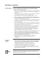
Introduction ................................................................................................................... 1
Overview .........................................................................................................................................1
Methods ......................................................................................................................... 3
General Cloning .............................................................................................................................3
Cloning into pRSET A, B, and C ..................................................................................................4
Expression .......................................................................................................................................8
Purification....................................................................................................................................11
Appendix...................................................................................................................... 12
Recipes...........................................................................................................................................12
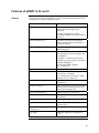
Map of pRSET A, B, and C..........................................................................................................14
Features of pRSET A, B, and C...................................................................................................15
Map of pRSET/lacZ .....................................................................................................................16
Transformation Protocol for TOP10F′ and BL21(DE3)pLysS ................................................17
Technical Support ........................................................................................................................18
Purchaser Notification.................................................................................................................19
iii
Kit Contents and Storage
Kit Contents
This kit contains the following reagents:
20 μg each of pRSET A, B, and C in TE buffer, pH 8.0* (40 μl each at 0.5 μg/μl)
1 stab TOP10F′
1 stab BL21(DE3)pLysS
1 stab BL21(DE3)pLysS containing the pRSET/lacZ control
*TE buffer, pH 8.0: 10 mM Tris-HCl, 1 mM EDTA, pH 8.0
Shipping and
Storage
This kit is shipped on wet ice. Upon receipt, store the plasmids at –20°C and the
stabs at 4°C.
Long-Term
Storage
For long-term storage of E. coli strains supplied as stabs with this kit, prepare
glycerol stocks as follows:
iv
1.
Grow the E. coli strain overnight in SOB medium overnight with antibiotic
selection when appropriate.
2.
Combine 0.85 ml of the overnight culture with 0.15 ml of sterile glycerol.
3.
Vortex and transfer to a labeled cryovial.
4.
Freeze the tube in liquid nitrogen or dry ice/ethanol bath and store at –80°C.
Accessory Products
The tables below lists related products that may be used with pRSET A, B, and C.
Introduction
Product
Application
Quantity
Cat. No.
One Shot TOP10F′ cells
Chemically competent cells for
transformation
20 × 50 μl
C3030-03
One Shot®
BL21(DE3)pLysS cells
Chemically competent cells for
transformation
20 × 50 μl
C6060-03
One Shot®
BL21(DE3)pLysE cells
Chemically competent cells for
transformation
20 × 50 μl
C6565-03
One Shot® BL21(DE3) cells Chemically competent cells for
transformation
20 × 50 μl
C6000-03
®
Anti-Xpress™ Antibody
Detection of recombinant proteins
50 μl
R910-25
Anti-Xpress™-HRP
Antibody
Detection of recombinant proteins
50 μl
R911-25
Anti-HisG Antibody
Detection of recombinant proteins
50 μl
R940-25
Anti-HisG-HRP Antibody
Detection of recombinant proteins
50 μl
R941-25
Anti-HisG-AP Antibody
Detection of recombinant proteins
125 μl
R942-25
Purification of recombinant proteins
50 ml
R801-01
150 ml
R801-50
250 units
E180-01
7.5 ml
R180-01
6 purifications
K850-01
™
ProBond Resin
™
EnterokinaseMax
EK-Away
™
™
ProBond Purification
System
Removal of N-terminal peptide
™
Removal of EnterokinaseMax
For native and denaturing purification
of recombinant proteins
Continued on next page
v
Accessory Products
Electrophoresis
Products
A large variety of pre-cast polyacrylamide gels and electrophoresis products
are available separately from Invitrogen for the separation and analysis of
recombinant proteins. Ordering information for the most widely used products
is provided below. For more detailed information, including size, concentration
and well formats available for pre-cast gel systems, visit www.invitrogen.com
or contact Technical Support (page 18).
Quantity
Cat. no.
1 box (10 gels)
NP0321BOX
1 box (10 gels)
EC6075BOX
10 ml
NP0007
250 ml
NP0008
20 ml
LC2676
SimplyBlue Safe-Stain
1L
LC6060
Colloidal Blue Staining Kit
1 kit
LC6025
1 unit
EI0002
Product
®
®
NuPAGE Novex 4–12% Bis-Tris Gels
®
Novex 10% Tris-Glycine Gels
®
NuPAGE LDS Sample Buffer (4X)
®
Novex Tris-Glycine SDS Sample Buffer (2X)
™
XCell SureLock™ Mini-Cell & XCell II™ Blot
Module
Media and
Reagents
In addition to the pre-cast polyacrylamide gel systems, Invitrogen offers a wide
range of pre-mixed media and reagents. Ordering information for the most
widely used products is provided below. For more detailed information, visit
www.invitrogen.com or contact Technical Support (page 18).
Product
S.O.C. Medium
®
(Miller’s LB Broth Base) Luria Broth Base,
powder
imMedia™ Amp Liquid
imMedia™ Amp Agar
UltraPure™ Sodium Dodecyl Sulfate (SDS)
UltraPure™ DNase/RNase-Free Water
vi
Quantity
Cat. no.
10 × 10 ml
15544-034
500 g
12795-027
20 pouches
(200 ml medium)
Q600-20
20 pouches
(8–10 plates)
Q601-20
500 g
15525-017
500 ml
10977-015
Introduction
Overview
Introduction
The pRSET vectors are pUC-derived expression vectors designed for high-level
protein expression and purification from cloned genes in E. coli. High levels of
expression of DNA sequences cloned into the pRSET vectors are made possible
by the presence of the T7 promoter. In addition, DNA inserts are positioned
downstream and in frame with a sequence that encodes an N-terminal fusion
peptide. This sequence includes an ATG translation initiation codon, a
polyhistidine tag that functions as a metal binding domain in the translated
protein, a transcript stabilizing sequence from gene 10 of phage T7, the Xpress™
epitope, and the enterokinase cleavage recognition sequence.
The metal binding domain of the fusion peptide allows simple purification of
recombinant proteins by Immobilized Metal Affinity Chromatography with
Invitrogen’s ProBond™ resin (available in bulk, see page v). The enterokinase
cleavage recognition site in the fusion peptide located between the metal
binding domain and the recombinant protein allows for subsequent removal of
this N-terminal fusion peptide from the purified recombinant protein.
Regulation of
Expression of the
Gene of Interest
Expression of the gene of interest from pRSET is controlled by the strong phage
T7 promoter that drives expression of gene 10 (Φ10). T7 RNA polymerase
specifically recognizes this promoter. For expression of the gene of interest, it is
necessary to deliver T7 RNA polymerase to the cells by either inducing
expression of the polymerase using the gratuitous inducer isopropyl β-Dthiogalactoside (IPTG), or infecting the cell with phage expressing the
polymerase. Once sufficient T7 RNA polymerase is produced, it binds to the T7
promoter and transcribes the gene of interest.
Regulation of
Expression of T7
RNA Polymerase
The BL21(DE3)pLysS strain is specifically included in the kit for expression of
T7 regulated genes. This strain carries the DE3 bacteriophage lambda lysogen.
This lambda lysogen contains the lacI gene, the T7 RNA polymerase gene under
control of the lacUV5 promoter, and a small portion of the lacZ gene. This lac
construct is inserted into the int gene, which inactivates the int gene. Disruption
of the int gene prevents excision of the phage (i.e. lysis) in the absence of helper
phage. The lac repressor represses expression of T7 RNA polymerase. Addition
of IPTG allows expression of T7 RNA polymerase.
The BL21(DE3)pLysE strain is also available. For more information on this
strain, BL21(DE3), and BL21(DE3)pLysS, see page 3.
Continued on next page
1
Overview, continued
There is always some basal level expression of T7 RNA polymerase. If a toxic
gene is cloned downstream of the T7 promoter, basal expression of this gene
may lead to reduced growth rates, cell death, or plasmid instability.
T7 lysozyme (produced from pLysS or pLysE) has been shown to bind to
T7 polymerase and inhibit transcription. This activity is exploited to reduce
basal levels of T7 RNA polymerase.
Regulation of T7
RNA Polymerase
by T7 Lysozyme
T7 lysozyme is a bifunctional enzyme. In addition to its T7 RNA polymerase
binding activity, it also cleaves a specific bond in the peptidoglycan layer of the
E. coli cell wall. This activity increases the ease of cell lysis by freeze-thaw
cycles prior to purification.
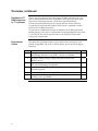
The table below describes the basic steps needed to clone and express your
protein using pRSET A, B, and C. For more details, please refer to the page(s)
indicated.
Experimental
Outline
Step
2
Action
Page
1
Propagate and maintain the empty pRSET A, B, and C vectors by
transforming them into a recA, endA E. coli host (i.e. TOP10F′).
3
2
Develop a cloning strategy to ligate your gene of interest into
pRSET A, B, or C.
3
Ligate your gene of interest into pRSET, transform into TOP10F′,
and select on 50–100 μg/ml ampicillin.
6
4
Sequence your construct to ensure that it is in frame with the
N-terminal peptide.
7
5
Perform a pilot expression using IPTG for induction.
8
6
Purify your recombinant protein by chromatography on metalchelating resin (e.g. ProBond™).
11
4–7
Methods
General Cloning
Introduction
The following information is provided to help you clone your gene of interest
into pRSET A, B, and C. For basic information on DNA ligations, E. coli
transformations, restriction analysis, DNA sequencing and DNA biochemistry,
see Current Protocols in Molecular Biology (Ausubel et al., 1994).
E. coli Host
For cloning and transformation, we recommend using a recA, endA strain such
as TOP10F′ (included in the kit). TOP10F′ cells are recA and endA making them
suitable for cloning, propagation, and maintenance.
Genotype of TOP10F′:
F′ {lacIq, Tn10(TetR)} mcrA (mrr-hsdRMS-mcrBC) 80lacZ M15 lac 74 recA1
araD139 (ara-leu)7697 galU galK rpsL (StrR) endA1 nupG.
BL21(DE3)pLysS is specifically designed for expression of genes regulated by
the T7 promoter. Do not use this strain for propagation or maintenance of your
plasmid.
Genotype of BL21(DE3)pLysS:
F-, ompT hsdSB (rB- mB-) gal dcm (DE3) pLysS (CamR)
Maintaining
pRSETA, B, and C
To propagate and maintain pRSET A, B, and C, use the supplied 0.5 μg/μl stock
solution in TE, pH 8.0 to transform a recA, endA E. coli strain like TOP10F’,
DH5α™-T1 R, TOP10, or equivalent. Select transformants on LB plates
containing 50–100 μg/ml ampicillin.
Be sure to prepare a glycerol stock of a transformant containing plasmid for
long-term storage (see page 7).
3
Cloning into pRSET A, B, and C
Introduction
The multiple cloning site of each version of pRSET is provided below and on
the following pages (see pages 5–6). To generate recombinant proteins that are
expressed correctly and contain the N-terminal fusion peptide, it is necessary to
clone in frame with the N-terminal peptide. To facilitate cloning, the pRSET
vector is provided in three different reading frames. They differ only in the
spacing between the sequences that code for the N-terminal peptide and the
multiple cloning site. For proper expression, determine which restriction sites
are appropriate for ligation.
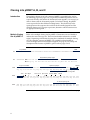
Multiple Cloning
Site of pRSET A
Below is the multiple cloning site for pRSET A. Restriction sites are labeled to
indicate the actual cleavage site. The boxed nucleotides indicate the variable
region. Sequencing and functional testing have confirmed the multiple cloning
site. The complete sequence of pRSET A is available for downloading at
www.invitrogen.com or from Technical Support (see page 18). For a map and
description of the features of pRSET A, please refer to pages 14–15.
T7 promoter
RBS
21
AATACGACTC ACTATAGGGA GACCACAACG GTTTCCCTCT AGAAATAATT TTGTTTAACT TTAAGAAGGA
91
GATATACAT ATG CGG GGT TCT CAT CAT CAT CAT CAT CAT GGT ATG GCT AGC ATG ACT
Met Arg Gly Ser His His His His His His Gly Met Ala Ser Met Thr
Polyhistidine (6xHis) region
Xpress Epitope
T7 gene 10 leader
148
EK recognition site
Xho I Sac I Bgl II
205
BamH I
GGT GGA CAG CAA ATG GGT CGG GAT CTG TAC GAC GAT GAC GAT AAG GAT CGA TGG GGA
Gly Gly Gln Gln Met Gly Arg Asp Leu Tyr Asp Asp Asp Asp Lys Asp Arg Trp Gly
Pst I Pvu II
Kpn I Nco I
EK cleavage site
EcoR I BstB I Hind III
TCC GAG CTC GAG ATC TGC AGC TGG TAC CAT GGA ATT CGA AGC TTG ATC CGG CTG CTA
Ser Glu Leu Glu Ile Cys Ser Trp Tyr His Gly ile Arg Ser Leu Ile Arg Leu Leu
T7 reverse priming site
262
ACA AAG CCC GAA AGG AAG CTG AGT TGG CTG CTG CCA CCG CTG AGC AAT AAC TAG CAT
Thr Lys Pro Glu Arg Lys Leu Ser Trp Leu Leu Pro Pro Leu Ser Asn Asn *** His
Continued on next page
4
Cloning into pRSET A, B, and C, continued
Multiple Cloning
Site of pRSET B
Below is the multiple cloning site for pRSET B. Restriction sites are labeled to
indicate the actual cleavage site. The boxed nucleotides indicate the variable
region. Sequencing and functional testing have confirmed the multiple cloning
site. The complete sequence of pRSET B is available for downloading at
www.invitrogen.com or from Technical Support (see page 18). For a map and
description of the features of pRSET B, please refer to pages 14–15.
T7 promoter
RBS
21
AATACGACTC ACTATAGGGA GACCACAACG GTTTCCCTCT AGAAATAATT TTGTTTAACT TTAAGAAGGA
91
GATATACAT ATG CGG GGT TCT CAT CAT CAT CAT CAT CAT GGT ATG GCT AGC ATG ACT
Met Arg Gly Ser His His His His His His Gly Met Ala Ser Met Thr
Polyhistidine (6xHis) region
Xpress Epitope
T7 gene 10 leader
148
BamH I
Xho I Sac I
GGT GGA CAG CAA ATG GGT CGG GAT CTG TAC GAC GAT GAC GAT AAG GAT CCG AGC TCG
Gly Gly Gln Gln Met Gly Arg Asp Leu Tyr Asp Asp Asp Asp Lys Asp Pro Ser Ser
EK recognition site
Bgl II
Pst I Pvu II
Kpn I Nco I
EK cleavage site
EcoR I BstB I Hind III
205
AGA TCT GCA GCT GGT ACC ATG GAA TTC GAA GCT TGA TCCGGCTGCT AACAAAGCCC
Arg Ser Ala Ala Gly Thr Met Glu Phe Glu Ala ***
261
GAAAGGAAGC TGAGTTGGCT GCTGCCACCG CTGAGCAATA ACTAGCATAA
T7 reverse priming site
Continued on next page
5
Cloning into pRSET A, B, and C, continued
Below is the multiple cloning site for pRSET C. Restriction sites are labeled to
indicate the actual cleavage site. The boxed nucleotides indicate the variable
region. Sequencing and functional testing have confirmed the multiple cloning
site. The complete sequence of pRSET C is available for downloading at
www.invitrogen.com or from Technical Support (see page 18). For a map and
description of the features of pRSET C, please refer to pages 14–15.
Multiple Cloning
Site of pRSET C
T7 promoter
RBS
21 AATACGACTC ACTATAGGGA GACCACAACG GTTTCCCTCT AGAAATAATT TTGTTTAACT TTAAGAAGGA
Polyhistidine (6xHis) region
91 GATATACAT ATG CGG GGT TCT CAT CAT CAT CAT CAT CAT GGT ATG GCT AGC ATG ACT
Met Arg Gly Ser His His His His His His Gly Met Ala Ser Met Thr
T7 gene 10 leader
Xpress Epitope
BamH I
148 GGT GGA CAG CAA ATG GGT CGG GAT CTG TAC GAC GAT GAC GAT AAG GAT CGA TGG ATC
Gly Gly Gln Gln Met Gly Arg Asp Leu Tyr Asp Asp Asp Asp Lys Asp Arg Trp Ile
EK recognition site
Xho I
Bgl II
Pst I Pvu II Kpn I Nco I EcoR I
EK cleavage site
BstB I Hind III
205 CGA CCT CGA GAT CTG CAG CTG GTA CCA TGG AAT TCG AAG CTT GAT CCG GCT GCT AAC
Arg Pro Arg Asp Leu Gln Leu Val Pro Trp Asn Ser Lys Leu Asp Pro Ala Ala Asn
T7 reverse priming site
262 AAA GCC CGA AAG GAA GCT GAG TTG GCT GCT GCC ACC GCT GAG CAA TAA CTA GCA
Lys Ala Arg Lys Glu Ala Glu Leu Ala Ala ALa Thr Ala Gln Gln ***
Ligation
Once you have determined a cloning strategy, digest the appropriate version of
pRSET with the selected restriction enzyme. Ligate your gene of interest into
pRSETA, B, or C using standard molecular biology techniques.
Transformation
After ligating your gene of interest into the appropriate pRSET vector, transform
the ligation mixture into competent TOP10F′. A detailed protocol for making
competent TOP10F′ cells and using them for transformation is provided in the
Appendix on page 17. Select 10–20 clones and analyze for the presence and
orientation of your insert.
Continued on next page
6
MEND
ION
AT
RECOM
Cloning into pRSET A, B, and C, continued
Making Frozen
Glycerol Stocks
We recommend that you sequence your construct to confirm that your gene is in
frame with the N-terminal tag and in the proper orientation. The T7 promoter
primer (Cat. no. N560-02) is available for sequencing your insert in pRSET A, B,
or C.
1.
Grow 1–2 ml of the E. coli strain to be frozen in SOB medium overnight with
antibiotic selection when appropriate.
2.
Combine 0.85 ml of the overnight culture with 0.15 ml of sterile glycerol
(sterilized by autoclaving).
3.
Mix well by vortexing.
4.
Transfer to an appropriate freezing vial (preferably a screw cap, air-tight
gasket).
5.
Freeze in an ethanol-dry ice bath or liquid nitrogen and then transfer to
–80°C for long-term storage.
7
Expression
Introduction
BL21(DE3)pLysS cells are included with the kit as the host for expression. You
will need pure plasmid DNA of your construct to transform into
BL21(DE3)pLysS for expression studies. Since each recombinant protein has
different characteristics that may affect optimal expression, it is helpful to do a
pilot expression to determine the best conditions for optimal expression of your
particular protein.
Preparation for
Expression
To express your recombinant protein from pRSET, transform the plasmid into
BL21(DE3)pLysS and select for ampicillin-resistant transformants (see page 17).
Before proceeding with the expression, streak out the BL21(DE3)pLysS
transformant containing the recombinant plasmid on LB containing 35 μg/ml
chloramphenicol and 50 μg/ml ampicillin. Chloramphenicol selects for
maintenance of the pLysS plasmid required for T7 lysozyme expression and
ampicillin selects for the pRSET plasmid (see Appendix for media recipes).
It is important to maintain BL21(DE3)pLysS strains on LB and chloramphenicol
as loss of the plasmid will increase basal levels of transcription. We recommend
preparing a frozen glycerol stock of untransformed BL21(DE3)pLysS
(see page 7).
Plasmid
Preparation
Plasmid DNA may be prepared using your method of choice. We recommend
the S.N.A.P.™ MiniPrep Kit (Cat. no. K1900-01) or the PureLink™ HiPure Plasmid
DNA Purification Kit (Cat. no. K2100-01) for isolation of pure plasmid DNA.
Positive Control
Vector
Included in the kit is a stab of E. coli strain BL21(DE3)pLysS containing
pRSET/lacZ. pRSET/lacZ is pRSET A with the β-galactosidase gene cloned into
the BamH I and Hind III sites for use as a positive control for expression.
β-galactosidase should appear as a band of approximately 120 kDa on a
denaturing polyacrylamide gel. The complete sequence of this vector is
available at www.invitrogen.com or from Technical Support (page 18).
Continued on next page
8
Expression, continued
Pilot Expression
Expression conditions will vary depending on the nature of your protein;
therefore, we recommend performing a time course experiment to optimize
expression of your recombinant protein.
1. Inoculate 2 ml of SOB containing ampicillin (50 μg/ml) and chloramphenicol
(35 μg/ml) with a single recombinant E. coli colony. Grow overnight at 37°C
with shaking.
2. The next day, inoculate 25 ml of SOB (it is not necessary to include antibiotics
for expression) to an OD600 of 0.1 with the overnight culture.
3.
Grow the culture at 37°C with vigorous shaking to an OD600 = 0.4–0.6.
4.
Remove a 1 ml aliquot of cells prior to IPTG induction, centrifuge the sample
in a microcentrifuge, and aspirate the supernatant. Freeze the cell pellet
at –20°C. This will be the time zero sample.
5. Add IPTG to a final concentration of 1 mM (0.25 ml of 100 mM IPTG stock to
25 ml culture) and continue to grow the cells. See page 12 for preparation of
the IPTG stock solution.
6. After 1 hour of incubation, remove a 1 ml sample, centrifuge as described in
Step 4, aspirate the supernatant, and freeze the cell pellet at –20°C. Continue
to take samples at 1 hour intervals for 4 to 6 hours.
7. When all time points have been collected, resuspend each pellet in 100 μl of
20 mM phosphate buffer at neutral pH, and freeze in liquid nitrogen or
methanol/dry ice (exercise caution when handling liquid nitrogen, it can
cause severe burns if it comes in contact with the skin, wear appropriate
protective equipment). Thaw the frozen lysate at 42°C.
8. Repeat this freeze-thaw two to three additional times and pellet the insoluble
protein in a microcentrifuge for 10 minutes at maximum speed at 4°C.
9. Remove the supernatant to a fresh labeled tube. To 100 μl of supernatant
sample, add an equal volume of 2X SDS-PAGE sample buffer. Resuspend the
pellet in 100 μl of 1X SDS-PAGE sample buffer.
10. Load 10–20 μl of each of the supernatant and pellet samples after
boiling for 5 minutes on an appropriate SDS-PAGE gel and
electrophorese.
Analysis of
Samples
1.
2.
3.
Stain the gel with Coomassie blue and look for a band of increasing intensity
in the expected size range for the recombinant protein. Use the uninduced
culture as a negative control. From this expression experiment, determine
the optimal time after IPTG induction to harvest the cells.
In addition, you may perform a western blot to confirm that the
overexpressed band is your desired protein (see next page).
Use the positive control to confirm that growth and induction were
performed properly. The pRSET/lacZ vector should produce an ~120 kDa
protein when induced with IPTG.
Expression of your protein with the N-terminal tag will increase the size of your
protein by approximately 3 kDa. Be sure to account for any additional amino
acids between the tag and your protein.
Continued on next page
9
Expression, continued
Detecting
Recombinant
Fusion Proteins
To detect expression of your recombinant fusion protein by western blot
analysis, you may use antibodies against the appropriate epitope available
from Invitrogen (see page v for ordering information) or an antibody to your
protein of interest. In addition, the Positope™ Control Protein (Cat. no. R900-50)
is available from Invitrogen for use as a positive control for detection of fusion
proteins containing an Xpress™ or HisG epitope. The ready-to-use
WesternBreeze® Chromogenic Kits and WesternBreeze® Chemiluminescent Kits
are available from Invitrogen to facilitate detection of antibodies by
colorimetric or chemiluminescent methods. For more information, please refer
to our website (www.invitrogen.com) or call Technical Support (see page 18).
Expressing
Recombinant
Protein
1.
2.
3.
4.
5.
6.
Troubleshooting
Expression
Problem
No or low
expression
10
Inoculate 2 ml of SOB containing ampicillin (50 μg/ml) and chloramphenicol
(35 μg/ml) with a single recombinant E. coli colony. Grow overnight at 37°C
with shaking (225 rpm).
The next day, inoculate 25 ml of SOB to an OD600 of 0.1 with the overnight
culture. Antibiotics are not required for expression. Please note that you may
increase the volume to produce more protein.
Grow the culture at 37°C with shaking (225 rpm) to an OD600 = 0.4–0.6.
Add IPTG to a final concentration of 1 mM (0.25 ml of 100 mM IPTG stock to
25 ml culture).
Grow the culture at 37°C with vigorous shaking for the optimal time
determined in pilot expression (see page 9).
Harvest the cells by centrifugation and either proceed directly to lysis or
freeze the cells at –80°C until ready for use.
Use the information provided in the table below to troubleshoot your expression
experiment.
Probable Cause
Possible Solution
Insert ligated into wrong
reading frame
Check sequence carefully and determine
which vector, pRSET A, B, or C is appropriate
with the restriction site selected
Kinetics of induction different
than expected
Try a longer time course for induction than
the 4–5 hours recommended
Not induced at OD600 0.4–0.6
Induce expression at OD600 0.4–0.6
IPTG solution is too old
Prepare a fresh solution of IPTG or use up to
10 mM IPTG
Protein is difficult to detect on
a Coomassie-stained gel
Perform a western blot using the
Anti-Xpress™ antibody for detection
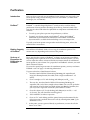
Purification
Introduction
Once you have expressed your recombinant fusion protein, you may purify your
fusion protein using a metal-chelating resin such as ProBond™ (available from
Invitrogen, Cat. no. R801-01).
ProBond™
ProBond™ is a nickel-charged Sepharose® resin that can be used for affinity
purification of fusion proteins containing the 6×His tag. Proteins bound to the
resin may be eluted with either low pH buffer or competition with imidazole or
histidine.
•
To scale up your pilot expression for purification, see below.
•
To purify your fusion protein using ProBond™, refer to the ProBond™
Purification System manual for instruction. The ProBond™ Purification
System manual is available for downloading at www.invitrogen.com.
To purify your fusion protein using another metal-chelating resin, refer to the
manufacturer’s instructions.
Binding Capacity
of ProBond™
One milliliter of ProBond™ binds at least 1 mg of recombinant protein. This
amount can vary depending on the nature of the protein.
Scale-up of
Expression for
Purification on
ProBond™
Please note that the capacity of ProBond™ is about 1 mg of protein per milliliter.
Depending on the expression level of your recombinant fusion protein, you may
need to adjust the culture volume to bind the maximum amount of recombinant
fusion protein to your column. For a prepacked 2 ml ProBond™ column, start with
50 ml of bacterial culture.
If you need to purify larger amounts of recombinant protein, you may need more
ProBond™ resin. See page v for ordering information.
To grow and induce a 50 ml bacterial culture:
1.
Inoculate 10 ml of SOB or LB containing 50–100 μg/ml ampicillin and
34 μg/ml chloramphenicol (if needed) with a single recombinant E. coli
colony.
2.
Grow overnight at 37°C with shaking (225–250 rpm) to OD600 = 1–2.
3.
The next day, inoculate 50 ml of SOB or LB containing 50-100 μg/ml
ampicillin with 1 ml of the overnight culture. Note: You can scale up further
and inoculate all of the 10 ml overnight culture into 500 ml of medium, but
you may need a larger bed volume for your ProBond™ column.
4.
Grow the culture at 37°C with shaking (225–250 rpm) to an OD600 = ~0.5
(2–3 hours). The cells should be in mid-log phase.
5.
Add 1 mM IPTG to induce expression.
6.
Grow at 37°C with shaking until the optimal time point determined by the
pilot expression is reached. Harvest the cells by centrifugation (3000 × g for
10 minutes at 4°C).
7.
At this point, you may proceed directly to purification, or store the cells for
future use at –80°C.
11
Appendix
Recipes
SOB (For 1 Liter)
To 950 ml of deionized water add:
20.0 g Tryptone
5.0 g Yeast Extract
0.5 g NaCl
186.0 mg KCl
1.
Mix the solution until dissolved.
2.
Adjust the pH to 7.0 with 5 N NaOH (approximately 0.2 ml).
3.
If making solid media (for plates or top agar), add 15 g of agar after
adjusting the pH.
4.
Adjust the volume to 1000 ml and sterilize by autoclaving.
5.
Once autoclaved, add 10 ml of sterile 1 M Mg2+
(e.g. 10 ml of sterile 1 M MgCl2 or sterile 1 M MgSO4).
SOC (For 1 Liter)
Follow recipe as per SOB. After autoclaving, let cool to about 60°C and
add 10 ml of 50% glucose. Mix the media well.
LB (For 1 Liter)
Component
Tryptone
Yeast Extract
NaCl
Agar
liquid
10 g
5g
10 g
-
plates
10 g
5g
10 g
15 g
top agar
10 g
5g
10 g
7g
1.
Combine the tryptone, yeast extract, and NaCl with 950 ml of deionized
water. Mix the solution until dissolved.
2.
Adjust the pH to 7.0 with 5 N NaOH (will take about 0.2 ml). If making solid
media (for plates or top agar) add the appropriate amount of agar after
adjusting the pH.
3.
Adjust volume to 1 liter with water.
4.
Sterilize by autoclaving.
5.
After autoclaving add antibiotic, if desired. Add chloramphenicol to a final
concentration of 10 μg/ml and ampicillin to a final concentration of
50 μg/ml.
Continued on next page
12
Recipes, continued
Antibiotics
Ampicillin:
Prepare a stock solution of 50 mg/ml in deionized water and filter sterilize it with
a 0.22 μm filter. To prepare selective medium, cool medium to ~50°C after
autoclaving, and add 1 ml of the ampicillin stock per liter of media (both liquid
and solid) for a final concentration of 50 μg/ml. Store the stock solution at –20°C.
Chloramphenicol:
Prepare a stock solution of 35 mg/ml in 100% ethanol. It is not necessary to filtersterilize. Store the stock solution at –20°C. To prepare selective medium, cool the
medium to ~50°C after autoclaving and add 1 ml of the stock solution per liter of
medium for a final concentration of 35 μg/ml.
100 mM IPTG
For 10 ml of a 100 mM solution:
Dissolve 0.24 g of IPTG in sterile, deionized water. Bring the final volume to 10 m
and filter sterilize (0.22 μm filter). Do not autoclave.
50 mM CaCl2
For 100 ml of a 50 mM solution:
Dissolve 0.56 g of anhydrous CaCl2 (MW = 111) in 100 ml of deionized water.
Filter sterilize (0.22 μm filter) or autoclave. Use this solution ice cold for
competent cell preparation.
13
Map of pRSET A, B, and C
The map below shows the features of pRSET A, B, and C. The complete
sequence of the vector is available for downloading from our website at
www.invitrogen.com or from Technical Support (see page 18).
BamH I
Xho I
Sac I*
Bgl II
Pst I
Pvu II
Kpn I
Nco I
EcoR I
BstB I
Hind III
pRSET A, B, and C
PT7
RBS ATG 6xHis Xpress Epitope EK
MCS
Stop
1
f
or
i
A,B,C
2.9 kb
ll
in
pUC ori
pRSET
Comments for pRSET A
2897 nucleotides
Am
*Version C does not contain Sac I
T7 promoter: bases 20-39
6xHis tag: bases 112-129
T7 gene 10 leader: bases 133-162
XpressTM epitope: bases 169-192
Multiple cloning site: bases 202-248
T7 reverse priming site: bases 295-314
T7 transcription terminator: bases 256-385
f1 origin: bases 456-911
bla promoter: bases 943-1047
Ampicillin (bla) resistance gene (ORF): bases 1042-1902
pUC origin: bases 2047-2720 (C)
14
i
pic
Features of pRSET A, B, and C
Features
The important elements of pRSET A, B, and C are described in the table below.
All features have been functionally tested.
Feature
Benefit
T7 promoter
Provides tight, dose-dependent
regulation of heterologous gene
expression.
Provides a binding site for most
T7 promoter primers for sequencing into
the insert.
Ribosome binding site
Optimally spaced from the multiple
cloning site for efficient translation of
the gene of interest.
Initiation ATG
Provides a translational initiation site for
the fusion protein.
N-terminal 6×His tag
Permits purification of recombinant
fusion protein on metal-chelating resins
(i.e. ProBond™).
In addition, it allows detection of the
recombinant protein with the Anti-HisG
Antibody (R940-25) or
the Anti-HisG-HRP Antibody
(Cat. no. R941-25)
T7 gene 10 sequence
Provides protein stability
™
N-terminal Xpress epitope tag
Allows detection of the fusion protein
by the Xpress™ Antibody
(Cat. no. R910-25) or the Xpress™-HRP
Antibody (Cat. no. R911-25)
Enterokinase cleavage site
Provides a site for efficient removal of
the fusion tag.
Multiple cloning site
Allows insertion of your gene of interest
and facilitates in cloning in frame with
the N-terminal epitope tag.
T7 reverse priming site
Allows sequencing of the insert.
T7 terminator
Permits efficient transcription
termination.
f1 origin
Allows single strand rescue of DNA
bla promoter
Allows expression of the ampicillin
resistance gene.
Ampicillin resistance gene
(β-lactamase)
Allows selection of the plasmid in E. coli.
pUC origin
High copy replication and growth in
E. coli.
15
Map of pRSET/lacZ
Description
pRSET/lacZ is a 5911 bp control vector expressing β-galactosidase. Note that galactosidase is fused to an N-terminal peptide containing the Xpress™ peptide,
6xHis tag and an enterokinase recognition site. The molecular weight is
approximately 120 kDa. The figure below summarizes the features of the
pRSET/lacZ vector. The complete sequence of the vector is available for
downloading from our website at www.invitrogen.com or from Technical
Support (see page 18).
PT7
RBS ATG 6xHis Xpress Epitope EK
f1
i
or
pUC or
pRSET/lacZ
5.9 kb
i
ll
in
Comments for pRSET/lacZ
5911 nucleotides
T7 promoter: bases 20-39
6xHis tag: bases 112-129
T7 gene 10 leader: bases 133-162
XpressTM epitope: bases 169-192
lacZ ORF: bases 199-3258
T7 reverse priming site: bases 295-314
T7 transcription terminator: bases 3270-3399
f1 origin: bases 3470-3925
bla promoter: bases 3957-4061
Ampicillin (bla) resistance gene (ORF): bases 4056-4916
pUC origin: bases 3930-5866 (C)
16
Am
i
pi c
lacZ
Stop
Transformation Protocol for TOP10F′ and BL21(DE3)pLysS
Introduction
This protocol is provided for your convenience. Other protocols may be suitable.
Use the table below to select the appropriate medium for use with TOP10F′ or
BL21(DE3)pLysS.
Strain
Protocol
Maintenance Medium
pRSET Selection Medium
TOP10F′
LB + 10 μg/ml tetracycline
LB + 50 μg/ml ampicillin
BL21(DE3)pLysS
LB + 35 μg/ml
chloramphenicol
LB + 50 μg/ml ampicillin +
35 μg/ml chloramphenicol
1.
Take the desired stab and streak out a small portion on the appropriate
maintenance medium and incubate at 37°C overnight. The stab should
remain viable for several months when stored at 4°C in the dark. We
recommend making a frozen glycerol stock for long-term storage (see page 7).
2.
Pick a single colony and transfer it into 100 ml of SOB medium in a 1 liter
flask (see page 12 for media recipes). Incubate the flask at 37°C with vigorous
shaking (> 200 cycles/minute in a rotary shaker).
3.
When the OD600 reaches approximately 0.5, collect the cells by centrifuging at
4000 rpm for 10 minutes in a 4°C rotor (Sorvall GSA).
4.
Resuspend the pellet in 10 ml of ice-cold 50 mM CaCl2. Keep the cells on ice
for at least 30 minutes.
5.
Centrifuge the CaCl2-treated cells in a 4°C rotor (Sorvall SS-34) at 4000 rpm
for 5 minutes. Gently resuspend the cells in 4 ml of ice-cold 50 mM CaCl2.
Keep the cells on ice.
6.
Aliquot 100 μl of the CaCl2-treated cells for each transformation into a
prechilled microcentrifuge tube. Store the cells at –80°C for long-term storage.
7.
For transformation, take one tube of 100 μl of competent cells (prepared
above) and add the plasmid DNA (10–100 ng) to the cells. Incubate the cells
on ice for 30 minutes.
8.
Heat shock cells at 42°C for 45 seconds (in a water bath). Return the tube(s) to
ice for 2 minutes.
9.
Add 1 ml of SOC media and incubate the culture(s) for 45 minutes at 37°C
with vigorous shaking (> 200 cycles/minute in a rotary shaker).
10. Plate the appropriate amount of cells onto SOB plates containing the
appropriate antibiotic selection for the plasmid (for pRSET vectors use
ampicillin).
Note: When selecting for transformants in BL21(DE3)pLysS, include 35 μg/ml
chloramphenicol in the plate.
For your convenience, One Shot® TOP10F′ or BL21(DE3)pLysS competent cells
are available for high efficiency transformation. See page v for more information.
17
Technical Support
Web Resources
Contact Us
Visit the Invitrogen website at www.invitrogen.com for:
•
Technical resources, including manuals, vector maps and sequences,
application notes, SDSs, FAQs, formulations, citations, handbooks, etc.
•
Complete technical support contact information
•
Access to the Invitrogen Online Catalog
•
Additional product information and special offers
For more information or technical assistance, call, write, fax, or email. Additional
international offices are listed on our website (www.invitrogen.com).
Corporate Headquarters:
5791 Van Allen Way
Carlsbad, CA 92008 USA
Tel: 1 760 603 7200
Tel (Toll Free): 1 800 955 6288
Fax: 1 760 602 6500
E-mail: [email protected]
Japanese Headquarters:
LOOP-X Bldg. 6F
3-9-15, Kaigan
Minato-ku, Tokyo 108-0022
Tel: 81 3 5730 6509
Fax: 81 3 5730 6519
E-mail: [email protected]
European Headquarters:
Inchinnan Business Park
3 Fountain Drive
Paisley PA4 9RF, UK
Tel: 44 (0) 141 814 6100
Tech Fax: 44 (0) 141 814 6117
E-mail: [email protected]
SDS
Safety Data Sheets (SDSs) are available at www.invitrogen.com/sds.
Certificate of
Analysis
The Certificate of Analysis (CofA) provides detailed quality control information for each
product. CofAs are available on our website. Go to www.invitrogen.com/support and
search for the Certificate of Analysis by product lot number, which is printed on the box.
Limited Warranty
Invitrogen (a part of Life Technologies Corporation) is committed to providing our
customers with high-quality goods and services. Our goal is to ensure that every customer
is 100% satisfied with our products and our service. If you should have any questions or
concerns about an Invitrogen product or service, contact our Technical Support
Representatives.
All Invitrogen products are warranted to perform according to specifications stated on the
certificate of analysis. The Company will replace, free of charge, any product that does not
meet those specifications. This warranty limits the Company’s liability to only the price of
the product. No warranty is granted for products beyond their listed expiration date. No
warranty is applicable unless all product components are stored in accordance with
instructions. The Company reserves the right to select the method(s) used to analyze a
product unless the Company agrees to a specified method in writing prior to acceptance
of the order.
Invitrogen makes every effort to ensure the accuracy of its publications, but realizes that
the occasional typographical or other error is inevitable. Therefore the Company makes no
warranty of any kind regarding the contents of any publications or documentation. If you
discover an error in any of our publications, report it to our Technical Support
Representatives.
Life Technologies Corporation shall have no responsibility or liability for any special,
incidental, indirect or consequential loss or damage whatsoever. The above limited
warranty is sole and exclusive. No other warranty is made, whether expressed or
implied, including any warranty of merchantability or fitness for a particular purpose.
18
Purchaser Notification
Limited Use Label
License
No: 22 Vectors and
Clones Encoding
Histidine Hexamer
This product is licensed under U.S. Patent Nos. 5,284,933 and 5,310,663 and
foreign equivalents from Hoffmann-LaRoche, Inc., Nutley, NJ and/or
Hoffmann-LaRoche Ltd., Basel, Switzerland and is provided only for use in
research. Information about licenses for commercial use is available from
QIAGEN GmbH, Max-Volmer-Str. 4, D-40724 Hilden, Germany.
Limited Use Label
License No: 30 T7
Expression System
The composition and/or use of this product may be claimed in U.S. Patent No.
5,693,489 licensed to Life Technologies Corporation by Brookhaven Science
Associates, LLC. The T7 expression system is based on technology developed
at Brookhaven National Laboratory under contract with the U.S. Department
of Energy, and is the subject of patents and patent applications assigned to
Brookhaven Science Associates, LLC (BSA,). By provisions of the Distribution
License Agreement granted to Life Technologies covering said patents and
patent applications, Life Technologies grants you a non-exclusive sub-license
under patents assigned to BSA for the use of this technology, including the
enclosed materials, based upon the following conditions: 1 – these materials
are to be used for non-commercial research purposes only. A separate license
under patents owned by BSA is required for any commercial use, including
the use of these materials for research purposes or production purposes by
any commercial entity. Information about commercial license may be obtained
from The Office of Technology Transfer, Brookhaven National Laboratory,
Bldg. 475D, P.O. Box 5000, Upton, New York 11973-5000. Phone (516) 3447134. 2 - No materials that contain the cloned copy of the T7 gene 1, the gene
for T7 RNA polymerase, may be distributed further to third parties outside of
your laboratory, unless the recipient receives a copy of this sub-license and
agrees to be bound by its terms. This limitation applies to strains BL21(DE3),
BL21(DE3)pLysS and BL21(DE3)pLysE, CE6, BL21-SI Competent Cells and
any derivatives that are made of them. You may refuse this sub-license by
returning this product unused in which case Life Technologies accept return
of the product with a full refund. By keeping or using this product, you agree
to be bound by the terms of this license.
19
Reference
Ausubel, F. M., Brent, R., Kingston, R. E., Moore, D. D., Seidman, J. G., Smith, J. A., and Struhl, K. (1994).
Current Protocols in Molecular Biology (New York: Greene Publishing Associates and WileyInterscience).
©2010 Life Technologies Corporation. All rights reserved.
For research use only. Not intended for any animal or human therapeutic or diagnostic use.
The trademarks mentioned herein are the property of Life Technologies Corporation or their respective owners.
.
20
Corporate Headquarters
5791 Van Allen Way
Carlsbad, CA 92008
T: 1 760 603 7200
F: 1 760 602 6500
E: [email protected]
For country-specific contact information, visit our web site at www.invitrogen.com