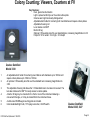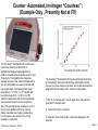
1
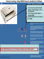
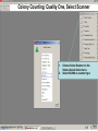
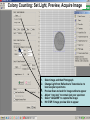
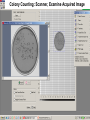
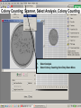
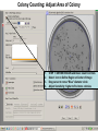
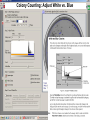
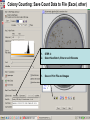
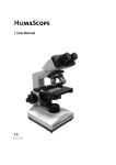
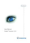
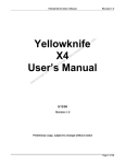
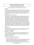
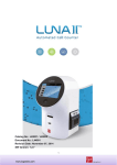
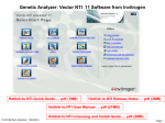
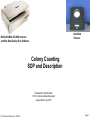
Darkfield
Viewers
BioRad/UMAX GS-800 Scanner
and Bio Rad Quality One Software
Colony Counting
SOP and Description
Prepared by: Bob Morrison
FVCC, Instrumentation Specialist
August 2008, July 2010
FVCC:BioTech;Morrison 7/15/2010
Page 1
Colony Counting: Using GS800 Scanner, Quality One Software
1.
2.
3.
Turn on Scanner and wait for both
lights to show steady green- this is
the ready state.
Turn on Host computer, logon as
Operator/SMET
Select Quality One to scan (acquire)
and image
Link to BioRad Quantity One 1-D Analysis Software Users Manual (pdf)
FVCC:BioTech;Morrison 7/15/2010
Page 2
Colony Counting: Quality One, Select Scanner
1.
2.
FVCC:BioTech;Morrison 7/15/2010
Choose Select Scanner on the
Volume Quick Guide menu
Select GS-800 as scanner type
Page 3
Colony Counting: Set Light, Preview, Acquire Image
1.
2.
3.
4.
5.
6.
Select image and then Photograph
Change Light from Reflective to Transmissive to
best see your specimens
Preview Scan and wait for image outline to appear
Adjust “crop box” to contain just your specimen
Select “ACQUIRE” to capture the image
Hit STOP if image preview fails to appear
Colony Counting: Scanner, Examine Acquired Image
Colony Counting; Scanner, Select Analysis, Colony Counting
1.
2.
Select Analysis
Select Colony Counting from Drop Down Menu
Colony Counting: Adjust Area of Colony
1.
2.
3.
4.
STEP 1: DEFINE REGION and Basic Count Controls
Select Icon to Define Region at Center of Image
Drag cursor to move “Blue” diameter circle
Adjust Sensitivity; higher to find more colonies
Colony Counting: Adjust Count vs. Density
1.
2.
If there is a clear peak on the left end of the
colony histogram, it is probably due to
background intensity or noise in the image.
Use Cutoff slide to adjust per notes below
Colony Counting: Adjust White vs. Blue
Colony Counting: Adjust counted items
Marked
Colonies
1.
2.
3.
Use STEP 3: to Adjust items in/out of the counts
Erase colony; then pick items from image that are
NOT part of the colonies
Make Colony; then pick items to be added as
colonies (yellow markers)
Colony Counting: Save Count Data to File (Excel, other)
1.
2.
STEP 4:
Select New Batch, Enter or edit filename
1.
Save or Print File and Images
Colony Counting: Viewers, Counters at FV
Key Features:
Even, glare-free illumination
Light is spread uniformly over the entire culture plate
Colonies are bright and easily distinguished
Adjustable dish holder for centering both round dishes and square culture plates
Adjustable focusing rod
Lens rotates a full 360º
Built-in tilt leg
Optional 1.5X auxiliary lens fits over standard lens, increasing magnification to 3X
Footprint: 12.5in wide x 14 in high x 12 in depth
Quebec Darkfield
Model 3330
1.
2.
3.
4.
5.
6.
An adjustable dish holder for centering round dishes with diameters up to 100mm and
square culture plates up to 100mm x 100mm.
An optional 1.5X auxiliary lens fits over the standard lens increasing magnification to
3.0X.
The adjustable focusing rod allows the 1.5X standard lens to be raised or lowered. The
lens also rotates a full 360º for ready access to culture plates.
A built-in tilt leg may be mounted in the front or rear of the instrument allowing a
convenient tilt angle, or it may be locked flat to the instrument base.
A white-ruled Wolffheugel counting plate is included.
Internal standard light bulb, 110V plug connection, On/Off switch.
FVCC:BioTech;Morrison 7/15/2010
Quebec Darkfield
Model 3325, 3327
Page 12
Counter: Automated, Invitrogen “Countess” ;
(Example Only , Presently Not at FV)
The Countess™ Automated Cell Counter uses
trypan blue staining combined with a
sophisticated image analysis algorithm to
produce accurate cell and viability counts in just
30 seconds. The algorithm also measures
average cell size of live, dead, and total cells to
give you all the data you need to proceed with
your experiments. The measurement range
extends from 1 x 10⁴ to 1 x 10⁷ cells/ml, with
an optimal range from 1 x 10⁵ to 4 x 10⁶
cells/ml, broader than that of a hemocytometer
(view technical notes for more comparison
data). The optimal cell size is between 5 µm to
60 µm (view validated cell lines). A handy
dilution calculator even helps you determine
how to prepare your sample for your next
passage or experiment.
FVCC:BioTech;Morrison 7/15/2010
The Countess™ Automated Cell Counter eliminates the tedium
and subjectivity of manual cell counting. Automated counting
frees up your time, reduces eye strain, and minimizes subjective
judgments that can lead to error. It takes 3 simple steps:
1. Mix 10 µl of sample with 10 µl of trypan blue, and pipet into
Countess™ chamber slide
2. Insert slide into the instrument
3. Press the "Count cells" button, results are displayed in 30
seconds
Page 13
: Other Protocols or Notes
•
Future home of other or more details protocols….
FVCC:BioTech;Morrison 7/15/2010
Page 14