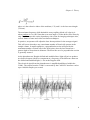
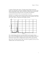
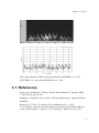
1
Cam1 and CapiScope User
Manual
KK Technology
Cam1 and CapiScope User Manual
by KK Technology
version 3.0 Edition
Published 03/07/2004
Copyright © 2004 by KK Technology
Table of Contents
I. CAM1 .....................................................................................................................i
1. Introduction.................................................................................................... 1
2. SAFETY PRECAUTIONS ............................................................................ 1
2.1. Electrical Safety .................................................................................. 1
2.2. Laser Safety ........................................................................................ 1
2.3. Certification......................................................................................... 2
3. Theory ............................................................................................................ 4
3.1. References........................................................................................... 8
4. CapiScope CAM1 Components................................................................... 11
5. CONNECTION DETAILS .......................................................................... 14
5.1. CAM1 ............................................................................................... 14
5.2. CapiScope PU Interface Card. CapiScope connector. ...................... 14
5.3. CapiScope PU Interface Card. ADC connector. ............................... 15
6. INSTALLATION ......................................................................................... 16
6.1. PU Connections ................................................................................ 16
6.2. Location ............................................................................................ 16
7. USING THE CAM1 .................................................................................... 17
7.1. Light Source...................................................................................... 17
7.2. Positioning and Focusing.................................................................. 17
7.3. Initial Checks .................................................................................... 19
7.4. Environment...................................................................................... 19
7.5. Holding the Finger ............................................................................ 20
7.6. Using adhesive pads.......................................................................... 20
7.7. Using Nail Glue ................................................................................ 22
7.8. Surface Preparation........................................................................... 23
8. Measuring Velocity Using the CAM1.......................................................... 24
8.1. Selecting a Capillary ......................................................................... 24
8.2. Taking Measurements ....................................................................... 26
9. Looking after the CAM1.............................................................................. 27
9.1. Maintenance and Cleaning................................................................ 27
9.2. Servicing ........................................................................................... 27
II. CapiScope .......................................................................................................... 28
10. Introduction................................................................................................ 29
11. Start Up ...................................................................................................... 30
3
11.1. Selecting functions.......................................................................... 30
11.2. Demo passcode ............................................................................... 31
11.3. Dynamic features passcode............................................................. 31
11.4. Select Profile ................................................................................... 32
12. Step by Step Guide..................................................................................... 33
12.1. Step 1. Displaying Video ................................................................ 33
12.2. Step 2. Dimension Lines ................................................................. 33
12.3. Step 3. Diameter measurement ....................................................... 34
12.4. Step 4. Measurements ..................................................................... 35
12.5. Step 5. Notes ................................................................................... 35
12.6. Step 6. Saving ................................................................................. 36
12.7. Step 7. Video................................................................................... 36
12.8. Step 8. Velocity ............................................................................... 38
13. Magnification Calibration .......................................................................... 39
14. Velocity Measurement ............................................................................... 41
14.1. Theory ............................................................................................. 41
14.2. Making Velocity Measurements ..................................................... 43
14.2.1. Measurement Line ............................................................... 43
14.2.2. Movement ............................................................................ 44
14.3. Calculation parameters.................................................................... 45
14.3.1. Calculate cbv........................................................................ 45
14.3.2. Subtract scans....................................................................... 45
14.3.3. Set sample rate ..................................................................... 45
14.3.4. CBV calculation parameters ................................................ 46
14.3.4.1. cbv method from linescan ......................................... 46
14.3.4.2. Number of correlations to smooth ............................ 46
14.3.4.3. Linescan sample periods between correlations......... 46
14.3.4.4. Correlation coeficient lower acceptable limit ........... 47
15. Movement Correction ................................................................................ 48
15.1. Introduction..................................................................................... 48
15.2. Automatic Movement Correction ................................................... 48
15.3. Manual Movement Correction ........................................................ 48
15.3.1. Tracking ............................................................................... 48
15.3.2. Dragging .............................................................................. 49
15.4. Reset movement correction offsets ................................................. 50
16. Software Reference.................................................................................... 51
16.1. Introduction..................................................................................... 51
4
16.2. Data Rate......................................................................................... 51
16.3. Clear All Traces .............................................................................. 52
16.4. Input channel................................................................................... 52
16.5. Input Settings Dialog ...................................................................... 52
16.5.1. Sample rate........................................................................... 53
16.5.2. Enable Input Buffer.............................................................. 53
16.5.3. Input Buffer Duration .......................................................... 54
16.5.4. Input buffer memory ............................................................ 54
16.5.5. Circular input buffer............................................................. 54
16.5.6. Stop on buffer full ................................................................ 54
16.5.7. FFT points............................................................................ 54
16.6. Laser On/Off ................................................................................... 55
16.7. Start Recording ............................................................................... 55
16.8. Stop Recording................................................................................ 55
16.9. Doppler Bandwidth......................................................................... 55
16.9.1. Increase Doppler Bandwidth................................................ 55
16.9.2. Lower Doppler Bandwidth .................................................. 56
16.10. Calculating Velocity...................................................................... 56
16.11. Calculating CBV from peak in spectrum...................................... 57
16.12. Calculate CBV from maximum frequency shift ........................... 57
16.13. Calculate CBV from envelope of power content .......................... 57
16.14. Find Average ................................................................................. 58
16.15. Threshold Dialog Control ............................................................. 58
16.16. Advanced analysis ........................................................................ 59
16.16.1. Signal strength ................................................................... 59
16.16.2. Power contour .................................................................... 59
16.16.3. Level contour ..................................................................... 59
16.16.4. Pulsatility Index ................................................................. 59
16.16.5. Resistance Index ................................................................ 60
16.16.6. Calculate Power Spectrum ................................................. 60
16.17. Charts ............................................................................................ 60
16.17.1. Introduction to charts ......................................................... 61
16.17.2. Axes ................................................................................... 61
16.17.2.1. Using the mouse...................................................... 61
16.17.3. Using the Keyboard ........................................................... 61
16.17.4. Traces ................................................................................. 62
16.17.4.1. Trace Properties ...................................................... 62
5
16.17.4.2. Selecting Trace Data ............................................... 62
16.17.4.3. Adding a Trace to a File.......................................... 62
16.17.5. Display Rate....................................................................... 63
16.17.6. Edit..................................................................................... 63
16.17.6.1. Edit Copy command ............................................... 63
16.17.6.2. Edit Cut command .................................................. 63
16.17.6.3. Edit Delete Command............................................. 64
16.17.6.4. Edit Paste command................................................ 64
16.17.7. Markers .............................................................................. 64
16.17.7.1. Event Markers ......................................................... 65
16.17.7.2. Edit Marker Dialog ................................................. 65
16.17.7.3. Edit New Marks ...................................................... 66
16.17.7.4. Marker Mode .......................................................... 66
16.17.7.5. Image Markers ........................................................ 66
16.17.8. Export data ......................................................................... 67
16.17.8.1. TEXT ...................................................................... 67
16.17.8.2. BINARY.................................................................. 67
16.17.9. FIR filter............................................................................. 68
16.17.10. Import data ....................................................................... 69
16.17.11. Keyboard.......................................................................... 69
16.17.12. Remove Pulse................................................................... 70
16.17.13. Selecting Data .................................................................. 71
16.17.14. Smoothing Dialog ............................................................ 71
16.17.15. Status Box ........................................................................ 71
16.17.15.1. Active Trace Name ............................................... 71
16.17.16. ToolBar ............................................................................ 72
16.17.17. Calculating Averages ....................................................... 72
16.17.18. Hide Trace........................................................................ 73
16.17.19. Increase Brightness .......................................................... 73
16.17.20. Decrease Brightness......................................................... 73
16.17.21. Smooth trace .................................................................... 74
16.17.22. Trace properties................................................................ 74
16.17.22.1. Trace Name ........................................................... 74
16.17.22.2. Y units................................................................... 75
16.17.22.3. X units................................................................... 75
16.17.22.4. Sample Rate .......................................................... 75
16.17.22.5. Trace length........................................................... 75
6
16.17.22.6. Axes X scale ......................................................... 75
16.17.22.7. Trace Start Position............................................... 75
16.17.22.8. Screen Y scale....................................................... 75
16.17.22.9. Screen Y offset...................................................... 76
16.17.22.10. Y scaling factor ................................................... 76
16.17.23. Trace Selection Dialog..................................................... 76
16.17.24. View ................................................................................. 76
16.17.24.1. Colour / mono Doppler spectrum ......................... 76
16.17.24.2. Inverse Colours ..................................................... 76
16.17.24.3. Minimum to background....................................... 77
16.17.25. Advanced Menu ............................................................... 77
16.17.26. Artifact Filter Dialog........................................................ 77
16.17.26.1. Artifact Limit ........................................................ 78
16.17.26.1.1. Absolute Value ........................................... 78
16.17.26.1.2. Percent........................................................ 78
16.17.26.2. Filter Action .......................................................... 78
16.17.26.2.1. No Action................................................... 78
16.17.26.2.2. Repeat last valid value ............................... 78
16.17.26.2.3. Set to zero & skip....................................... 79
16.18. Menu Commands .......................................................................... 79
16.18.1. File Menu commands......................................................... 79
16.18.1.1. New command (File menu) .................................... 79
16.18.1.2. Open command (File menu) ................................... 79
16.18.1.3. Close command (File Menu) .................................. 79
16.18.1.4. Save command (File Menu).................................... 80
16.18.1.5. Save As command (File menu)............................... 80
16.18.1.6. Export (Dimension List) ......................................... 81
16.18.1.7. Export (Image) ........................................................ 81
16.18.1.8. Export as avi ........................................................... 81
16.18.1.8.1. AVI Output compression.............................. 81
16.18.1.9. Import avi................................................................ 83
16.18.1.9.1. AVI Output compression.............................. 83
16.18.1.10. Import Image......................................................... 85
16.18.1.11. Open Cytoscan file................................................ 85
16.18.1.12. Print Preview command (File menu) .................... 86
16.18.1.13. Exit command (File menu) ................................... 86
16.18.2. Edit menu ........................................................................... 86
7
16.18.2.1. Cut command (Edit menu)...................................... 86
16.18.2.2. Copy commad (Edit menu)..................................... 86
16.18.2.3. Paste command (Edit menu) ................................... 87
16.18.2.4. Clear All Dimensions.............................................. 87
16.18.2.5. Notes ....................................................................... 87
16.18.2.6. Smooth Selection Menu.......................................... 87
16.18.3. View Menu......................................................................... 87
16.18.3.1. View Toolbar command .......................................... 88
16.18.3.2. Video Input Control ................................................ 88
16.18.3.3. Show Crosshair ....................................................... 88
16.18.3.4. Grid ......................................................................... 88
16.18.3.5. 2D View Menu ........................................................ 88
16.18.3.5.1. Always on Top ............................................. 88
16.18.4. Settings menu..................................................................... 88
16.18.4.1. Calibrate Magnification .......................................... 89
16.18.4.2. Magnification .......................................................... 89
16.18.4.3. Units........................................................................ 89
16.18.4.4. Timer....................................................................... 89
16.18.4.5. Grid Settings ........................................................... 90
16.18.5. Tools Menu ........................................................................ 90
16.18.5.1. Pan Image................................................................ 90
16.18.5.2. Region of Interest (ROI) ......................................... 90
16.18.5.3. Origin ...................................................................... 90
16.18.5.4. Counting.................................................................. 90
16.18.5.5. Dimension Lines ..................................................... 91
16.18.5.6. Freehand dimension lines ....................................... 91
16.18.5.7. Grab Live Video...................................................... 91
16.18.5.8. Freeze Video Input.................................................. 91
16.18.5.9. Start Timer .............................................................. 91
16.18.6. Window Menu.................................................................... 91
16.18.6.1. New command (Window menu) ............................. 92
16.18.6.2. Cascade command (Window menu) ....................... 92
16.18.6.3. Tile command (Window menu) .............................. 92
16.18.6.4. Window Arrange Icons Command.......................... 92
16.18.6.5. Dimension List........................................................ 92
16.18.7. Help Menu ......................................................................... 92
16.19. Toolbar .......................................................................................... 93
8
16.20. Status Box ..................................................................................... 93
17. Troubleshooting ......................................................................................... 95
17.1. CapiScope does not start................................................................. 95
17.2. No video signal ............................................................................... 96
17.3. ......................................................................................................... 96
Index........................................................................................................................ 98
9
List of Tables
5-1. 7 way BINDER 712 socket .............................................................................. 14
5-2. 9 way female ’D’ connector ............................................................................. 14
5-3. 9 way male ’D’ connector ................................................................................ 15
16-1. Export data Header Format............................................................................. 68
16-2. FIR 1 second RC filter .................................................................................... ??
16-3. Keyboard commands and shortcuts ................................................................ 69
16-4. Analysis of compressors for AVI file output using 128 frame test file (.mve
filesize=56,231,940 bytes) ............................................................................... 81
16-5. Velocity results after video compression ........................................................ 83
List of Figures
3-1. CAM1 Schematic ............................................................................................... 4
3-2. Beam Profile at Focal Point ................................................................................ 5
1
I. CAM1
Table of Contents
1. Introduction.......................................................................................................... 1
2. SAFETY PRECAUTIONS.................................................................................. 1
3. Theory ................................................................................................................... 4
4. CapiScope CAM1 Components ........................................................................ 11
5. CONNECTION DETAILS................................................................................ 14
6. INSTALLATION................................................................................................ 16
7. USING THE CAM1........................................................................................... 17
8. Measuring Velocity Using the CAM1............................................................... 24
9. Looking after the CAM1 ................................................................................... 27
Chapter 1. Introduction
This User Manual describes the CAM1 Capillary Anemometer CapiScope System.
This system consists of the CAM1 probe and the CapiScope Processing Unit.
The CAM1 measures blood cell velocity using a low power near infrared laser. This
is focused to a 10 micron diameter spot which can be positioned onto the apex of
any capillary that is visible, or barely visible through the skin surface. The built in
CCD camera allows continuous monitoring of the capillary position on the
CapiScope monitor.
The low power laser beam is reflected by blood cells at the focal point moving
perpendicular to the skin surface. The frequency of the reflected beam is Doppler
shifted, the shift being directly proportional to the velocity of the reflecting blood
cell. This is detected in the CAM1.
The Doppler signal is captured and digitised by the CapiScope Processor Unit. The
software processes the signal to extract the Capillary Blood cell Velocity (CBV) and
provides the user display, control and data storage.
The CapiScope software can also record and play back video and includes functions
for capillary diameter,density and velocity measurement using video correlation.
1
Chapter 2. SAFETY PRECAUTIONS
2.1. Electrical Safety
Warning
The CAM1 is designed to be connected only to the CapiScope
PU Processing Unit. The CapiScope PU Processing Unit must
only be connected to other EN 60601 compliant equipment in
order to maintain compliance.
Warning
DO NOT use near water, wet locations or outdoors.
Warning
DO NOT OPERATE IN THE PRESENCE OF FLAMMABLE
MATERIALS. POSSIBLE EXPLOSION HAZARD EXISTS.
This is particularly relevant to uses in operating theatres
where inflammable anaesthetic gases may be present.
2.2. Laser Safety
Warning
Caution- use of controls or adjustments or performance of
procedures other than those specified herein may result in
hazardous radiation exposure.
1
Chapter 2. SAFETY PRECAUTIONS
The CAM1 contains a low power infra-red semiconductor laser diode.
The maximum accessible output power from the CAM1 is 1.5mW.
The output wavelength is nominally 780 nm.
The laser beam is focused to a spot about 5mm in front of the CAM1 L300
objective. The L300 objective has a numerical aperture of 0.14 Beam divergence
from the focal point is about 16 degrees.
The CAM1 is supplied with the following labels ( black on yellow background)
fixed on one side:
Users should read section 10 ’Safety Precautions’ of EN 60825 : 1991 ’Radiation
safety of laser products, equipment classification, requirements and user’s guide’.
This publication does not think it is necessary to appoint a laser safety officer for
class 3A products (such as the CAM1). However the user should check local
requirements.
In normal use the CAM1 is positioned such that it is impossible to view the laser
beam directly. For other uses the user should take precautions to prevent continuous
viewing of the laser beam.
Although the CAM1 satisfies the Maximum Permissible Exposure levels (MPE) for
skin exposure, the user should be aware that the power density is very high at the
focal point. The main biological hazard from infra red radiation is considered to be
due to heating. With the CAM1 in normal use, the total power is low & tissue mass
large, skin tissue and red blood cell absorption at 780nm is low, and the power at the
focal point will be much reduced in normal use due to scattering within the tissue.
The CAM1 uses continuous wave (cw) radiation so there are no thermoacoustic
transients. Latent or cumulative [photochemical] effects of laser radiation have not
been found prevalent.
2
Chapter 2. SAFETY PRECAUTIONS
2.3. Certification
This product meets the provisions of Council Directive 93/42/EEC (Medical
Devices Directive) Annex VII. It complies with standards:
EN 60601-1:1990 Medical electrical equipment. CLASS I TYPE B.
EN 60825-1:1994 Safety of laser products. CLASS 3A.
3
Chapter 3. Theory
There are several methods available for measuring blood cell velocity within single
capillaries: flying spot, frame to frame, and photometric correlation. They are all
image based techniques and can only measure flows in vessels lying parallel to the
surface. Since the measurement is derived from the image, good, high contrast
images are necessary. This is not always possible to obtain in many subjects and
using standard video equipment restricts the maximum range of measurement to the
order of 2 mm/s.
Measuring velocities using the laser Doppler technique is well established and there
are many applications in the study of fluid flow. The technique was first
demonstrated in 1964 (YEH et al., 1964).
The first in vivo application of laser Doppler was the measurement of the velocity of
blood cells in an 80 micron diameter retinal artery of an albino rabbit (RIVA et al.,
1972). In 1974 Mishina et al. described a dual beam Laser Doppler Microscope
(Mishina et al.,1974). This was used to demonstrate the measurement of velocity in
a 70 micron diameter venule in the web of a frog’s foot by transmission through the
tissue (Mishina et al., 1976). Another laser Doppler microscope anemometer (Einav
et al., 1975) was used to measure velocity profiles in arterioles 65-98 microns in
diameter.
All the above techniques measure velocities parallel to the tissue surface, which like
the imaging techniques restricts measurements to such sites as the nailfold or
forearm.
The CAM1 will measure red blood cell velocities in small vessels, particularly the
capillaries of skin, but also the surface capillaries of any organ. Skin tissue is
relatively transparent and the apex of capillaries can be viewed using a microscope
with high illumination. For maximum contrast between red blood cells and the
surrounding tissue, green light of 525 nm is used.
4
Chapter 3. Theory
Figure 3-1. CAM1 Schematic
A near infrared (780nm) laser produces a 5x10 µm elliptical spot, with a power of
about 1 mW.
Figure 3-2. Beam Profile at Focal Point
When the CAM1 is positioned and focused so that the laser beam is on a vertical
arterial or venous limb of a capillary loop, a small fraction of the laser light will be
backscattered by the column of blood cells and collected by lens l2. Lens l2 will
also collect laser light reflected from the surrounding tissue.
Laser light backscattered by a moving blood cell will shift the frequency of the
light. The amount of the shift will be proportional to blood cell velocity:
5
Chapter 3. Theory
where n is the refractive index of the medium (1.33) and λ is the laser wavelength
(780 nm).
The maximum frequency shift obtainable at any capillary blood cell velocity is
when texttheta is 0 or 180. Note that even at an angle of 18 the shift will be down by
only 5%. Of course this cosine dependency also exists for imaging techniques also,
such as frame to frame and video correlation techniques.
In practice, an operator will adjust the laser beam position for the strongest signal.
This will occur when there are a maximum number of blood cells present in the
sample volume. In small capillaries, a perpendicular section will provide the
maximum number of blood cells in the focal point, since the laser beam has a
greater depth of focus than its diameter. Therefore the use of a perpendicular section
is generally assured.
At the photodetector, Doppler shifted and unshifted laser light will mix to produce
an electrical output with an ac component at a frequency of the difference between
the shifted and unshifted light, i.e. the at the Doppler shift.
The electrical signal from the photodetector is amplified and filtered within the
CAM1. The output from the CAM1 is converted by the CAM1/PC interface card at
up to 100,000 samples per second.
6
Chapter 3. Theory
A sample of Doppler shift is shown. The high frequency Doppler shift can be
clearly seen, along with low frequency amplitude variations due to the transit of the
blood cell through the sample volume. In situations with large gaps between blood
cells the Doppler shift component will be weak and the lower frequency
components will dominate. This can cause problems at low flows.
When there is a continuous stream of blood cells these produce bursts with random
amplitude and phase. This leads to a broadening of the power spectrum, and a lot of
power in the ’pedestal’ component of the Doppler spectrum.
The resulting Doppler shift power spectrum (DSPS) from this signal is shown
above, with a well defined narrow peak. The power scale is uncalibrated. The figure
below shows how the DSPS is displayed by the CAM1 software. The raw data
shown above is taken from one sample point at about the 44 seconds point in the
figures below. The highly pulsatile nature of the CBV in capillaries can be seen. The
calculated CBV trace is shown below right.
7
Chapter 3. Theory
First results using the CAM1 have been published by MORRIS et al., 1996;
STÜCKER et al., 1996; and ALTMEYER et al., 1997.
3.1. References
Altmeyer P., Hoffmann K., Stücker: ’Kutane Mikrozirkulation’, Springer, ISBN
3-540-62564-X, pp 188-199.
Bollinger A., Fagrell B. (Eds.)(1990): ’Clinical Capillaroscopy’. Hogrefe & Huber
Publishers.
Bonner, R. F., Clem, T. R., Bowen, P. D. and Bowman, R. L. (1981):
’Laser-Doppler continuous real-time monitor of pulsatile and mean blood flow in
tissue microcirculation’, in Born G. V. R., Melling A., Whitelaw J. H. (1978):
8
Chapter 3. Theory
’Laser Doppler microscope for blood velocity measurements’. Biorheology, 15. pp
163-172.
Chen, S., Chu, B. and Nossal, R. (Eds.): ’NATO advanced study institutes series,
Series B physics, (Plenum Press New York, USA) pp 685-701.
Cochrane T., Earnshaw J C., (1978): ’Practical laser Doppler microscopes’. J. Phys.
E: Sci. Instrum., Vol 11. pp196-198.
Drain L E. (1980): ’The Laser Doppler Technique’. John Wiley & Sons.
Einav S., Berman H. J., Fuhro R. L., DiGiovanni P. R., Fridman J. D., Fine S.
(1975): ’Measurement of blood flow in vivo by laser Doppler anemometry through
a microscope’. Biorheology, 12. pp 203-205.
Einav S., Berman H. J., Fuhro R. L., DiGiovanni P. R., Fine S., Fridman J. D.
(1975): ’Measurement of velocity profiles of red blood cells in the microcirculation
by laser Doppler anemometry’. Biorheology, 12. pp 207-210.
Einav S., Berman H. J. (1988): ’Fringe mode transmittance laser Doppler
microscope anemometer: its adaptation for measurement in the microcirculation’. J.
Biomed. Eng. 10. Pp 393-399.
Einav S., Berman H. J., Dean H. C. (1989): ’Fringe mode reflectance laser Doppler
microscope system’. J. Biomed. Eng. 11. pp 57-62.
Eiju T., Matsuda K., Ohtsubo J., Honma K., Shimizu K., (1981): ’A frequency
shifting of LDV for blood velocity measurements by a moving wedged glass’.
Applied Optics Vol 20. No.22. pp 3833-3837.
Eiju T., Nagai M., Matsuda K., Ohtsubo J., Homma K., Shimizu K., (1993):
’Microscopic laser Doppler velocimeter for blood velocity measurement’. Optical
Engineering 32(1), pp15-20.
Holloway, G. A. and Watkins, D. W. (1977): ’Laser Doppler measurement of
cutaneous blood flow’, J. Invest. Dermatol., 69, 306-309.
Koyama T., Horimoto M., Mishina H., Asakura T. (1982): ’Measurements of blood
flow velocity by means of a laser Doppler microscope’. Optik, 61(4), pp 411-426.
Meyer M.F., Schatz H. (1998). ’INFLUENCE OF METABOLIC CONTROL AND
DURATION OF DISEASE ON MICROVASCULAR DYSFUNCTION IN
DIABETES ASSESSED BY LASER DOPPLER ANEMOMETRY’. Exp Clin
Endocrinol Diabetes106 pp 395-403.
9
Chapter 3. Theory
Mishina H., Asakura T., Nagi S., (1974): ’A Laser Doppler Microscope’. Opt.
Commun. 11. pp99-102.
Mishina H., Ushizaka T., Asakura T. (1976): ’A laser Doppler microscope’. Optics
and Laser Technology, June. pp 121-127.
Morris S J, Kunzek S, Shore A C. (1996) The effect of acetylcholine on finger
capillary pressure and capillary flow in healthy volunteers Journal of Physiology
(1996), 494.1, pp 307-313.
Nilsson, G. E., Tenland, T. and ÖBERG, P. Å (1980): ’A new instrument for
continuous measurement of tissue blood flow by light beating spectroscopy’, IEEE
Trans., BME-27, 12-19.
Nilsson, G. E. (1984): ’Signal processor for laser Doppler tissue flowmeters’, Med.
& Biol. Eng. & Comput., 22, 343-348.
Petrig B L., Riva C E., Grunwald J E., (1984): ’Computer Analysis of Laser
Doppler Measurements in retinal Blood Vessels’. Invest. Ophthalmol. Vis. sci.
25(Suppl.), pp7.
Petrig B. L., Riva C. E. (1988): ’Retinal laser Doppler velocimetry: towards its
computer-assisted clinical use’. Applied Optics. Vol 27 No 6. pp 1126-1134.
Riva C., Ross B., Benedek G. (1972): ’Laser Doppler measurements of blood flow
in capillary tubes and retinal arteries’ Investigative Ophthalmology November 1972.
pp 936-944.
Stern, M. D. (1975): ’In vivo evaluation of microcirculation by coherent light
scattering’, Nature, 254, 56-58.
Stücker M, Baier V, Reuther T, Hoffmann K, Kellam K, Altmeyer P. (1996):
Capillary Blood Cell Velocity in Human Skin Capillaries Located Perpendicularly
to the Skin Surface: Measured by a new Laser Doppler Anemometer Microvascular
Research 52, pp 188-192.
Ushizaka T., Asakura T., (1983): ’Measurements of flow velocity in a microscopic
region using a transmission grating’. Applied Optics Vol 22 No 12. pp1870-1874.
Yeh Y., Cummins H. Z., (1964): Applied Physics Letters, 4, pp176-8.
10
Chapter 4. CapiScope CAM1
Components
A CapiScope CAM1 system comprises the following components:
•
CAM1
Includes CCD camera, laser, laser driver electronics, detector, detector
electronics, optics, special microscope objective.
•
CapiScope PU Processing Unit
This provides power, User interface and processing for the CAM1.
11
Chapter 4. CapiScope CAM1 Components
•
XYZ Micropositioners
These provide fine focusing and positioning for the CAM1.
•
Workplatform
This provides a strong rigid stand for the CAM1.
•
Finger Vice
Enables a finger to be held in position under the CAM1.
•
Light Source
An array of ultra bright light emitting diodes provides light for imaging with the
CCD camera.
•
Cables
•
•
CAM1-PU Cable
Special cable for connection between the CAM1 and the IPS module.
•
ADC-ADC Cable
12
Chapter 4. CapiScope CAM1 Components
Special cable for connection to the CAM1 ADC Analogue to Digital interface
card.
13
Chapter 5. CONNECTION DETAILS
5.1. CAM1
Table 5-1. 7 way BINDER 712 socket
pin
1
2
3
4
5
6
7
function
+10V supply ( typically 20mA ). [Range 8V to 15V regulated].*
-10V supply ( typically 20mA, 80 mA LEDs on ) [Range -10V to -12V
regulated].
+5V +/-10% supply ( typically 4 mA laser and LEDs off; 70 mA laser
on; 140mA laser and LEDs on).
supply 0V.
Doppler shift signal output
LED control input. (0V - 3V)
Laser disable. 0V = laser on; open circuit or 5V = laser off.
* CCD camera supply = 12V 1.6W [Range 10.5 to 15V regulated].
5.2. CapiScope PU Interface Card. CapiScope
connector.
Table 5-2. 9 way female ’D’ connector
pin
1
2
3
4
5
function
+10V supply output.
-10V supply output.
+5V supply output.
supply 0V
Doppler shift signal input
14
Chapter 5. CONNECTION DETAILS
pin
6
7
8
9
function
CCD 0V return.
CCD +12V supply.
LED brightness control output.
Laser enable output. laser on = 0V; laser off = 5V.
5.3. CapiScope PU Interface Card. ADC
connector.
Table 5-3. 9 way male ’D’ connector
pin
1
2
3
4
5
6
7
8
9
function
NC
NC
NC
signal ground.
Doppler signal output.
NC
NC
LED brightness control output.
Laser control input.
15
Chapter 6. INSTALLATION
6.1. PU Connections
There are two connectors on the interface card: a 9 pin "CapiScope" and a 25 pin
"ADC". The top "CapiScope" connector provides the control and CAM1 inputs. The
CAM1-PU lead with the single male 9 way connector fits here. The two connectors
on the other end fits to the CAM1 probe and the CCD camera ’DC IN/SYNC’.
Warning
Do NOT use any other cable to extend the cable supplied with
the CAM1. Doing so WILL damage the CAM1, the PU and
also the interface board. Although the connectors are the
same type as used for serial RS232 cables, the internal wiring
from to connector to connector is different.
The other 25 pin "ADC" connector on the interface card provides output of the
Doppler signal for the ADC card. A short ADC-ADC cable connects the two
"ADC" connectors.
6.2. Location
Ideally, locate the PU on a different surface to the CAM1 to prevent any problems
with vibrations from the PU cooling fans and feeedback from the speakers.
Make sure the cooling inlet and outlets are kept clear to maintain efficient cooling
by the fans.
16
Chapter 7. USING THE CAM1
7.1. Light Source
An array of high intensity light emmitting doides (LEDs) within the CAM1 lens
provides the illumination for the iaging of the capillaries. The brightness of the
LED array is adjusted by the knob on the front panel of the CapiScope PU.
It is usually best to adjust the CapiScope video input settings first. (See Video input
control). Set the gain to 1.0, and the brightness and contrast to midpoint.
If you adjust the LEd brightness level when looking at the skin surface, then apply
oil, you will then need to increase the LED brightness. Usually, once set the LED
brightness doesnt need to be adjusted very often, you might find sing the Video
input control easier.
Pigmented tissues will need consideral higher LED brightness levels.
If the LED brightness level is too high, then the CCD will become saturated.
Adjusting the Video Input Control will just change the grey level of the saturated
image. i.e. you will get flat areas with no detail and adjusting the Video Input
Control brightness will just make the flat area darker. In this case you will need to
lower the LED brightness.
If the LED brightness is too low and you compensate by increasing the Video Input
Control brightness and/or contrast, then the image will be noisier (grainier) than it
needs to be. Also the CAM1 laser spot will appear brighter than it needs to.
17
Chapter 7. USING THE CAM1
7.2. Positioning and Focusing
Approximately adjust the three micropositioners to their midway position. Adjust
the post stop
Warning
Always make sure the post clamp is clamped, before
loosening the post stop.
The function of the post stop is to set an initial coarse focusing position, and to
prevent the CAM1 falling when the post clamp is released. Set the post stop so that
the bottom of the illumination LEDs are just about 5 mm above the surface of the
subject. The post clamp allows the CAM1 to be rotated about the post without
changing the focusing position. Always make sure the post stop is clamped before
loosening the post clamp.
18
Chapter 7. USING THE CAM1
Fine focusing is achieved using the ’Z’ micropositioner. The focal point is about 5
mm below the bottom of the standard lens. X and Y movement is achieved using the
’X’ and ’Y’ micropositioners. The standard micropositioners provide about 10 mm
of travel in each axis.
7.3. Initial Checks
Check the CAM1 image by focusing onto a static object. The image should be crisp
and sharp and there should be no visible movement. Sometimes vibrations can be
transmitted through the bench, eg from the PU cooling fan.
Switch on the laser. If a loudspeaker is connected there is likely to be a lot of loud
noise. This is primarily laser noise caused by the strong reflection from the static
object back into the laser cavity. There should be no noise on removal of the object.
Any movements, such as tapping the workplatform, should also produce a signal.
7.4. Environment
All environmental and subject preconditioning protocols recommended for Laser
Doppler Perfusion measurements should be considered. Capillary blood flow is
influenced by many factors so it is best to control as many known influences as
possible. These include:
19
Chapter 7. USING THE CAM1
•
using a temperature controlled room,
•
acclimatising the subject for a set period before any measurements are taken,
•
prohibiting the intake of coffee or smoking before or during a measurement,
•
reducing stress,
•
using a consistent position: supine, sitting or standing, To reduce movement
artifacts it is best to have the subject laying in a supine position.
7.5. Holding the Finger
Place wadding under the finger to bring the top surface above the edges of the finger
vice.
To help reduce unwanted movement during a recording, place double sided
adhesive tape to both sides of the finger vice.
Push the sliding jaw of the finger vice to clamp the finger in position. Avoid
applying too much pressure which will occlude blood flow.
20
Chapter 7. USING THE CAM1
7.6. Using adhesive pads
Locate the desired position and then rotate the CAM1 to one side by loosening the
post clamp. Always make sure the post stop is secure first before loosening the post
clamp. Apply a double sided adhesive pad either to the skin, or to the underside of
the plastic plate on the end of the restraint arm.
21
Chapter 7. USING THE CAM1
Lower the restraint arm so that the plastic plate makes contact with the finger. Some
pressure may be needed to get the adhesive to stick to the skin surface, but once it
has stuck, avoid applying too much downward pressure when the restraint arm is
clamped.
7.7. Using Nail Glue
For measurements close to the nailfold, movement can be reduced by gluing the
fingernail to the restraint arm. One end of the restraint arm has been shaped to fit the
curved surface of the finger nail. Apply one very small drop of ’nail adhesive’
(Ethyl Cyanoacrylate) onto the finger nail. Lower the restraint arm down so that the
shaped end of the arm is in contact with the finger nail. After about 20 seconds, a
very strong bond will have formed. Carefully apply acetone to release the glue after
the experiment. It may take several seconds for the acetone to weaken the bond.
Avoid acetone contact with the skin.
Rotate the CAM1 back into position.
Make sure the subject is comfortable and that the whole arm is well supported to
reduce unwanted movements.
For imaging nailfold capillaries, position the finger with the illumination pointing at
right angles to the finger. When the light is incident from the finger tip, there will be
too much surface reflection from the curved surface.
22
Chapter 7. USING THE CAM1
7.8. Surface Preparation
Place a drop of liquid paraffin onto the finger to reduce surface reflections. Make
sure that the liquid paraffin is at skin temperature to avoid any unwanted
physiological disturbances to blood flow. Although some workers have used clear
nail varnish this has been found to give a strong surface reflection of the laser. Also
the cooling effect of the evaporating solvent could have an effect on capillary flow.
The disadvantage of the paraffin oil is that it seems to be absorbed by the skin, and
needs replenishing during a long measurement.
This surface preparation is only for the benefit of the video image, it is not
necessary for the CBV measurement. If there is a lot of noise during a
measurement, try adding more oil to give a thicker layer so that surface reflections
are further from the focal point (i.e. weaker).
23
Chapter 8. Measuring Velocity Using
the CAM1
8.1. Selecting a Capillary
First the laser position will need to be marked on the video monitor. Focus onto the
surface of the skin before applying any oil. The laser beam should be just visible as
a small bright spot in the centre of the video image. It may be necessary to turn
down the light source. If you have the CapiScope option mark the laser spot with
the crosshair, otherwise mark circle on the video monitor using a dry marker pen.
Position a perpendicular capillary loop under the laser beam. Choose a capillary
where there is little surface reflection of the laser. Too much surface reflection will
feedback into the laser causing a lot of ’noise’. This sounds like loud crackling. Too
much surface reflection may also cause tissue surface movements to dominate the
detected signal. Try adding more optical matching oil, or slightly adjust the angle of
the skin tissue. It may still be possible to acquire good measurements but it is
difficult and tiring if using the audio output to position the beam. When the beam is
moved off the capillary a zero or close to zero flow should be shown with little
noise.
Note that it is quite common for there to be little or no capillary flow when the room
is cool or the subject is nervous. In this case there will be no difference when the
laser beam is on or off position. Therefore when first using the CAM1, experiment
with subjects with warm hands, or stimulate high velocities by pinching or scraping
the surface. Faster flows also give a much stronger signal, so making them easier to
practice with. Slow velocities require that the subject is very still, otherwise it will
be difficult to distinguish between capillary flow and tissue movement.
Position over the top of the arterial or venous limb. For low flows this may be
determined by noting the direction of flow on the video monitor. For higher flows or
skin with poor images this may not be possible. One should then either measure
both limbs, or allow for a lower overall average reading assuming that half of
measurements may be from the arterial limb and half from the venous limb.
24
Chapter 8. Measuring Velocity Using the CAM1
This image shows some of the possible sites which should give good Doppler
signals. Note the capillary in the centre of the image. This capillary loop folds over
near the surface, but the arterial and venous limbs should give good signals. Even
though they cannot be visualised in the image because of scattering in the tissue, the
longer wavelength laser is scattered less and enough signal can sometimes be
obtained to give good measurement.
The four measurement points in the bottom centre should be treated with care.
Since the two capillaries are so close together, some signal may be picked up from
the adjacent vessel. This should be visible in the Doppler spectrum as two peaks in
the spectrum.
In this image, taken close to the nailfold, capillaries are folding over at the tops of
the loops. Here the laser should be positioned at the point where the capillaries start
to bend.
At the nailfold, the flow is perpendicular to the laser beam, so there will be no
Doppler shift. However, light may be reflected along the axis of the capillary and be
shifted. Try the apex of capillaries which are twisted sideways, or points on tortuous
25
Chapter 8. Measuring Velocity Using the CAM1
loops which travel perpendicular to the surface and show up as darker points.
8.2. Taking Measurements
Reduce the bandwidth
down to the lowest level to maximise the resolution. It
can be switched at any time during the recording. Too low a bandwidth will cause
clipping of the signal. Press
to raise the bandwidth. Typically most resting
velocities will be less than 2 mm/s, therefore the lowest bandwidth of 6.25 kHz will
be best.
When measuring low flows (< 4mm/s) the audio monitoring from the loudspeaker is
very useful for maintaining the laser beam position on the capillary. Above this the
Doppler shift is out of the audio range so it becomes more difficult to judge a
’clean’ signal although it is still possible to asses signal strength. Use the audio
mixer utility supplied with the sound card to adjust the line volume.
A good signal gives an almost whistling sound from the loudspeaker, and a clear
narrow peak in the Doppler spectrum. In this type of signal there is clearly only one
velocity and provided the subject’s movement can be restricted, a continuous
pulsatile signal like the one shown above should be possible.
Note that it quite normal to find there is no flow in many capillaries, even though
they are filled. The duration of no-flow periods can be quite long if the subject is
slightly stressed or cold. Therefore it is a good idea to use a higher room
temperature to help ensure an initial resting flow.
26
Chapter 9. Looking after the CAM1
9.1. Maintenance and Cleaning
The objective lens should be kept clean not only for good optical images but also to
minimise laser reflections which will degrade the CAM1 performance. Remember
that this is a precision optic and should be cleaned with great care. First gently
brush away any dust using a soft brush or a piece of gauze. To remove fingerprints
or grease, gently clean using a lens tissue lightly moistened with lens cleaner, or
pure alcohol ( ethyl alcohol or methyl alcohol ). Observe sufficient caution when
handling methanol & ethanol.
Also remember that the CAM1 contains carefully aligned optics so always handle
with care!
When not in use cover and store in a place free from moisture and fungus.
9.2. Servicing
There are no user serviceable parts. Never attempt to disassemble! If a problem
does arise please contact KK Research Technology Ltd. ( Email:
supportKKTechnology.com ) or your local distributor.
27
II. CapiScope
Table of Contents
10. Introduction...................................................................................................... 29
11. Start Up............................................................................................................. 30
12. Step by Step Guide........................................................................................... 33
13. Magnification Calibration ............................................................................... 39
14. Velocity Measurement ..................................................................................... 41
15. Movement Correction...................................................................................... 48
16. Software Reference .......................................................................................... 51
17. Troubleshooting................................................................................................ 95
Chapter 10. Introduction
The CapiScope Image Acquisition and Analysis software provides a simple and
uncomplicated means to capture and store capillaroscopy images from video onto a
Personal Computer. The video source can be a live signal direct from the KK
Technology CAM1 Capillary Anemometer, KK Technology VCS Video
Capillaroscopy System, any video camera, or a pre-recorded signal from a VCR.
Previously digitised video sequences can also be imported from avi files and also
read directly from a Cytometrics Cytoscan via a network connection. Live video
can be displayed on the computer screen.
Input brightness and contrast can be controlled using slider controls to optimise the
dynamic range of the image input signal. A single frame or short video sequences
can be captured by a mouse click, keyboard, CAM1 marker or external trigger. By
using the mouse, capillaries can be marked or dimensioned from live images, frozen
images, or images previously saved to disk. Capillary density (either number or
linear density) can be quickly calculated. Dimension and calculated data can be
easily exported to other Window applications or spreadsheets, such as Excel, for
further statistical analysis.
The optional Dynamic measurement version adds capillary blood cell velocity
measurement, automatic movement correction, and continuous densiometry
measurements.
The actual CapiScope file (.cs1, single image, .dcs video sequence) contains the
filename of the image data, together with the dimensions list, comments and capture
time. The image data is saved in a proprietry raw binary file (.kkg, single image or
.mve video sequence), or optionally, windows bitmap (bmp) or tif formats.
Video can be exported as an avi file.
The optical magnification can be calibrated seperately for the x axis and y axis.
Dimensions can then be shown in either calibrated units or pixels.
29
Chapter 11. Start Up
When starting CapiScope, you may be presented with one or all three dialog boxes
described below.
11.1. Selecting functions
This dialog box appears when there are no profiles, or initialisation files (.ini), in the
program directory, or no options have been selected.
•
CAM1 Capillary Anemometer Only select this item if this is the demonstration
version, or if a CAM1 interface card is installed in the computer. Selecting it
when the CAM1/PC interface card is not fitted, can cause the computer to lock
up. It is ok to select this in the demonstration version, since the interface card and
input data are simulated.
•
CapiScope Capillaroscopy Analysis If this is selected, and this is not the demo
version, then a security key is required which fits into the parallel printer port of
the computer. If there is not a security key, then you will be prompted for a
passcode, which will enable this option for the current month. You can get a
passcode from KKTechnology or your local KK Technology distributor.
30
Chapter 11. Start Up
•
Dynamic Capillaroscopy Again, this option requires a security key, or an
additional passcode to evaluate it for one month.
This dialog box should only appear once. The values chosen will be set in the
profile. To change them you need to edit the profile (.ini file in program directory).
11.2. Demo passcode
Enter the passcode number here. Include the minus sign if neccessary. The passcode
is valid until the end of the calendar month.
11.3. Dynamic features passcode
31
Chapter 11. Start Up
An additional passcode is required if the dynamic features are enabled. Enter the
passcode number here. Include the minus sign if neccessary. The passcode is valid
until the end of the calendar month.
11.4. Select Profile
Different profiles can be selected. These store user settings and calculation
parameters and magnification settings. Using different profiles could be used, for
example, by different users, so that settings are not unexpectedly changed. Or they
can be used to automatically provide the correct setings for different experimental
setups.
32
Chapter 12. Step by Step Guide
12.1. Step 1. Displaying Video
Launch CapiScope by clicking on its icon. Start grabbing images by clicking on the
Grab video button. You can adjust the Contrast and Brightness of the Input
image using the Video Input control. Click on View on the menu and select Video
Input control.
Change the video input gain by selecting from the list of available gains. Press the
pan button. Click on the image and drag with the left mouse button. The image
should move with the mouse.
12.2. Step 2. Dimension Lines
Click on the
measurement button, and then click on the
counting button.
Then click on capillaries using the left mouse button. Each point has a number next
to it. From the Windows menu, select Dimension List. This will open the dimension
list window. Add some more points to the video window. Note how each new point
is added to the dimension list.
Click on the
dimension line button. Click the left mouse button on the image.
Move to a new point and click to add a new node. Note how the cursor position is
displayed in the second indicator pane in the staus bar. The third indicator pane
shows the x and y distance from the last node. To end the line double click the left
33
Chapter 12. Step by Step Guide
mouse button or press escape. The start position and the total length is added to the
dimension list.
Select Grey Level from the Window menu. This will open a Grey Level chart
window. Go back to the Live Video window and add a dimension new line. Notice
how the grey level profile along the line is displayed in the Grey Level window.
Note too that it is updated twice per second. Press the
button to freeze the
image. Click on the first point on any dimension line. This now becomes the active
line (all nodes on the line will be shown as crosses), and the grey level profile will
be shown in the Grey Level chart. Click on a dimension line and drag it by holding
down the left mouse button, the Grey Level profile will be continously updated as
the line is moved.
12.3. Step 3. Diameter measurement
Draw a line perpendicular to a vessel. Right click on the first point of the line, and
select Set line width from the pop-up menu. Set a width in pixels. In this image the
width is 40 pixels.
Click on the
diameter measurement button. This will open the grey level
window to show the grey level along the line, averaged over the width of the line.
The detected vessel diameter is highlighted, and the dialog box shows the calcualted
vessel diameter.
34
Chapter 12. Step by Step Guide
To delete the active dimension line press the Delete key.
12.4. Step 4. Measurements
Click on the
Region of Interest (ROI) button. Click and drag the mouse whilst
holding down the left mouse button. This will create a region of interest. The size of
the ROI will be displayed in the status bar as it is drawn. To see the size again, click
on the ROI button. To move the ROI click on the ROI and drag while holding down
the left mouse button. To calculate the number of capillaries and length of
capillaries within the ROI press the
button. This will open the Image Results
window. Add more counts or lines to the image, or move or create a new ROI. Press
the calculate button again. The new results will be added to the Image Results
window. Click on the
Show grid button to display the grid. Press it again to
remove the grid. The grid size can be adjusted using the Settings | Grid menu
option. Click the
origin button. Now click the left mouse button on the image.
The grid will now be aligned with the new origin, and the start positions in the
dimension list will now be relative to the new origin. Double click the right mouse
button on the image. This now will reset the position of the target crosshair.
12.5. Step 5. Notes
Select Notes... from the Edit menu. The Properties dialog will be shown:
35
Chapter 12. Step by Step Guide
Add whatever comments you like to the notes edit box. The image will be
automatically given a filename when the document is saved for the first time. Only
change this if you require a seperate name for the image.
12.6. Step 6. Saving
Press the
File Save button. If you have a Region of Interest (ROI) a prompt
will ask if you want to save just the ROI or the whole image. Enter a new filename,
eg test. CapiScope will create a CapiScope file test.cs1, and an image file
test.kkg. Whenever test.cs1 is loaded, CapiScope will look for test.kkg and
load that as the corresponding image. CapiScope will ask if you want to open the
newly created file. Press No this time. Select File Open, and open the test.cs1
image. This should load the test.kkg image and all the dimensions previously
saved.
12.7. Step 7. Video
Open a video sequence file using file, Open... and choosing file types of Videos
(*.dcs).
The demo.dcs file in the capidemo directory on the CD is a good example.
36
Chapter 12. Step by Step Guide
See which direction the blood flow is travelling using the play (
) button.
Draw a line on a vessel as described above, following the direction of flow.
If you make any mistakes whilst drawing the line then just click on the first point of
the line and press the Del key on the keyboard.
Click on Window, Linescan.to open the linescan window, which will be used to
calculate the velocity.
Click on the title bar of the video window to reactivate it, then press the play (
button again, this time the velocity will be calculated as the video sequence is
played.
)
Click on the title bar of the linescan window to reactivate it, then press the TAB key,
untill the status box shows that the "cbv" trace (black trace) is activated. Spot
measurements of capillary cell velocity can be read off from here.
37
Chapter 12. Step by Step Guide
12.8. Step 8. Velocity
You can measure the average velocity by marking a section of the trace by dragging
the mouse with the left mouse button held down.
Press the equals (=) key or button to calculate the average cbv over the marked
period.
Note that periods where the correlation is poor, cbv is set to zero, and zero points,
are, by default, excluded from the average result. The ratio of data points with good
correlation to poor correlation is shown in the % good column.
The coloured trace in the linescan window is a correlellogram trace. High
correlation is shown as red, and low correlation as blue, with a rainbow spectrum
for values in between. Velocity is on the vertical axis.
38
Chapter 13. Magnification Calibration
Line up Graticule with Capiscope video output and focus as usual. Move the
Graticule so as much of the 1/10 mm scale (x-axis) and the 1/100-inch scale (y-axis)
is displayed on the output window. Freeze the video output with the
on the
button (Measurement) and then on the
icon; click
button (Dimension line).
Click on the start of the furthest left black line on the mm (x-axis) scale and then on
the start of the furthest right black line on the mm scale. This will be dimension line
number 1. Then click on the bottom of the topmost black line on the inch (y-axis)
scale and then on the bottom of the lowest black line on the inch scale. This will be
dimension line number 2.
Click Calibrate Magnification to bring up the Calibration dialog box. The box
should have the 2 lines with the measurements for the true length alongside them.
Adjust the settings for each line so that the length corresponds to that on the
Graticule (Note that the default units for each axis is um so each division in the inch
39
Chapter 13. Magnification Calibration
scale (y-axis) can be approximated to 254 um each). Tick Set as default
magnification and press OK.
You can check the validity of the calibration from Settings Magnification where the
x scale and y scale measurements should be as close as possible.
40
Chapter 14. Velocity Measurement
14.1. Theory
The dynamic capillaroscopy option enables capillary blood cell velocity to
measured from live or recorded video using a spatial correlation technique.
Measurements can be made in real time, directly from the subject. However, it is
usually more convenient to record onto good quality video tape or, for best quality,
directly into the computer memory, or for longer sequences, directly to the hard
disk. This also allows more than one vessel to be measured from the same image
sequence. Note that the digitised video is not compressed, and will use about 10
Mbyte per second for a full frame.
Velocity is measured by using the mouse to draw a line along the vessel. The vessel
can have any orientation, and does not even need to be straight, although straight
vessels are more likely to give good results. The line thickness can be adjusted so
that it covers the width of the capillary. This gives an average of the pattern over the
whole width. i.e. the image of the vessel is projected onto a 1 pixel thick line. Note
that the image itself has already effectively projected 3D into a 2D image (thicker
vessels appear darker), and the averaging across the width of the vessel reduces the
2D information down to 1 dimension.
The grey level profile along the line is taken for each field every 1/50th of a second
(1/60th second for NTSC based systems). Note that standard video formats have 25
(or 30) frames per second, but each frame is made from 2 interlaced fields. Even
numbered lines make the even field, and odd numbered lines make the odd field.
Usually, each field is captured seperately and then sent whilst the next field is being
captured.
41
Chapter 14. Velocity Measurement
The grey level pattern along each line is compared to the pattern from the next field
(or several fields later for very low velocities).
The comparison is performed by calculating the correlation coefficient for every
possible shift of the previous grey level profile relative to the new profile. The shift
which produces the highest correlation, and which can be seen as the peak in the
correlation (the y scale is multiplied by 1000) in the following figure, indicates the
distance that the pattern travelled between the two grey level profile measurements.
42
Chapter 14. Velocity Measurement
Since the time laspe between the two grey level profiles is known (ie 1/50th second)
the velocity is easily calculated. CapiScope displays the correlation, along with the
velocity trace (cbv) as a colour map, showing red for a high correlation through to
blue for low correlation. If the correlation is below a preset limit then it is rejected,
and a zero cbv value is set at that point.
14.2. Making Velocity Measurements
See capistep.xml of the Step by Step Chapter, for a quick tutorial on making
velocity measurements.
43
Chapter 14. Velocity Measurement
14.2.1. Measurement Line
The velcoity calculation requires a measurement line. Most of the time, CapiScope
can determine which line to use, but in the case of several dimension lines, or
several video sequences open at the same time, you will need to specify which line
to use.
Right click on the first node of the line. This should open a small menu. Click on
"measurement line" to make this line the current measurement line.
Warning
All data in the linescan window will be lost without warning
when switching to another line.
There are a few points to consider when creating measurement lines.
•
Maximum velocity is half the line length multiplied by number of fields per
second (i.e. 50 or 60). This is reduced if the sample rate or number of linescan
samples between correlations are not both 1.
•
A straight line avoids the distortions which occurs at bends.
•
Horizontal lines have twice the resolution as vertical lines.
•
Using a line which is wider than the vessel, might help when there are movement
artifacts, but the signal, and hence correlation will be poorer, since it will be
averaged with useless background information.
If the vessel is large (> 30 um) then make the line cover only the central third of
the vessel, otherwise the correlation will tend to pick out the slower rolling
leukocytes along the vessel wall.
•
The line needs to be at least twice as long as typical pattern (erythrocyte gap or
leukocyte). Note that the resolution of the cbv velocity measurement is half the
length of the measurement line.
44
Chapter 14. Velocity Measurement
14.2.2. Movement
It is important to have no movement in the video sequence. If the subject moves, it
is possible to reposition the line by dragging the first node of the measurement line
using the mouse and left mouse button. This can be done whilst the sequence is
running, or by stopping the video sequence, repositioning the measurement line,
then resuming the sequence by clicking on the play button.
14.3. Calculation parameters
There are several settings which control and effect the velocity correlation
measurements. These are found in the Settings menu whilst the Linescan window is
active.
14.3.1. Calculate cbv
This menu item needs to be checked to enable cbv calculation in the linescan
window. If it off, then only the grey level pattern along the line is recorded.
14.3.2. Subtract scans
This is required to be on for most measurements except very slow flows. When
checked each grey level profile is subtracted from the grey level profile of the
previous field. This removes any static pattern which might give an overwhelming
zero velocity correlation.
14.3.3. Set sample rate
The grey level profile along the line is, by default, sampled on every field of the
video sequence. This setting alows a lower sampling rate for lower velocity
measurements. It is recommended to leave this at the default value of 1 unless you
are measuring for a long period (eg directly from the "live video" window with
45
Chapter 14. Velocity Measurement
truely live video or from video tape) and so need to reduce the memory
requirements of the linescan window.
14.3.4. CBV calculation parameters
This dialog box enables fine tuning of the velocity measurement.
14.3.4.1. cbv method from linescan
This contains some experimental options. Please leave at the default "Spatial
Correlation" setting
14.3.4.2. Number of correlations to smooth
This provides a smothing function to the correlations. Without smoothing, the cbv
trace would have many intermittent short spikes. Increasing the number of
corrleations smoothed, reduces the peak correlation coeficient. Therfore the
correlation coeficient limit will need to be adjusted to a lower value.
46
Chapter 14. Velocity Measurement
14.3.4.3. Linescan sample periods between correlations
Increase this value for lower velocities, to improve resolution.
14.3.4.4. Correlation coeficient lower acceptable limit
Use this parameter to remove noise from low correlations. Values below this limit
will give a zero cbv value. Note that zero values are not included in the average
calculations (but this can be changed see artifact filter).
47
Chapter 15. Movement Correction
15.1. Introduction
Movement artifacts in video sequences can be corrected either automatically, or
manually by dragging the mouse. Individual images, selected images, or the whole
video sequence can be corrected. These functions can be accessed using the Tools
menu when a video sequence window is active.
Although good results can be obtained, it always much better to try and make sure
there is no movement in the first place, especially if velocity measurements are
going to be made. For fast movements it will probably be better to split the images
from frame into fields. There will still be bluring of the image, but this can be
reduced beforehand by increasing the shutter speed on the CCD and increasing the
light level to compensate.
15.2. Automatic Movement Correction
This function will try to automatically correct any movements in the video
sequence. It uses a least squares fit pixel by pixel so it can take quite some time
processing full frame video. It is best to experiment on a short (say 5 second) video
sequence to get a feel for the performance on your computer. When slected, a dialog
box allows some parameters to be adjusted so that you can trade off between
processing time and correction accuracy.
15.3. Manual Movement Correction
There are two methods of manually correcting movement, "Tracking" and
"Dragging"
48
Chapter 15. Movement Correction
15.3.1. Tracking
This function allows you to track an identifying feature on the image using the left
mouse button. It is better than the Dragging function for manually correcting
movement in video sequences.
You may also want to slow down the playback speed to make it easier to react.
Move the video to your desired start position, then click down the left mouse button
on some feature in the image that you can follow with the mouse. If you want to
correct a sequence of images, keep the left button down, so that the total correction
so far is remembered for following images, then press the ’P’ key to play the
sequence. Follow the movement with the left mouse button down as the video plays.
Release the left mouse button as soon as you want to stop corrections. If the image
has stopped moving around, you will probably still want to keep the left button
depressed until you reach the end of the video so that the remaining images are
corrected too.
Note that this method uses the distance of the mouse from the position of the left
button click as an offset to adjust to the current image’s offset. When the button is
released, no offset is applied. When you click again, a new offset correction is
started.
15.3.2. Dragging
This function allows you to drag the image into the correct position using the left
mouse button. It is probably most useful for correcting individual images, but can
be used on sequences. If using on a sequence, it helps to have a dimension mark or
line marking a feature than then be draged back to the marking dimension line.
You may also want to slow down the playback speed to make it easier to react.
Move the video to your desired start position, then click down the left mouse button
and drag the image to its corrected postion. If you want to correct a sequence of
images, keep the left button down, so that the total correction so far is remembered
for following images, then press the ’P’ key to play the sequence. Drag the image
into position as the video plays. Release the left mouse button as soon as you want
to stop corrections. If the image has stopped moving around, you will probably still
49
Chapter 15. Movement Correction
want to keep the left button depressed until you reach the end of the video so that
the remaining images are corrected too.
Note that this method uses the distance of the mouse from the position of the left
button click as an offset to adjust to the current image’s offset. When the button is
released, no offset is applied. When you click again, a new offset correction is
started.
15.4. Reset movement correction offsets
Use the "Reset offsets" function in the "Edit" menu to clear all offsets back to zero.
This works wether the offsets were created by the automatic or manual methods.
To clear just a range of images, first set the start and end image numbers in the
"Edit", "Select images...".
50
Chapter 16. Software Reference
16.1. Introduction
The CAM1 software has a ’MONITOR’ window which displays the contents of the
input buffers. There are two input buffers, the Doppler spectrum and a capillary
blood cell velocity (CBV) trace. Either buffer can be enabled,disabled or hidden.
The length of each buffer can be individually preset by the user. The buffers can be
circular (i.e. they automatically overwrite themselves when the end is reached), or
they can be set to stop recording as soon as one buffer becomes full. By default the
buffers are circular.
The whole contents of both buffers can be saved to disc, or part of one buffer can be
copied to another file or a new file via the clipboard. Data is NOT saved unless
explicitly saved by the user.
The CBV trace has minimal memory requirements, and can be set to a much longer
duration than the Doppler trace which requires large amounts of memory (about
600 kbytes/minute! ). The advantage of saving the Doppler spectrum is that the
quality of the CBV can be seen.
16.2. Data Rate
By default the data rate is set to 20 samples per second. The maximum data rate
possible depends on the processing power of the computer and graphics card and
the bandwidth (sample rate). 512 data points from the Doppler shift signal is always
used to calculate each sample, (256 points for a 64 point FFT) therefore a slow
Doppler sample rate means the data takes longer to acquire. For example the lowest
bandwidth (6.25kHz)takes about 70ms for 512 samples. The ADC sample rate is
hardware controlled but the processing of each 512 sample block to produce the
velocity measurement is achieved by software polling of a hardware timer. The
hardware timer is polled continuously whilst waiting for user input. If the PC
cannot process and display the data fast enough then missing samples will be filled
in by copying the latest value into all missing data points. This can be seen in the
51
Chapter 16. Software Reference
trace as horizontal lines in the output at high x scale magnification.Because of this
any user operations which delay the software polling by over one sample period
will cause missing data and flats in the trace.
16.3. Clear All Traces
Use this command to clear all traces to zero. Useful for clearing old data away from
the input buffers before recording fresh data.
Only available for the CAM1 monitor window.
Note the traces themselves are not deleted, but are cleared and all values set to zero.
Any unsaved data will be lost forever.
16.4. Input channel
Use this command to select an alternative source for the Doppler input. This could
be used to take measurements from Doppler signal previously recorded onto the
audio track of a video recorder for example.
This enables the raw Doppler signal to be recorded together with the video image
of the capillary under investigation. Data can be later captured and analysed. This
can help the analysis for subjects where the is a lotof movement and hence a lot of
signal dropout, or where a measurement isto be made over a long period, and disc
storage is a concern.
Note that the maximum velocity that can be recorded will be limited by the
frequency range of the video recorder. (20Hz -20 kHz for a goodquality VCR
equivalent to a maximum of about 5.8 mm/s).
52
Chapter 16. Software Reference
16.5. Input Settings Dialog
Use this dialog to edit the parameters of the input buffers of the currently active
CAM1 monitor.
16.5.1. Sample rate
Any sample rate from 4 to 100 samples per second can be set here. Default value is
20 samples per second.
This is the rate at which data is calculated and saved, NOT the rate at which the raw
Doppler spectrum is sampled. That is controlled by the Doppler Bandwidth
controls.
Using high samples rates will use vast amounts of memory if saving the Doppler
spectrum is enabled.
Also the highest practicable sample rates depends on the performance of your PC.
Missing sample points are filled in with the last sample. This will show as
horizontal ’flats’ in the recording (it is unlikely that two or more sequential points
will ever be exactly the same). These are also caused by user operations or other
running tasks which occupy the processor for longer than one sample period.
See also Data Rates.
53
Chapter 16. Software Reference
16.5.2. Enable Input Buffer
Check this to enable the recording of this input buffer.
16.5.3. Input Buffer Duration
Set the desired duration for the input buffer. All data is stored in memory until saved
to disk by the user. Note that storage of the Doppler spectrum requires obscene
amounts of memory!
The two buffers can have different durations.
16.5.4. Input buffer memory
This displays the amount of memory (RAM) required by the buffer at the current
sample rate and buffer duration settings. Especially useful for the Doppler spectrum
which consumes a lot of memory. Too big an input buffer may result in poor
performance as the Windows virtual memory manager switches memory to & from
disc.
16.5.5. Circular input buffer
This setting allows the CAM1 monitor to run continuously only saving the latest
period as set in the duration settings.
16.5.6. Stop on buffer full
Causes the CAM1 monitor to stop as soon as the buffer becomes full.
16.5.7. FFT points
Choose between 64 point and 256 point FFT calculations. Obviously the 64 point
will have lower frequency resolution and lower memory requirements, but it does
54
Chapter 16. Software Reference
allow faster and longer recordings.
16.6. Laser On/Off
This command (F9) switches the Laser on or off. No warm up time is required for
the laser.
16.7. Start Recording
Use this command to start recording data from the CAM1 into the inputbuffers
(Monitor window). The data is not saved to disc unless explicitlyrequested by the
user. Save by either selecting data, copying to clipboard, and pasting into another
file, or by savingthe whole input buffer using the Save command.
16.8. Stop Recording
Stops the current recording session.
The data is not saved to disc unless explicitly requested by the user.Save by either
selecting data, copying to clipboard, and pasting into another file, or by saving the
whole input buffer using the Save command.
16.9. Doppler Bandwidth
16.9.1. Increase Doppler Bandwidth
Use this command to increase the Doppler bandwidth. Maximum bandwidth
is 50kHz.
55
Chapter 16. Software Reference
If the Doppler bandwidth is too low then the signal will be clipped or aliased back
into the set bandwidth.
The bandwidth is controlled by switching the sample rate of the raw Doppler signal.
There are no antialiasing filters. It is recommended to start witha high bandwidth
and switch to the lowest necessary for the measurement.
16.9.2. Lower Doppler Bandwidth
Use this command to lower the Doppler bandwidth. Minimum bandwidth is
6.25kHz.
If the Doppler bandwidth is too low then the signal will be clipped or aliased back
into the set bandwidth.
The bandwidth is controlled by switching the sample rate of the raw Doppler signal.
There are no antialiasing filters. It is recommended to start with a high bandwidth
and switch to the lowest necessary for the measurement.
16.10. Calculating Velocity
Use this command to recalculate the velocity from the currently active Doppler
spectrum.
Either all or a selection of the Doppler spectrum can be used.
The resulting trace can be output to a new trace, or replace the values in an existing
trace. The existing trace must have the same sample rate as the source Doppler
spectrum. Sections of an existing trace may be replaced enabling different detection
thresholds to be used for different signal strengths in the recorded Doppler
spectrum.
A modeless Threshold dialog control enables the threshold to be adjusted to the
optimum level. Press the apply button to recalculate the velocity.
56
Chapter 16. Software Reference
The Artifact Filter can also be activated to control the action to take for signal
dropouts and noise spikes.
The method previously set in the Calculations | Velocity menu will be used.
16.11. Calculating CBV from peak in spectrum
This method calculates the capillary blood cell velocity (CBV) by finding the
frequency component in the power spectrum with the strongest signal.
All frequencies above the highest frequency which is higher than the threshold are
ignored, and the peak is searched for in only the higher50% of frequencies below
this.
This method is best for good signals with a strong peak and a characteristic
’whistling’ sound, it also exhibits a lower dependence on the threshold setting than
the Maximum method.
16.12. Calculate CBV from maximum
frequency shift
This method calculates the capillary blood cell velocity (CBV) by finding the
maximum frequency component in the power spectrum which is stronger than the
threshold value.
This method detects the envelope of the Doppler Shift and is best for signals
without a strong peak, perhaps due to not being directly above a perpendicular
section of capillary, or where the is a sharp bend in the capillary at the measurement
site, or where there are many gaps between blood cells or aggregates of cells.
16.13. Calculate CBV from envelope of power
57
Chapter 16. Software Reference
content
This method calculates the capillary blood cell velocity (CBV) by finding the
frequency component in the power spectrum below which the sum of thesignal
power is the preset percentage of the total power.
This method is best for signals with varying signal strength, and enables one setting
to be used for a set of measurements. It is no good where there is signal dropout or
where the CBV falls to zero.
16.14. Find Average
Use this function to calculate a CBV trace from the ’average’ Dopplershift. This is
the same as the SPEED signal from Laser Doppler Flux monitors such as the Moor
Instruments MBF3, DRT4 etc.SPEED = FLUX / CONC
where
FLUX is the frequency weighted sum of the Doppler shift spectrum.
CONC is the sum of the Doppler shift spectrum.
This function allows a comparison with Laser Doppler Flux monitors calibration
standards, but should not be used normally, since it is not appropriate for
measurements in single vessels.
Set the threshold to zero points where the average power level in a spectrum is too
low.
The output is in calibrated kHz. The mm/s scale should not be used since this is
only applicable to measurements in single vessels.
16.15. Threshold Dialog Control
Use this dialog to control the detection threshold for velocity recalculations.
Click on the slider using the left mouse button and drag. The top of the slider
corresponds to the value for the ’brightest’ colour in the spectrum.This will change
when the Doppler Brightness commands are used. The bottom of the slider is zero.
58
Chapter 16. Software Reference
Press the Apply button to recalculate. The result will be entered in the previously
selected trace.
Use the Artifact Filter Dialog control to specify the action to take for low signal
levels and noise spikes.
16.16. Advanced analysis
16.16.1. Signal strength
Use this command to create a new trace, calculated from the signal strength of the
selection, or all of the currently active Doppler spectrum.
16.16.2. Power contour
Use this command to calculate ten power contours from the currently active
Doppler spectrum.
Ten new traces will be created.
This calculation may take some time for long traces.
16.16.3. Level contour
Use this command to calculate and create any number of contours from the
currently active Doppler spectrum.
This calculation may take some time for very long traces.
16.16.4. Pulsatility Index
Use this function to calculate and create a Pulsatility Index trace from a CBV trace.
This is calculated using the following formula:
Pulsatility Index = ( Peak Systolic - Minimum Diastolic ) / Mean CBV
59
Chapter 16. Software Reference
The traces created during the calculation are left in place to help identify any
artifacts. These traces may be deleted if desired.
The mean is created automatically using the Remove Pulse function.
For the detection of the peak systolic and minimum diastolic, you will be prompted
for a Window size. This is the number of data points before and after the current
point, which must be all lower or all higher than the current point, in order for that
point is considered a peak or a minimum.
16.16.5. Resistance Index
Create and calculate the resistance index for the current selection of the currently
line active trace.
Resistance Index = ( Peak systolic - minimum diastolic) / ( Peaksystolic )
The systolic and diastolic traces created during the calculations are left to help
identify any artifacts. These can be deleted if desired.
Select Edit, Smooth Selection, to smooth the resulting Resistance Index trace if
desired.
16.16.6. Calculate Power Spectrum
Use this function to calculate a 256 point colour coded power spectrum from the
currently active line trace.
Creates 1 PSD point for each block of 512 data points in the sourcetrace.
Note the DC value is not saved, since WCAM1 saves the bandwidth in the 0 Hz bin.
60
Chapter 16. Software Reference
16.17. Charts
16.17.1. Introduction to charts
Charts are documents that contain data in the form of traces. A chart can contain
many traces. Traces can be moved between charts or duplicated, via the clipboard.
To copy a section of a trace to another chart, first select a section of the trace, copy
to the clipboard, then paste into the other chart.
Traces can be smoothed (low pass filtered) and averaged. The results of average
calculations are placed in Results documents. These can be saved to disc. Selected
results from a Results document can be copied to a spreadsheet such as Excel via
the clipboard.
16.17.2. Axes
16.17.2.1. Using the mouse
The x and y scales can be altered by clicking on the
respective axes.
or
buttons on the
The view of the data can be scrolled along the x axis by using the ScrollBar on the
bottom of the Trace window.
The left and right arrow buttons on the scroll bar scroll all the traces by one pixel.
Clicking with the left button on either side of the scroll box, the traces are scrolled
3/4 of the visible view. Dragging the scroll box scrolls the traces accordingly.
The y scale can be offset by clicking on the y axis with the left mouse button and
dragging up or down. The y axis controls only effects the traces in the same Y
group as the currently active trace.
61
Chapter 16. Software Reference
16.17.3. Using the Keyboard
The x axis scale can be increased or decreased by using the ’+’ and ’-’ keys.
Use the up and down cursor keys for the y axis scale.
Use the Page Up, Page Down, Home, End, cursor left and right keys to scroll along
the x axis.
See Keyboard below.
16.17.4. Traces
A trace window or file can contain more than one trace. Each trace has its own y
axis, but only one y axis is visible at any one time. To activate a trace click on it
using the left mouse button, or use the TAB key to activate the next trace. The
currently active trace is indicated by its name displayed in the Status
16.17.4.1. Trace Properties
The trace properties can be altered by clicking the trace with the right mouse button
or using the Trace Properties command in the edit menu. A Trace Properties Dialog
box allows properties such as the trace name and trace colour to be altered. Note
that some colours may not be printed on non-colour printers.
16.17.4.2. Selecting Trace Data
Click on the trace to select data by using the left mouse button. Drag the mouse
whilst holding the left button down. Selected data will be highlighted.
Use the Copy Tool Bar button, <CTRL><Insert> keys or <CTRL>C keys to copy
the data into the clipboard. In the current version this is stored in a private format
which can only be used by CapiScope.
Use the ’=’ Tool Bar button to calculate averages etc. the results will be appended to
the last activated Results window, or if no results windows are open then a new
results file will be created.
62
Chapter 16. Software Reference
16.17.4.3. Adding a Trace to a File
Use the Paste Tool Bar button or <SHIFT><Insert>, or <CTRL>V keys to insert
data from the clipboard into the currently active trace window. If the window
contains a marked selection then the data will be copied to that location, otherwise
it is copied to the start.
16.17.5. Display Rate
Data can be displayed at any rate independent of the data rate. Additionally two or
more windows can be used to show the data at different rates. For example use
Window|Duplicate Window to produce two views of the CAM1 monitor window.
One could be set to a slow display rate to show long term trends whilst using the
other window at a fast display rate to show the pulsatile component.
Display rate is controlled by using the x axis magnify/minify buttons.
16.17.6. Edit
16.17.6.1. Edit Copy command
Use this command to copy selected data from the active trace onto the clipboard.
This command is unavailable if there is no data currently selected.
Copying data to the clipboard replaces the contents previously stored there.
Note that when copying large 2D colour traces, there will be two copies in memory.
Also if this is then pasted into another document, there could be three copies in
memory. This could cause the system to become very sluggish. It is a good idea to
copy a small section of a line trace into the clipboard to release at least one copy
from memory.
Shortcuts: Tool Bar
; Keys: CTRLC, CTRLInsert
63
Chapter 16. Software Reference
16.17.6.2. Edit Cut command
Use this command to remove the currently selected data from the active trace or the
whole active trace if there is no data currently selected and put it on the clipboard.
The ’cut’ data is set to zero in the trace if a selection was used (ie memory is still
used).
The active trace is removed from the chart if no selection was made.
Cutting data to the clipboard replaces the contents previously stored there.
Shortcuts: Tool Bar
.Keys: CTRLX; ShiftDelete
16.17.6.3. Edit Delete Command
Use this command to remove the currently active trace from the chart.
If part of a trace is selected, then the selection is set to zero, no memory is freed.
All unsaved data will be lost forever.
Not available in the monitor window.
16.17.6.4. Edit Paste command
Use this command to insert a copy of the clipboard contents at the insertion point or
the beginning of the Chart (t=0) if there is no selection. This command is
unavailable if the clipboard is empty.
Traces cannot be pasted into the Monitor window.
Shortcuts: Tool Bar:
Keys: CTRLV,SHIFTInsert
64
Chapter 16. Software Reference
16.17.7. Markers
16.17.7.1. Event Markers
Event Markers are small flags which can be used to mark events, artifacts, or
Captured Images using CapiScope during a recording. Markers can also be added
later to highlight a feature.
An Event Marker contains a label, which is shown on the flag, and a notelet, which
can contain more detailed information. Event Markers are stored with the Chart,
and are also Copied / Pasted to/from the clipboard together with the active trace.
Image Markers contain the filename of the captured image in the notelet.
Markers are added to a recording by pressing the space bar. This will generate a
marker with a sequential number for the label, and will add the time and date to the
notelet.
Alternatively, a marker can be created by pressing any key, the character from that
key will be used for the label, and the time and date will be added to the notelet.
Image markers are added by pressing the F4 function key.
Markers can be added to a Chart by switching to Marker Mode and then clicking
with the left mouse button to place a marker at the current cursor position.
Any marker can be edited by clicking on it with the right mouse button. This will
open the Edit Marker box.
Markers can be edited as soon as they are created by enabling the Edit New Marks
option in the Settings menu.
16.17.7.2. Edit Marker Dialog
65
Chapter 16. Software Reference
This dialog allows the properties of the selected Event Marker to be edited.
The first line is the text displayed on the Event Marker flag, and the large edit box
allows more descriptive notes to be added and saved with the marker. (Use <Ctrl>
<Return> to add a newline to the note).
The marker is not updated until the Update button is clicked.
The dialog will show the properties of any newly selected marker if a different
marker is selected whilst the Edit Marker Dialog is open. This makes it quicker to
view the contents of many markers.
16.17.7.3. Edit New Marks
Enable this option to always open up the Edit Markers dialog box whenever a new
marker is created. This is particularly useful if descriptive markers are required
during recording. The suggested procedure is:
(i) Press the spacebar to create the new marker. The markers position is set to the
time at which the spacebar is pressed.
(ii) Type in the label for the new marker and press return.
The only disadvantages using this method are that one or two data points may be
lost whilst Windows creates the Edit Markers dialog box, and that the Doppler
Spectrum trace is not redrawn when the dialog box is closed (WCAM1 disables the
Doppler trace redrawing whilst recording to minimise lost data).
Note the new marker is not redrawn with the new label until recording is stopped.
16.17.7.4. Marker Mode
Use this to enable/disable the marker mode. When enabled, clicking the left mouse
button will add an Event Marker to that position in the Chart.
16.17.7.5. Image Markers
If the KK Technology CapiScope™ imaging software is running, then images can
be captured and saved to disc automatically from CAM1. A marker is also saved in
66
Chapter 16. Software Reference
the CAM1 chart, along with the unique filename of the captured image. Clicking on
the marker with the left mouse button will reload the image into CapiScope
automatically.
To create an image marker press the F4 function key.
The image will be saved in the current working directory of CapiScope.
The filename is created from the system clock which only has a resolution of 1
second. Therefore attempts to save more than one image in quick succession will
result in the second image overwriting the first image.
16.17.8. Export data
Outputs trace data to a file in either binary or ASCII text. The ASCII text file can be
loaded directly into Excel, the binary format is for users who wish to write their
own data analysis programs.
16.17.8.1. TEXT
1. Output is in rows, each row corresponding to one pixel on the display x axis.
2. Each trace is output in a separate column(s).
3. Each Doppler spectrum is output in 256 columns (one column for each
frequency)
4. Warning: Doppler spectrums will produce very large text files!
5. If ’no selection’ is chosen, then data from left to right of the active view is used
(even if the trace ends part way through).
16.17.8.2. BINARY
1. Every point (within selection if applicable) is output irrespective of the x axis
scale.
2. If a trace is not in the selection (if applicable) then it will not be output.
3. Each complete trace is output one after the other.
67
Chapter 16. Software Reference
4. Data is always output as ’raw’ values.
5. File Format: [HEADER] DATA [HEADER] DATA ..... [HEADER] DATA
6. HEADER FORMAT (total 128 bytes):
Table 16-1. Export data Header Format
type
long
long
int
int
double
double
double
double
double
double
char[]
bytes
4
4
2
2
8
8
8
8
8
8
68
description
no. of x points
no. of y points
no. of bytes per element
’?’ (reserved for future versions)
x scaling factor
x offset
y scaling factor
y offset
z scaling factor
z offset
trace name (NULL terminated)
7. DATA FORMAT For line traces, the data consists of sequential array of elements,
the first being the oldest point, the last is the youngest.
For 2D traces, the data is a sequential array of 256 or 64 point arrays.
Each 256/64 point array being one spectrum (0Hz - bandwidth).
16.17.9. FIR filter
Enables multi-tapped filtering of the current selection. A dialog box allows to enter
the filename containing the weights for each tap.
For unity gain, the sum of the weights should equal 1.
68
Chapter 16. Software Reference
For example, the following filter is similar to a 1 second RC filter but without the
phase lag:
Table 16-2. FIR 1 second RC filter
WEIGHT
0.06
0.09
0.1
0.15
0.2
0.15
0.1
0.09
0.06
This command destroys the original data in the selection of the trace!
16.17.10. Import data
Enables a new trace to be created from an arbitrary ASCII or binary source file.
Once the data has been imported, use the Trace Properties editor to enter the correct
scalings.
See also Export Data.
16.17.11. Keyboard
The following list shows keyboard commands and shortcuts for using CapiScope.
Table 16-3. Keyboard commands and shortcuts
F1
shiftF1
Help
Context Help
69
Chapter 16. Software Reference
F2
shiftF2
F4
F6
shift F6
F8
F9
F10
TAB
up cursor
down cursor
+
left cursor
right cursor
page up
page down
Home
End
ctrlN
ctrlO
ctrlS
ctrlP
ctrlC
ctrlInsert
ctrlV
shiftInsert
ctrlX
shiftdelete
Increase bandwidth
Decrease Bandwidth
Capture Image and save to disc
Next window
Previous window
Calculate Average
Laser ON/OFF
Start/Stop recording
Activate next trace
magnify y scale (halve full scale)
minify y scale (double full scale)
magnify x scale
minify x scale
scroll one pixel right
scroll one pixel left
scroll left 3/4 screen
scroll right 3/4 screen
move to start of active trace
move to end of active trace
File New
File Open
File Save
File Print
edit copy
edit copy
edit paste
edit paste
edit cut
edit cut
70
Chapter 16. Software Reference
16.17.12. Remove Pulse
Use this function to filter the currently active line trace. (Does not work for Doppler
spectrums). Designed to remove the pulsatile component. Does not introduce any
phase lag.
This command destroys the original data in the selection of the trace!
16.17.13. Selecting Data
Select data for Averaging or copying to the clipboard, or for Smoothing, or other
calculations, by pressing down the left mouse button and dragging, whilst holding
down the left button. Selected data will be highlighted.
If the active trace changed when the left button was pressed, switch back to the
desired active trace using the TAB key. Switching traces using the TAB key
preserves the selection.
16.17.14. Smoothing Dialog
Set the time constant of the single pole rc filter to apply to the currently active trace.
Four common time constants are provided, or any other value can be set in the
’other’ edit control.
Zero is a valid value. ( No filtering will be applied, but the conditions set in the
Artifact Filter Dialog will still apply.)
16.17.15. Status Box
71
Chapter 16. Software Reference
16.17.15.1. Active Trace Name
The name of the currently active trace is shown here on the status box. Use the TAB
key to change the active trace. Use the Trace Properties box to change the name
16.17.16. ToolBar
The Tool Bar is a feature common to many Windows applications. It allows quick
access to most of the more commonly used menu commands.
The tool bar is displayed across the top of the application window, below the menu
bar. The tool bar provides quick mouse access to many tools used in CAM1,
Sometimes some of the Tool Bar buttons will be inactive ( dimmed ). Usually this is
when the button is not applicable to the currently active window.
To hide or display the Tool Bar, choose Toolbar from the View menu (ALT, V, T).
16.17.17. Calculating Averages
Use this command to recalculate averages. This command is only available when
data is selected.
Averages, minimum and maximum are entered into the most recently active Results
file. If no result file is open then a new one is created automatically. Each new
average calculation is appended to the Result file.
72
Chapter 16. Software Reference
Data is taken from the active trace, from the leftmost selected point up to the
rightmost point. Zero points are excluded from the average, but this can be changed
using the Artifact Filter.
Dropout and noise artifacts can be excluded from the result by using the Artifact
Filter.
Data can be selected from the result file and copied to any other Windows
application. Perhaps the primary use will be to copy results into a spreadsheet
application such as Excel.
16.17.18. Hide Trace
Use this command to hide the currently active trace. The trace remains active
although it is hidden from view. Activating a hidden trace automatically reveals it.
16.17.19. Increase Brightness
Use this command to increase the ’brightness’ of the currently active Doppler
spectrum.
This actually reduces the value associated with the brightest colour of the current
palette.
This only effects the displaying of the spectrum in the current view, it does not alter
the actual data.
16.17.20. Decrease Brightness
Use this command to reduce the ’brightness’ of the currently active Doppler
spectrum.
This actually increases the value associated with the brightest colour of the current
palette.
73
Chapter 16. Software Reference
This only effects the displaying of the spectrum in the current view, it does not alter
the actual data.
16.17.21. Smooth trace
Use this command to apply a single pole RC filter to the selection of the
currently active trace.
The Smoothing box allows the time constant to be selected.
The Artifact Filter controls the action to take for signal dropouts and noise spikes.
This command destroys the original data in the selection of the trace!
16.17.22. Trace properties
This dialog allows properties of the currently active trace to be altered.
This dialog can be invoked from the edit menu, or by clicking the right mouse
button over a trace.
74
Chapter 16. Software Reference
16.17.22.1. Trace Name
Name used to describe the active trace. Displayed in the Status Box.
16.17.22.2. Y units
Y units of the active trace. This is a text string which can be edited by the user.
16.17.22.3. X units
The x units label. This is a text string which can be edited by the user.
16.17.22.4. Sample Rate
This shows the interval between samples in x units. Normally this should not be
altered since it would have been set when the trace was recorded. (Or in the original
trace from which this trace has been derived).
See also Data Rate.
16.17.22.5. Trace length
This shows the number of samples in the active trace.
16.17.22.6. Axes X scale
The scaling of the x units to screen pixels.
16.17.22.7. Trace Start Position
Alter this to shift the trace start position. Don’t change the start positions of traces
in the Monitor window.
75
Chapter 16. Software Reference
16.17.22.8. Screen Y scale
The scaling of y units to screen pixels. Note that the Doppler spectrum y scale has
to result in the trace being displayed as a power of 2 pixels high.
16.17.22.9. Screen Y offset
The offset used to shift the active trace in the y axis, in screen pixels. This value
does not change with y scale changes. Easier to change by clicking the left mouse
button on the y axis and dragging. (Set Y Group to -1 to offset just this trace.)
16.17.22.10. Y scaling factor
The scaling factor used to scale the stored data to the y axis scale.
16.17.23. Trace Selection Dialog
Select desired trace from list of all traces available. Some traces may be empty (
have been deleted using Edit command ) and some may not be appropriate for the
operation. The command should fail safely if you make a bad choice.
16.17.24. View
16.17.24.1. Colour / mono Doppler spectrum
Use this command to switch all Doppler traces in all views between colour and
monochrome display.
Remember to switch to mono before printing onto a mono printer.
76
Chapter 16. Software Reference
16.17.24.2. Inverse Colours
This function reverses the colour coding of the Doppler spectrum. Useful for
printing out onto non-colour printers, either to save ink or toner, or to improve the
visual appearance.
16.17.24.3. Minimum to background
Use this set the colour of the lowest values to the same colour as the background.
This is sometimes useful for printouts in order to either improve the appearance, or
to conserve printer ink or toner.
16.17.25. Advanced Menu
This menu provides advanced functions, some of which are primarily for
development purposes. To access this menu press [Ctrl]A.
WARNING: some of the undocumented functions do not have full error checking.
Make sure important data is saved before using any of these commands.
16.17.26. Artifact Filter Dialog
The Artifact Filter dialog controls the action (if any) to take when the signal is above
or below the specified levels. Used by the Average and Smoothing commands.
The low limit is primarily used for excluding dropout signals from average
calculations. The default value, and ’Set to zero and skip’ should be suitable for
77
Chapter 16. Software Reference
many cases. The dropouts may be from loss of signal due to tissue movement or
gaps in the erythrocytes passing through the capillary.
The high limit is primarily used for excluding noise spikes generally caused by laser
reflections from the tissue surface. This is not as effective as the low level filter,
since it is difficult to distinguish between the sharp rise from the cardiac pulse and
noise. Using an absolute value on small sections at a time may be most appropriate.
To apply only the artifact filter to a trace, Smooth the trace using a zero time
constant.
16.17.26.1. Artifact Limit
Enter a value here for the limit of acceptable values. Either absolute values or
percent of the last valid value can be used.
16.17.26.1.1. Absolute Value
Set the limit to an absolute value. Units of the currently active trace in the current
view are used. Any values above or below this will be considered as artifact. Action
to be taken is set by the Filter Action.
16.17.26.1.2. Percent
The percentage of the last valid value is used as the artifact limit. All values above
or below this level are treated as artifact.
16.17.26.2. Filter Action
16.17.26.2.1. No Action
Ignore this artifact limit and calculate all points.
78
Chapter 16. Software Reference
16.17.26.2.2. Repeat last valid value
The last value within the artifact limits is substituted for points falling outside the
artifact limits.
16.17.26.2.3. Set to zero & skip
If the output of the operation is a trace then the result for any points falling above or
below the limits will be set to zero.
In the Smoothing operation the artifact value is ignored and does not influence the
smoothed trace. (No compensation for the missing time is made).
For the Average calculations the point is simply ignored.
16.18. Menu Commands
16.18.1. File Menu commands
16.18.1.1. New command (File menu)
Use this command to create a new Image, Video or Chart document in CapiScope.
You can open an existing document with the Open Command.
Shortcuts Toolbar: Keys: CTRL+N
16.18.1.2. Open command (File menu)
Use this command to open an existing document in a new window. You can create
new documents with the New Command.
Shortcuts Toolbar: Keys: CTRL+O
79
Chapter 16. Software Reference
16.18.1.3. Close command (File Menu)
Use this command to close all windows containing the active document. CapiScope
suggests that you save changes to your document before you close it. If you close a
document without saving, you lose all changes made since the last time you saved
it. Before closing an untitled document, CapiScope displays the Save As dialog box
and suggests that you name and save the document.
16.18.1.4. Save command (File Menu)
Use this command to save the active document to its current name and directory.
When you save a document for the first time, CapiScope displays the Save As
dialog box so you can name your document. If you want to change the name and/or
directory of an existing document before you save it, choose the Save As
Command. By default the image will be saved with the same name as the
CapiScope document, but with a kkg extension (a KK Technology format). If you
want the image to be saved with a different filename and/or format then use the
Edit, Notes... function to change the image filename before saving the document.
Note only the kkg format is supported for saving the ROI.
See Export.
To always save in a different format, edit the profile(s) (ini files in the program
folder) using notepad. Add the following line:
imagetype=.bmp
Currently, the available formats are kkg (default KK Technology), bmp (windows
bitmap), and tif (tagged image format).
Video sequence data is saved with a .mve extension (a KK Technology format). To
save in an avi format use the Export as avi command (File menu)
Shortcuts Toolbar:
Keys: CTRL+S
16.18.1.5. Save As command (File menu)
Use this command to save and name the active document. CapiScope displays the
Save As dialog box so you can name your document. To save a document with its
80
Chapter 16. Software Reference
existing name and directory, use the Save command. To save an image in a different
format see the Save command and the Export conmand.
16.18.1.6. Export (Dimension List)
Use this function to save the dimension list as a TAB seperated text file which can
be easily opened as an Excel spreadsheet. see Also Export Image
16.18.1.7. Export (Image)
Use this function to save either the whole image, or if you currently have a Source
ROI, just part of the image to disc. You will be prompted for a filename for the new
image file. Only the kkg format image files are fully supported at present. TIFF and
BMP files are only supported as whole images, NOT the ROI. If the currently active
window is a chart or Dimension list, then the data from that will be exported. See
Export Dimensions
16.18.1.8. Export as avi
Use this function to export the video sequence as an avi format file. You will be
prompted to select one of the available CODECs (COmpressor/DECompressor)
available on your computer. Note that not all CODECs can compress, some are just
decompressors, and also, some may not be available on other computers; they may
have been installed with another application. For uncompressed output, click on
cancel.
16.18.1.8.1. AVI Output compression
Table 16-4. Analysis of compressors for AVI file output using 128 frame test file
(.mve filesize=56,231,940 bytes)
CODEC
Compression
Quality
Filesize (bytes)
Subjective
Quality
81
Chapter 16. Software Reference
CODEC
Compression
Quality
all
VDOnet™
VDOWave
Cinepak Codec by all
Radius™
Intel™Indeo®
all
Video R3.2
Intel™Indeo®IYUVall
Filesize (bytes)
Subjective
Quality
FAILED TO COMPRESS
786,432
(2/10)
FAILED TO COMPRESS
FAILED TO COMPRESS
Intel™ 4.2.0 Video all
Microsoft™ Video 1 lowest
FAILED TO COMPRESS
508,928
(1/10)
Microsoft™ Video 1 highest
35,151,872
Microsoft™ RLE
Microsoft™ H263
Microsoft™ H261
Intel™Indeo®
Video 5.04
Intel™Indeo®
Video 5.04
Intel™Indeo® 4.5
Intel™Indeo® 4.5
Intel™Indeo®
Video 5.10
Intel™Indeo®
Video 5.10
Divx MPEG4 Low
Motion
Divx MPEG4 Low
Motion
Divx MPEG4 High
Motion
all
0 - 100
0 - 100
lowest
56,789,504
(8/10)
FAILED TO COMPRESS
FAILED TO COMPRESS
292,352
(5/10)
highest
7,201,280
(10/10)
lowest
highest
lowest
2,600,960
14,127,104
2,473,984
(4/10)
(10/10)
(8/10)
highest
11,022,336
(9/10)
lowest (1)
7,680
(0/10)
highest (6000)
1,200,128
(10/10)
lowest (1)
7,680
(0/10)
(7/10)
82
Chapter 16. Software Reference
CODEC
Compression
Quality
Divx MPEG4 High highest (6000)
Motion
MainConcept™ DV N/A
Codec 2.0.4
Brooktree™ YUV N/A
411 Raw(16)
Full Frames
N/A
(uncompressed)
Filesize (bytes)
353,792
Subjective
Quality
(7/10)
FAILED TO COMPRESS
84,352,512
(10/10)
56,231,940
(10/10)
16.18.1.9. Import avi
Use this to import avi files into a video sequence. The CODEC used to create the
avi file needs to have been installed on the computer. If it runs in the Microsoft
Mediaplayer, then it should load. Some avi files with compressed video may have
incorrect headers and will not be decompressed. Mediaplayer probably guesses in
these cases, but CapiScope will only correctly formatted files.
16.18.1.9.1. AVI Output compression
Velocity and quality comparison of a good quality video sequence (Demo.mve, 300
images, File Size: 86,020,804 bytes) using various video codecs. Tested with a
20um wide line from (304.8,116) to (296,199.2)
Table 16-5. Velocity results after video compression
CODEC
name
Quality of
image
Velocity
(um)
% GOOD
Velocity
error (%)
Cinpak
Codec by
Radius™
Lowest
650.45
32
76.546
% of
Uncompressed file
size
1.677
83
Chapter 16. Software Reference
CODEC
name
Quality of
image
Cinpak
Highest
Codec by
Radius™
Microsoft™ Lowest
Video 1
Microsoft™ Highest
Video 1
Microsoft™ Lowest
RLE
Microsoft™ Highest
RLE
Intel™
Lowest
Indeo®
Video 4.5
Intel™
Highest
Indeo®
Video 4.5
Intel™
Lowest
Indeo®
Video 5.04
Intel™
Highest
Indeo®
Video 5.04
Intel™
Lowest
Indeo®
Video 5.1
Intel™
Highest
Indeo®
Video 5.1
Divx MPEG4 Lowest
Low Motion
Velocity
(um)
% GOOD
Velocity
error (%)
650.45
32
76.546
% of
Uncompressed file
size
1.677
0
0
-100
0.862
337.69
87
-8.344
62.506
368.43
93
0
100.452
368.43
93
0
100.452
197.98
64
-46.264
0.676
177.39
61
-51.853
16.555
299.59
97
-18.685
0.593
349.76
91
-5.067
14.545
209.5
84
-43.137
12.812
162.96
60
-55.769
14.545
0
0
-100
0.014
84
Chapter 16. Software Reference
CODEC
name
Quality of
image
Divx MPEG4 Highest
Low Motion
Divx MPEG4 Lowest
High Motion
Divx MPEG4 Highest
High Motion
Brooktree™ Lowest
YUV 411
Raw(16)
Brooktree™ Highest
YUV 411
Raw(16)
Full Frames Lowest
(uncompressed, no
codec)
Full Frames Highest
(uncompressed, no
codec)
Velocity
(um)
% GOOD
Velocity
error (%)
512.92
96
39.218
% of
Uncompressed file
size
3.0127
0
0
-100
0.014
526.81
91
42.988
1.494
368.12
92
-0.084
149.993
368.12
92
-0.084
149.993
368.43
93
0
100
368.43
93
0
100
16.18.1.10. Import Image
Use this function to import an image into the currently active image. If the ROI is
not empty, then it is possible to import into the destination ROI. The image size is
not changed, or clipped by the destination ROI, but its top left corner is aligned with
the destination ROI top left hand corner.
85
Chapter 16. Software Reference
16.18.1.11. Open Cytoscan file
See the OPS Analysis User Guide for details.
16.18.1.12. Print Preview command (File menu)
Use this command to display the active document as it would appear when printed.
When you choose this command, the main window will be replaced with a print
preview window in which one or two pages will be displayed in their printed
format. The print preview toolbar offers you options to view either one or two pages
at a time; move back and forth through the document; zoom in and out of pages;
and initiate a print job. Note the image will usually be printed with much better
colour resolution than shown in preview mode.
16.18.1.13. Exit command (File menu)
Use this command to end your CapiScope session. You can also use the Close
command on the application Control menu. CapiScope prompts you to save
documents with unsaved changes.
Shortcuts Keys: ALT+F4
16.18.2. Edit menu
16.18.2.1. Cut command (Edit menu)
NOT IMPLEMENTED YET FOR IMAGES - only available for charts. Use this
command to remove the currently selected data from the document and put it on the
clipboard. This command is unavailable if there is no data currently selected.
Cutting data to the clipboard replaces the contents previously stored there.
Shortcuts Toolbar:
. Keys: CTRL+X
86
Chapter 16. Software Reference
16.18.2.2. Copy commad (Edit menu)
NOT IMPLEMENTED YET - only available for charts. Use this command to copy
selected data onto the clipboard. This command is unavailable if there is no data
currently selected. Copying data to the clipboard replaces the contents previously
stored there.
Shortcuts Toolbar:
Keys: CTRL+C
16.18.2.3. Paste command (Edit menu)
NOT IMPLEMENTED YET - only available for charts. Use this command to insert
a copy of the clipboard contents at the insertion point. This command is unavailable
if the clipboard is empty.
Shortcuts Toolbar:
Keys: CTRL+V
16.18.2.4. Clear All Dimensions
Use this command to clear all dimensions from the current document.
16.18.2.5. Notes
Use this to add notes to your CapiScope document. Also allows the image filename
to be changed. The notes are printed with the image.
16.18.2.6. Smooth Selection Menu
Use this command to smooth the resulting Resistance Index trace
87
Chapter 16. Software Reference
16.18.3. View Menu
16.18.3.1. View Toolbar command
Use this command to display and hide the Toolbar, which includes buttons for some
of the most common commands in CapiScope. A check mark appears next to the
menu item when the Toolbar is displayed. See Toolbar for help on using the toolbar.
16.18.3.2. Video Input Control
Use this command to Hide/Show the video input control.
16.18.3.3. Show Crosshair
Use this command to Hide/Show the Target Crosshair. (Used for marking the
CAM1 laser beam position). To repostion the target crosshair, double click the right
mouse button.
16.18.3.4. Grid
Use this to toggle the grid on or off. See Grid Settings to adjust spacing, style and
colour of the grid.
16.18.3.5. 2D View Menu
16.18.3.5.1. Always on Top
Use this to make CapiScope always visible, even if another application is active.
88
Chapter 16. Software Reference
16.18.4. Settings menu
16.18.4.1. Calibrate Magnification
Use this to calibrate the total optical magnification. Before calling this, create two
straight dimension lines. Using something of a known length, create one horizontal
line to calibrate the x axis, and one vertical line to calibrate the y axis. ( You can use
the same object, rotating it 90 degrees after marking one of the lines ). In the
Calibrate Magnification dialog box, enter the line numbers for the x axis and y axis
calibration lines, and also their known true length, using your prefered user units.
The magnication scaling will then be calculated. To see the resulting values, use the
Magnification... option in the Settings menu.
16.18.4.2. Magnification
This opens a dialog box showing the current magnification scaling factors for the x
and y axis. Usually these are set automatically using the Calibrate Magnification
function.
16.18.4.3. Units
This allows one to choose between using calibrated user defined units, or pixels. All
dimensions will be displayed using the current setting. The label for the user units is
just a text label and has no other significance. It is recommended to use units so that
dimensions and cursor position etc will be in the range of 1 to 1000. This is so that
significant digits are displayed in the dimension list, and status bar. See Calibrating
Magnification.
16.18.4.4. Timer
Use this to open the Timer Dialog and alter the timer settings. Number of images
and time between images can be set. Also the base filename can be set. Each image
will be saved using the base name plus two digits for the number in the sequence. If
a file with the same name already exists, it will be overwritten without warning. The
Start Timer button is disabled until the number of images has been set. TIP: a quick
89
Chapter 16. Software Reference
way to view the saved images is to open the Windows Explorer, and position it next
to the CapiScope window. Then the image files can be dragged onto CapiScope
from the Windows Explorer using the left mouse button.
16.18.4.5. Grid Settings
Use this to set the Grid style ( lines or points ), colour and spacing. The Grid always
passes through the Origin. Use the Grid command to hide/show the grid.
16.18.5. Tools Menu
16.18.5.1. Pan Image
Use this to pan the image in its window, by dragging with the left mouse button.
Note that it not possible to pan when the window is maximised. Also it is not
possible to pan the image beyond the actual screen area. e.g. the top left corner of
the image cannot be panned beyond the top left of the screen, even though it cannot
be seen because it is outside the CapiScope window.
16.18.5.2. Region of Interest (ROI)
Use this to create a region of interest (ROI). Click and drag with the left mouse
button to create the ROI. To move the ROI drag by holding down the left mouse
button. To remove the ROI, select this function and just click the left mouse button.
16.18.5.3. Origin
Use this to set the origin (0, 0) in the image. All dimension starting points are
relative to the origin. Also the Grid passes through the origin.
90
Chapter 16. Software Reference
16.18.5.4. Counting
Use this mode to count capillaries by clicking with the left mouse button. A cross
will mark each capillary counted, and the dimension list will show the location of
each count. A count of the number of dimensions is shown on the status bar.
16.18.5.5. Dimension Lines
Use this to create straight dimension lines. Click with the left mouse button to add
another node, double click or press escape key to end the line. The start position and
total calibrated, or pixel length is shown in the dimension list.
16.18.5.6. Freehand dimension lines
Use this to create freehand dimension lines. Click with the left mouse button and
drag whilst holding the left mouse button. The start position and total length in
calibrated units or pixels is shown in the dimension list.
16.18.5.7. Grab Live Video
Use this to start and stop live video. The current image will be destroyed if it has not
been saved. WARNING: there is no prompt to warn about losing the current image
if it has not already been saved!
16.18.5.8. Freeze Video Input
Use this to freeze and capture the video image. The image is NOT automatically
saved to disc.
16.18.5.9. Start Timer
Use this to start the capture and saving of a timed sequence of images. It is disabled
until a valid number of images has been set. See Timer... for setting the timer.
91
Chapter 16. Software Reference
16.18.6. Window Menu
16.18.6.1. New command (Window menu)
Use this command to open a new window with the same contents as the active
window. You can open multiple document windows to display different parts or
views of a document at the same time. If you change the contents in one window, all
other windows containing the same document reflect those changes. When you
open a new window, it becomes the active window and is displayed on top of all
other open windows.
16.18.6.2. Cascade command (Window menu)
Use this command to arrange multiple opened windows in an overlapped fashion.
16.18.6.3. Tile command (Window menu)
Use this command to arrange multiple opened windows in a non-overlapped
fashion.
16.18.6.4. Window Arrange Icons Command
Use this command to arrange the icons for minimized windows at the bottom of the
main window. If there is an open document window at the bottom of the main
window, then some or all of the icons may not be visible because they will be
underneath this document window.
16.18.6.5. Dimension List
Use this to Show/Hide the Dimension List.
92
Chapter 16. Software Reference
16.18.7. Help Menu
Click on Help to open this User Guide.
Click on About... to see what the current version of Capiscope you are using.
16.19. Toolbar
To hide or display the Toolbar, choose Toolbar from the View menu (ALT, V, T).
The toolbar can be moved to any edge of the CapiScope window, or dragged away
to become a floating toolbar. Click on the toolbar background with the left mouse
button and drag to its new position. A floating toolbar can be resized by dragging its
edge or corner.
16.20. Status Box
93
Chapter 16. Software Reference
The status box displays information on the currently active window.
The indicators show ( in order from top to bottom):
The cursor position in the currently selected coordinates. If not in pixels, then the
coordinates will be in calibrated units.
Grey Level Image grey level (0-255) at current cursor position.
Object size, Image size, or ROI size in calibrated units or pixels.
Input voltage range. (black 0 to white 255).
Approximate Frame rate in frame per second.
94
Chapter 17. Troubleshooting
17.1. CapiScope does not start
•
Check that that CAM1 has not been enabled in the ini profile file when there is no
CAM1 interface card fitted. Edit all .ini files in the program directory (usually
c:\Program Files\KK Technology\CapiScope ). Change the line:
camEN=1 to camEN=0.
•
If CapiScope does not terminate normally, the frame grabber may be not be freed
properly. For the PX610, use the PXClear program in the PX5 program group on
the start menu. For the Matrox, you may need to shutdown the computer.
•
The Matrox and Microsoft DLL files have been installed correctly, in either the
CapiScope directory or the Windows, or Windows\System directory.
•
The correct SuperPro software key has been fitted.
•
If using the USB superpro key, or WindowsNT with the parallel port key, install
the superpro drivers. Select "Run..." from the start menu, and enter:
D:\legacy\setup.exe /USB(replace ’D:’ with your CDROM drive
name).
•
You are entering the correct codes when evaluating demo versions. A new code is
required every month, and there are two codes: one to enable the demo version of
the basic CapiScope then a second code to enable the optional dynamic features.
•
Check enough memory has been reserved for the Imagenation drivers. Select
"Run.." from the windows start menu, and type regedit and edit the key
"HKEY_LOCAL_MACHINE\system\currentControlSet\services\Vxd\PX5_95\memory
size". This should be set to 0x200000 (hexdecimal). You will need to reboot the
computer for any changes to take effect. Alternatively, reinstall CapiScope, and
make sure the default 0x200000 memory is set in the Imagenation setup program.
•
For the Matrox MetoerII check memory allocation by right click on the "My
computer" icon on the desktop. Select "properties", "Device Manager" tab. Select
the MeteorII and click on Properties. In the Driver tab, it is possible to change the
memory setting. Make sure it is at least 0x200000.
95
Chapter 17. Troubleshooting
•
If you get the following:
this is probably because of an old version of WPX5_95.DLL in the CapiScope
folder. Copy the newer file from c:\PX5\bin\WPX5_95.DLL into C:\KK
Technology\CapiScope\.
•
17.2. No video signal
•
If no video is visable, check the frames per second indicator on the status box. It
should give a value fluctuating around fps=25.0 (PAL European system) or
fps=30.0 (NTSC US system).
•
The video input is connected to the right input. For the ImagenationPX610, there
are two BNC connectors. The lowest BNC is a trigger input, the top BNC (closest
to the multipin ’D’ connector) is the video input.
•
The video source has been switched on. i.e. the IPS Isolator/Power supply (mains
switch is on rear panel) and VCR.
•
If using an LCD monitor, check the brightness and contrast settings. Sometimes
the low contrast when out of focus just gives the impression of no video signal.
•
96
Chapter 17. Troubleshooting
17.3.
•
97
Index
2D View menu, 88
always on top, 88
Advanced analysis, 59
Average, 72
avi, 81
CODEC, 81
CODEC comparison, 83
compression, 81
Bandwidth, 55
increase, 55
reduce, 56
Brightness, 73
Buffers, 51
Calculation parameters, 45
Calibration Validity, 40
CAM1
capillary anemometer, 30
introduction, 1
Capillary
positioning, 24
selecting, 24
Capiscope
capillaroscopy analysis, 30
file types, 29
introduction, 29
startup intructions, 30
status box, 93
step by step guide, 33
toolbar, 93
Cbv
calculating, 57
calculation, 45
CAM1 trace, 51
Certification, 3
Charts
Axes, 61
introduction, 61
scales, 61
Checks, 19
Cleaning, 27
Components, 11
Connection, 14
Connector
ADC-ADC, 16
CAM1-PU, 16
Contour
level, 59
power, 59
Data
export, 67
export (binary), 67
export (text), 67
import, 69
selecting, 71
Data Rate, 51
Demo, 31
Dialog
artifact filter, 77
smoothing, 71
Threshold, 58
Display rate, 63
DLL missing, 95
Doppler
bandwidth, 55
positioning, 25
recording, 52
shift envelope, 57
spectrum, 54
spectrum brightness, 73
theory, 4
Edit
98
copy, 63
cut, 64
delete, 64
paste, 64
Edit menu, 86
clear dimensions, 87
copy, 87
cut, 86
notes, 87
paste, 87
smooth selection, 87
Environment, 19
Export
avi, 81
data, 67
dimension list, 81
image, 81
FFT points settings, 54
File menu, 79
close, 80
exit, 86
export avi, 81
export dimension list, 81, 81
import avi, 83
import image, 85
new, 79
open, 79
print preview, 86
save, 80
save as, 80
FIR filter Dialog, 68
Focusing, 18
Hardware Timer, 51
Help menu, 93
Holding finger, 20
with adhesive pads, 21
with glue, 22
Import
avi, 83
data, 69
Ini files, 95
Input
channel, 52
settings, 53
Input Buffer, 54
circular, 54
duration, 54
memory, 54
Keyboard
shortcuts, 69
using in charts, 62
Laser
noise, 19
on/off, 55
safety, 1
specification, 5
LCD monitor, 95
Light Source, 17
Location
position, 16
Magnification Calibration, 39
Maintenance, 27
Markers, 65
edit dialog, 65
edit new, 66
enable and disable, 66
event, 65
image, 65
image create, 66
Measurements, 26
Menu
advanced, 77
edit, 63
tools, 90
99
Movement, 45
movement correction
automatic, 48
dragging, 49
Introduction, 48
manual, 48
resetting offsets, 50
tracking, 49
Password, 31
Positioning, 18
Power Spectrum, 60
Pulsatility Index, 59
Recording
start, 55
stop, 55
References, 8
Remove pulse, 71
Resistance Index, 60
Safety, 1
Sample rate
Input Settings, 53
Trace Properies dialog, 75
Servicing, 27
Settings menu, 89
calibrate, 89
grid, 90
magnification, 89
timer, 89
units, 89
Signal Strength, 59
Status Box, 71
Storage, 27
Superpro key, 95
Surface preparation, 23
Toolbar, 72
Tools menu
counting, 91
dimension lines, 91
freehand dimension lines, 91
freeze video input, 91
grab live video, 91
origin, 90
pan image, 90
region of interest, 90
timer, 91
Trace
adding, 63
clear all, 52
copying, 61
dialog, 74
hide, 73
length, 75
name, 75
properties, 74
properties dialog, 62
selecting data, 62
selection dialog, 76
smoothing, 74
start position, 75
view, 76
window, 62
troubleshooting, 95
Using
keyboard, 62
mouse, 61
VCR, 52
Velocity
calculating, 56
making measurements, 43
measurement theory, 41
View menu, 88
display toolbar, 88
input control, 88
show crosshair, 88
100
show grid, 88
Window menu, 92
arrange, 92
cascade, 92
dimension list, 92
new, 92
tile, 92
WPX5_95.DLL, 95