1
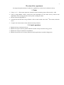
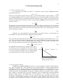
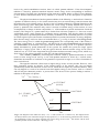
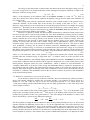
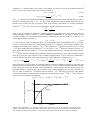
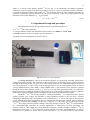
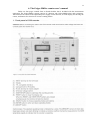
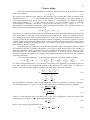
Vilnius University
Faculty of Physics
Department of Solid State Electronics
Laboratory of Applied Nuclear Physics
Experiment No. 5
INVESTIGATION OF RADIOACTIVE EQUILIBRIUM AND
MEASURING THE HALF-LIFE OF
137m
Ba
by Andrius Poškus
(e-mail: [email protected])
2013-06-04
Contents
The aim of the experiment
3
1. Tasks
3
2. Control questions
3
3. Theoretical background
4
3.1. The law of radioactive decay
4
3.2. Nuclear gamma radiation
4
3.3. Radioactive equilibrium in a system of two nuclides
6
4. Experimental setup and procedure
8
5. Analysis of measurement results
10
6. The Geiger-Müller counter user’s manual
11
7. Linear fitting
15
3
The aim of the experiment
Investigate the phenomenon of radioactive equilibrium, test the law of radioactive decay.
1. Tasks
1. Using a 137Cs / 137Ba isotope generator, obtain the eluate containing atoms of the nuclide 137mBa.
2. Using a Geiger-Müller counter, measure the time dependence of the eluate activity and the time
dependence of the isotope generator activity after creating the eluate.
3. Plot the measured dependences.
4. Calculate the half-life and average lifetime of the second excited state of the 137Ba nucleus, and their
errors.
5. Compare the measurement results with theoretical predictions.
2. Control questions
1.
2.
3.
4.
Explain the law of radioactive decay.
Explain the origin of nuclear gamma radiation and the concept of nuclear energy levels.
Explain the concept of radioactive equilibrium.
Explain the operation of an isotope generator and its applications in medical diagnostics.
4
3. Theoretical background
3.1. The law of radioactive decay
It is known that the number of atoms of a radioactive nuclear species (nuclide) decreases
exponentially with time:
N (t ) = N 0 e−λt ,
(3.1)
where N(t) is the number of atoms (nuclei) at time t, N0 is the initial number of nuclei (at time t = 0), and
λ is the decay constant. The decay constant determines the rate at which the number of radioactive nuclei
decreases: the larger the decay constant, the faster the radioactive decay. Another way to specify the rate
of decrease of the number of radioactive nuclei is by specifying the half-life, which is related to the decay
constant as follows:
ln2
T1/ 2 =
.
(3.2)
λ
The half-life is the time needed for the number of radioactive nuclei to decrease by half. Yet another
quantity that can be used to describe the decay rate is the average lifetime of the nuclide. It is equal to the
average time that has to pass until a given nucleus undergoes the decay. The average lifetime (τ) is the
inverse of the decay constant:
1
τ= .
(3.3)
λ
Equation (3.1) is the formulation of the so-called “law of radioactive decay”. An equivalent way
to formulate it is to take time derivatives of both sides of the equation:
dN
= −λ N .
(3.4)
dt
This is the first-order differential equation whose solution corresponding to initial condition N(0) = N0 is
(3.1). The left side of this equation is opposite to the rate of decay (the number of atoms decaying per unit
time). The rate of decay is the so-called activity of the sample. Since N decreases with time, from
Eq. (3.4) it follows that activity also decreases exponentially with time:
dN
Φ (t ) ≡ −
= λ N (t ) = λ N 0 e − λ t .
(3.5)
dt
The logarithm of this function is a straight line (see Fig. 1).
lnΦ
Its intercept gives the logarithm of the initial activity
−λt
ln(λN0e ) = ln(λN0) − λt
ln(λ N0), and its slope is the opposite of the decay constant ln(λN0)
(−λ). Thus, analysis of the decay curve is a simple method
of determining the decay constant λ and the half-life T1/2.
0
t
Fig. 1. Time dependence of the
natural logarithm of activity
3.2. Nuclear gamma radiation
Gamma rays or gamma radiation (denoted as γ) are electromagnetic radiation of high frequency
(very short wavelength). As a rule of thumb, the term “gamma radiation” is usually applied when the
wavelength is much less than the distance between atoms of solid matter (i. e. much less than 10−8 cm).
From the point of view of quantum mechanics, electromagnetic radiation can be interpreted as a flux of
elementary particles called the photons. The photons of gamma radiation are frequently called “γ quanta”
(here, “quanta” is the plural form of the noun “quantum”). The wavelength range of gamma radiation
partially overlaps with the wavelength range of X-ray radiation (e. g., see Table 1 in Section 3 of
descriptions of Experiments No. 1 and No. 2). This is because in practice the distinction between those
two types of electromagnetic radiation is based mainly on their physical origin and not on their
wavelength. If the electromagnetic radiation is caused by quantum transitions between nuclear energy
5
levels or by particle annihilation reactions, then it is called “gamma radiation”. If the electromagnetic
radiation is caused by quantum transitions between atomic energy levels corresponding to innermost
electron shells of an atom or by deceleration of fast electrons in matter, then it is called “X-ray radiation”
(a more detailed information about X-ray radiation is in Section 4 of the description of Experiment
No. 1).
The physical mechanism of nuclear gamma radiation is the following. A nucleus that is formed as
a product of radioactive decay or of a nuclear reaction may be in an excited energy state (this means that
the nucleus has excess internal energy). In such a case it eventually undergoes a quantum transition to the
main (lowest-energy) state. During this transition, the excess energy is usually emitted in the form of a
photon (γ quantum). This transition only causes a decrease of internal energy of the nucleus; it is not
accompanied by a change of nuclear composition (i. e. of the numbers of protons and neutrons inside the
nucleus). The energies of γ quanta emitter by a nucleus form a discrete sequence (i. e. there are several
well-defined energy values instead of a continuous energy spectrum). This discrete nature of nuclear
energy spectrum is a quantum phenomenon caused by localization of the constituent particles of a system
in a small region of space (the nuclear constituent particles are protons and neutrons). In energy diagrams,
those discrete energy values are shown as horizontal lines (e. g. see Fig. 2) and are called energy levels.
The lowest energy level is called the ground energy level. The corresponding state of the nucleus is
called the “ground state” or “principal state”. All other levels are called excited levels and correspond to
the excited states of the nucleus. The typical interval between energy levels of a quantum system is
mainly determined by spatial dimensions of the systems: the smaller the system, the larger typical
differences of energy levels. That is why the typical intervals between nuclear energy levels (100 to
1000 keV) are 102 to 106 times larger than the typical intervals between atomic energy levels, which vary
from a few eV (for valence electrons) to a few keV (for inner-shell electrons).
Thus, nuclear γ radiation is electromagnetic radiation caused by quantum transitions of excited
nuclei to lower energy levels (in Fig. 2, one such quantum transition is shown by a vertical arrow).
Sometimes the transition of a nucleus to the ground level proceeds in stages, as a series of transitions to
lower excited levels.
The opposite transitions (from lower to higher energy levels) are also possible. However, since
those transitions involve an increase of the internal energy of the nucleus, they can not happen
spontaneously (unlike the transitions to lower levels, which do not require any external influence). They
can only happen as a result of an external event that supplies energy to the nucleus, such as a collision
with an energetic subatomic particle. A process that is the inverse of the process leading to nuclear γ
radiation is absorption of a photon.
7/2+
137
y. )
Cs (30.04 m.
β- 0.512 MeV
(94.6%)
11/2-
137m
Ba (2.6 min)
β- 1.174 MeV
(5.4%)
1/2+
0.6617 MeV
0.2835 MeV
3/2+
137
0
Ba
137
137
Fig. 2. The diagram of the radioactive decay Cs → Ba (cesium-137 → barium-137). The diagram shows
the decay half-lives, the maximum energies of the β particles (i. e. of the electrons emitted from the nucleus),
the probabilities of the two β-decay channels, the lowest energy levels of the 137Ba nucleus and the most
probable quantum transition between 137Ba energy levels.
6
The energy of the photon that is emitted when the nucleus drops from the higher energy level E1
to a lower energy level E2 (or absorbed when the nucleus undergoes the opposite transition) is equal to the
difference of the two energy levels:
hν = E1 − E 2 ,
(3.6)
−34
where ν is the frequency of the radiation, and h is the Planck constant: h = 6.626 ⋅ 10 J⋅s. Thus, in
order for a nucleus to be able to absorb a photon, the photon’s energy must be equal to the difference of
two energy levels.
The average time until the spontaneous transition of an excited nucleus to the ground level is
called the “lifetime” of the excited state of the nucleus. If is usually of the order of (10−14 − 10−6) s.
However, some nuclei stay in an excited state for a much longer period of time. For example, the average
lifetime of a 137Ba nucleus in the second excited level (which is formed as a result of the β decay of 137Cs)
is 221 s. Such unusually long-lived excited states are called the metastable states and denoted by a letter
“m” after the nuclear mass number (for example, “137mBa”).
Emission of a photon is usually the most probable mechanism by which an excited nucleus loses
the excess energy, but it is not the only possible one. Two other mechanisms of energy loss are possible,
too: internal conversion and internal pair creation. Internal conversion is a process whereby an excited
nucleus transfers the excess energy directly to an electron of the atom to which is belongs. Since the
energy transferred to the electron is much larger than the binding energy of an atomic electron, the
electron is ejected from the atom with a kinetic energy that is approximately equal to the nuclear
excitation energy. In the case of the previously-mentioned second excited level of 137Ba, there is about
10 % probability of energy loss by means of internal conversion. Internal pair creation is process
whereby the excitation energy of a nucleus is transformed into total relativistic energy of two particles
that did not exist prior the nuclear transition to the ground state. Those two particles are the electron and
the positron (the positron is the electron’s antiparticle). From the definition of the total relativistic energy
(3.7)
E = mc2,
where m is the relativistic mass of the particles, it follows that internal pair creation only becomes
possible when the excitation energy is larger than 2m0c2 = 1.02 MeV, where m0 is the rest mass of the
electron.
Gamma radiation is also emitted during some annihilation reactions. Annihilation is process that
occurs when a subatomic particle collides with its respective antiparticle. As a result of this process, both
initial particles disappear (hence the term “annihilation”, which is defined as "total destruction" or
"complete obliteration" of an object). Since energy and momentum must be conserved, the particles are
not actually made into nothing, but rather into new particles. Some or all of the new particles may be
photons. Since their energies are usually of the order of hundreds of keV or more, those photons are also
classified as γ radiation. The annihilation reaction that is easiest to achieve in practice is the annihilation
of an electron and a positron.
3.3. Radioactive equilibrium in a system of two nuclides
Let us discuss a mixture of two radioactive nuclides, where one nuclide (the “primary nuclide”)
decays into another radioactive nuclide (the “secondary nuclide”). An example of such a system is a
mixture of 137Cs and 137mBa (see Fig. 2). In this case, the primary nuclide is 137Cs, which decays into
137m
Ba (the secondary nuclide), which then decays into the stable ground state of 137Ba. This decay chain
can be written as follows:
λ1
λ2
N 1 ⎯⎯→
N 2 ⎯⎯→
N3,
(3.8)
137
where N1 is the number of the nuclei of the primary nuclide (in this case, Cs), N2 is the number of the
nuclei of the secondary nuclide (in this case, 137mBa), and N3 is the number of the nuclei of the secondary
nuclide’s decay product (in this case, ground state of 137Ba). The change of the numbers N1 and N2 with
time is described by a system of two differential equations:
dN 2
dN1
= −λ1 N1 and
= λ1 N 1 − λ 2 N 2 .
dt
dt
(3.9)
The first equation is simply the law of radioactive decay for the primary nuclide (see Eq. 3.4). The second
equation includes an additional positive term (λ1 N1) on the right-hand side. This term reflects the fact
that, in addition to the radioactive decay of the secondary nuclide (this process decreases N2), there is
another process that influences N2: production of the secondary nuclide due to radioactive decay of the
primary nuclide (this process increases N2, therefore the corresponding term on the right-hand side of the
second equation is positive). The solution of the system of differential equations (3.9) depends on initial
7
conditions, i. e. on initial values of N1 and N2. For example, if values of N1 and N2 at the initial moment of
time (t = 0) are N1(0) and 0, respectively, then the solution is
−λ t
N1 (t ) = N1 (0)e 1 ,
(3.10a)
λ1
−λ t
−λ t
N 2 (t ) = N1 (0)
e 1 −e 2 .
(3.10b)
λ2 − λ1
If (λ2 > λ1), then the second exponential function in parentheses decreases faster than the first one. After a
time that is much larger than 1 / (λ2 − λ1), the second term becomes much smaller than the first one,
therefore the second term may be ignored. Then N2(t) becomes proportional to a single exponential
−λ t
function e 1 , as is N1(t). Thus, the ratio of quantities of both nuclides becomes constant:
(
)
N2
λ1
.
=
N 1 λ 2 − λ1
(3.11)
Such a state of a mixture of radioactive nuclides belonging to a single decay chain, when the ratios of
nuclide quantities remain constant, is called radioactive equilibrium. If the decay constant of the
secondary nuclide (λ2) is much larger than the decay constant of the primary nuclide (λ1), then Eq. 3.11
can be approximately re-written as
λ 2 N 2 ≈ λ1 N 1 .
(3.12)
I. e., the decay rate of the secondary nuclide (λ2N2) is practically equal to its production rate λ1N1, which,
in turn, is equal to the decay rate of the primary nuclide (i. e. to activity of the primary nuclide). This
condition is satisfied in the system of 137Cs/137mBa, because the 137mBa decay half-life (153 s) is about
6 ⋅ 106 times smaller than decay half-life of 137Cs (30 years), i. e. the 137mBa decay constant is about 6 ⋅ 106
times larger than decay constant of 137Cs (see the relation between decay constant and decay half-life,
Eq. 3.2).
If the radioactive equilibrium is artificially disturbed by removing a part of the secondary nuclide
from the system, then the amount of the secondary nuclide begins to grow until the equilibrium is
restored. This increase of N2 can be derived from Eq. 3.10b, taking into account that λ2 >> λ1:
λ1
λ
−λ t
−λ t
− λ t λ1 ⎛
− ( λ − λ )t
−λ t
N 2 (t ) = N1 (0)
e 1 − e 2 = N1 (0)e 1
⎜1 − e 2 1 ⎞⎟ ≈ N1 1 1 − e 2 . (3.13)
λ2 − λ1
)
(
⎠
λ2 − λ1 ⎝
λ2
(
)
Since in practice it is much easier to count the number of particles emitted by radioactive nuclei per unit
time than to measure the number of those nuclei directly, is it more useful to formulate the latter result in
terms of activities of the two nuclides. This transformation is done using the fact that current activity of a
particular nuclide is equal to the current number of its nuclei multiplied by its decay constant (see
Eq. 3.5). Thus, a removal of 137mBa nuclei from the mixture 137Cs/137mBa causes a decrease of the number
γ photons emitted per second (because all photons are emitted due to decay 137mBa → 137Ba), but then it
begins to grow according to the equation
−λ t
−λ t
A2 (t ) ≡ λ 2 N 2 (t ) = λ1 N 1 1 − e 2 ≡ A1 1 − e 2 ,
(3.14)
Number
γ photons
per unit
time
γ kvantųof
skaičius
per laiko
vienetą
(
) (
)
A1
A2(t)
-1
A1(1-e )
1/λ2
time t t
Laikas
Fig. 3. Time dependence of γ radiation intensity after removing a part of the secondary nuclide from the
mixture consisting of a long-lived primary nuclide and a short-lived secondary nuclide (it is assumed that only
the secondary nuclide emits γ photons). A1 is activity of the primary nuclide, which is practically constant
8
where A1 is activity of the primary nuclide 137Cs (see Fig. 3). At equilibrium, the number of photons
emitted per second is equal to the primary nuclide’s activity A1, which is practically constant. Therefore,
A1 should be measured either before removing 137mBa from the mixture or after complete restoration of
radioactive equilibrium. Having measured A1, the decay constant of 137mBa can be determined from the
measured values of the difference A1 − A2(t), because
−λ t
A1 − A2 (t ) = A1 e 2 .
(3.15)
4. Experimental setup and procedure
The equipment used for this experiment consists of the following devices:
137
1. A Cs/137mBa isotope generator.
2. A Geiger-Müller counter (the dead time of that counter is τd ≈ 0.001 s ≈ 1.7·10-5 min).
3. Isotrak ratemeter (its user’s manual is given in Section 6).
A general view of the equipment is shown in Fig. 4.
Fig. 4. General view of the equipment
An isotope generator is a device for chemical separation of a short-lived secondary nuclide from
a long-lived primary nuclide. The separation of the two nuclides is achieved by passing a special solution
through the generator. In the case of a 137Cs/137mBa isotope generator, the mentioned solution consists of
0.9 % NaCl and 4·10–6 % HCl. In Fig. 4, the isotope generator is seen as small cylinder placed in front of
a Geiger-Müller detector (also called a “Geiger-Müller tube”). The electrons (“beta particles”) emitted
during the decay of the primary nuclide (e. g. see Fig. 2) are much less penetrating than γ rays, therefore
those electrons are completely absorbed in the plastic cover of the isotope generator. Only the γ photons
produced by quantum transitions of the secondary nuclide escape from the isotope generator.
Inside the 137Cs/137mBa isotope generator, a material containing 137Cs is deposited onto a special
organic polymer layer (called “the substrate”), which facilitates transfer of Ba ions into a solution
containing a sufficiently high concentration of Na (sodium) ions. I. e., the Na ions take up the positions
on the substrate previously occupied by Ba ions, whereas Ba ions enter the solution. Such a chemical
reaction is called the ion exchange. In this way, a majority of 137Ba atoms can be removed from the
137
Cs/137mBa isotope generator with the solution. The same principle is used in 99Mo/99mTc (molybdenum99 / technetium-99m) generators for medical applications of radioactivity. In those generators, the
primary nuclide is 99Mo, which has a decay half-life of 66 hours, and the secondary nuclide is 99mTc (a
metastable excited state of 99Tc), which has a decay half-life of 6 hours. Molibdenum-99 (as well as
cesium-137) is a product of nuclear fission (which takes place, e. g., in nuclear reactors). The solution
9
containing short-lived secondary nuclide obtained from such a generator is injected into blood of a
patient, and then its γ radiation is recorded. In this way, the nuclide’s distribution inside the patient’s body
can be determined, which, in turn, may provide information about various medical conditions (such as
tumors). One of the reasons why the isotope generators are useful in medicine is that the long half-life of
the primary nuclide makes transportation over long distances easy, whereas the short half-life of the
secondary nuclide makes the intravenous injections less dangerous (the activity of the secondary nuclide
decays rapidly and therefore does not do any significant damage to the biological tissues).
The measurement procedure
1. Switch on and prepare the ratemeter with the Geiger-Müller tube. In this experiment, the automatic
counting mode must be used, with the gate time of 60 s (see the ratemeter’s user’s manual in Section
6). Since the measurement results must be stored in the ratemeter’s memory, which can contain no
more than 50 counts, it is important to erase any data stored in the memory before starting the
experiment (this is done by pressing the “Start/Stop” and “Reset” buttons simultaneously). The state
of the ratemeter after preparing it for the measurements must be such that it is enough to press the
“Start/Stop” button to start a sequence of counts.
Warning! Wear disposable gloves when manipulating the radioactive eluate. After the experiment, drop
the gloves into a bin.
2. Extract the nuclide 137mBa is from the 137Cs/137mBa generator. This must be done in the following way:
a) attach a plastic tube to a syringe, then take approximately 2 ml of the eluting solution into the
tube, then detach the tube from the syringe,
b) unscrew the protective cap of the isotope generator (it is at the side of the generator where the
label “Isotope generator” is) and screw the syringe with the solution in its place,
c) take off the protective cap that is at the opposite side of the isotope generator (there is no screw
thread there, so that it is enough to pull the cap) and place the isotope generator together with the
syringe above an empty flask,
d) push the plunger of the syringe until all solution passes through the generator and is collected in
the flask (this step should take about 10 or 20 s),
e) put both protective caps back on the isotope generator and place it in front of the Geiger-Müller
tube as shown in Fig. 4,
f) place the flask with the radioactive eluate at a distance of 2 m or more from the counter (so that it
does not influence the measurement results),
g) start the sequence of counts by pressing the button “Start/Stop” on the ratemeter.
3. Do 30 counts, each count 1 minute-long. The results must be written down in the form of a table.
4. Empty the flask with the eluate (which is no longer radioactive at this point) into a sink. Repeat
Step 2, but now place the flask with the radioactive eluate in front of the detector, whereas the isotope
generator must be placed at a distance of at least 2 m from the detector.
5. Do 10 counts, each count 1 minute-long. Again, write down the results in the form of a table.
6. Place the flask at a distance of at least 2 m from the detector. Do 10 counts of the background.
7. Empty the flask with the eluate into a sink, rinse the flask with water.
8. Show the measurement results (in table format) to the laboratory supervisor for signing.
10
5. Analysis of measurement results
1. Correct the counts taking into account the so-called “dead time” of the detector (it is the time that has
to pass after detecting a particle until the next particle can be detected). I. e., if N' is the measured
number of particles in 1 min and τd is the dead time (in minutes), then the true number of particles in
1 min is
N′
N=
.
1 − N ′τ d
2. Calculate the average background.
3. Subtract the average background from the corrected results corresponding to the radioactive eluate.
4. Plot the time dependence of the natural logarithm of the numbers obtained in previous step (it should
be plotted as a scatter graph). Then calculate the decay constant λ of 137mBa and its error, using the
method of linear fitting, and plot the fitting line in the same graph. Calculate the average lifetime and
decay half-time of 137mBa and their errors (use Eq. 3.2 and 3.3). Linear fitting can be done using
various computer programs, or it can be done “by hand”, using a calculator (see Section 7).
5. Calculate the average value of the last 10 counts of the 137Cs/137mBa isotope generator (i. e.
measurements No. 21 – 30). At this stage of the measurements, intensity of γ radiation of the isotope
generator has been completely restored, therefore this average is equal to the initial γ radiation
intensity of the generator (prior to nuclide separation).
6. Subtract each of the first ten counts of the 137Cs/137mBa isotope generator from the initial intensity
obtained in previous step. Then plot and analyze the resulting ten numbers in the same way as in Step
4 of this section.
7. Compare the calculation results of Step 4 and Step 6 with each other and with the exact value of
137m
Ba half-life (153 s).
8. Discuss the general shape of both measured time dependences.
11
6. The Geiger-Müller counter user’s manual
Below are four pages scanned from an Isotrak booklet that is included with the measurement
equipment. The Geiger-Müller counter consists of a detector (the Geiger-Müller tube) and a ratemeter.
The ratemeter has a built-in the adjustable power supply for the detector, an LCD display for showing the
counts, and buttons for selection of various counting modes.
12
13
14
15
7. Linear fitting
The aim of linear fitting is determination of the least squares estimates of the coefficients A and B
of the linear equation
y = A + B ⋅ x,
(7.1)
The essence of the method of least squares is the following. Let us assume that a data set consists of the
argument values x1, x2, …, xn-1, xn and corresponding values of the function y(x). A typical example is a
set of measurement data. In such a case, n is the number of measurements. The measured function
values will be denoted y1, y2, …, yn. The “theoretical” value of y at a given argument value xk is a function
of the unknown coefficients A and B (see (7.1)), hence we can write y(xk) = y(xk; A,B) (k = 1, 2, …, n).
The problem of estimating the coefficients A and B is formulated as follows. The most likely values of A
and B are the values that minimize the expression
n
F ( A, B ) ≡ ∑ ⎡⎣ y ( xk ; A, B ) − yk ⎤⎦ .
2
(7.2)
k =1
Expression (7.2) is the sum of squared deviations of theoretical values from the measured ones (hence the
term “least squares”). That sum is also called “the sum of squared errors” (SSE). This expression always
has a minimum at certain values of A and B. However, even if the form of the theoretical function y(x)
correctly reflects the true relationship between the measured quantities y and x, those “optimal” values of
A and B, which correspond to the minimum SSE, do not necessarily coincide with the true values of A and
B (for example, because of measurement errors). The method of least squares only allows estimation of
the most likely values of A and B.
Everything that was stated above about the method of least squares also applies to the case when
the theoretical function is nonlinear. Regardless of the form of that function and of the number of
unknown coefficients, a SSE expression of the type (7.2) must be minimized. However, when y(x) is the
linear function (7.1), this problem can be solved analytically (i.e., A and B can be expressed using
elementary functions), but when y(x) is nonlinear, this problem can only be solved numerically (applying
an iterative procedure).
If y(x) is the linear function (7.1), then the SSE expression (7.2) can be written as follows:
n
n
n
n
n
n
k =1
k =1
k =1
k =1
k =1
k =1
F ( A, B ) ≡ ∑ ( A + Bxk − yk ) 2 = nA2 + B 2 ∑ xk2 + ∑ yk2 + 2 AB ∑ xk − 2 A∑ yk − 2 B ∑ xk yk .
(7.3)
It is known that partial derivatives of a function with respect to all arguments at a minimum point are
zero. After equating to zero the partial derivatives of the expression (7.3) with respect to A and B, we
obtain a system of two linear algebraic equations with unknowns A and B. Its solution is
n
⎛ n ⎞⎛ n
⎞
n∑ xk yk − ⎜ ∑ xk ⎟⎜ ∑ yk ⎟
⎝ k =1 ⎠⎝ k =1 ⎠ ,
B = k =1
2
n
⎛ n ⎞
2
n∑ xk − ⎜ ∑ xk ⎟
k =1
⎝ k =1 ⎠
n
1
B n
A = ∑ yk − ∑ xk .
n k =1
n k =1
(7.4)
(7.5)
The B coefficient is called the “slope” of the straight line, and the A coefficient is called the “intercept”.
The standard deviations (or “errors”) of those two coefficients are calculated according to formulas
ΔA =
Fmin ⎛
x2 ⎞
⎟,
⎜1 +
n(n − 2) ⎝ Dx ⎠
(7.6)
Fmin
,
n(n − 2) Dx
(7.7)
ΔB =
where Fmin is the minimum value of the SSE (7.3), i.e., the value of SSE when A and B are equal to their
optimal values (7.4) and (7.5), x is the average argument value:
x=
1 n
∑ xk ,
n k =1
(7.8)
and Dx is the variance of the argument values:
Dx =
1 n
1 n
( xk − x ) 2 = ∑ xk2 − x 2 .
∑
n k =1
n k =1
(7.9)