1
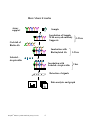
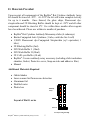
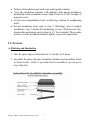
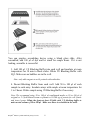
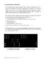
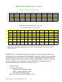
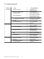
RayBio® Rat Cytokine Antibody Array
G Series
Patent Pending Technology
User Manual (Revised Aug. 25, 2008)
RayBio® Rat Cytokine Antibody Array G Series 2 (Cat# AAR-CYT-G1-4)
RayBio® Rat Cytokine Antibody Array G Series 2 (Cat# AAR-CYT-G2-4)
RayBio® Custom Rat Cytokine Antibody Array G Series 1 (Cat# AAR-CUST-G)
RayBio® Rat Cytokine Antibody Array G Series 1 Service (Cat# AAR-SER-G)
Please read the manual carefully before you start your experiment
RayBiotech, Inc.
We Provide You With Excellent
Protein Array Systems And Service
Tel: (Toll Free) 1-888-494-8555 or 770-729-2992; Fax: 1-888-547-0580;
Website: www.raybiotech.com Email: [email protected]
RayBiotech, Inc.
RayBio® Rat Cytokine Antibody Array G Series Protocol
TABLE OF CONTENTS
I.
Introduction……..……………………………...
2
How It Works………………………..…………
4
Materials Provided……………………………..
5
Additional Materials Required…………………
5
III. Overview and General Considerations…………
6
A. Preparation of Samples………………………
6
B. Handling Glass Chips ……………………….
6
C. Incubation……………………………………
7
IV. Protocol…………………………………………
7
A. Blocking and Incubation…………………….
7
B. Detection…………………………………….
10
Interpretation of Results………………………..
11
VI. Troubleshooting Guide…………………………
15
VII. Reference List………………………………….
16
II.
V.
Cytokine Antibody Arrays are RayBiotech patent-pending technology.
RayBio® is the trademark of RayBiotech, Inc.
RayBio® Mouse Cytokine Antibody Array G series
1
I. Introduction
All cell functions, including cell proliferation, cell death and differentiation,
as well as maintenance of health status and development of disease, are
controlled by many genes and signaling pathways. New techniques such as
cDNA microarrays have enabled us to analyze the global gene expression 1-3.
However, almost all cell functions are executed by proteins, which cannot be
studied by DNA and RNA alone. Experimental analysis clearly shows a
disparity between the relative expression levels of mRNA and their
corresponding proteins 4. Therefore, it is critical to analyze the protein
profile. Currently, two-dimensional polyacrylamide SDS page coupled with
mass spectrometry is the mainstream approach to analyzing multiple protein
expression levels 5,6. However, the requirement of sophisticated devices and
the lack of quantitative measurements greatly limit its broad application.
Thus, no simple, cost effective, and rapid method of analysis of multiple
protein expression levels has been available to researchers until now.
Our RayBio® Rat Cytokine Antibody Array is the first commercially
available rat cytokine antibody array system 7-11. By using the RayBiotech
system, scientists can rapidly and accurately identify the expression profiles
of multiple cytokines in several hours inexpensively.
The RayBiotech kit (G series) is a glass chip format. The kit provides a
highly sensitive approach to simultaneously detect multiple cytokine
expression levels from cell culture supernatant, patient’s serum, tissue lysate
and other sources. The arrays are manufactured using non-contact arrayer.
The experimental procedure is simple and can be performed in any
laboratory. The signals from G series arrays are detected using a laser
scanner.
Besides the products listed in this manual, RayBiotech also provides
RayBio® Mouse Cytokine Antibody Array G series 1000 for simultaneous
detection of 96 mouse cytokines in single experiment. RayBiotech also
provides RayBio® Human Cytokine Antibody Array G series 2000 which is
the only product available in the market that can detect 174 human cytokines
in single experiment. We also provide quantitative antibody arrays which can
RayBio® Mouse Cytokine Antibody Array G series
2
quantitatively measure multiple protein expression in antibody array
technology.
Pathway-specific array systems allow investigators to focus on the specific
problem and are becoming an increasingly powerful tool in cDNA
microarray system. RayBiotech’s first protein array system, known as
RayBio® Rat Cytokine Antibody Array, is particularly useful compared with
the Rat cytokine cDNA microarray system. Besides the ability to detect
protein expression, RayBiotech’s system is a more accurate reflection of
active cytokine levels because it only detects secreted cytokines, and no
amplification step is needed. Furthermore, it is much simpler, faster,
environmentally friendlier, and more sensitive.
Simultaneous detection of multiple cytokines undoubtedly provides a
powerful tool to study cytokines. Cytokines play an important role in innate
immunity, apoptosis, angiogenesis, cell growth and differentiation 12.
Cytokines are involved in most disease processes, including cancer and
cardiac diseases. The interaction between cytokines and the cellular immune
system is a dynamic process. The interactions of positive and negative
stimuli, and positive as well as negative regulatory loops are complex and
often involve multiple cytokines.
RayBio® Mouse Cytokine Antibody Array G series
3
Here’s how it works
Array
support
Sample
Incubation of Sample
With arrayed antibody
Supports
Cocktail of
Biotin-Ab
Incubation with
Biotinylated Ab
1-2 hrs
1-2 hrs
Labeled –
streptavidin
Incubation with
Labeled- streptavidin
2 hrs
Detection of signals
Data analysis and graph
RayBio® Mouse Cytokine Antibody Array G series
4
II. Materials Provided
Upon receipt, all components of the RayBio® Rat Cytokine Antibody Array
kit should be stored at -200C. At -200C the kit will retain complete activity
for up to 6 months. Once thawed, the glass chips, Fluorescent dyestreptavidin and 2X Blocking Buffer should be kept at –200C and all other
component should be stored at 40C. Use within three months after reagents
have been thawed. Please use within six months of purchase.
• RayBio® Rat Cytokine Antibody Microarray slides (4 subarrays)
• Biotin-Conjugated Anti-Cytokines (1 tube, each tube for 4 well)
• 1,500X Fluorescent dye-Conjugated Streptavidin (cy3 equivalent, 1
tube)
• 2X Blocking Buffer (5ml)
• 20X Wash Buffer I (30ml)
• 20X Wash Buffer II (30ml)
• 2X Cell Lysis Buffer (10 ml)
• RayBio® G series antibody array accessory (including slide incubation
chamber, Gasket, Protective cover, Snap-on sides and adhesive film)
• Manual
Additional Materials Required
•
•
•
•
•
Orbital shaker
Laser scanner for fluorescence detection
Aluminum foil
Distilled water
Plastic box
Antibody
Array
Blank
Layout of Rat G series
Barcode
4 arrays in one glass chip
®
RayBio Mouse Cytokine Antibody Array G series
5
III. Overview and General Considerations
A. Preparation of Samples
• Use serum-free conditioned media if possible.
• If serum-containing conditioned media is required, use serum as control
since many types of sera contain cytokines.
• For cell lysates and tissue lysates, we recommend using 1X Cell Lysis
Buffer to extract proteins from cell or tissue (e.g. using homogenizer).
After extraction, spin the sample and save supernatant for experiment.
Determine protein concentration. Dilute 2X Cell Lysis Buffer with H2O
(we recommend adding proteinase inhibitors to Cell Lysis Buffer
before use). Prepare relative concentrated lysate since we recommend
diluting lysate at least 10 folds with Blocking Buffer for array assay.
• We recommend using
50 to 100 µl of cell culture supernatant
or
50 to 100 µl of original or 5-fold diluted serum or plasma
or
20-200 µg of protein for cell lysates and tissue lysates.
If you experience high background, you may further dilute your sample.
B. Handling glass chips
• The microarray slides are sensitive, do not touch the surface. Hold the
slides by the edges only.
• Handle all buffers and slides with latex free gloves.
• Avoid breaking glass slide.
• Handle glass chip in clean environment
C. Incubation
• Completely cover array area with sample or buffer during incubation,
and cover the incubation chamber with adhesive film or plastic sheet
protector to avoid drying.
• Avoid foaming during incubation steps.
RayBio® Mouse Cytokine Antibody Array G series
6
• Perform all incubation and wash steps under gentle rotation.
• Cover the incubation chamber with adhesive film during incubation,
particularly when incubation is more than 2 hours or 50 µl of sample or
reagent is used.
• Avoid cross-contamination from overflowing solution to neighboring
wells.
• Several incubation steps such as step 3 (blocking), step 4 (sample
incubation), step 9 (biotin-Ab incubation) or step 12(Fluorescent dyestreptavidin incubation) may be done at 40C for overnight. Please make
sure to cover the incubation chamber tightly to prevent evaporation.
IV. Protocol
A. Blocking and Incubation
1. Take the glass chip out from the box. Let air dry for 2 hours.
2. Assemble the glass chip into incubation chamber and incubation frame
as shown below. (Note: if you slide has be assembled, you can go to
step 3 directly).
RayBio® Mouse Cytokine Antibody Array G series
7
You can practice assembling device using a blank glass slide. After
assembled, add 100 µl of dye and let stand for couple hours. If it is not
leaking, assemble is successful.
3. Add 100 µl 1 X Blocking Buffer into each well and incubate at room
temperature for 30 min to block slides. Dilute 2X Blocking Buffer with
H2O. Make sure no bubbles are in the well.
Note: only add reagents to wells printed with antibodies.
4. Decant Blocking Buffer from each well. Add 50 to 100 µl of each
sample to each array. Incubate arrays with sample at room temperature for
1 to 2 hours. Dilute sample using 1X Blocking Buffer if necessary.
Note: We recommend using 50 to 100 µl of conditioned media or 50 to 100 µl of
original or 2-5 fold diluted serum or plasma or 10-200 ug of protein for cell lysates
and tissue lysates. Dilute the lysate at least 10 folds with 1 X blocking buffer to
make a total volume of 50 to 100 µl. Make sure there is no bubble in the wells.
RayBio® Mouse Cytokine Antibody Array G series
8
Note: The amount of sample used depends on the abundance of cytokines. More
samples can be used if signals are too weak. If signals are too strong, the sample can
be diluted further.
Note: Incubation may be done at 40C for overnight.
5. Decant the samples from each well, and wash 5 times with 150 µl of
1X Wash Buffer I at room temperature with gentle shaking. 2 min per
wash. Dilute 20X Wash Buffer I with H2O. Completely remove wash
buffer I in each wash step.
Note: avoid solution flowing into neighboring wells.
6. Put the glass chip with frame into a box with 1X Wash Buffer I (cover
the whole glass slide and frame with Wash Buffer I), and wash at room
temperature with gentle shaking for 20 min.
7. Decant the 1X Wash Buffer I from each well, Put the glass chip with
frame into the box with 1XWash Buffer II (cover the whole glass slide
and frame with Wash Buffer II), and wash 2 times at room temperature
with gentle shaking for 5 min. Remove all of Wash Buffer II in the
well. Dilute 20X Wash Buffer II with H2O.
8. Prepare working solution for biotin-conjugated antibodies. After brief
spinning,
Add 300 µl of 1x blocking buffer to the Biotin-Conjugated Antibody
tube. Mix gently.
Note: the diluted biotin-conjugated antibodies can be stored at 40C for 2-3 days.
9. Add 70 µl of diluted biotin-conjugated antibodies to each
corresponding well. Incubate at room temperature for 2 hours.
Note: incubation may be done at 40C for overnight.
10.
Wash as directed in steps 5 and then wash 3 times with 150 µl of
1X Wash Buffer II at room temperature with shaking. 2 min per wash.
Completely remove wash buffer II in each wash step.
RayBio® Mouse Cytokine Antibody Array G series
9
11.
Add 70 µl of 1,500 fold diluted Fluorescent dye-conjugated
streptavidin (after brief spinning, add 1.5 ml of Blocking Buffer to
Fluorescent dye-conjugated streptavidin tube) to each array. Cover the
incubation chamber with Adhesive film. Cover the plate with
aluminum foil to avoid exposure to light or incubate in dark room.
12. Incubate at room temperature for 1 to 2 hours.
Note: incubation may be done at 40C for overnight.
13. Wash with Wash Buffer I twice as directed in steps 5.
B. Fluorescence Detection
1. Decant excess Wash Buffer from wells.
2. Disassemble the slide out of the incubation frame and chamber.
3. Place the whole slide in a 50 ml centrifuge tube, add enough Wash
Buffer I (about 30 ml) to cover the whole slide and gently shake at
room temperature for 10 minutes. Decant Wash Buffer I. Repeat Wash
Buffer I once. Wash with Wash Buffer II (about 30 ml) with gentle
shake at room temperature for 10 minutes. Or wash using slide
chamber. Rinse the slide with distilled H2O.
4. Remove water droplets by centrifuge at 1,000 rpm for 3 minutes and
then let slide dry completely in air at least 20 minutes (protect from
light). Make sure the slides are absolutely dry before the scanning
procedure.
5. Image the signals using laser scanner such Axon GenePix using cy3
channel.
Note: we recommend scanning slides right after experiment. You also can store the
slide at –200C in dark for several days. If you do not have a laser scanner,RayBioteh
can provide service for you. Just simply send your slide to us and we will take care
of it.
RayBio® Mouse Cytokine Antibody Array G series
10
V. Interpretation of Results:
The following figure shows RayBio® Rat Cytokine Antibody Array G 1
probed with different cell culture supernatant. The images were captured
using laser scanner. The biotin-conjugated protein produces positive
signals, which can be used to identify the orientation and to compare the
relative expression levels among the different wells.
The signal intensities obtained from laser scanner can simply be imported
into our analysis tool. The analysis tool will help you:
•
•
•
•
•
•
•
Locate your signal intensities to antibody array map
Link the protein to website for more detailed information on the particular protein
Protein list sorting
Average signal intensities
Subtract background
Normalize the data from different samples
Obtain protein level comparison charts among different samples
This analysis tool is very simple and affordable, which will not only assist
in compiling and organizing your data, but also reduces your calculations
to a “copy and paste” step.
Conditioned medium
RayBio® Mouse Cytokine Antibody Array G series
Negative Control
11
If you do not use our RayBio® Analysis Tool, you can locate the cytokines
by referring to RayBio® Rat cytokine Antibody Array G series 1.
Normalization and comparison
For biomarker discovery or for analysis of large number of arrays, great
attention must be paid to the normalization. Our antibody array design
includes several controls for normalization and comparison of arrays
performing in different membranes and different experiments (for more
information please read the reference 17).
Positive control. Positive control is biotinylated protein. It can be used to
normalize the streptavidin incubation step. If the positive signals from
different array membranes are similar, positive control is a simple and
effective way for normalization.
Negative control. Negative control is BSA. Normally, it should only give a
background reading.
RayBio® Mouse Cytokine Antibody Array G series
12
RayBio® Rat Antibody Array G - Series 1
Detect 19 cytokines in one experiment.
a
b
c
d
e
f
g
h
i
1
Pos 1
Pos 2
Pos 3
Neg
Neg
CINC-2
CINC-3
CNTF
Fractalkine
2
Pos 1
Pos 2
Pos 3
Neg
Neg
CINC-2
CINC-3
CNTF
Fractalkine
3
GM-CSF
IFN-γ
IL-1α
IL-1β
IL-4
IL-6
IL-10
LIX
Leptin
4
GM-CSF
IFN-γ
IL-1α
IL-1β
IL-4
IL-6
IL-10
LIX
Leptin
5
MCP-1
MIP-3α
β-NGF
TIMP-1
TNF-α
VEGF
Neg
Neg
Pos 1
6
MCP-1
MIP-3α
β-NGF
TIMP-1
TNF-α
VEGF
Neg
Neg
Pos 1
RayBio® Rat Cytokine Antibody Array G series 2
(for simultaneous detection of 34 rat cytokines)
A
B
C
D
E
F
G
H
I
J
K
L
1
POS 1
POS 2
POS 3
NEG
Activin A
Agrin
B7-2/CD86
beta-NGF
CINC-1
CINC-2alpha
CINC-3
CNTF
2
POS 1
POS 2
POS 3
NEG
Activin A
Agrin
B7-2/CD86
beta-NGF
CINC-1
CINC-2alpha
CINC-3
CNTF
3
Fas Ligand
Fractalkine
GM-CSF
ICAM-1
IFN-gamma
IL-1alpha
IL-1beta
IL-1 R6
IL-2
IL-4
IL-6
IL-10
4
Fas Ligand
Fractalkine
GM-CSF
ICAM-1
IFN-gamma
IL-1alpha
IL-1beta
IL-1 R6
IL-2
IL-4
IL-6
IL-10
Thymus
Chemokine-1
Thymus
Chemokine-1
5
IL-13
Leptin
LIX
L-Selectin
MCP-1
MIP-3alpha
MMP-8
PDGF-AA
Prolactin R
RAGE
6
IL-13
Leptin
LIX
L-Selectin
MCP-1
MIP-3alpha
MMP-8
PDGF-AA
Prolactin R
RAGE
7
TNF-alpha
VEGF
NEG
NEG
NEG
NEG
NEG
NEG
NEG
NEG
NEG
POS 1
8
TNF-alpha
VEGF
NEG
NEG
NEG
NEG
NEG
NEG
NEG
NEG
NEG
POS 1
TIMP-1
TIMP-1
Abbreviations: IP-10, Interferon-inducible protein-10; LAP, latency associated peptide (TGF-β1); LIF, leukocyte
inhibitory factor. MMP, Matrix Metalloproteinase; Pos, positive control; Neg, negative control. All other are used
standard abbreviations.
RayBiotech, Inc., the protein array pioneer company, strives to research and develop
new products to meet demands of the biomedical community. RayBio’s patent-pending
technology allows detection of over 180 cytokines, chemokines and other proteins in a
single experiment. Our format is simple, sensitive, reliable and cost effective. Products
include: Cytokine Arrays, Chemokine Arrays, ELISA kits, Phosphotyrosine kits,
Recombinant Proteins, Antibodies, and custom services.
1. Antibody arrays
Cytokine antibody array
Human cytokine antibody arrays
Mouse cytokine antibody arrays
Rat cytokine antibody arrays
Pathway- or disease-focused antibody arrays
RayBio® Mouse Cytokine Antibody Array G series
13
Inflammation antibody array
Angiogensis antibody array
Chemokine antibody array
Growth factor antibody array
MMP antibody array
Atherosclerosis antibody array
Quantibody arrays for quantitative measurement of cytokine and other protein
concentraton
Phosphorylation antibody arrays
Biotin label-based antibody arrays for high density antibody arrays.
Antibody analysis tool, software
2.
3.
4.
5.
6.
7.
8.
ELISA
Cell-based phosphorylation assay
Custom antibody arrays
Antibody
Recombinant protein
Peptide
Protein arrays
RayBiotech also provides excellent custom service:
1. Antibody arrays
2. Protein arrays
3. Peptide synthesis
4. Production of recombinant protein and antibody
5. Peptide arrays
6. Phosphorylation arrays
7. ELISA
Just simply send your samples and we will do the assay for you.
Technology transfer program
Have you developed technologies or reagents interested to the scientific and research
community? RayBiotech can help you commercialize your technologies, reagents and
dream.
RayBio® Mouse Cytokine Antibody Array G series
14
VI. Troubleshooting guide
Problem
Weak signal
High background
Cause
Recommendation
Inadequate detection
Check laser power
and PMT parameters
Inadequate reagent volumes
or improper dilution
Check pipetters and
ensure correct preparation
Short incubation times
Ensure sufficient incubation
Time and change sample
incubation step to overnight
Too low protein concentration
in sample
Don’t make too low dilution
Or concentrate sample
Improper storage of kit
Store kit at suggested
temperature
Make sure correct
amount of antibodies
Excess of biotinylated antibodies
Excess of streptavidin
Make sure correct
amount of streptavidin
Inadequate detection
Check laser power
And PMT parameters
dust
Work in clean environment
Increase wash time and
use more wash buffer
Insufficient wash
Uneven signal
Bubbles formed during
incubation
Avoid bubble formation
during incubation
Arrays are not completed
Covered by reagent
Completely cover arrays
with solution
RayBio® Mouse Cytokine Antibody Array G series
15
Reference List
1. LPS induces the interaction of a transcription factor, LPS-induced TNF-a factor,
and STAT6(B) with effects on multiple cytokines. Xiaoren Tang, Deborah Levy
Marciano, Susan E. Leeman and Salomon Amar. PNAS. April 5, 2005 vol. 102
no. 14 5132-5137
2. HIV-1-mediated apoptosis of neuronal cells: Proximal molecular mechanisms of
HIV-1-induced encephalopathy. Yan Xu, Joseph Kulkoshy, Roger j. Pomerantz.
PNAS. 2004 May 4, 2004 Vol. 101 No. 18.
3. Synergistic increases in intracellular Ca(2+), and the release of MCP-1,
RANTES, and IL-6 by astrocytes treated with opiates and HIV-1 Tat. GLIA.
2005 Apr 15;50(2):91-106.
4. Bone Marrow Stroma Influences Transforming Growth Factor-β Production in
Breast Cancer Cells to Regulate c-myc Activation of the Preprotachykinin-I Gene
in Breast Cancer Cells. Hyun S. Oh, Anabella Moharita… Pranela Rameshwar.
CANCER RESEARCH. 64, 6327-6336, September 1, 2004
5. Recombinant Herpes Simplex Virus Type 1 (HSV-1) Codelivering Interleukin12p35 as a Molecular Adjuvant Enhances the Protective Immune Response
against Ocular HSV-1 Challenge JOURNAL OF VIROLOGY. Mar. 2005 Vol.
79, No. 6.
6. Dysregulated Inflammatory Response to Candida albicans in a C5-Deficient
Mouse Strain. Alaka Mullick, Miria Elias, Serge Picard, Philippe Gros.
Infection and Immunity, Oct. 2004, p. 5868-5876, DOI:
10.1128/IAI.72.10.5868-5876.2004
7. Leukotriene B4 Strongly Increases Monocyte Chemoattractant Protein-1 in
Human Monocytes Li Huang, Annie Zhao, Frederick Wong, Julia M. Ayala,
Jisong Cui Arteriosclerosis, Thrombosis, and Vascular Biology. 2004;24:17831788
8. Human CD1d-unrestricted NKT cells release chemokines upon Fas engagement.
Martin Giroux and François Denis. Yan Xu, Joseph Kulkoshy, Roger j.
Pomerantz. Blood. prepublished online September 2, 2004; DOI 10.1182/blood2004-04-1537
RayBio® Mouse Cytokine Antibody Array G series
16
9. Monitoring the response of orthotopic bladder tumors to granulocyte macrophage
colony-stimulating factor therapy using the prostate-specific antigen gene as a
reporter. Wu Q, Esuvaranathan K, Mahendran R. Clinical Cancer Research.
2004 Oct 15; 10(20):6977-84.
10. Neuroglial activation and neuroinflammation in the brain of patients with autism
(p NA). Diana L. Vargas, Caterina Nascimbene, Chitra Krishnan, Andrew W.
Zimmerman, Carlos A. Pardo. Annals of Neurology. 2005 Jan 1; DOI:
10.1002/ana.20315
11. Cytokine profiling of macrophages exposed to Porphyromonas gingivalis, its
LPS or its FimA. Qingde Zhou, Tesfahun Desta, Dana T. Graves and Salomon
Amar. Infection and Immunity (IAI). 2005 Feb;73(2):935-43.
12. Veto-like activity of mesenchymal stem cells: functional discrimination
between cellular responses to alloantigens and recall antigens. Rameshwar P.
Journal of Immunology. 2003 Oct 1;171(7):3426-34.
13. Cytokine responses elicited in endothelial cells after treatment with a specific
toxin. Jaya Pandey. BioCompare Product Review. May 13, 2004
14. Proteomic Characterization of the Interstitial Fluid Perfusing the Breast Tumor
Microenvironment. A Novel Resource for Biomarker and Therapeutic Target
Discovery. Julio E. Celis, Pavel Gromov, Teresa Cabezón, José M. A. Moreira,
Noona Ambartsumian, Kerstin Sandelin, Fritz Rank, and Irina Gromova.
Molecular Cellular Proteomics. April 2004; 11(3):328-39.
15. Increased Expression and Secretion of Interleukin-6 in Patients with Barrett’s
Esophagus.. Katerina Dvorakova, Harinder Garewal Clinical Cancer Research.
2004 Mar 15;10(6):2020-8.
16. Antibody array-generated profiles of cytokine release from THP-1 leukemic
monocytes exposed to different amphotericin B formulations. Turtinen LW, Prall
DN, Bremer LA, Nauss RE, Hartsel SC. Antimicrobial Agents Chemotherapy.
2004 Feb;48(2):396-403.
17. The promise of cytokine antibody arrays in drug discovery process. R.-P.
Huang, W. Yang, D. Yang, L. Flowers, I. R. Horowitz, X. Cao and R. Huang.
Expert opinion on drug discovery. (2005) 9:601-615.
RayBio® Mouse Cytokine Antibody Array G series
17
Note:
RayBio® is the trademark of RayBiotech, Inc.
Cytokine protein arrays are RayBiotech patent-pending technology.
This product is intended for research only and is not to be used for clinical
diagnosis. Our produces may not be resold, modified for resale, or used to
manufacture commercial products without written approval by RayBiotech,
Inc.
Under no circumstances shall RayBiotech be liable for any damages arising
out of the use of the materials.
Products are guaranteed for three months from the date of purchase when
handled and stored properly. In the event of any defect in quality or
merchantability, RayBiotech’s liability to buyer for any claim relating to
products shall be limited to replacement or refund of the purchase price.
RayBio® Mouse Cytokine Antibody Array G series
18
This product is for research use only.
©2008 RayBiotech, Inc.
RayBio® Mouse Cytokine Antibody Array G series
19