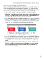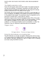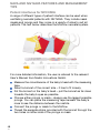
1
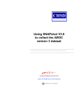
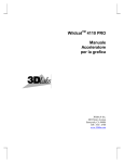
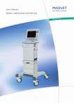
POCKET GUIDE
NAVA® and NIV NAVA in neonatal settings
EMPTY
Table of contents
1
2
3
4
5
6
Introduction and background facts
Invasive ventilation with NAVA
Non invasive ventilation with NAVA
NAVA and NIV NAVA features and management tips
Glossary
References
|
|
|
|
|
|
4
18
30
46
52
54
3
1.2
INTRODUCTION AND BACKGROUND FACTS
Introduction
Neonatal care is a specialized field focusing on babies in the first
few weeks of life. Both in terms of size and development, these
patients have special requirements.
This pocket guide aims to present Neurally Adjusted Ventilatory
Assist (NAVA) in both its invasive and non invasive forms to users
working with neonatal intensive care. In order to avoid confusion
and information overload, other modes of ventilation used in the
context of neonatal care are described separately, so that the user
can concentrate on understanding and learning to use NAVA. It
should of course be noted that this pocket guide cannot replace the
appropriate user’s manual.
The first chapter provides a brief description of the physiological
background to NAVA, as well as certain important central concepts,
such as the Edi signal and the NAVA level, and an outline of the
workflow prior to beginning patient ventilation with NAVA or NIV
NAVA. It is then followed by two main chapters, one focusing on
patient ventilation using invasive NAVA and the other on the non
invasive mode, and concludes with a final shared chapter presenting
some of the unique features of both modes, along with a few useful
management tips relevant to neonatal care.
Background and basic concepts
Neurally Adjusted Ventilatory Assist, in both its invasive and non
invasive forms, can be used on all patients requiring ventilatory
assistance (neonatal, pediatric and adult patients), provided that the
electrical signal from the brain to the diaphragm is intact and that
there is no contraindication for insertion/exchange of a nasogastric
tube.
4
1.2
INTRODUCTION AND BACKGROUND FACTS
NAVA delivers assist in proportion to and in synchrony with the
patient’s respiratory efforts. These efforts are reflected by the Edi
signal, which represents the electrical activity of the diaphragm, the
body’s principal breathing muscle. Understanding the Edi signal and
its use in NAVA and NIV NAVA is essential to successful ventilation
using these modes.
Physiology of the Edi signal
During normal respiration, a spontaneous breath starts with an
impulse generated by the respiratory center in the brain. This impulse
is then transmitted via the phrenic nerves and electrically activates
the diaphragm (excitation), leading to a muscle contraction. The
diaphragm contracts into the abdominal cavity, which leads to a
descending movement, creating a negative alveolar pressure and
an inflow of air.
Muscular contraction of the diaphragm is always preceded by an
electrical impulse and this electrical activation is controlled by nerve
stimuli, and ultimately by the respiratory center in the brain.
5
1.2
INTRODUCTION AND BACKGROUND FACTS
The signal that excites the diaphragm is proportional to the integrated
output of the respiratory center in the brain and controls the depth
and cycling of the breath.
Using the Edi signal in NAVA
When NAVA is used with the SERVO-i ventilator, the electrical activity
of the diaphragm, the Edi signal, is captured by a special catheter
(the Edi Catheter), which is fitted with an array of electrodes. Like
an ordinary feeding tube, the Edi Catheter is placed in the esophagus.
In neonates, the diaphragm, laryngeal and chest wall muscles work
in concert to augment the function of every component and thereby
protect the fragile tissues in the lungs.
1.
2.
3.
4.
6
1.2
Edi Catheter (with electrodes marked in black – the first is the
reference electrode, the others are measuring electrodes and
the distance between the measuring electrodes is the Inter
Electrode Distance or IED)
Esophageal wall
Diaphragm
Stomach
INTRODUCTION AND BACKGROUND FACTS
The Edi signal that is picked up by the electrodes on the Edi Catheter
is filtered and processed by the Edi Module. The Edi signal is
measured in microvolts 62.5 times per second. The processed Edi
signal is relayed to the SERVO-i ventilator which will, depending on
the NAVA level chosen, then deliver assist to the patient in proportion
to and in synchrony with this signal.
Basically, NAVA uses the Edi signal to control the ventilator and
assist the patient’s breathing in proportion to and synchrony with
his or her own effort.
The Edi signal serves as a respiratory vital sign in that it provides:
continuous monitoring of the respiratory drive
decision support for adjusting assist and unloading
objective criteria for intubation and extubation decisions.
The efficacy of the respiratory muscles and the degree of respiratory
demand will determine the degree of respiratory center output.
7
1.2
INTRODUCTION AND BACKGROUND FACTS
In a healthy subject, the low amplitude of diaphragm excitation
reflects the fact that neuroventilatory coupling is highly efficient and
that only about 5% of maximum capacity is used.
The signal is displayed on the ventilator screen, enabling the user
to monitor this vital sign and to observe and follow the synchrony
between patient and ventilator.
8
1.2
INTRODUCTION AND BACKGROUND FACTS
An example of an Edi curve for a single patient breath is presented
in the diagram below. The vertical green lines represent Edi signals,
sampled at a rate of 62.5 times per second.
When the preset Edi trigger level is reached, the ventilator will start
to deliver assist in proportion to the Edi signal, using the preset NAVA
level as the factor by which the signal is multiplied to ensure
continuous proportionality. Both NAVA modes, invasive and non
invasive, are triggered by an increase in the Edi signal from its lowest
value, known as Edi min, rather than a specific Edi level.
In the diagram below, the Edi min is 0.3 µV and the trigger level 0.5,
which means that the ventilator will be triggered at an absolute level
of 0.8 µV.
9
1.2
INTRODUCTION AND BACKGROUND FACTS
NAVA and NIV NAVA also employ a pneumatic trigger, based on
flow or pressure, as a secondary trigger source. In combination with
the Edi trigger, this operates on a first-come-first-served basis.
The ventilator will continue to multiply each of the subsequent
measured Edi signals (the individual green lines in the diagram above)
by the preset NAVA level, resulting in a pressure curve that follows
the Edi signal in a smooth and consistent way.
10
1.2
INTRODUCTION AND BACKGROUND FACTS
The pressure delivered is derived from the following formula:
NAVA level x (Edi signal - Edi min) + PEEP
The pressure curve in both NAVA and NIV NAVA follows the Edi
signal pattern. When the Edi signal has fallen to 70% of its peak
value, the patient is allowed to exhale and the ventilator no longer
offers any assist until the next breath is initiated and the trigger level
is again reached.
11
1.2
INTRODUCTION AND BACKGROUND FACTS
As long as the patient has an Edi Catheter in position, the Edi signal
can in addition be monitored in all modes of ventilation, invasive and
non invasive, as well as in Standby, including values for both Edi
peak and Edi min.
The values are also trended in all modes, as well as in Standby.
The NAVA level
The NAVA level is the factor by which the Edi signal is multiplied to
adjust the amount of assist delivered to the patient. This assist is
thus proportional to the patient’s Edi and as such, it follows a
physiological pattern.
12
1.2
INTRODUCTION AND BACKGROUND FACTS
The set NAVA level reflects the amount of work of breathing that the
SERVO-i ventilator will take over from the patient. The appropriate
NAVA level varies for different patients since they require different
assist levels. It may also need adjusting over time in the same patient.
The NAVA level is typically set to between 1.0 and 4.0 cmH20/µV.
The range of settings is 0.0 to 15.0 cmH20/µV.
Insertion and positioning of the Edi Catheter
Select the appropriate Edi Catheter for the patient. You need to know
the patient’s height and weight. The table below provides more
details.
8 Fr 100 cm
IED (Inter
Electrode
Distance)
8 mm
< 55 cm
1.0 - 2.0 kg
6 Fr 50 cm
6 mm
< 55 cm
0.5 - 1.5 kg
6 Fr 49 cm
6 mm
Patient height Patient weight
45 - 85 cm
Edi Catheter
size
Insert the Edi Module into the SERVO-i and connect the Edi Cable.
Perform the Edi Module function check.
13
1.2
INTRODUCTION AND BACKGROUND FACTS
Measure the distance from the bridge of the Nose (1) to the Earlobe
(2) and then to the Xiphoid process (3). This is the NEX measurement.
Make a note of it.
Calculate the insertion distance (Y) for the Edi Catheter. This will
depend on whether the Edi Catheter is inserted orally or nasally, as
well as on the size of the Edi Catheter. Use the appropriate table as
shown below.
Insertion distance Y for nasal insertion
Fr/cm
Calculation of Y
14
1.2
8 Fr 100 cm
NEX cm x 0.9 + 8 = Y cm
6 Fr 50 cm
NEX cm x 0.9 + 3.5 = Y cm
6 Fr 49 cm
NEX cm x 0.9 + 2.5 = Y cm
INTRODUCTION AND BACKGROUND FACTS
Insertion distance Y for oral insertion
Fr/cm
Calculation of Y
8 Fr 100 cm
NEX cm x 0.8 + 8 = Y cm
6 Fr 50 cm
NEX cm x 0.8 + 3.5 = Y cm
6 Fr 49 cm
NEX cm x 0.8 + 2.5 = Y cm
Examples:
-
Infant – height 40 cm, weight 900 g
Selected Edi Catheter – 6 Fr 49 cm
Insertion – nasal
NEX – 12 cm
Insertion distance Y = 12 x 0.9 + 2.5 = 12.3 cm
Infant – height 46 cm, weight 1.8 kg
Selected Edi Catheter – 6 Fr 50 cm
Insertion – oral
NEX – 15 cm
Insertion distance Y = 15 x 0.8 + 3.5 = 15 cm
Dip the Edi Catheter into water for a few seconds. Do NOT use
lubricants as this may destroy the Edi Catheter coating and interfere
with the measurement of the Edi signal.
Insert the Edi Catheter to the Y value calculated above.
Connect the Edi Catheter to the Edi Cable.
Open the “Neural access” menu and select “Edi Catheter positioning”
to confirm the position of the Edi Catheter.
15
1.2
INTRODUCTION AND BACKGROUND FACTS
Verify the position of the Edi Catheter by analyzing the ECG
waveforms. Ideally, P and QRS waves are present in the top leads,
while the P waves gradually decrease and disappear in the lower
leads, where QRS amplitude also decreases. Check that the Edi
scale is fixed and that it is set appropriately (greater than or equal
to 5 µV).
If Edi deflections are present, observe which leads are highlighted
in blue.
-
-
-
-
16
1.2
If the second and third leads are highlighted in blue, secure the
Edi Catheter in this position after marking it at its final position
and making a note of the distance in centimeters.
If the top leads are highlighted, pull out the Edi Catheter in steps
corresponding to the Inter Electrode Distance (IED, measured in
millimeters) until the blue highlight appears in the center. Do not
exceed four times the IED, Mark the Edi Catheter at its final
position.
If the bottom leads are highlighted, insert the Edi Catheter further
in steps corresponding to the IED until the blue highlight appears
in the center. Again, do not exceed four times the IED. Mark the
Edi Catheter at its final position.
If the Edi signal is very low, there will be no blue highlights. If this
happens, evaluate the Edi signal as described below.
INTRODUCTION AND BACKGROUND FACTS
Secure the Edi Catheter in position once the position has been
verified. Check first that the marking on the Edi Catheter is in the
right place and observe the ECG waveforms and their blue highlights.
Make sure that the Edi Catheter is not secured to the endotracheal
tube.
Record the insertion length.
Important: Always follow hospital routines to check the position
of the Edi Catheter when it is used as a gastric feeding tube.
Evaluate the Edi signal. A low or absent Edi may be due to any of
the following:
hyperventilation
sedation
muscle relaxants
neural disorders
If possible, perform an expiratory hold and verify that the positive
Edi deflection coincides with a negative deflection in the pressure
waveform.
Edi Catheter positioning may be reconfirmed after 1-2 hours if minor
adjustments are necessary.
SUMMARY
Select Edi Catheter and measure NEX, calculating the insertion
distance, Y.
Dip Edi Catheter in water and insert.
Verify the position in the positioning window.
Secure the Edi Catheter.
17
1.2
INVASIVE VENTILATION WITH NAVA
Starting and running NAVA
The information below covers the procedures involved in starting
and running the NAVA mode.
Press “Neural access” and select “NAVA preview”.
The gray curve then displayed on the user interface below shows
the estimated pressure based on the Edi and the set NAVA level.
18
1.2
INVASIVE VENTILATION WITH NAVA
Simply press “NAVA level” and use the main rotary dial to set it
appropriately. The NAVA level range is 0 to 15 cmH2O/µV. One way
to select the initial NAVA level is to try the level that will produce the
same pressure as that used in the current ventilation mode, or
perhaps slightly lower. By accepting and pressing “Close”, the
selected NAVA level is saved to the NAVA ventilation mode window.
19
1.2
INVASIVE VENTILATION WITH NAVA
To set the parameters for operating the NAVA mode, choose “NAVA”
in the “Select ventilation mode” window. This opens the “Set
ventilation mode” parameters window. The NAVA level displayed
here is the initial one saved (as outlined in the point above) from the
“NAVA preview” window.
The other two basic parameters are PEEP (cmH2O) and oxygen
concentration (%).
Trigg. Edi has a default setting of 0.5 µV and the range 0-2 µV. The
value set here is the one that will trigger the ventilator to assist the
patient.
20
1.2
INVASIVE VENTILATION WITH NAVA
The remaining parameters refer to NAVA Pressure Support (NAVA
(PS) – Trigger Sensitivity, Inspiratory Cycle off (%) and PS above
PEEP (cmH2O). NAVA (PS) is the mode to which the ventilator
automatically switches under certain specific circumstances (see
diagram and information in the section below). It is therefore
important to set adequate values here; otherwise the patient’s Edi
signal may be affected and the ventilator may be unable to switch
back to NAVA. NAVA (PS) and Backup are described in greater detail
later in this chapter.
Finally, the column for “Backup ventilation” has three parameters
that can be set by the user – PC above PEEP (cmH2O), Resp. Rate
and Ti/I:E. Default settings are used for the following parameters:
Infant parameters
Ti/I:E ratio
0.5 s/1:2
Respiratory rate
30 breaths/minute
Inspiratory rise time
0.15 s/5%
The backup pressure level, respiratory rate and inspiratory time or
I:E ratio are all adjustable. The default settings can be adjusted in
the Edit startup configuration window.
21
1.2
INVASIVE VENTILATION WITH NAVA
Set appropriate alarm limits in the Alarm profile window. The apnea
alarm for infants can be set between 5 and 45 seconds.
It is important to set a suitable upper limit for pressure. The maximum
available pressure level is 5 cmH2O below the preset upper pressure
limit. Appropriate values should also be set for the patient’s minute
volume, respiratory rate and end expiratory pressure. The alarms’
sound level and the apnea time are also adjusted here.
SUMMARY
Select NAVA preview via "Neural access".
Adjust the NAVA level.
Accept and press "Close" to save the initial NAVA level.
Open the NAVA mode window and adjust settings.
Set appropriate values for NAVA (PS) and NAVA Backup.
Accept and proceed with NAVA ventilation. Remember to set
appropriate alarm limits.
22
1.2
INVASIVE VENTILATION WITH NAVA
Switching between NAVA, NAVA (PS) and NAVA (Backup)
When running invasive NAVA, the ventilator has several modes
between which it switches freely under specific conditions. These
are described and explained here.
23
1.2
INVASIVE VENTILATION WITH NAVA
Switching from NAVA to NAVA (PS)
The Edi respiratory rate differs from the pneumatic respiratory
rate by more than 25% for at least 5 seconds. The calculated
respiratory rates are based on the last 20 seconds.
The Edi Ti/Ttot is more than 0.5, calculated over the last 20
seconds if the Edi Catheter position is classified as invalid.
The Edi Ti/Ttot is more than 0.6, calculated over the last 20
seconds if the Edi Catheter position is classified as valid.
The Edi Catheter is disconnected.
There is ECG leakage into the Edi signal.
Switching from NAVA (PS) to NAVA
The Edi respiratory rate differs from the pneumatic respiratory
rate by less than 20%.
At least 7 of the last 10 breaths are classified as being in
synchrony with the Edi signal.
Note: Pneumatic respiratory rate and Ti/Ttot are shown on the
user interface. Edi respiratory rate and Edi Ti/Ttot are not shown
on the user interface.
Switching from NAVA to NAVA (Backup)
Apnea with a permanently low Edi signal and no pneumatic trigger.
Switching from NAVA (Backup) to NAVA
An adequate Edi signal can be detected.
24
1.2
INVASIVE VENTILATION WITH NAVA
Switching from NAVA (PS) to NAVA (Backup)
When the ventilator is in NAVA (PS) for any of the above reasons,
and the pneumatic trigger can no longer be detected, the
ventilator will switch to backup mode (Pressure Control).
There are, however, certain restrictions on the number of times
the ventilator may switch back and forth automatically between
NAVA and NAVA (Backup). The ventilator will thus lock in backup
ventilation if:
- the patient switches between NAVA and NAVA (Backup) more
than three times in the space of two minutes OR
- the patient only triggers a single breath with the Edi signal to
interrupt each of two consecutive backup periods.
A dialog box will then open where the user may either review the
ventilatory settings or return to the supported mode by pressing
one of the two buttons displayed.
Alarms for invasive NAVA
The following are high priority alarms.
Asynchrony alarm
In case of asynchrony, the ventilator will switch back and forth
without triggering an alarm until one of the following conditions is
fulfilled:
-
the ventilator has been in NAVA (PS) for more than 120 seconds;
there have been six switches from NAVA to NAVA (PS) in the last
five minutes.
25
1.2
INVASIVE VENTILATION WITH NAVA
In either case, the asynchrony alarm will be activated and the
message “Pneumatic-Edi out of synch” will appear, since the
measured Edi signal is out of phase with the pressure and flow
signals generated by the patient. There will also be a message to
tell the user to check the Edi Catheter position. In addition, the user
should check and if necessary adjust the trigger settings. The
ventilation mode can also be changed.
When the asynchrony alarm has been activated, the ventilator will
as usual search for synchrony indices. As soon as synchrony is
re-established, the message “Pneumatic-Edi synch restored” will be
displayed. Press the OK button or wait for ten seconds, and the
ventilator will switch back to NAVA. You can also choose to return
to NAVA manually by pressing the “Back to NAVA” key on the user
interface if the patient’s Edi signal is in synchrony with the pneumatic
breath.
If asynchrony is still detected, a message will appear followed by a
question.
Users then need to confirm that they wish to return to NAVA.
No Edi monitoring alarm
The message “Edi monitoring not active” appears when the NAVA
mode is activated without an Edi Module being connected or if the
Edi Catheter is not properly connected. If this occurs, simply insert
the Edi Module or adjust the Edi Catheter.
Regulation pressure limited message
"Regulation pressure limited" appears as a text message activated
during NAVA at a level 5 cmH20 below the set Upper Pressure Limit.
If you then increase the NAVA level, the ventilator will make a beeping
sound to draw your attention to the message. The maximum available
pressure level is thus 5 cmH20 below the preset upper pressure limit.
26
1.2
INVASIVE VENTILATION WITH NAVA
One way to manage the NAVA level
When ventilating neonates with NAVA, treatment may normally be
started at a NAVA level that will ensure that the pressure delivered
is the same as, or slightly lower than, the set Pressure Support level.
The NAVA level is then gradually titrated to a level that will ensure
comfortable unloading of the patient. The Edi signal provides the
necessary information for performing this procedure efficiently, as
summarized below. Throughout this procedure, the patient should
be carefully observed for signs of discomfort or hemodynamic
instability.
Once a reliable Edi signal with a satisfactory amplitude has been
obtained, activate the NAVA preview window and set a NAVA
level that ensures that the pressure delivered is equal to or slightly
below the set Pressure Support level.
Set an Upper Pressure Limit that is acceptable to the patient.
The NAVA level is then increased in steps of 0.1 cm H2O/µV with
the aim of achieving a decrease in the Edi signal. If the aim is, for
example, to unload 50% of the patient’s work of breathing, the
target for the decrease in the Edi signal will be 50%.
27
1.2
INVASIVE VENTILATION WITH NAVA
The Edi signal is followed carefully while the NAVA level is slowly
increased in small increments. The Edi signal helps the clinician
to quantify and continuously evaluate the patient’s respiratory
work.
Once the target is reached, the Edi signal should be followed
without changing the NAVA level. A decrease in the Edi signal
with a maintained tidal volume indicates an improvement in
neuromuscular coupling.
One way to wean patients from invasive NAVA
This chapter describes one procedure that can be followed when
weaning a patient from NAVA.
When the patient is on NAVA, an objective measure of the patient’s
progress becomes available – the Edi signal. The first sign to look
for is a decline in the Edi signal with unchanged tidal volumes. This
represents an improvement in neuromuscular coupling in the sense
that diaphragm performance is unchanged at a lower level of
stimulation.
As soon as this situation is achieved, the airway pressure will be
lower. The patient is in effect giving us a signal that he or she is now
ready to be weaned. For all practical purposes, the patient can then
be left with an essentially unchanged NAVA level and will wean him
or herself off the ventilator, while we watch the progress in the shape
of a falling Edi signal.
The process may however be speeded up to allow earlier
disconnection from the ventilator. The procedure suggested below
is one example of how this may be done.
28
1.2
INVASIVE VENTILATION WITH NAVA
Weaning procedure example:
1.
2.
3.
4.
5.
The first sign that it is possible to wean the patient is a decline
in the Edi signal with maintained tidal volume.
Please remember the following differential diagnosis – sedation
bolus or increase in maintenance dose of opiates/sedatives. If
an increase in sedation is not the cause of the decline in the
Edi signal, however, then the decrease provides confirmation
of an improvement in neuromuscular coupling. The mechanical
efficiency of the diaphragm has improved and the patient will
be receiving less support – weaning has in effect been started
automatically.
When the patient is stable and the tidal volume is unchanged
while the Edi signal is declining or unchanged, reduce the NAVA
level in steps of 0.1-0.2 cmH2O/µV.
If VT is reduced and the Edi signal increases disproportionally,
go back to the previous setting. The reaction may indicate one
of the two following situations:
a. The patient is not yet ready to be weaned. Allow the patient
to rest on the previous setting and try again later.
b. The initial assist is too low (look for a slow upstroke in the
Edi and flow curve, and an increase in neural Ti).
Weaning progress can be monitored by the decline in the Edi
signal and peak pressure.
SUMMARY
While keeping the NAVA level unchanged, verify a decrease in
the Edi amplitude with a maintained tidal volume.
Decrease the NAVA level in steps of 0.1-0.2 cmH2O/µV. Observe
a decrease in peak pressure minus PEEP with a maintained
tidal volume.
29
1.2
NON INVASIVE VENTILATION WITH NAVA
Introduction to NIV NAVA
Synchronized non invasive respiratory assist enables smooth
transition to natural breathing and provides proportional assist in
synchrony with the patient’s own breathing efforts. Patient and
ventilator are thus in synchrony.
NIV NAVA does not rely on a pneumatic signal. Both triggering and
cycle off of the breath rely on the Edi signal and are therefore
independent of leakage.
There are several types of patient interface. For neonates, the most
commonly used interfaces are nasal prongs or nasal masks, although
nasopharyngeal tubes may also be used (see management tips).
Monitoring the respiratory drive
The Edi signal serves as a respiratory vital sign in that it provides:
continuous monitoring of the respiratory drive
decision support for adjusting assist and unloading
objective criteria for intubation and extubation decisions.
30
1.2
NON INVASIVE VENTILATION WITH NAVA
Starting and running NIV NAVA
Settings
To access NIV NAVA, the user must be in the Standby position and
select the relevant patient category (infant) and non invasive
ventilation. Please note that when NIV is selected, the frame on the
screen turns from grey to yellow.
In the Select Ventilation Mode window, the user selects NIV NAVA
and the Set Ventilation Mode window opens. This is where the
parameters are set for NIV NAVA.
31
1.2
NON INVASIVE VENTILATION WITH NAVA
Basic settings:
NAVA level – range 0.0 to 15.0 cmH2O/µV. Default – 0.5
cmH2O/µV. For NIV NAVA, the estimated pressure delivered will
be: NAVA level x (Edi peak – Edi min) + PEEP + 2 cmH2O
PEEP – range 2 to 20 cmH2O
O2 concentration – range 21 to 100%
Trigg. Edi – range 0.0 to 2.0 µV
Backup ventilation for NIV NAVA is Pressure Control.
32
1.2
NON INVASIVE VENTILATION WITH NAVA
Backup parameters
Infant parameters
Ti/I:E ratio
0.5 s/1:2
Respiratory rate
30 breaths/minute
Inspiratory rise time
0.15 s/5%
The backup settings should be chosen so as to ensure adequate
ventilation in case of apnea. It is important to consider that apnea
is particularly frequent in infants, particularly premature infants, and
the backup settings should be made with this in mind. When suitable
values have been selected, press Accept.
Before starting ventilation by pressing the Start button in the bottom
left corner of the user interface, it is important to check the alarm
profile.
It is important to set a suitable upper limit for pressure. The maximum
available pressure level is 5 cmH2O below the preset upper pressure
limit. Appropriate values should also be set for the patient’s minute
volume, respiratory rate and end expiratory pressure. The alarms’
sound level and the apnea time are also adjusted here.
33
1.2
NON INVASIVE VENTILATION WITH NAVA
The Regulation pressure limited message appears when Ppeak is 5
cmH2O below the upper pressure limit. The maximum available
pressure level is thus 5 cmH2O below the preset upper pressure limit.
The maximum peak pressure is 32 cmH2O.
Since leakage often varies during non invasive ventilation, alarms
may be activated frequently, which may be perceived as disturbing,
particularly to the patient. It is therefore possible to set audible alarms
to “Audio off” by pressing the bell on the relevant alarm. This applies
to all patient related alarms except the high pressure alarm. When
appropriate values have been set, press Accept.
If the patient is already being ventilated in NIV PS or NIV PC, the
initial NAVA level can be set in the NAVA preview window, which is
opened via the Neural access fixed key. There are thus different
ways to start NIV NAVA.
34
1.2
NON INVASIVE VENTILATION WITH NAVA
SUMMARY
If NIV NAVA is the first ventilation choice, the NAVA preview
window cannot be used. The NAVA level is instead set in the
Set ventilation mode window by choosing a low level and then
gradually increasing to a level at which the patient is adequately
unloaded. The process of setting and optimizing the NAVA level
is discussed in a separate section below.
If the patient is being ventilated with NIV PS, NIV PC, or an
invasive mode other than invasive NAVA, the NAVA preview
window can also be used – the user presses “NAVA level” in
this window and uses the main rotary dial to set it appropriately.
Generally, the first NAVA level should be low and then slowly
increased in steps of 0.1 cmH2O/µV.
Running NIV NAVA
After starting ventilation by pressing the Start button in the bottom
left corner of the user interface, a waiting position for NIV NAVA will
be initiated, which gives the user an opportunity to adjust the
ventilator and/or the patient interface (i.e. the mask, prongs or other).
The time shown is counted by the ventilator from the time that the
waiting position is entered.
Ventilation begins when one of the following criteria has been met:
the ventilator detects that the patient is connected to the ventilator
the user presses the Start ventilation soft key.
35
1.2
NON INVASIVE VENTILATION WITH NAVA
If neither of these criteria has been met within two minutes, an alarm
will be activated to remind the user that it is appropriate to start
ventilation. During the waiting phase, all audible patient related alarms
are deactivated and no ventilation is delivered. There is however a
bias flow of 7.5 l/min.
When the Start ventilation button is pressed, NIV NAVA will be started
without further delay.
The screen shot below shows the SERVO-i ventilator running in NIV
NAVA. The screen displays the pressure, flow, volume and Edi curves,
as well as measured values relating to each curve on the right. Note
that the Edi deflections are synchronous with the assist delivered.
36
1.2
NON INVASIVE VENTILATION WITH NAVA
NIV NAVA alarms
No patient effort alarm
If the Edi signal disappears, the ventilator will sound a high priority
alarm after the set apnea time and the message ”No patient effort”
will appear on the screen. The ventilator will automatically switch to
Backup ventilation.
The backup mode for NIV NAVA is Pressure Control and the function
of the third and fourth direct access knobs at the bottom of the
screen is changed so that they can be used to adjust the respiratory
rate and PC above PEEP.
Once the ventilator detects a valid Edi signal again, it will
automatically return from backup ventilation to NIV NAVA (please
note that the ventilator will only return to NIV NAVA if triggered by
the Edi, not if there is a pneumatic trigger). There is no limit on the
number of times the ventilator can switch back and forth between
NIV NAVA and backup ventilation.
37
1.2
NON INVASIVE VENTILATION WITH NAVA
Leakage out of range alarm
The ventilator will compensate for leakage of up to 25 l/min for
infants. If leakage is excessive (>25 l/min for infants during expiration)
or if the patient is disconnected, the ventilator will pause and issue
a high priority alarm. A dialog appears, stating that leakage is too
high and recommending that the patient circuit should be checked.
The message Leakage out of range also appears at the top of the
screen. When this happens, a constant disconnect flow is delivered.
The flow depends on the setting of the Disconnect flow function in
the Edit startup configuration window. The default value is 7.5 l/min.
38
1.2
NON INVASIVE VENTILATION WITH NAVA
Once leakage has been reduced or the patient has been reconnected,
ventilation will automatically resume and the screen dialog will
disappear after three breaths. It is also possible to start ventilation
manually by pressing the “Resume ventilation” button. This brings
the ventilator out of its pause position and returns it to NIV NAVA,
but a message (“Operator-initiated breath”) will appear and the alarm
will still be active.
It may be noted that NIV NAVA is less sensitive to autotriggering at
high leakage levels because the pneumatic trigger is deactivated
when 60% of the maximum leakage compensation is reached.
Disconnect flow
The Disconnect flow can be set in the Edit startup configuration
window. The following settings are possible:
Low flow - 7.5 l/min
High flow - 15 l/min for infants
Disabled - No pause in ventilation in case of high leakage. The
SERVO-i will continue to deliver assist even when leakage is
excessive. The Leakage out of range alarm will then go from high
priority to medium priority.
39
1.2
NON INVASIVE VENTILATION WITH NAVA
There is also a special low priority leakage fraction alarm for the NIV
NAVA infant option. It is activated if leakage is > 95%. This feature
is useful when ventilating infants with NIV NAVA, as it gives personnel
the possibility to adjust prongs or other non invasive interfaces that
have come loose.
The leakage fraction alarm function is enabled via Biomed and this
function is only available in the NIV NAVA infant option.
Check catheter position/RR and HR coupling alarm
In this high priority alarm, RR refers to the respiratory rate and HR
to the heart rate. This alarm is activated when there is leakage of the
ECG signal into the Edi signal. The ventilator switches automatically
to backup ventilation. The Edi Catheter position should be checked
by going to the Edi Catheter positioning window (via the Neural
access fixed key), where adjustments can be made to the position
of the Edi Catheter. The alarm will then disappear and the ventilator
should automatically return to NIV NAVA.
40
1.2
NON INVASIVE VENTILATION WITH NAVA
Unreliable Edi signal alarm
A further high priority alarm is ”Unreliable Edi signal”, which occurs
in cases of extreme asynchrony between the detected Edi signal
and the pneumatic parameters. The alarm is activated if one or more
of the following conditions are fulfilled during NIV NAVA ventilation:
the Edi respiratory rate differs from the pneumatic respiratory rate
by more than 25% for at least 5 seconds, assuming that leakage
is low. The calculated respiratory rates are based on the last 20
seconds.
the Edi Ti/Ttot is more than 0.5, calculated over the last 40
seconds, if the Edi Catheter position is classified as invalid.
the Edi Ti/Ttot is more than 0.6, calculated over the last 40
seconds, if the Edi Catheter position is classified as valid.
Note: The pneumatic respiratory rate and Ti/Ttot are shown on
the user interface. The Edi respiratory rate and Ti/Ttot are not
shown on the user interface.
This alarm does not cause the ventilator to switch automatically to
backup ventilation. Instead the user should press the alarm fixed
key for more details.
When the “Bell” fixed key on the right of the user interface is pressed,
a dialog box opens in which the user is recommended to check the
Edi Catheter position and asked if he wishes to turn the alarm audio
off for the duration of the alarm situation. Once the Edi Catheter
position has been corrected, the alarm should disappear and NIV
NAVA ventilation should continue normally.
41
1.2
NON INVASIVE VENTILATION WITH NAVA
Check catheter position/Edi invalid alarm
Another high priority alarm concerns the Edi Catheter and occurs
when there is no valid Edi signal for the ventilator to work with, for
example if the Edi Catheter or Edi cable have been accidentally
disconnected. The ventilator then switches to backup ventilation and
a dialog box opens. As soon as the Edi Catheter has been
reconnected and the ventilator detects a valid Edi signal, the
ventilator will switch back to NIV NAVA.
Apnea audio delay
It is also possible to set an Apnea audio delay to between 0 and 30
seconds. Again, this only applies to infants. If, for example, the user
sets the apnea audio delay to 20 seconds, while the apnea time has
been set to 10 seconds, the SERVO-i will, after 10 seconds with no
Edi signal, activate the “No patient effort” visual alarm and display
the message “Alarm audio paused”. If the Edi signal does not return
within the next 20 seconds, a high priority audio alarm signal will be
activated.
42
1.2
NON INVASIVE VENTILATION WITH NAVA
One way to manage the NAVA level
When ventilating in NIV NAVA, the treatment is normally started at
a low NAVA level and then gradually titrated to the level that will
ensure comfortable unloading of the patient. The Edi signal provides
the information needed to perform this procedure efficiently.
If possible, start by setting the NAVA level to 0 (for 30-60
seconds). The resulting Edi peak value that is then registered will
provide information that allows the clinician to assess the total
muscle load, as well as any muscular weakness.
The NAVA level is then increased in steps of 0.1 cm H2O/µV with
the aim of achieving a decrease in the Edi signal. If the aim is, for
example, to unload 50% of the patient’s work of breathing, the
target for the decrease in the Edi signal will be 50%.
The Edi signal is followed carefully while the NAVA level is slowly
increased in small increments. The Edi signal helps the clinician
to quantify and continuously evaluate the patient’s respiratory
work.
Once the target is reached, the Edi signal should be followed
without changing the NAVA level. A decrease in the Edi signal
with a maintained tidal volume indicates an improvement in
neuromuscular coupling.
43
1.2
NON INVASIVE VENTILATION WITH NAVA
One way to wean patients from NIV NAVA
As for invasive NAVA, weaning from NIV NAVA is largely a matter of
self-weaning.
The Edi signal can be used during ventilation with NIV NAVA as an
objective measure of the patient’s progress. The first sign to look
for is a decline in the Edi signal with unchanged tidal volumes. This
represents an improvement in neuromuscular coupling in the sense
that diaphragm performance is unchanged at a lower level of
stimulation.
As soon as this situation is achieved, the airway pressure will be
lower. The patient is in effect giving us a signal that he or she is now
ready to be weaned. For all practical purposes, the patient can then
be left with an essentially unchanged NAVA level and will wean him-or
herself off the ventilator, while we watch the progress in the shape
of a falling Edi signal.
The process may however be speeded up to allow earlier
disconnection from the ventilator. The procedure suggested below
is one example of how this may be done.
Weaning procedure example:
1.
2.
44
1.2
The first sign that it is possible to wean the patient is a decline
in the Edi signal with maintained tidal volume.
Please remember the following differential diagnosis – sedation
bolus or increase in maintenance dose of opiates/sedatives. If
an increase in sedation is not the cause of the decline in the
Edi signal, however, then the decrease provides confirmation
of an improvement in neuromuscular coupling. The mechanical
efficiency of the diaphragm has improved and the patient will
be receiving less support – weaning has in effect been started
automatically.
NON INVASIVE VENTILATION WITH NAVA
3.
4.
5.
When the patient is stable and the tidal volume is unchanged
while the Edi signal is declining or unchanged, reduce the NAVA
level in steps of 0.1-0.2 cmH2O/µV.
If VT is reduced and the Edi signal increases disproportionately,
go back to the previous setting. The reaction may indicate one
of the two following situations:
a. The patient is not yet ready to be weaned. Allow the patient
to rest on the previous setting and try again later.
b. The initial assist is too low (look for a slow upstroke in the
Edi and flow curve, and an increase in neural Ti).
Weaning progress can be monitored by the decline in the Edi
signal and peak pressure.
SUMMARY
While keeping the NAVA level unchanged, verify a decrease in
the Edi amplitude with a maintained tidal volume.
Decrease the NAVA level in steps of 0.1-0.2 cmH2O/µV. Observe
a decrease in peak pressure minus PEEP with a maintained
tidal volume.
45
1.2
NAVA AND NIV NAVA FEATURES AND MANAGEMENT
TIPS
The NAVA respiration cycle
Since the Edi signal varies with each breath, the assist pressures
will vary accordingly. In summary, inspiration will start when the
patient triggers a breath and gas flows into the lungs at a varying
pressure proportional to the patient’s Edi signal.
The breath may be triggered either by the Edi or pneumatically, by
flow or pressure. The assist to the patient will remain proportional
to the patient’s Edi signal. The maximum time for inspiration is 2.5
seconds for adults and 1.5 seconds for infants.
Expiration begins when the Edi decreases below 70% of the peak
value during ongoing inspiration (40% for low Edi signals), or when
the pressure increases 3 cmH2O above the inspiratory target
pressure, or if the Upper Pressure Limit is exceeded, or when the
maximum time for inspiration specified above is exceeded.
Trigger colors
T
Edi trigger indicator
T
Flow/pressure trigger indicator
Using the Edi Catheter as a feeding tube
The Edi Catheter is a single-use gastric feeding tube with an array
of 10 electrodes (nine measuring and one reference electrode). The
Edi catheter has been validated for use for 5 days, both for feeding
and when using the NAVA function.
46
1.2
NAVA AND NIV NAVA FEATURES AND MANAGEMENT
TIPS
Noting Edi Catheter insertion length
Remember to mark the Edi Catheter at its final position and make a
note of the final distance in centimeters in the patient chart.
Suctioning
During suctioning, or in case of patient disconnection, it is important
to use the Suction Support function to avoid activating the
asynchrony alarm (see the Alarms chapter below). The function is
not used when a closed suction system is in use.
What happens if…
… the patient is apneic:
In case of apnea, the SERVO-i will automatically switch to Backup
ventilation, which is the Pressure Control mode. The apnea time is
set under the alarm profile.
… the Edi Catheter has been accidentally removed:
If the Edi Catheter is pulled out by mistake, the SERVO-i will
automatically switch to NAVA (PS) for invasive NAVA or to NAVA
(Backup) for non invasive NAVA.
… there is no Edi signal, or if it is low:
Check that the Edi Catheter is correctly placed and connected by
using the Edi Catheter Positioning Window. If it is, check the patient
for signs that the PS level is too high, or of hyperventilation or
oversedation.
The Ventilation Record Card
The Ventilation Record Card enables patient data and screen shots
to be transferred from the SERVO-i to a computer, where they can
be accessed. This feature is useful for teaching as well as for
diagnostic purposes. For more details, refer to the relevant SERVO-i
training material and user manuals.
47
1.2
NAVA AND NIV NAVA FEATURES AND MANAGEMENT
TIPS
Patient interfaces for NIV NAVA
A range of different types of patient interface can be used when
ventilating neonatal patients with NIV NAVA. They include nasal
masks and prongs and they come in a variety of sizes to suit all
patients. The text below describes the Miniflow neonatal system.
For more detailed information, the user is referred to the relevant
User’s Manual from Medin Innovations GmbH.
Measure the circumference of the baby’s head with the measuring
tape.
Select a bonnet of the correct size – it has to fit loosely.
Put the bonnet on the baby’s head – pull the bonnet as far down
towards the baby’s eyes as possible.
Choose either prongs or mask – always use the largest possible
prongs. You can place the measuring tape beneath the baby’s
nose to see the distance between the nostrils.
Connect the prongs or mask to the Miniflow.
Thread the separate strips provided with the bonnet through the
two holes on either side of the prongs or mask.
48
1.2
NAVA AND NIV NAVA FEATURES AND MANAGEMENT
TIPS
Choose the most suitable angle for the Miniflow connection –
either 45° for babies weighing more than 1000 g or 60º for babies
weighing less than 1000 g.
Open the Velcro tube fastener on the bonnet.
Place the tubes on either side of the central part of the Velcro
tube fastener.
Either place the mask over the baby’s nose, or insert the prongs
halfway into the baby’s nostrils.
Fasten the strips onto the bonnet below the baby’s ears.
Fixate the tubes in the Velcro fastener on the bonnet.
If desired, tie the top of the bonnet using the bonnet’s white fabric
strips.
Important:
-
If prongs are used, the semicircular edge of the base must point
towards the baby’s mouth.
Insert the prongs only halfway into the baby’s nostrils.
The base of the prongs should not be in contact with either the
baby’s nose or the skin below the nose.
Do not pull the strips too tight.
The Miniflow’s tubes should be angled so that the highest point
is located at the baby’s nose to prevent any water from flowing
back to the baby’s airways.
Please note, before starting ventilation with NIV NAVA, it is vital to
prepare ahead and have all necessary interfaces and connections
ready for use.
Disconnection of the Edi Module
If the Edi Module becomes disconnected, the SERVO-i will
automatically switch to NAVA (PS) in invasive NAVA or to PC in non
invasive NAVA. An alarm will also be activated.
49
1.2
NAVA AND NIV NAVA FEATURES AND MANAGEMENT
TIPS
Scale settings
It is important to verify that the Edi scale is fixed and set appropriately
(i.e. greater than or equal to 5 µV). Avoid clipping the Edi signal by
imposing too low an upper limit on the scale.
NAVA and Heliox
NAVA can be used together with Heliox.
Trends
Trends are extremely useful when following a patient’s progress on
NAVA.
Coughing and hiccups
If a patient is suffering from hiccups, they may trigger the ventilator
and cause a short assist delivery. If a patient coughs, NAVA will
provide assist during the inspiration phase prior to the cough, and
the expiratory valve will open during the coughing itself. The usual
safety mechanisms, such as the Upper Pressure Limit, are in place
to handle coughing while using the NAVA mode.
Titrating the PEEP level
An important parameter to optimize when ventilating patients in
NAVA is the PEEP level. A titration procedure for optimizing the PEEP
level has been described in an article by Brander and colleagues.
The article concludes that during NAVA ventilation using an adequate
NAVA level, increasing PEEP reduces respiratory drive, and
monitoring the ratio of tidal volume to Edi signal (the neuroventilatory
index) during PEEP changes allows identification of a PEEP level at
which tidal breathing occurs at minimal Edi cost.
50
1.2
NAVA AND NIV NAVA FEATURES AND MANAGEMENT
TIPS
NIV NAVA – independent of leakage
This mode of ventilation is based on a neural signal (the Edi signal)
and the triggering and cycle off of the breath are thus unaffected by
leakage.
Monitoring the Edi signal – a respiratory vital sign
The Edi signal can be monitored in all modes of ventilation, as well
as in the standby position. This includes Edi peak and Edi min values
on a breath-by-breath basis, and these are trended and can be
followed in the trend graphs. This enables clinicians to follow an
important respiratory parameter easily and reliably at a level not
previously accessible in practical clinical work.
Since the Edi signal reflects the patient’s own respiratory drive and
neural effort, it can be used as a respiratory vital sign that is
continually monitored. It provides crucial information about the
patient’s ability or inability to breathe spontaneously, as well as about
synchrony, or lack of synchrony, between patient and ventilator.
51
1.2
GLOSSARY
List of terms
CMV - Controlled Mechanical Ventilation.
ECG – electrocardiogram, a recording of the electrical activity of the
heart.
Edi – the electrical activity of the diaphragm. Varying versions of the
acronym EAdi are sometimes used to denote the same thing.
Edi peak – the highest value of the Edi signal during a single breath
cycle.
Edi min – the lowest value of the Edi signal during a single breath
cycle.
Fr – abbreviation for French. The French catheter scale is commonly
used to measure the outer diameter of cylindrical medical instruments
including catheters. In millimeters, the diameter is equal to the
number of French units divided by 3.
IED – inter-electrode distance, the distance in millimeters between
two measuring electrodes on the Edi Catheter.
MV – minute volume.
MVe – expiratory minute volume.
NAVA – neurally adjusted ventilatory assist, an optional mode of
ventilation for the SERVO-i that mimics normal respiration and
enhances patient-ventilator interaction.
NAVA level – the “gain factor” by which the patient’s Edi signal is
multiplied to deliver assist in proportion to the patient’s own breathing
effort.
NEX – measurement developed specifically to help with the insertion
and positioning of the Edi Catheter. The distance measured is from
the bridge of the Nose to the Earlobe and then to the tip of the
Xiphoid process.
NIV NAVA – non invasive neurally adjusted ventilatory assist.
P mean – mean airway pressure.
P peak – maximum inspiratory pressure.
52
1.2
GLOSSARY
PEEP – positive end expiratory pressure, measured in cmH20.
Ti/Ttot – ratio of inspiration time to total breathing cycle time.
Trigg. Edi – neurally triggered assist (i.e. triggered by the patient’s
Edi signal rather than pneumatically) is triggered by an increase in
the Edi from the Edi min, rather than at an absolute level.
VT – tidal volume, i.e. the volume inspired and expired with each
normal breath.
VTe – expiratory tidal volume.
VTi – inspiratory tidal volume.
53
1.2
REFERENCES
Beck J, Gottfried SB, Navalesi P, Skrobik Y, Comtois N, Rossini M,
Sinderby C. Electrical activation of the diaphragm during pressure
support ventilation in acute respiratory failure. Am J Respir Crit Care
Med 2001; 164(3): 419-424.
Navalesi P, Costa R. New modes of mechanical ventilation:
proportional assist ventilation, neurally adjusted ventilatory assist,
and fractal ventilation. Curr Opin Crit Care 2003; 9(1): 51-58.
Lellouche F. et al.: A multicenter randomized trial of computer-driven
protocolized weaning from mechanical ventilation. Am J Respir Crit
Care Med, 2006 Oct 15; 174(8): 894-900.
Lecomte F, Brander L, Jalde F, Beck J, Qui H, Elie C, Slutsky AS,
Brunet F, Sinderby C. Physiological response to increasing levels of
neurally adjusted ventilatory assist (NAVA). Respir Physiol Neurobiol.
2009 Apr 30;166(2):117-24.
Sinderby C, Beck J, Spahija J, de Marchie M, Lacroix J, Navalesi P,
Slutsky AS. Inspiratory muscle unloading by neurally adjusted
ventilatory assist during maximal inspiratory efforts in healthy
subjects. Chest 2007; 131(3): 711-717.
Vargas F. Neural trigger and cycling off during helmet pressure
support ventilation: the epitome of the perfect patient ventilator
interaction? Intensive Care Med. 2008.
Moerer O, Beck J, Brander L, Costa R, Quintel M, Slutsky AS, Brunet
F, Sinderby C. Subject-ventilator synchrony during neural versus
pneumatically triggered non-invasive helmet ventilation. Intensive
Care Med. 2008.
Laghi F. NAVA: brain over machine? Intensive Care Med. 2008.
54
1.2
REFERENCES
Colombo D, Cammarota G, Bergamaschi V, De Lucia M, Della Corte
F, Navalesi P. Physiologic response to varying levels of pressure
support and neurally adjusted ventilatory assist in patient with acute
respiratory failure, Intensive Care Med 2008.
Brander L, Leong-Poi H, Beck J, Brunet F, Hutchison SJ, Slutsky
AS, Sinderby C. Titration and implementation of Neurally Adjusted
Ventilatory Assist in critically ill patients. Chest (official journal of the
American College of Chest Physicians), November 2008.
Lecomte F, Brander L, Jalde F, Beck J, Qui H, Elie C, Slutsky AS,
Brunet F, Sinderby C. Physiological response to increasing levels of
neurally adjusted ventilatory assist (NAVA). Respir Physiol Neurobiol
2009; Apr. 30; 166(2): 117-124.
Nava S, Hill N. Non-invasive ventilation in acute respiratory failure.
Lancet 2009; 374: 250-59.
Spahija J, de Marchie M, Albert M, Bellemare P, Delisle S, Beck J,
Sinderby C. Patient-ventilator interaction during pressure support
ventilation and neurally adjusted ventilatory assist. Crit Care Med
2010 Vol 38, No. 2, 518-526.
Passath C, Takala J, Tuchscherer D, Jakob S, Sinderby C and
Brander L. Physiological response to changing positive end-expiratory
pressure during neurally adjusted ventilatory assist in sedated,
critically ill adults, CHEST (official journal of the American College of
Chest Physicians) 2010 Apr. 30.
Beck J, Reilly M, Grazelli G. Patient-ventilator interaction during
neurally adjusted ventilatory assist in low birth weight infants. Pediatr
Res, 65, 2009.
Zhu LM, Shi ZY, Ji G. Application of neurally adjusted ventilatory
assist in infants who underwent cardiac surgery for congential heart
disease. Zhongguo Dand Dai Er Ke Za Zhi 11, 2009.
55
1.2
REFERENCES
Breatnach C, Conlon NP. A prospective crossover comparison of
neurally adjusted ventilatory assist and pressure-support ventilation
in a pediatric and neonatal intensive care unit population. Pediatr
Crit Care Med 11, 2010.
Leiter JC, Manning HL. The Hering-Breuer reflex, feedback control
and mechanical ventilation : the promise of neurally adjusted
ventilatory assist. Crit Care Med 38, 2010.
Bengtsson JA, Edberg KE. Neurally adjusted ventilatory assist in
children: an observational study. Pediatr Care Med 11, 2010.
Websites:
www.criticalcarenews.com
www.medicalterms.com
56
1.2
© Maquet Critical Care AB 2010. All rights reserved. • MAQUET reserves the right to modify the design and specifications contained herein without prior notice.
MAQUET Medical Systems USA
45 Barbour Pond Dr
Wayne, NJ 07470
www.maquetusa.com
ca.maquet.com
GETINGE GROUP is a leading global provider of products and systems that
contribute to quality enhancement and cost efficiency within healthcare and
life sciences. We operate under the three brands of ArjoHuntleigh, GETINGE
and MAQUET. ArjoHuntleigh focuses on patient mobility and wound
management solutions. GETINGE provides solutions for infection control within
healthcare and contamination prevention within life sciences. MAQUET
specializes in solutions, therapies and products for surgical interventions,
interventional cardiology and intensive care.
MCV00008110 REVB