1
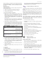
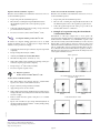
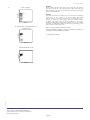
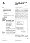
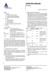
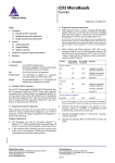
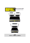
CD4+ CD62L+ T Cell
Isolation Kit II
mouse
Order no. 130-093-227
Index
1.1 Principle of MACS® Separation
1. Description
1.1 Principle of MACS® Separation
1.2 Background and product applications
1.3 Reagent and instrument requirements
2. Protocol
2.1 Sample preparation
2.2 Magnetic labeling of non-CD4+ T cells
2.3 Magnetic separation:
Depletion of non-CD4+ T cells
2.4 Magnetic labeling of CD4+CD62L+ T cells
2.5 Magnetic separation:
Positive selection of CD4+CD62L+ T cells
3. Example of a separation using the
CD4+CD62L+ T Cell Isolation Kit II
1. Description
Components
1 mL CD4+ T Cell Biotin-Antibody Cocktail
II, mouse:
Cocktail of biotin-conjugated monoclonal
anti-mouse antibodies against CD8a, CD45R,
CD11b, CD25, CD49b, TCRγ/δ, and Ter-119.
The isolation of mouse CD4+CD62L+ T cells is performed in a twostep procedure. First, non-CD4+ T cells are indirectly magnetically
labeled with a cocktail of biotin-conjugated antibodies and AntiBiotin MicroBeads. The labeled cells are subsequently depleted by
separation over a MACS® Column.
In the second step, CD4+CD62L+ T cells are directly labeled with
CD62L (L-selectin) MicroBeads and isolated by positive selection
from the pre-enriched CD4+ T cell fraction.
The magnetically labeled CD4+CD62L+ T cells are retained on the
column and eluted after removal of the column from the magnetic
field.
Depletion of non-CD4+ T cells
1. Indirect magnetic labeling of nonCD4+ T cells with the CD4+ T Cell
Biotin-Antibody Cocktail II and AntiBiotin MicroBeads.
2. Magnetic separation using an LS
Column or the autoMACS Separator
(program "Depletes").
Flow-through fraction:
pre-enriched CD4+ T cells
Positive selection of CD4+CD62L+ T cells
2 mL Anti-Biotin MicroBeads:
MicroBeads conjugated to monoclonal antibiotin antibody (isotype: mouse IgG1).
1. Direct magnetic labeling of
CD4+CD62L+ T cells with CD62L
(L-selectin) MicroBeads.
2 mL CD62L (L-selectin) MicroBeads:
MicroBeads conjugated to monoclonal antimouse CD62L (L-selectin; isotype: rat IgG2a)
antibody.
2. Magnetic separation using an MS
Column or the autoMACS Separator
(program "Possel").
Size
For 1×10⁹ total cells.
Product format
All components are supplied in buffer containing
stabilizer and 0.05% sodium azide.
Storage
Store protected from light at 2–8 °C. Do not freeze.
The expiration date is indicated on the vial label.
140-002-112.02
Miltenyi Biotec GmbH
Friedrich-Ebert-Straße 68, 51429 Bergisch Gladbach, Germany
Phone +49 2204 8306-0, Fax +49 2204 85197
[email protected]
www.miltenyibiotec.com
Single-cell suspension
Elution from column:
CD4+CD62L+ T cells
1.2 Background and product applications
The CD4+CD62L+ T Cell Isolation Kit II has been developed for
the isolation of CD4+CD62L+ T helper cells from spleen and lymph
nodes.
CD62L (L-selectin) is highly expressed on naive T cells and downregulated upon activation. It is also expressed on a small subset of
memory T helper cells, the central memory T cells, which can be
distinguished from naive T helper cells by their high expression
of CD44. Furthermore, CD62L is expressed on most thymocytes,
naive CD8+ T cells, B cells, dendritic cells, macrophages, NK cells,
neutrophils, eosinophils, regulatory T cells, and TCRγ/δ+ T cells.
For isolation of CD4+CD62L+ T helper cells, the non-T helper cells
as well as regulatory T cells and TCRγ/δ+ T cells are depleted by
indirect magnetic labeling using a cocktail of lineage-specific biotinconjugated antibodies against CD8a (Ly-2), CD45R (B220), CD49b
(DX5), CD11b (Mac-1), and Ter-119, as well as antibodies against
Miltenyi Biotec Inc.
2303 Lindbergh Street, Auburn, CA 95602, USA
Phone 800 FOR MACS, +1 530 888 8871, Fax +1 530 888 8925
[email protected]
page 1/4
Order no. 130-093-227
CD25 and TCRγ/δ in combination with Anti-Biotin MicroBeads.
Subsequently, CD4+CD62L+ T cells are positively selected from
the enriched CD4+ T helper cell fraction with CD62L (L-selectin)
MicroBeads.
▲ Note: Dead cells may bind non-specifically to MACS MicroBeads. In case of high
numbers of dead cells, removal of dead cells by density gradient centrifugation or the
Dead Cell Removal Kit (# 130-090-101) is recommended.
2.2 Magnetic labeling of non-CD4+ T cells
Example applications
Isolation of CD4+CD62L+ T cells from single cell suspensions of
spleen and lymph nodes for:
●
●
●
analysis of T cell activation by antigen-presenting cells;
studies on cytokine expression and receptor signaling;
adoptive transfer experiments.
1.3 Reagent and instrument requirements
●
Buffer: Prepare a solution containing phosphate-buffered saline
(PBS) pH 7.2, 0.5% bovine serum albumin (BSA), and 2 mM
EDTA by diluting MACS BSA Stock Solution (# 130-091-376)
1:20 with autoMACS™ Rinsing Solution (# 130-091-222). Keep
buffer cold (4−8 °C). Degas buffer before use, as air bubbles could
block the column.
▲ Note: EDTA can be replaced by other supplements such as anticoagulant citrate
dextrose formula-A (ACD-A) or citrate phosphate dextrose (CPD). BSA can be
replaced by other proteins such as gelatine, mouse serum, or fetal calf serum. Buffers or
media containing Ca2+ or Mg2+ are not recommended for use.
MACS Columns and MACS Separators: Depletion of non-CD4+
T cells is performed on an LS Column. The subsequent positive
selection of CD4+CD62L+ T cells is performed on an MS Column.
Depletion and positive selection can also be performed by using the
autoMACS Separator.
●
Column
Max. number of
Max. number of
labeled leukocytes total leukocytes
Separator
Depletion
108
2×10⁹
LS
MidiMACS, QuadroMACS,
VarioMACS, SuperMACS
Positive selection
2×10⁸
MS
107
MiniMACS, OctoMACS,
VarioMACS, SuperMACS
▲ Work fast, keep cells cold and use pre-cooled solutions. This will
prevent capping of antibodies on the cell surface and non-specific cell
labeling.
▲ Volumes for magnetic labeling given below are for up to 108
leukocytes. When working with fewer than 108 cells, use the same
volumes as indicated. When working with higher cell numbers, scale
up all reagent volumes and total volumes accordingly (e.g. for 2×10⁸
leukocytes use twice the volume of all indicated reagent volumes and
total volumes).
▲ For optimal performance it is important to obtain a single-cell
suspension before magnetic separation. Pass cells through 30 µm
nylon mesh (Pre-Separation Filters, # 130-041-407) to remove cell
clumps which may clog the column.
1. Determine the number of leukocytes.
2. Centrifuge cells at 300×g for 10 minutes. Aspirate supernatant
completely.
3. Resuspend cell pellet in 400 µL of buffer per 108 total cells.
4. Add 100 µL of CD4+ T Cell Biotin-Antibody Cocktail II per
108 total cells.
5. Mix well and refrigerate for 10 minutes (4 – 8 °C).
6. Add 300 µL of buffer and 200 µL of Anti-Biotin MicroBeads per
108 total cells.
7. Mix well and refrigerate for 15 minutes (4 – 8 °C).
8. Wash cells by adding 10 mL of buffer and centrifuge at 300×g for
10 minutes at 4 – 8 °C. Aspirate supernatant completely.
9. Resuspend up to 108 cells in 500 µL of buffer.
Depletion and positive selection
autoMACS
2×10⁸
4×10⁹
autoMACS
▲ Note: For higher cell numbers, scale up buffer volume accordingly.
10. Proceed to magnetic separation (2.3).
2.3 Magnetic separation:
Depletion of non-CD4+ T cells
▲ Note: Column adapters are required to insert certain columns into the VarioMACS™
or SuperMACS™ Separators. For details see the respective MACS Separator data sheet.
●
●
●
(Optional) Fluorochrome-conjugated antibodies, for flowcytometric analysis e.g. CD4-FITC (# 130-091-608), CD4-PE
(#130-091-607), CD4-APC (#130-091-611), CD62L-PE (# 130091-794), CD62L-APC (# 130-091-805), CD25-PE (# 130-091013), and CD44 antibodies.
1. Place LS Column in the magnetic field of a suitable MACS
Separator. For details see LS Column data sheet.
(Optional) Propidium iodide (PI) or 7-AAD for flow-cytometric
exclusion of dead cells.
2. Prepare column by rinsing with 3 mL of buffer.
(Optional) Pre-Separation Filters (# 130-041-407) to remove cell
clumps.
4. Collect unlabeled cells which pass through and wash column
with 3×3 mL of buffer. Perform washing steps by adding buffer
three times. Only add new buffer when the column reservoir is
empty. Collect total effluent. This contains the unlabeled preenriched CD4+ T cell fraction.
2. Protocol
2.1 Sample preparation
140-002-112.02
Prepare a single-cell suspension from spleen and lymph nodes using
standard methods.
Depletion with LS Column
3. Apply cell suspension onto the column.
5. Proceed to 2.4 for the isolation of CD4+CD62L+ T cells.
▲ Note: Red blood cell lysis or density gradient centrifugation is not necessary,
since red blood cells are depleted in the first magnetic separation step on the basis
of expression of Ter-119, a red blood cell restricted surface antigen of the mouse.
Unless otherwise specifically indicated, Miltenyi Biotec
products and services are for research use only and not for
diagnostic or therapeutic use.
page 2/4
Order no. 130-093-227
Depletion with the autoMACS™ Separator
Positive selection with the autoMACS™ Separator
▲ Refer to the autoMACS™ User Manual for instructions on how to
use the autoMACS Separator.
▲ Refer to the autoMACS™ User Manual for instructions on how to
use the autoMACS Separator.
1. Prepare and prime the autoMACS Separator.
1. Prepare and prime the autoMACS Separator.
2. Place the tube containing the magnetically labeled cells in
the autoMACS Separator. Choose the separation program
"Depletes".
2. Place the tube containing the magnetically labeled cells in the
autoMACS Separator. Choose the separation program "Possel".
3. Collect the unlabeled fraction from outlet port neg1. This is the
pre-enriched CD4+ T cell fraction.
4. Proceed to 2.4 for the isolation of CD4+CD62L+ T cells.
2.4 Magnetic labeling of naive CD4+ T cells
▲ Volumes for magnetic labeling given below are for an initial
starting cell number of up to 108 leukocytes. For larger initial cell
numbers, scale up volumes accordingly.
1. Centrifuge the cells at 300×g for 10 minutes. Aspirate supernatant
completely.
2. Resuspend cell pellet in 800 µL of buffer.
3. Example of a separation using the CD4+CD62L+
T Cell Isolation Kit II
CD4+CD62L+ T cells were isolated from a mouse spleen cell
suspension using the CD4+CD62L+ T Cell Isolation Kit II, an LS and
an MS Column, a MidiMACS™ and a MiniMACS™ Separator. The
cells were fluorescently stained with CD4-FITC (# 130-091-608) and
CD62L-APC (# 130-091-805) for detection of naive T cells (a) and
with CD62L-APC and CD44-PE for detection of central memory T
cells (b). Additionally, cells were stained with CD25-PE (# 130-091013) to illustrate the removal of CD25+CD62L+ regulatory T cells (c).
Cell debris and dead cells were excluded from the analysis based on
scatter signals and PI fluorescence.
Before separation
Add 200 µL of CD62L (L-selectin) MicroBeads.
5. Wash cells by adding 10 mL of buffer and centrifuge at 300×g for
10 minutes. Aspirate supernatant completely.
6. Resuspend up to 108 cells in 500 µL of buffer.
(b)
(a)
CD62L-APC
4. Mix well and refrigerate for 15 minutes (4 – 8 °C).
CD62L-APC
3.
3. Collect the positive fraction from outlet port pos1. This is the
enriched CD4+CD62L+ T cell fraction.
7. Proceed to magnetic separation (2.5).
CD4-FITC
2.5 Magnetic separation:
Positive selection of CD4+CD62L+ T cells
3. Apply cell suspension onto the column.
CD62L-APC
2. Prepare column by rinsing with 500 µL of buffer.
CD62L-APC
Pre-enriched CD4+ T cells after depletion of
non-CD4+ T cells
Positive selection with MS Columns
1. Place MS Column in the magnetic field of a suitable MACS
Separator. For details see MS Column data sheet.
4. Wash column with 3×500 µL of buffer. Perform washing steps by
adding buffer three times. Only add new buffer when the column
reservoir is empty.
CD44-PE
CD4-FITC
5. Remove column from the separator and place it on a suitable
collection tube.
CD62L-APC
Isolated CD4+CD62L+ T cells
CD62L-APC
6. Pipette 1 mL of buffer onto the column. Immediately flush out
the magnetically labeled CD4+CD62L+ T cells by firmly pushing
the plunger into the column.
CD44-PE
140-002-112.02
CD4-FITC
Unless otherwise specifically indicated, Miltenyi Biotec
products and services are for research use only and not for
diagnostic or therapeutic use.
page 3/4
CD44-PE
Order no. 130-093-227
(c)
CD62L-APC
Before separation
Warnings
Reagents contain sodium azide. Under acidic conditions sodium azide yields hydrazoic
acid, which is extremely toxic. Azide compounds should be diluted with running water
before discarding. These precautions are recommended to avoid deposits in plumbing where
explosive conditions may develop.
Warranty
CD25-PE
Pre-enriched CD4+ T cells after depletion of
non-CD4+ T cells
The products sold hereunder are warranted only to be free from defects in workmanship
and material at the time of delivery to the customer. Miltenyi Biotec GmbH
makes no warranty or representation, either expressed or implied, with respect to
the fitness of a product for a particular purpose. There are no warranties, expressed
or implied, which extend beyond the technical specifications of the products.
Miltenyi Biotec GmbH’s liability is limited to either replacement of the products or refund of
the purchase price. Miltenyi Biotec GmbH is not liable for any property damage, personal
injury or economic loss caused by the product.
MACS is a registered trademark of Miltenyi Biotec GmbH.
CD62L-APC
autoMACS, MidiMACS, MiniMACS, OctoMACS, QuadroMACS, SuperMACS, and
VarioMACS are trademarks of Miltenyi Biotec GmbH.
© 2007 Miltenyi Biotec GmbH.
CD25-PE
CD62L-APC
Isolated CD4+CD62L+ T cells
CD25-PE
140-002-112.02
Unless otherwise specifically indicated, Miltenyi Biotec
products and services are for research use only and not for
diagnostic or therapeutic use.
page 4/4