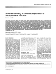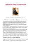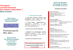
1
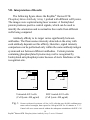
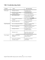
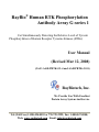
RayBio® Human RTK Phosphorylation
Antibody Array G-series 1
For Simultaneously Detecting the Relative Level of Tyrosin
Phosphorylation of Human Receptor Tyrosine Kinases (RTKs)
User Manual
(Revised Mar 12, 2008)
(Cat#: AAH-PRTK-G1-4 and AAH-PRTK-G1-8)
RayBiotech, Inc.
We Provide You With Excellent
Protein Array System And Service
Tel:(Toll Free) 1-888-494-8555 or 770-729-2992; Fax: 1-888-547-0580;
Web: www.raybiotech.com Email: [email protected]
RayBiotech, Inc.
RayBio® Huamn RTK Phosphorylation Antibody Array G-series 1
Protocol
TABLE OF CONTENTS
I.
Introduction……..……………………………...
2
How It Works………………..…………………
3
II. Materials Provided……………………………..
4
III. Additional Materials Required…………………
5
IV. Reagent Preparation…………………………….
5
V. Overview and General Considerations………… 7
A.Preparation of Samples………………………
7
B. Handling Glass Chips……..…………..……..
8
C. Incubation……………………………………
8
VI. Protocol…………………………………………
9
A. Dry the Array Chips…………………………. 9
B. Blocking and Incubation…………………….. 9
C. Fluorescence Detection……………………… 13
VII. Interpretation of Results……………………….. 14
VIII.Troubleshooting Guide………………………… 16
IX. Reference List………………………………….
17
1
RayBio® Human RTK Phosphorylation Antibody Array G-series 1 Protocol
I. Introduction
Protein phosphorylation plays an unusually prominent role in
cell signaling, development and growth. The RayBio® Human
RTK Phosphorylation Antibody Array G-series 1 is a very rapid,
convenient and sensitive assay to simultaneous detect multiple
protein phosphorylations and can be used to monitor activation or
function of important biological pathways.
RayBiotech is committed to developing a series of
phosphorylation antibody arrays. Our first product in this series is
RayBio® Human RTK Phosphorylation Antibody Array 1 which is
specifically designed for simultaneously identifying the relative
levels of phosphorylation of 71 different Human Receptor Tyrosine
Kinases (RTKs) in cell lysate. By monitoring the changes in protein
tyrosine phosphorylation in your experimental model system, you
can verify pathway activation in your cell lines without spending
excess time and effort in performing an analysis of
immunoprecipitation and/or Western Blot.
To use the RayBio® Human RTK Phosphorylation Antibody
Array G-series 1, treated or untreated cell lysate is added
into antibody array glass chip. The antibody array chips are washed
and biotinylated anti-phosphotyrosine antibodies are used to detect
phosphorylated tyrosines on activated receptors. After incubation
with Fluorescent dye-Conjugated Streptavidin (cy3 equivalent),
image the signals using laser scanner, such as the Axon GenePix,
using the cy3 channel.
2
RayBio® Human RTK Phosphorylation Antibody Array G-series 1 Protocol
Here’s how it works
+
Sample
Antibody array
chips
Incubation of Sample with
arrayed antibody chips
2 hrs
Biotinylated antiPhosphotyrosine
Incubation with
Biotinylated
anti-Phosphotyrosine
LabeledStreptavidin
Incubation with labeled
Streptavidin
Data analysis
and graph
3
RayBio® Human RTK Phosphorylation Antibody Array G-series 1 Protocol
2 hrs
2 hrs
II. Materials Provided
Upon receipt, the kit should be stored at –20 °C to -80 °C. Please use
within 6 months from the date of shipment. After initial use 2X Cell
Lysis Buffer, Blocking Buffer, 20X Wash Buffer I, 20X Wash
Buffer II, Biotin-Conjugated Anti-phosphotyrosine and Fluorescent
dye-Conjugated Streptavidin (cy3 equivalent) should be stored at 4
°C to avoid repeated freeze-thaw cycles. Array I Glass Chip,
Protease Inhibitor Cocktail and Phosphatase Inhibitor Cocktail Set II
should be kept at –20 ° to -80°C. Use within three months after
initial use.
• RayBio® Human RTK Phosphorylation Antibody Array Gseries 1 Glass Chip with Frame (each slide with 4 Subarrays, 1
slide for 4 subarray chips, and 2 for 8 subarray chips)
• 2X Cell Lysis Buffer (5 ml)
• Protease Inhibitor Cocktail (1/2 tubes, 1 tube for 4-subarrary
chips, and 2 for 8-subarray chips)
• Phosphatase Inhibitor Cocktail Set II (1/2 tubes, 1 tube for 4subarrary chips, and 2 for 8-subarray chips)
• Blocking Buffer (8 ml)
• 20X Wash Buffer I (30ml)
• 20X Wash Buffer II (30ml)
• Biotin-Conjugated Anti-phosphotyrosine (1/2 tubes, 1 tube for
4-subarrary chips, and 2 for 8-subarray chips)
• Fluorescent dye-Conjugated Streptavidin (cy3 equivalent) (1/2
tubes, 1 tube for 4-subarrary chips, and 2 for 8-subarray chips)
• Wash Buffer III (20 ml)
• Adhesive film
4
RayBio® Human RTK Phosphorylation Antibody Array G-series 1 Protocol
III. Additional Materials Required
•
•
•
•
•
•
•
Shaker
Laser scanner for fluorescence detection
Aluminum foil
Distilled water
Plastic box
50 ml Centrifuge tube
Isopropanol (2-propanol)
Layout of Array Glass Chip
Array
Blank
Barcode
4 arrays in one glass chip
IV. Reagent Preparation.
1. Protease Inhibitor Cocktail: Briefly spin down the Protease
Inhibitor Cocktail tube before use. Add 60 µl of 1x Lysis Buffer
into the vial to prepare a 100X Protease Inhibitor Cocktail.
5
RayBio® Human RTK Phosphorylation Antibody Array G-series 1 Protocol
2. Phosphatase Inhibitor Cocktail Set II: Briefly spin down the
Phosphatase Inhibitor Cocktail Set II tube before use. Add 180 µl
of 1X Lysis Buffer into the vial to prepare 25X Phosphatase
Inhibitor Cocktail Set II Concentrate. Dissolve the powder
thoroughly by a gentle mix.
3. 2X Cell Lysis Buffer: Cell lysis buffer should be diluted 2-fold
with deionized or distilled water. Add 20 µl of Protease Inhibitor
Cocktail Concentrate and 80 µl of Phosphatase Inhibitor Cocktail
Set II Concentrate into 1.9 ml of 1X Lysis Buffer before use. Mix
well.
4. 20X Washing Buffer I or II : If the 20X Wash Concentrate
contains visible crystals, warm to room temperature and mix
gently until dissolved. Dilute 25 ml of Wash Buffer Concentrate
into deionized or distilled water to yield 500 ml of 1X Wash
Buffer.
5. Biotinylated anti-Phosphotyrosine: Briefly spin the Detection
Antibody tube before use. Add 65 µl of Blocking Buffer into the
tube to prepare a Biotinylated Anti-phosphotyrosine Concentrate.
Pipette up and down to mix gently (the concentrate can be stored
at 40C for 5 days). Add 30 µl of Detection Antibody Concentrate
into a tube with 570 µl of Blocking Buffer. Mix gently to prepare
1X Biotinylated Anti-phosphotyrosine.
6. Fluorescent dye-Conjugated Streptavidin (cy3 equivalent): briefly
spin the Fluorescent dye-Conjugated Streptavidin before use.
Add 50 µl of Blocking Buffer into the tube to prepare a
Streptavidin Concentrate. Pipette up and down to mix gently.
Add 10 µl of Streptavidin Concentrate into a tube with 1 ml of
Blocking Buffer. Mix gently to prepare 1X Streptavidin solution.
6
RayBio® Human RTK Phosphorylation Antibody Array G-series 1 Protocol
V. Overview and General Considerations
A. Preparation of Samples
The cell can be prepared using following conventional way.
For attached cells, remove supernatant from cell culture, wash cells
twice with cold 1X PBS (for suspension cells, pellet the cells by
spinning down the cells at 1500 rpm for 10 min) making sure to
remove any remaining PBS before adding Lysis Buffer containing
Protease Inhibitor Cocktail and Phosphatase Inhibitor Cocktail Set
II. Solubilize the cells at 2x10⁷ cells/ml in 1X Lysis Buffer. Pipette
up and down to resuspend the cells and rock the lysates gently at 2–
8 °C for 30 minutes. Transfer extracts to microcentrifuge tubes and
centrifuge at 14,000 x g for 5 min.
It is recommended that sample protein concentrations be determined
using a total protein assay. For incubation with the Phosphorylation
Antibody Array I, use cell lysates in 50–1000 µg/ml of
concentration (as starting point, we recommended to use 400 µg/ml
of cell lysate, dilute the lysate at least 5-folds with Blocking Buffer).
Lysates should be used immediately or aliquoted and stored at –70
°C. Thawed lysates should be kept on ice prior to use.
If you experience high background, you may further dilute your
sample. If signals are too week, the cell lysates can be pretreated by
immunoprecipitations before incubation with array membranes.
Immunoprecipitations can be done using anti-phosphotyrosine and
protein A.
7
RayBio® Human RTK Phosphorylation Antibody Array G-series 1 Protocol
B. Handling glass chips
• The microarray slides are sensitive, do not touch the array
surface by tips, forceps or hand. Hold the slides by the edges
only.
• Handle all buffers and slides with latex free gloves.
• Avoid breaking glass slide.
• Clean environment
C. Incubation
• Completely cover array area with sample or buffer during
incubation, and cover the incubation chamber with adhesive
film or plastic sheet protector to avoid drying.
• Avoid foaming during incubation steps.
• Perform all incubation and wash steps under gentle rotation.
• Cover the incubation chamber with adhesive film during
incubation, particularly when incubation is more than 2 hours
or less than 50 µl of sample or reagent is used.
• Avoid cross-contamination from overflowing solution to
neighboring wells.
• Several incubation steps such as step 3 (sample incubation),
step 8 (biotin-Ab incubation) or step 11 (Fluorescent dyeConjugated Streptavidin incubation) may be done at 4 °C for
overnight. Please make sure to cover the incubation chamber
tightly to prevent evaporation.
• Avoid array slide to exposure to light since step 9 in page 11.
8
RayBio® Human RTK Phosphorylation Antibody Array G-series 1 Protocol
VI. Protocol
A. Dry the Glass Chip
Open the box containing glass chip with frame and take it out,
and then let it air dry for 1 hour in clean environment before use.
Note: Protect the chip from dust or others contaminants.
B. Blocking and Incubation
1. Add 100 µl of 1 X Blocking Buffer into each well and incubate
at room temperature with gentle shaking for 30 min to block
slides. Make sure no bubbles are in the well.
Note: Only add reagents to wells printed with antibodies.
2. Decant Blocking Buffer from each well (make sure to remove
all of buffer). Add 100 µl of each sample into appropriate
wells. Incubate arrays with sample at room temperature with
gentle shaking for 2 hours or 4 °C for overnight.
Note: We recommended using 100 µl of cell lysates in 50–1000
ug/ml of concentration (as starting point, we
recommended to use 400 µg/ml of concentration of cell
lysate). Dilute the lysate at least 5 folds with blocking
buffer. Make sure there is no bubble in the wells.
Note: The amount of sample used depends on the abundance of
target proteins. More sample can be used if signals are
9
RayBio® Human RTK Phosphorylation Antibody Array G-series 1 Protocol
too weak. If signals are too strong, the sample can be
diluted further.
Note: Incubation may be done at 4 °C overnight.
Note: The cell lysates can be pretreated by
immunoprecipitations before incubation with array
membranes if signals are too week. Immunoprecipitations
can be done using anti-phosphotyrosine and protein A.
The elution samples from protein A can be diluted with
Blocking Buffer and then incubate with array membranes.
3. Decant the samples from each well, and wash 3 times with
100 µl of 1X Wash Buffer I at room temperature with gentle
shaking. 5 min per wash.
Note: avoid solution flowing into neighboring wells.
4. Put the glass chip with frame into a box with Wash Buffer I
(cover the whole glass slide and frame with Wash Buffer I),
and wash at room temperature with gentle shaking for 20 min.
5. Decant the Wash Buffer I from each well, Put the glass chip
with frame into the box with Wash Buffer II (cover the whole
glass slide and frame with Wash Buffer II), and wash 2 times
at room temperature with gentle shaking for 5 min.
6. Remove all of Wash Buffer II in the well. Add 100 µl of 1x
Biotin-conjugated Anti-phosphotyrosine to each
corresponding well. Incubate at room temperature with gentle
shaking for 2 hours.
10
RayBio® Human RTK Phosphorylation Antibody Array G-series 1 Protocol
7. Decant the antibody solution and wash as directed in steps 4,
Wash 3 times.
8. Wash as directed in steps 5.
9. Remove all of Wash Buffer II in the well. Add 100 µl of 1X
Fluorescent dye-Conjugated Streptavidin to each subarray.
Cover the incubation chamber with Adhesive film. Cover the
plate with aluminum foil to avoid exposure to light or
incubate in dark room (the array slide also needs to avoid
exposure to light in the following step 12, 13 and 14).
10. Incubate at room temperature with gentle shaking for 2 hours.
Note: Incubation may be done at 4 °C for overnight.
11. Decant the streptavidin solution and disassemble the slide out
of the incubation frame and chamber.
12. Gently put the glass chip into a 50 ml centrifuge tube or a
plastic box with 40 ml of 1X Wash Buffer I. Gently roll over
the tube or shake for 5 min. Remove the wash buffer. Repeat
2 times for a total of 3 washes.
11
RayBio® Human RTK Phosphorylation Antibody Array G-series 1 Protocol
13. Wash the glass chip with 40 ml of Wash Buffer II. Repeat one
time for a total of two washes. 5 min per wash.
14. Finally wash the glass chip with 40 ml of deionized or
distilled water.
Note: You may assemble the glass chip into an incubation
chamber by following step. You may want to practice assembling
the device with a blank glass slide.
1. Apply slide to incubation chamber barcode facing upward as in
step 1.
2. Gently snap one edge of a snap-on side as shown in step 2.
3. Gently press other of side against lab bench and push in
Direction shown in step 3.
4. Repeat with the other side.
1
3
2
4
12
RayBio® Human RTK Phosphorylation Antibody Array G-series 1 Protocol
C. Fluorescence Detection
1. Put the glass chip into a 50 ml centrifuge tube, dry the glass
chip by centrifuge at 1,000 rpm for 3 minutes. Or dry the
glass chip by a compressed N2 stream. Or let glass chip dry
completely in clean air condition (protect from light). Make
sure the slides are absolutely dry before the scanning
procedure.
Image the signals using laser scanner, such as the Axon
GenePix, using the cy3 channel.
Note:
We recommend scanning slides right after
experiment.You also can store the slide at –20 °C in
dark for several days .If you do not have a laser
scanner, we can provide service for you. Just simply
send your slide to us and we will take care of it.
Note:
Put the glass chip into a tube with 40 ml of 30% Wash
Buffer III in isopropanol (add 15 ml of Wash Buffer
III into a tube with 35 ml of isopropanol, mix well )
and incubate for 10 min at room temperature if the
background is not even or too high (cover the tube
with aluminum foil to avoid exposure to light or
incubate in dark room). Dry the slide and re-scan the
slide.
13
RayBio® Human RTK Phosphorylation Antibody Array G-series 1 Protocol
VII. Interpretation of Results:
The following figure shows the RayBio® Human RTK
Phosphorylation Antibody Array 1 probed with different cell lysates.
The images were captured using laser scanner. A biotinylated
protein produces positive control signals, which can be used to
identify the orientation and to normalize the results from different
wells being compared.
Antibody affinity to its target varies significantly between
antibodies. The fluorescence intensity detected on the array with
each antibody depends on this affinity; therefore, signal intensity
comparison can be performed only within the same antibody/antigen
system and not between different antibodies. Certain proteins
containing phosphorylated tyrosine may not be recognized by
biotinylated anti-phosphotyrosine because of steric hindrance of the
recognition site.
EGFR
ErbB2
EGFR
ErbB3
Untreated A431 cells
(Cell lysate: 400 µg/ml)
Fig. 1.
ErbB2
ErbB3
EGF treated A431 cells
(Cell lysate: 400 µg/ml)
Human epidermoid carcinoma cell line, A431 cells that were 80-90% confluent were
serum starved overnight, then exposed to 100 ng/ml EGF for 10 minutes at 37 °C.
Control cells were serum starved without the subsequent stimulation with EGF. Cell
14
RayBio® Human RTK Phosphorylation Antibody Array G-series 1 Protocol
lysates were prepared following the "Sample Preparation" portion of our protocol IV.
To use the RayBio®Human RTK Phosphorylation Antibody Array G-series 1, treated
or untreated cell lysate was added into antibody array glass chips. The antibody array
chips were washed and biotinylated anti-phosphotyrosine antibody was used to detect
phosphorylated tyrosines on activated receptors. After incubation with Fluorescent
dye-Conjugated Streptavidin, the signals were visualized by laser scanner using the
cy3 channel.
Untreated Treated
EGFR
Untreated Treated Untreated Treated
ErbB2
ErbB3
Fig. 2. Immunoprecipitations were done using anti-EGFR, ErbB2 and
ErbB3 monoclonal antibodies and Protein A. Immunoblots were
incubated with a biotinylated anti-phosphotyrosine monoclonal
antibody to detect phosphorylated target protein receptors. Bands
were visualized with Streptavidin-HRP followed by
chemiluminescent detection substrate.
RayBio® Human RTK Phosphorylation Antibody Array G-series 1
Map
.
1
2
3
4
5
6
7
8
A
B
C
D
E
F
G
H
I
J
K
L
M
N
O
POS 1
POS 1
POS 1
POS 2
POS 2
POS 2
POS3
POS3
POS3
ABL1
ABL1
ABL1
ACK1
ACK1
ACK1
NEG
NEG
NEG
NEG
NEG
NEG
ALK
ALK
ALK
Axl
Axl
Axl
Blk
Blk
Blk
BMX
BMX
BMX
Btk
Btk
Btk
Csk
Csk
Csk
Dtk
Dtk
Dtk
EGFR
EGFR
EGFR
EphA1
EphA6
EphA1
EphA6
EphA1
EphA6
EphA2
EphA2
EphA2
EphA3
EphA3
EphA3
EphA4
EphA4
EphA4
EphA5
EphA5
EphA5
EphA7
EphA7
EphA7
EphA8
EphA8
EphA8
EphB1
EphB1
EphB1
EphB2
EphB2
EphB2
EphB3
EphB3
EphB3
EphB4
EphB4
EphB4
EphB6
EphB6
EphB6
ErbB2
ErbB2
ErbB2
ErbB3
ErbB3
ErbB3
ErbB4
ErbB4
ErbB4
FAK
FAK
FAK
FER
FER
FER
FGFR2
FGFR2
FGFR2
Fgr
Fgr
Fgr
FRK
FRK
FRK
FGFR1
Fyn
FGFR1
Fyn
FGFR1
Fyn
FGFR2
Hck
FGFR2
Hck
FGFR2
Hck
(α isoform) (α isoform) (α isoform)
9
10
11
12
13
14
15
16
HGFR
JAK2
HGFR
JAK2
HGFR
JAK2
IGF-I R
IGF-I R
IGF-I R
Insulin R
Insulin R
Insulin R
Itk
Itk
Itk
JAK1
JAK1
JAK1
JAK3
JAK3
JAK3
LCK
LCK
LCK
LTK
LTK
LTK
Lyn
Lyn
Lyn
MATK
MATK
MATK
M-CSFR
M-CSFR
M-CSFR
MUSK
MUSK
MUSK
NGFR
NGFR
NGFR
PDGFR-β PDGFR-β PDGFR-β
PDGFR-α PDGFR-α PDGFR-α
PYK2
PYK2
PYK2
RET
RET
RET
ROR1
ROR1
ROR1
ROR2
ROR2
ROS
ROS
ROS
RYK
RYK
RYK
SCFR
SCFR
SCFR
SRMS
SRMS
SRMS
SYK
SYK
ROR2
SYK
Tec
Tec
Tec
Tie-1
Tie-1
Tie-1
Tie-2
Tie-2
Tie-2
TNK1
TNK1
TNK1
TRKB
TRKB
TRKB
TXK
TXK
TXK
Tyk2
Tyk2
Tyk2
TYRO10
TYRO10
TYRO10
VEGFR2
VEGFR2
VEGFR2
NEG
NEG
NEG
VEGFR3
VEGFR3
VEGFR3
ZAP70
ZAP70
ZAP70
NEG
NEG
NEG
NEG
NEG
NEG
POS4
POS4
POS4
15
RayBio® Human RTK Phosphorylation Antibody Array G-series 1 Protocol
VIII. Troubleshooting Guide
Problem
Weak signal
Cause
Inadequate detection
Inadequate reagent volumes
or improper dilution
High background
Uneven signal
Recommendation
Check laser power
and PMT parameters
Check pipettors and
ensure correct preparation
Short incubation times
Ensure sufficient incubation
time and change sample
incubation step to overnight
Too low protein concentration
in sample
Don’t make too low dilution
Or concentrate sample
Improper storage of kit
Store kit at suggested
temperature
Excess of biotinylated antibodies
Make sure to use the correct
amount of antibodies
Excess of streptavidin
Make sure to use the correct
amount of streptavidin
Inadequate detection
Check laser power
and PMT parameters
Inadequate wash
Increase the volume of wash
buffer and incubation time
Bubbles formed during
incubation
Avo id bubble formation
during incubation
Arrays are not completely
covered by reagent
Completely cover arrays
with solution
16
RayBio® Human RTK Phosphorylation Antibody Array G-series 1 Protocol
IX. Reference List
1.
Profiling receptor tyrosine kinase activation by using Ab
microarrays. Nielsen UB, Cardone MH, Sinskey AJ, MacBeath G,
and Sorger PK. PNAS. 2003;100(16):9330-9335.
2. A Prototype Antibody Microarray Platform to Monitor Changes
in Protein Tyrosine Phosphorylation. Gembitsky DS, Lawlor K,
Jacovina A, Yaneva M, and Tempst P. Mol Cell Proteomics.
2004;3:1102–1118.
3. Analysis of receptor signaling pathways by mass spectrometry:
Identification of Vav-2 as a substrate of the epidermal and platelet
derived growth factor receptors. Pandey A, Podtelejnikov AV,
Blagoev B, Bustelo XR, Mann M, and Lodish HF. PNAS. 2000;
97(1);179–184.
4. Reduced T-cell and dendritic cell function is related to
cyclooxygenase-2 overexpression and protaglandin e(2) secretion
in patients with breast cancer". Pockaj BA, Basu GD. Annal Surg
Oncol. 2004;3:327-344.
5. Cytokine Antibody Arrays: A Promising Tool to Identify
Molecular Targets for Drug Discovery. Huang RP. Comb Chem
High Throughput Screen. 2003,;6:79-99.
6. Connexin suppresses human glioblastoma cell growth by downregulation of monocyte chemotactic protein 1, as discovered using
protein array technology. Huang R, Lin Y, Wang CC, J et al.
Cancer Res. 2002;62:2806-2812.
17
RayBio® Human RTK Phosphorylation Antibody Array G-series 1 Protocol
7. Profiling of cytokine expression by biotin-labeled-based protein
arrays. Lin Y, Huang R, Chen L-P, et al. Proteomics. 2003, 3:
1750-1757.
8. A novel method for high- throughput protein profiling from
conditioned media and patient’s sera. Huang RP, Huang R, Fan
Y, and Lin Y. Ana. Biochem. 2001;294(1):55-62.
18
RayBio® Human RTK Phosphorylation Antibody Array G-series 1 Protocol
RayBiotech, Inc., the protein array pioneer company, strives to research
and develop new products to meet demands of the biomedical community.
RayBio’s patent-pending technology allows detection of over 180 cytokines,
chemokines and other proteins in a single experiment. Our format is simple,
sensitive, reliable and cost effective. Products include: Cytokine Arrays,
Chemokine Arrays, ELISA kits, Phosphotyrosine kits, Recombinant
Proteins, Antibodies, and custom services.
Antibody Array
Cytokine Antibody Array: Simultaneous detection up to 200 proteins
(cytokine, chemokine, growth factor, adipokine, angiogenic factor,
protease) in one experiment
Phosphorylation Antibody Array
•
•
RTK antibody array
EGFR phosphorylation antibody arrays
Label based antibody array: Simultaneous detection more than 500
proteins in one experiment
Quantibody Array: Quantitative measurement of multiple protein levels
Protein Array
ELISA
Cell-Based Phosphorylation ELISA
Tissue MicroArray
Protein: Cytokine, Chemokine, Adiplokine, Angiogenic factor, Virus, bacteria and
infectious disease protein, hormone, Enzyme, other
Peptide
Antibody: Cytokine, Adipokine, Angiogenic factor, Signal transduction,
Transcription factor, Receptor, Adhesion molecule, Virus, bacteria and other
infectious agents, Secondary antibody, Tag antibody, Immunoglobulin,
Hormone, Cell surface, Protease, other
Assay service: just simply send your samples and get data in 1 to 2 weeks.
Antibody array, Protein array, ELISA, Quantibody array
Antibody production: highest quality with very competitive price
Monoclonal antibody, Recombinant antibody, Polyclonal antibody, Phase
display, Antibody angineering, Antibody conjugation
Recombinant protein production
Assay development
Antibody array, Protein array, Peptide array, ELISA, Phosphorylation assay
Tissue array
Array printing
Contact and non-contact arrayers. All kinds of substrates of your choice
including glass slides, membranes and plates.
19
RayBio® Human RTK Phosphorylation Antibody Array G-series 1 Protocol
Note:
20
RayBio® Human RTK Phosphorylation Antibody Array G-series 1 Protocol
This product is for research use only.
©2004 RayBiotech, Inc.
21
RayBio® Human RTK Phosphorylation Antibody Array G-series 1 Protocol