1
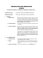
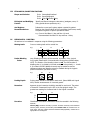
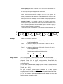
DIVISION OF PERIODONTICS
USER MANUAL FOR
USER MANUAL
FOR
RE-EVALUATION /SPT PATIENTS
SUPPORTIVE PERIODOTAL
MAINTENANCE
Division of Periodontics USER MANUAL: RE-EVALUATION / SPT PATIENTS
2008-09
USER MANUAL FOR CLINICAL PERIODONTOLOGY
2008-2009
Periodontal Re-evaluation OR Supportive Periodontal Treatment (SPT)
Student must circle:
Re-evaluation PSR classification OR SPT classification
Student must enter:
Student name, Patient name, Chart number and Date
I.
HISTORY:
A.
Medical History:
Based on a current medical history, check (√) "Clear" or "No". If
"No" specify condition. Note any prescription drugs and over-thecounter (OTC) medications that the patient is taking. Determine
the drug's function in the CPS, and make a decision if any special
precautions and/or oral manifestations can arise due to the
medication before treatment commences.
B.
Periodontal History:
Circle Yes/No for previous prophylaxis and/or periodontal surgery
— Give dates.
C.
Tobacco-Use History:
Ask the patient if s/he currently uses tobacco in any form. If yes,
“What form of tobacco do you use?” If they do not currently use
tobacco, ask whether they did previously. Document the average
number of cigarettes used each day and approximately the
number of years that the person has used tobacco.
If the patient is a current tobacco-user, advise her/him of the
oral/general health consequences of smoking, strongly advise
her/him to quit and ask her/him how interested s/he is in
quitting. This question will assist you in determining the patient’s
stage of ‘readiness to quit’ and how you can help to move
her/him closer to quitting.
II.
ORAL OBSERVATIONS:
A.
Soft/Hard Tissues:
Record general oral soft and hard tissue changes, and request
an oral pathology consult, as needed.
B.
Gingiva:
Describe generalized color, contour and consistency of gingiva,
both in health and disease, as well as unusual localized gingival
changes.
C.
Minimal Attached Gingiva:
Record all sites with less than 2 mm of attached gingiva.
2
III. ETIOLOGICAL/MODIFYING FACTORS:
Plaque and Calculus:
Circle: Generalized/Localized
Circle: Supra/Subgingival
Circle: Light, Moderate, Heavy
Etiological and Modifying
Factors:
Identify and Record iatrogenic restorations, (amalgam, crown, illfitting partial denture prosthesis, etc.).
Oral HygieneRecommendations:
Indicate the current oral hygiene regimen reported by patient.
Based on the clinical findings of the re-evaluation/recall exam, modify
the patients regimen to improve periodontal status.
e.g. Current: Mod Bass 1 X day and floss 1 X week
Recommended: Mod Bass 2 X day and floss 1 X day
IV.
PERIODONTAL CHARTING:
All teeth are to be evaluated in sequence using the following parameters:
Missing teeth:
Cross out missing teeth with a vertical line.
e.g.
18
17
16
B
L
Pocket Bleeding
Index:
(PBI): Bleeding on probing will be recorded as present, if the gingival
unit (pocket) bleeds within 30 seconds after initial probing (Vander-velden,
1979). The location of the bleeding is done in red. The total number of
bleeding sites (n) is divided by the total number of units probed (n ÷ # teeth
X 4) to obtain the PBI. Additional records may be needed to monitor
subsequent plaque and bleeding scores for feedback during the hygienic
phase of treatment. (See back of chart.)
e.g.
18
17
B
L
Probing Depth:
Probing depth should be recorded for each tooth, (Buccal MBD and Lingual
MLD). Record measurements of 4 mm and greater.
Recession:
Apparent gingival recession should be recorded for each tooth. The extent
of recession is measured from the CEJ to the free gingival margin. The
greatest mm reading for the Buccal and for the Lingual is recorded.
e.g.
Furcation:
18
17
B
2 mm
2 mm
L
3 mm
2 mm
Classification of furcation involvement should be recorded in the following
manner:
Class I (Dip): Furcation concavity is present. Furcation concavity can be detected
with the probe; however, the furcation probe cannot enter the furcation area. In
many cases, this type of defect cannot be detected radiographically. Charted as I in
appropriate box on diagram.
3
Class II (Cave): This lesion is essentially a cave or cul-de-sac lesion with the roof
of the furcation clinically detectable. Furcation probe will enter the furcation area
but cannot pass through to the opposite side of the tooth.
The depth of the horizontal entry into the furcation will vary. This determines
whether the furcation involvement is shallow class II or deep class II. The
difference between shallow class II and deep class II would be an empirical one.
However, notation should be made on the chart indicating shallow class II or deep
(e.g. IIS/IID) class II furcation involvement. A slight radiolucency in the furcation
area may be seen on the radiograph. Charted as IIS/IID directly in appropriate box
on diagram.
Class III (Tunnel): It is essentially a through and through or tunnel lesion.
Furcation probe will pass between the roots through the entire furcation. In this
type of furcation involvement, the inter-radicular bone is completely absent. A
definite radiolucency in the furcation area is usually visible on the radiograph.
Charted as III in appropriate box on diagram.
*Please note that this is a classification of the extent of furcation involvement. The
numerals I, II, and III do not represent millimeter measurements.
e.g.
B
L
Mobility:
I I
II
I
I
III
III
Mobility is recorded for each tooth.
Normal
=
movement of the crown of the tooth, less than .5 mm in
buccal-lingual direction
Class I
=
movement of the crown of the tooth .5 mm to 1.0 mm in
buccal-lingual direction
Class II
=
movement of the crown of the tooth 1.0 mm to 2.0 mm in
buccal-lingual direction
Class III =
movement of more than 2.0 mm in mesial-distal or buccallingual direction and/or vertical depression.
e.g.
Plaque Control
Record:
II/D
II/S
18
17
I
II
(PCR): The presence of dental plaque is recorded on four surfaces of
each tooth (DBML). If, after swishing with a plaque disclosing tablet, disclosed
plaque in contact with the gingival margin can be removed with the
explorer/probe from the tooth surface, that surface should be marked in black
on the record form.
The PCR score is determined by dividing the total number of marked surfaces
(n) by the total number of available surfaces (n ÷ # teeth X 4) present.
(O'Leary, et al, 1972).
Our goal in teaching plaque control procedures is to reduce plaque
accumulation until it is found on 15% or less of the available tooth surfaces.
Surgical procedures should not be initiated until the patient reaches that level.
The PCR is recorded at each session. Additional PCR records are to be used
for this purpose. (See back of chart).
4
V.
AREAS OF PERSISTENT PATHOLOGY:
Student must write, if bleeding areas on probing are present regardless of the pocket depths,
residual deep pockets, mobility, gingival deformity (soft tissue craters and hyperplastic gingiva),
gingival inflammation, gingival suppuration, furcations, mucogingival involvement, sensitivity, trauma
from occlusion, caries, perio/endo lesion, and TM disorder, etc.
VI. PROGNOSIS:
Update the overall and individual tooth prognosis.
TREATMENT PLAN:
The treatment plan for patients undergoing Post-Treatment Assessment / Periodontal Maintenance
Exam will have a definitive surgical treatment plan or a supportive periodontal therapy plan
formulated. The plan must be discussed and approved in writing by the instructor. Upon
completion of the periodontal re-evaluation/SPT examination and instructor feedback, the student
should disclose the patient and record his/her plaque score. Individualized instructions based on
the clinical examination with emphasis on the bleeding score and plaque score should be provided.
Patients are to be given a brush and asked to remove visible plaque. Students are to hold the hand
mirror for the patient and observe the entire brushing procedure, directing the patient to the
problem areas. Patients will then be given the most appropriate tools for interproximal plaque
removal. The student will observe while the patient removes all interproximal plaque. Once the
student is satisfied that the patient has removed the plaque, the student should commence with
indicated scaling/root planing. Upon completion of scaling and root planing, students may carry out
a selective rubber cup polish to remove extrinsic stain. It is the responsibility of the student
that the mouth be “plaque-free” upon completion of periodontal post-treatment
assessment / periodontal maintenance treatment appointment. Clinic instructors will
assess the removal of both hard and soft deposits from the teeth. In addition, they will ask the
patient questions which will help to determine how well the patient understands the oral hygiene
instructions that were given. e.gs.: “What is it that you are going to do to keep your teeth clean
between now and your next appointment?, What was the most important thing you learned today
about how to keep your teeth cleaner?”.
Following completion of active therapy, the date for the post-treatment assessment /periodontal
maintenance treatment appointment must be specified.
VII. TOBACCO-USE INTERVENTION:
For a patient who is a current tobacco-user, the student will discuss oral/general consequences of
tobacco use and will strongly advise him/her to quit, e.g. “As your oral care provider, I strongly
advise you to quit.” Enter the date that you give this advice. There are individualized tobacco use
cessation materials available on the wall by the Dispensary, including information about where
they can be referred for individual/group counseling, nicotine replacement therapy, test for
nicotine dependence, tips on quitting, etc.