1
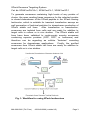
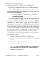
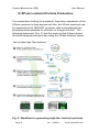
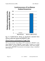
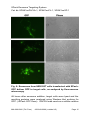
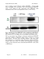
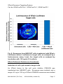
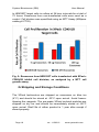
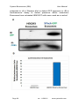
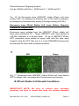
XPack™ Exosome Protein Engineering Technology Cat #s: XPAK1xxPA/VA-1, XPAK1xxCL-1, XPAK1xxXP-1 User Manual Store at -20ºC upon arrival A limited-use label license covers this product. By use of this product, you accept the terms and conditions outlined in the Licensing and Warranty Statement contained in this user manual. XPack Exosome Targeting System Cat. #s XPAK1xxPA/VA-1, XPAK1xxCL-1, XPAK1xxXP-1 Contents I. Introduction ..............................................................................2 A. XPack Overview ..................................................................2 B. Uses of XPack Technology .................................................2 II. XPack Lentivector System .......................................................4 A. List of Constructs and Pre-Packaged Particles ..................4 B. XPack Transfection Protocol ...............................................5 D. XPack Lentiviral Particle Production ...................................8 E. Transduction of Pseudoviral Particles into Target Cells ....11 F. XPack Sample Data ..........................................................12 G. Shipping and Storage Conditions .....................................18 H. Related Products...............................................................19 III. XPack Stable Cell Lines .....................................................19 A. XPack Stable Cell Line Validation Data ............................19 B. XPack Stable Cell Growth Conditions ..............................21 C. Shipping and Storage Conditions .....................................22 IV. XPack Purified Exosomes ..................................................22 V. Frequently Asked Questions ..............................................23 VI. References .........................................................................23 VII. Technical Support ..............................................................26 VIII. Licensing and Warranty information ..................................27 888-266-5066 (Toll Free) 650-968-2200 (outside US) Page 1 System Biosciences (SBI) I. User Manual Introduction A. XPack Overview Exosomes are nanosized membrane vesicles secreted by most cell types in vivo and in vitro. They are produced by the inward budding of multivesicular bodies (MVBs) and subsequently released from the cell into the microenvironment following the fusion of MVBs with the plasma membrane. Exosomes are extracellular nanoshuttles that facilitate communication between cells and organs and are found in various biofluids including blood, urine, amniotic fluid, breast milk, malignant ascites fluid, and cerebrospinal fluid (CSF). Exosomes contain distinct subsets of RNAs and proteins depending upon the cell type from which they are secreted, making them useful for biomarker discovery. Additionally, their natural function as cell to cell communication vehicles makes them attractive for use as therapeutic shuttles to deliver biological molecules or drugs to target disease cells. SBI has identified a specific peptide sequence that targets a protein to the interior exosomal membrane, allowing them to be packaged into exosomes for secretion. This “XPack” peptide sequence has been incorporated into the XPack cloning and expression lentivectors, allowing for reporter proteins or any protein of interest to be loaded into exosomes within a producer cell line, then delivered to target cells of choice. Additionally, stable XPack cell lines have been generated that serve as a constant source of exosomes loaded with reporter protein cargo, enabling tracking of exosome dynamics in both human cells and in vivo murine models. Purified, ready to use exosomes from these stable cell lines are also available as part of the comprehensive XPack system. B. Uses of XPack Technology The XPack technology allows for cell-mediated generation of ready to use exosomes packed with any protein of choice. These exosomes can then be used to efficiently deliver proteins to target cells to alter or supplement biological pathways or be used to study exosome trafficking in vivo. Page 2 ver. 1-150316 www.systembio.com XPack Exosome Targeting System Cat. #s XPAK1xxPA/VA-1, XPAK1xxCL-1, XPAK1xxXP-1 To generate exosomes containing high levels of any protein of choice, the open reading frame sequence for the selected protein is cloned downstream of the XPack peptide in the XPack cloning lentivector, which is suitable for transient transfection assays as well generation of lentiviral particles for downstream production of XPack stable cell lines. After transfection or transduction, exosomes are isolated from cells and are ready for addition to target cells in culture or in vivo studies. The XPack stable cell lines have been validated to continuously secrete exosomes containing reporter proteins (GFP, RFP, or luciferase) and therefore can be regarding as cellular “factories” secreting exosomes for downstream applications. Finally, the purified exosomes from XPack stable cell lines are ready for addition to target cells or in vivo studies. Fig. 1: Workflow for using XPack lentivectors 888-266-5066 (Toll Free) 650-968-2200 (outside US) Page 3 System Biosciences (SBI) II. User Manual XPack Lentivector System A. List of Constructs Particles and Pre-Packaged Cat# Description Size XPAK100PA-1 XPack Cloning Lentivector 10 µg XPAK110PA-1 XPack GFP Lentivector 10 µg XPAK120PA-1 XPack RFP Lentivector 10 µg XPAK130PA-1 XPack Luciferase Lentivector XPack GFP Pre-Packaged Lentivirus 10 µg 2 x 25 ul (> 1 x 10^6 IFUs) XPAK120VA-1 XPack RFP Pre-Packaged Lentivirus 2 x 25 ul (> 1 x 10^6 IFUs) XPAK130VA-1 XPack Luciferase PrePackaged Lentivirus 2 x 25 ul (> 1 x 10^6 IFUs) XPAK110VA-1 NOTE: ExoQuick-TC and Exo-FBS are not provided with the XPack vectors and can be purchased separately. The following ExoQuick-TC products are recommended for exosome concentration prior to addition to target cells. Cat# Description Size EXOTC10A-1 ExoQuick-TC for Tissue Culture Media and Urine ExoQuick-TC for Tissue Culture Media and Urine 10 ml EXOTC50A-1 Page 4 ver. 1-150316 50 ml www.systembio.com XPack Exosome Targeting System Cat. #s XPAK1xxPA/VA-1, XPAK1xxCL-1, XPAK1xxXP-1 EXO-FBSHI50A-1 Exosome-depleted FBS media supplement - Heat Inactivated 50 ml IMPORTANT NOTE: Be sure to culture your exosome producer cell lines in media that does not contain standard FBS. There are high levels of bovine exosomes present in FBS. Instead, use SBI’s Exo-FBS Heat-Inactivated Exosomedepleted FBS Media Supplement (cat#EXO-FBSHI-50A-1) in place of standard FBS media supplements. B. XPack Transfection Protocol Transfection of Exosome Producer Cells: 1. Seed exosome producer cells in culture dish of choice to reach 70-80% confluency after 24 hours using media compatible with the cells of choice. Because standard FBS contains high levels of bovine exosomes, be sure to use SBI’s Exosome-depleted FBS Media Supplement to ensure that exosomes isolated after cell transfection are not contaminated by bovine exosomes. Return cells to incubator. 2. 24 hours later, mix XPack vector with transfection reagent of choice and follow appropriate protocol to achieve transfection of target cells. An example transfection reaction using SBI’s PureFection (Cat# LV750A-1) in a 6-well plate of cells at 70-80% confluency: a. Mix 5 uL PureFection reagent, 2.5 ug XPack Lentivector, and 200 uL serum-free media in sterile 1.5 mL Eppendorf tube. 888-266-5066 (Toll Free) 650-968-2200 (outside US) Page 5 System Biosciences (SBI) User Manual b. Vortex briefly and incubate at room temperature for 15 minutes. c. Add entire volume to 6-well of cells in a total volume of 2-3 ml media. 3. Change media after 24 hours. 4. Isolate exosomes in 48-96 hour window post-transfection Isolation of XPack Exosomes and Addition to Target Cells: 1. Remove cell culture media and place in 15 mL or 50 mL centrifuge tube. 2. Add ExoQuick-TC at 1:5 the volume of cell culture media. 3. Mix by inversion and incubate at 4°C overnight. 4. Spin centrifuge tubes at 3,000 x g for 30 minutes at room temperature or 4°C (temperature does not affect exosome yield). Discard supernatant and resuspend exosome containing pellet in 100 uL PBS. 5. Measure exosome yield using A280 on Nanodrop. Adjust concentration to 1 ug/uL. 6. Add exosomes to cell culture dish containing target cells. For target cells (>1.5x10^5 cells) in a 6 well plate format, 250 ug exosomes is sufficient to visualize efficient delivery of XPack GFP using fluorescence microscopy. The number of exosomes required to discern effects in target cells may vary by cell type and by the specific phenotype being assayed; therefore, optimization of specific experimental conditions may be needed. Page 6 ver. 1-150316 www.systembio.com XPack Exosome Targeting System Cat. #s XPAK1xxPA/VA-1, XPAK1xxCL-1, XPAK1xxXP-1 C. Cloning of XPack Proteins into XPack MCS: 1. The XPack MCS has the following sequence, with restriction enzyme sites (XhoI, BamHI, NotI, EcoRI, NheI, and PstI) in bold and underlined: CTCGAGttGGATCCaaGCGGCCGCgaGAATTCtc GCTAGCatCTGCAG 2. To clone fusions in frame, note that the XPack tag is 32 nucleotides long at the DNA sequence level, making it necessary to add 1 nucleotide to the 5’ end of your ORF to generate an in frame fusion. Then, count the number of nucleotides from the start of the MCS to where first nucleotide in the initial codon of your inserted ORF will fall, and add as many nucleotides as needed to make that number a multiple of 3. Add 1 to this number (to generate an in frame XPack tag), and add that number of nucleotides to your forward PCR primer between the 5’ enzyme site and your ORF sequence. 3. Make sure that any added nucleotides do not generate a premature stop codon. Example: Using EcoRI as the 5’ restriction site: Number of nucleotides from start of MCS to end of EcoRI site: 32 Add 1 to make 33, a multiple of 3 Add 1 to keep XPack fusion in frame 1 + 1 = 2: Add 2 nucleotides (noted as X) to forward PCR primer between EcoRI site and ORF priming sequence: 5’-GAATTC-XX-ORF site 888-266-5066 (Toll Free) 650-968-2200 (outside US) Page 7 System Biosciences (SBI) User Manual D. XPack Lentiviral Particle Production For researchers looking for sustained, long-term expression of the XPack construct in their desired cell line, the XPack construct can be transfected into HEK293T producer cells and packaged into pseudolentiviral particles for infection of a target cell line. The following schematic (Fig. 2) and the protocol that follows shows the lentiviral production process using the XPack lentiviral vector. Fig. 2: Workflow for generating high-titer lentiviral particles Page 8 ver. 1-150316 www.systembio.com XPack Exosome Targeting System Cat. #s XPAK1xxPA/VA-1, XPAK1xxCL-1, XPAK1xxXP-1 1. Transfection of XPack plasmids into HEK293T equivalent) producer cells (or 6 a) 18 - 24 hours prior to transfection, seed 7.0 – 8.0 x10 293T cells per 150mm cell culture plate in standard growth media w/o antibiotics. Cells should be ~80% confluent by next day. b) During transfection day, mix 45 µl of pPACKH1 packaging plasmid mix as provided in the LentiStarter 2.0 Kit and 4.5 µg of XPack lentivector in 1.6 ml of serum-free DMEM by pipetting. c) Add 55 µl PureFection into the same tube. Vortex for 10 seconds. Note: If using other transfection reagents (e.g. Lipofectamine 2000) please follow suggested guidelines for 150mm plates. d) Incubate mixture at room temperature for 15 minutes. e) Add mixture drop-wise to the dish, and swirl to disperse evenly throughout the plates. f) Change the medium ~12 hours (or next day) after transfection. g) At 48 hours and 72 hours after transfection, collect the medium (which now contains pseudoviral particles) into a 50-ml sterile, capped conical centrifuge tube. Centrifuge at 3000 x g for 15 minutes at room temperature to pellet cell debris. Transfer the viral supernatant into a new tube. Caution: You are working with infectious pseudoviral particles at this stage. Please follow the recommended guidelines for working with BSL-2 biosafety agents. 2. Concentration of Pseudoviral Particles 888-266-5066 (Toll Free) 650-968-2200 (outside US) Page 9 System Biosciences (SBI) User Manual The PEG-it™ Virus Precipitation Solution in the LentiStarter 2.0 Kit provides a simple and highly effective means to concentrate lentiviral particles. PEG-it is a formulation of polyethylene glycol optimized for the precipitation of lentiviral-based particles. The PEG-it Virus Precipitation Solution is provided as a 5x solution. 1. Transfer supernatant containing virus to a sterile vessel and add 1 volume of cold PEG-it Virus Precipitation Solution (4ºC) to every 4 volumes of virus supernatant. (Example: 5ml PEG-it with 20ml viral supernatant). 2. Refrigerate overnight (at least 12 hours). Viral supernatants mixed with PEG-it Virus Precipitation Solution are stable for up to 4-5 days at 4°C. 3. Centrifuge supernatant/PEG-it mixture at 1500 × g for 30 minutes at 4ºC. After centrifugation, the virus particles may appear as a beige or white pellet at the bottom of the vessel. 4. Discard the supernatant into a suitable biohazard waste container. Spin down residual PEG-it solution by centrifugation at 1500 × g for 5 minutes. Remove all traces of fluid by aspiration, taking great care not to disturb the precipitated lentiviral particles in pellet. 5. Resuspend lentiviral pellets in 1/500 to 1/1000 of original volume of pooled virus supernatant using cold, sterile Phosphate Buffered Saline (PBS) or DMEM containing 25mM HEPES buffer at 4ºC. For example, if you performed 2 collections from 2 x 150mm plates (20ml per plate), this would be approximately 80ml of media. You would resuspend the resulting pellet in 80-160 µl of 1X PBS or DMEM. 6. Aliquot in cryogenic vials and store at -80°C until ready for use. 7. The resulting pseudoviral particles can be accurately titered using SBI’s UltraRapid Global Titering Kit (Cat #LV961A-1) Page 10 ver. 1-150316 www.systembio.com XPack Exosome Targeting System Cat. #s XPAK1xxPA/VA-1, XPAK1xxCL-1, XPAK1xxXP-1 http://www.systembio.com/lentiviral-technology/deliverysystems/ultrarapid/overview. E. Transduction of Pseudoviral Particles into Target Cells For efficient transduction of target cells, the negative charges present in the virus envelope protein and the cell surface must be neutralized. SBI’s TransDux reagent (provided in the LentiStarter 2.0 Kit) is a non-toxic, proprietory formulation that promotes cellvirus contact and subsequent fusion by negating these charges. The following protocol can be utilized for delivery of virus to your target cells. The following protocol is for infection of target cells in a single well of a 24-well plate – if using larger vessels please scale up reagents accordingly. Day 1 1. Plate 75,000 cells per well into a single well of a 24-well plate in cell culture medium. Make sure that cells are well-dispersed and are not clumped together. Include wells for negative (noninfected) cells. Note: If infecting target cells for the first time or an optimal MOI is not known, please titrate virus at varying MOIs (1, 5, 10 and 20, etc.) to optimize transduction using a positive control virus with a fluorescent marker such as SBI’s pre-packaged positive transduction control (Cat #CD511VB-1). Day 2 2. Cells should be between 70-80% confluent. Aspirate medium from cells. 3. Combine culture medium with TransDux to a 1X final 888-266-5066 (Toll Free) 650-968-2200 (outside US) Page 11 System Biosciences (SBI) User Manual concentration. For example, add 2.5 μl of TransDux to 500 µl culture medium and then transfer to each well. If using other types of transduction reagents (e.g. Polybrene) please dilute the reagent to a final working concentration of 2-8 μg/ml. 4. Add XPack virus at desired MOI to each well and swirl to mix, for negative control wells only add media/viral transduction reagent. Day 3 5. Aspirate off medium and add complete growth medium to cells. Day 5 7. Virus should be integrated into the host cell genome by this time, and should be expressing the XPack construct for packaging into exosomes. F. XPack Sample Data XPack Proteins are Packaged into Exosomes: XPack GFP was transfected into HEK293T cells as described above. GFP localization patterns within exosome producer cells were visualized on a Leica DMI300B fluorescence microscope using a standard GFP filter set. XPack-GFP The characteristic localization pattern observed in exosome producer cells is shown in Figure 3. Fluorescence is detected along the cell periphery, which indicate budding exosomes, and in a brightly fluorescent spot in the cell interior, which indicates the MVB. Page 12 ver. 1-150316 www.systembio.com XPack Exosome Targeting System Cat. #s XPAK1xxPA/VA-1, XPAK1xxCL-1, XPAK1xxXP-1 Fig. 3: Visualization of XPack-GFP exosome producing HEK293T cells. Exosomal presence of GFP in exosomes secreted from HEK293T XPack-GFP producing cells (Fig. 4) was determined using Western blot using a primary antibody against Turbo GFP antibody (Thermo Scientific catalog # PA5-22688). An antibody against the cholinesterase transfer protein (CETP; Abcam catalog # ab19012) was used as an exosomal marker. Fig. 4: Western blot on exosomes secreted from XPack-GFP transfected HEK293T cells. Exosomal presence of firefly luciferase in exosomes secreted from HEK293T XPack-Luciferase producing cells was determined using Promega’s Luciferase Assay System (Promega catalog # E1501). Notably, the size of firefly luciferase is ~60 KDa, indicating that mid to large size proteins can be packaged into exosomes efficiently using the XPack system (Fig. 5 below) 888-266-5066 (Toll Free) 650-968-2200 (outside US) Page 13 System Biosciences (SBI) User Manual Fig. 5: Luminescence assay on exosomes secreted from XPack-Luciferase transfected HEK293T cells. XPack Proteins are Delivered to Target Cells: 250 ug exosomes from XPack GFP transfected HEK293T cells were added to naïve HEK293T target cells in culture in a 6-well plate format. 24 hours after exosome addition, cells were imaged on a Leica DMI300B fluorescence microscope using a standard GFP filter set. Page 14 ver. 1-150316 www.systembio.com XPack Exosome Targeting System Cat. #s XPAK1xxPA/VA-1, XPAK1xxCL-1, XPAK1xxXP-1 Fig. 6: Exosomes from HEK293T cells transfected with XPackGFP deliver GFP to target cells, as analyzed by fluorescence microscopy. 48 hours after exosome addition, target cells were lysed and the resulting proteins were analyzed using Western blot probing for GFP (+XPack GFP Exos). GAPDH was used as a cellular marker 888-266-5066 (Toll Free) 650-968-2200 (outside US) Page 15 System Biosciences (SBI) User Manual and a loading control (Abcam catalog #ab9485). Control cells were incubated with exosomes from non-transfected HEK293T cells (+ NT Exos) or with exosomes from HEK293T cells transfected with a GFP overexpression vector (+ GFP Exos). Fig. 7: Exosomes from HEK293T cells transfected with XPackGFP deliver GFP to target cells, as analyzed by Western blot. 250 ug exosomes from XPack Luciferase transfected HEK293T cells were added to HEK293T target cells in culture in a 6-well plate format. 24 hours after exosome addition, cells were lysed and subjected to a luciferase assay (Promega catalog # E1501). Control cells were either not treated with exosomes (Untreated Cells) or with exosomes from untransfected HEK293T cells (Cells + HEK Exos). Page 16 ver. 1-150316 www.systembio.com XPack Exosome Targeting System Cat. #s XPAK1xxPA/VA-1, XPAK1xxCL-1, XPAK1xxXP-1 Fig. 8: Exosomes from HEK293T cells transfected with XPackLuciferase deliver Luciferase to target cells, as analyzed by a luminescence assay. Inset, live target cells as analyzed by incubation with 150 ng/mL D-Luciferin. XPack Delivered Proteins are Functional: Induction of cell cycle arrest in target cells An XPack fusion to the cell cycle inhibitor CDKN1B was transfected into HEK293T cells. Exosomes were isolated, and a range of 5 ug to 100 ug of XPack-CDKN1B exosomes were added 888-266-5066 (Toll Free) 650-968-2200 (outside US) Page 17 System Biosciences (SBI) User Manual to HEK293T target cells in culture at 24 hour intervals for a total of 72 hours. Exosomes from non-transfected cells were used as a control. Cell division was quantified using an MTT Assay (Millipore catalog # CT02). Fig. 9: Exosomes from HEK293T cells transfected with XPackCDKN1B inhibit cell division, as analyzed by a MTT cell growth assay. G. Shipping and Storage Conditions The XPack lentivectors are shipped on exosomes on blue ice (4°C) and should be stored at -20°C upon arrival. Avoid freezethawing the reagents. The pre-made XPack lentiviral particles are shipped on dry ice and should be immediately stored at -80°C upon arrival. Shelf life of either product is 1 year after receipt if stored properly. Page 18 ver. 1-150316 www.systembio.com XPack Exosome Targeting System Cat. #s XPAK1xxPA/VA-1, XPAK1xxCL-1, XPAK1xxXP-1 H. Related Products SBI offers a number of exosome research products. You can review them here: http://www.systembio.com/exosomes • • • • • III. ExoQuick exosome isolation reagents Exo-FBS exosome-depleted media supplement Detect and quantitate exosomes with ExoAB, ELISA, and EXOCET kits Purify exosome RNA and profile by qPCR with SeraMir Kit Discover novel exoRNA biomarkers with Exo-NGS next-gen sequencing services XPack Stable Cell Lines Stable HEK293T cell lines have been created that constitutively secrete exosomes packaged with GFP (Cat #XPAK110CL-1) and Firefly Luciferase (Cat #XPAK120CL-1). These cell lines were generated by transduction of XPack lentiviral particles into each parental cell line. Exosomes from XPack stable cell lines have been validated to contain the specified reporter protein by luciferase assay and Western blot. A. XPack Stable Cell Line Validation Data Exosomes from XPack Stable Cell Lines are Loaded with Reporter Proteins: After a stable cell population was generated using puromycin selection, exosomes were isolated from HEK293T XPack stable cell line media using ExoQuick-TC. The exosomes were 888-266-5066 (Toll Free) 650-968-2200 (outside US) Page 19 System Biosciences (SBI) User Manual subjected to (A) a Western blot to detect GFP presence or (B) a luminescence assay to detect presence within exosomes. Exosomes from untreated HEK293T cells were used as a control. Page 20 ver. 1-150316 www.systembio.com XPack Exosome Targeting System Cat. #s XPAK1xxPA/VA-1, XPAK1xxCL-1, XPAK1xxXP-1 Fig. 10: A) Exosomes from HEK293T stable XPack cell lines contain GFP, as assayed by Western blot, or B) Firefly Luciferase, as assayed by luminescence assay (Promega catalog # E1501) Exosomes from XPack Stable Cell Lines Deliver Reporter Proteins to Target Cells: Exosomes were isolated from the HEK293T XPack stable cell lines using ExoQuick-TC. Target HEK293T cells were plated at 100,000 cells/well in a 24-well plate. 50 ug of HEK293T XPack GFP exosomes were added to target cells and the cells were imaged using the GFP filter set on a Leica DMI300B fluorescence microscope 24 hours after exosome addition. Fig. 11: Exosomes from HEK293T stable XPack cell lines deliver GFP to target cells, as assayed by fluorescence microscopy B. XPack Stable Cell Growth Conditions IMPORTANT NOTE: Be sure to culture your exosome producer cell lines in media that does not contain standard 888-266-5066 (Toll Free) 650-968-2200 (outside US) Page 21 System Biosciences (SBI) User Manual FBS. There are high levels of bovine exosomes present in FBS. Instead, use SBI’s Exo-FBS Exosome-depleted FBS Media Supplement (cat#EXO-FBS-50A-1) in place of standard FBS media supplements. Parental Cell Line HEK293T Base Media Supplement(s) Dulbecco’s 10 % Fetal Bovine Modified Serum; Antibiotics of Eagle’s Medium choice (DMEM) C. Shipping and Storage Conditions The XPack stable cell lines are shipped on dry ice and should be stored in the vapor phase of liquid nitrogen dewar immediately upon arrival. IV. XPack Purified Exosomes Exosomes have been purified from each stable cell line described above. These exosomes are ready to use for addition to target cells, for in vivo tracking studies, or for other applications. Prepackaged, purified exosomes containing GFP (Cat #XPAK120XP1) or firefly luciferase (XPAK130XP-1) from HEK293T cells are available. These exosomes have been validated to contain the specified reporter protein by luciferase assay and Western blot and were obtained from cells grown in exosome-depleted FBS; therefore, they are free of contaminating bovine exosomes. The XPack purified exosomes are shipped on blue ice (-20°C) and should be stored at -20°C upon arrival. Avoid freeze-thawing the Page 22 ver. 1-150316 www.systembio.com XPack Exosome Targeting System Cat. #s XPAK1xxPA/VA-1, XPAK1xxCL-1, XPAK1xxXP-1 reagents. Shelf life of the product is 1 year after receipt if stored in -20°C. V. Frequently Asked Questions Q. How long and in what condition should I store exosomes after isolation from exosome generating cell line? After exosomes are isolated with ExoQuick-TC, the pellet can be stored at -80°C for 1 year. After resuspension in PBS, it can be stored at 4°C for 2 weeks or -20°C for 3 months. Q. How many exosomes should I add to my target cells? 250 ug of exosomes (as determined by A280 on NanoDrop) is sufficient to see efficient delivery of XPack reporter proteins on target cells in a 6-well plate format. The number of exosomes required in culture dishes of other size can be scaled up or down proportionally to the difference in total cell number relative to one well of a 6 well plate. Example: HEK293T cells 6 well seeding density: 400,000 cells 24 well seeding density: 100,000 cells 100,000/400,000 = ¼ number of cells 250 ug exosomes x ¼ = 62.5 ug exosomes for use in 24 well plate format. VI. References György B, Hung ME, Breakefield XO, Leonard JN. Therapeutic applications of extracellular vesicles: clinical promise and open questions. Annu Rev Pharmacol Toxicol. 2015;55:439-64. doi: 888-266-5066 (Toll Free) 650-968-2200 (outside US) Page 23 System Biosciences (SBI) User Manual 10.1146/annurev-pharmtox-010814-124630. Epub 2014 Oct 3. PubMed PMID: 25292428. van der Meel R, Fens MH, Vader P, van Solinge WW, EniolaAdefeso O, Schiffelers RM. Extracellular vesicles as drug delivery systems: lessons from the liposome field. J Control Release. 2014 Dec 10;195:72-85. doi: 10.1016/j.jconrel.2014.07.049. Epub 2014 Aug 2. Review. PubMed PMID: 25094032. Coleman BM, Hill AF. Extracellular vesicles - Their role in the packaging and spread of misfolded proteins associated with neurodegenerative diseases. Semin Cell Dev Biol. 2015 Feb 20. pii: S1084-9521(15)00034-8.doi:10.1016/j.semcdb.2015.02.007. [Epub ahead of print] Review. PubMed PMID:25704308. Yao Y, Wei W, Sun J, Chen L, Deng X, Ma L, Hao S. Proteomic analysis of exosomes derived from human lymphoma cells. Eur J Med Res. 2015 Jan 29;20(1):8 PubMed PMID: 25631545; PubMed Central PMCID: PMC4329659. Yang J, Wei F, Schafer C, Wong DT. Detection of tumor cellspecific mRNA and protein in exosome-like microvesicles from blood and saliva. PLoS One. 2014 Nov 14;9(11):e110641. doi: 10.1371/journal.pone.0110641. eCollection 2014. PubMed PMID: 25397880; PubMed Central PMCID: PMC4232306. Zhao X, Wu Y, Duan J, Ma Y, Shen Z, Wei L, Cui X, Zhang J, Xie Y, Liu J. Quantitative proteomic analysis of exosome protein content changes induced by hepatitis B virus in Huh-7 cells using SILAC labeling and LC-MS/MS. J Proteome Res. 2014 Dec 5;13(12):5391-402. doi: 10.1021/pr5008703. Epub 2014 Oct 8. PubMed PMID: 25265333. Revenfeld AL, Bæk R, Nielsen MH, Stensballe A, Varming K, Jørgensen M. Diagnostic and prognostic potential of extracellular vesicles in peripheral blood. Clin Ther. 2014 Jun 1;36(6):830-46. doi: 10.1016/j.clinthera.2014.05.008. PubMed PMID: 24952934. Page 24 ver. 1-150316 www.systembio.com XPack Exosome Targeting System Cat. #s XPAK1xxPA/VA-1, XPAK1xxCL-1, XPAK1xxXP-1 Zhang L, Wrana JL. The emerging role of exosomes in Wnt secretion and transport. Curr Opin Genet Dev. 2014 Aug;27:14-9. doi: 10.1016/j.gde.2014.03.006. Epub 2014 May 8. Review. PubMed PMID: 24791688. Drake RR, Kislinger T. The proteomics of prostate cancer exosomes. Expert Rev Proteomics. 2014 Apr;11(2):167-77. doi: 10.1586/14789450.2014.890894. Epub 2014 Feb 25. PubMed PMID: 24564711. Soldevilla B, Rodríguez M, San Millán C, García V, FernándezPeriañez R,Gil-Calderón B, Martín P, García-Grande A, Silva J, Bonilla F, Domínguez G.Tumor-derived exosomes are enriched in ΔNp73, which promotes oncogenic potential in acceptor cells and correlates with patient survival. Hum Mol Genet. 2014 Jan 15;23(2):467-78. doi: 10.1093/hmg/ddt437. Epub 2013 Sep 18. PubMed PMID:24067531. Camacho L, Guerrero P, Marchetti D. MicroRNA and protein profiling of brain metastasis competent cell-derived exosomes. PLoS One. 2013 Sep 16;8(9):e73790.doi: 10.1371/journal.pone.0073790. eCollection 2013. PubMed PMID: 24066071; PubMed Central PMCID: PMC3774795. Raimondo F, Morosi L, Corbetta S, Chinello C, Brambilla P, Della Mina P, Villa A, Albo G, Battaglia C, Bosari S, Magni F, Pitto M. Differential protein profiling of renal cell carcinoma urinary exosomes. Mol Biosyst. 2013 Jun;9(6):1220-33. doi: 10.1039/c3mb25582d. Epub 2013 Mar 19. PubMed PMID: 23511837. de Jong OG, Verhaar MC, Chen Y, Vader P, Gremmels H, Posthuma G, Schiffelers RM, Gucek M, van Balkom BW. Cellular stress conditions are reflected in theprotein and RNA content of endothelial cell-derived exosomes. J Extracell Vesicles. 2012 Apr 888-266-5066 (Toll Free) 650-968-2200 (outside US) Page 25 System Biosciences (SBI) User Manual 16;1. doi: 10.3402/jev.v1i0.18396. eCollection 2012. PubMed PMID: 24009886; PubMed Central PMCID: PMC3760650. VII. Technical Support For more information about SBI products and to download manuals in PDF format, please visit our web site: http://www.systembio.com For additional information or technical assistance, please call or email us at: System Biosciences (SBI) 265 North Whisman Rd. Mountain View, CA 94043 Phone: (650) 968-2200 (888) 266-5066 (Toll Free) Fax: (650) 968-2277 E-mails: General Information: [email protected] Technical Support: [email protected] Ordering Information: [email protected] Page 26 ver. 1-150316 www.systembio.com XPack Exosome Targeting System Cat. #s XPAK1xxPA/VA-1, XPAK1xxCL-1, XPAK1xxXP-1 VIII. Licensing and Warranty information Limited Use License Use of the XPack system (i.e., the “Product”) is subject to the following terms and conditions. If the terms and conditions are not acceptable, return all components of the Product to System Biosciences (SBI) within 7 calendar days. Purchase and use of any part of the Product constitutes acceptance of the above terms. The purchaser of the Product is granted a limited license to use the Product under the following terms and conditions: The Product shall be used by the purchaser for internal research purposes only. The Product is expressly not designed, intended, or warranted for use in humans or for therapeutic or diagnostic use. The Product may not be resold, modified for resale, or used to manufacture commercial products without prior written consent of SBI. This Product should be used in accordance with the NIH guidelines developed for recombinant DNA and genetic research. Purchase of the product does not grant any rights or license for use other than those explicitly listed in this Licensing and Warranty Statement. Use of the Product for any use other than described expressly herein may be covered by patents or subject to rights other than those mentioned. SBI disclaims any and all responsibility for injury or damage which may be caused by the 888-266-5066 (Toll Free) 650-968-2200 (outside US) Page 27 System Biosciences (SBI) User Manual failure of the buyer or any other person to use the Product in accordance with the terms and conditions outlined herein. Limited Warranty SBI warrants that the Product meets the specifications described in this manual. If it is proven to the satisfaction of SBI that the Product fails to meet these specifications, SBI will replace the Product or provide the purchaser with a credit. This limited warranty shall not extend to anyone other than the original purchaser of the Product. Notice of nonconforming products must be made to SBI within 30 days of receipt of the Product. SBI’s liability is expressly limited to replacement of Product or a credit limited to the actual purchase price. SBI’s liability does not extend to any damages arising from use or improper use of the Product, or losses associated with the use of additional materials or reagents. This limited warranty is the sole and exclusive warranty. SBI does not provide any other warranties of any kind, expressed or implied, including the merchantability or fitness of the Product for a particular purpose. SBI is committed to providing our customers with high-quality products. If you should have any questions or concerns about any SBI products, please contact us at (888) 266-5066. © 2015 System Biosciences (SBI), All Rights Reserved. Page 28 ver. 1-150316 www.systembio.com