1
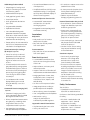
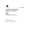
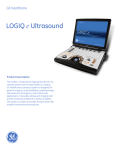
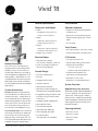
Vivid T8
™
General Specifications
User Interface
Dimensions and Weight
Operator Keyboard
• Width:
- Keyboard: 500 mm (19.7 in)
- Caster: 720 mm (28.3 in)
• Support for international keyboard
character sets
• Depth:
- Maximum: 810 mm (31.9 in)
- Caster: 800 mm (31.5 in)
• Interactive back lighting for control
panel
• Height:
- Maximum: 1495 mm (58.9 in)
- Minimum: 1410 mm (55.5 in)
• Weight: 58 kg, 128 lbs
• Ergonomic full size hard key layout
• Six TGC pods
Touch Screen
• 8.4" high-resolution, color, touch screen
• Interactive dynamic software menu
Electrical Power
LCD Monitor
• Nominal input voltage:
100-240 VAC, frequency 50/60 Hz
• 19" high-resolution LCD
• Optional articulating monitor arm
The Vivid T8 is our Multipurpose
Cardiovascular System designed for
reliability in busy environments for
cardiac and shared service imaging,
with the additional capabilities of 2D,
adult, pediatric, fetal/obstetrics, vascular/peripheral vascular, urological,
abdominal, transcranial, small parts,
musculoskeletal, and transesophageal
applications.
• Power consumption maximum:
400 VA with peripherals
• Optional LCD translation:
- +25˚/–90˚ tilt on LCD
- Swivel to side viewing direction
System Architecture
• Optional on-board storage for
thermal printer, integrated speakers
Product Description
Ease of Use features help make the
Vivid T8 a productive cardiovascular
ultrasound system – simplification of
Vivid software and touch panel help
make working seamless in any environment. Stress Echo, Auto EF and Scan
assist help allow for a productive
environment with added efficiency.
True Scan Raw Data is GE’s innovative
technology that allows for advanced
processing on archived images by
applying many of the same scan controls and advanced quantitative tools
available during the original exam.
Console Design
• Four active probe ports
• Fold down and rotation lock
mechanism for transportation
• ECG port
• Resolution: 1280 x 1024
• Integrated HDD (500G)
• Brightness, contrast adjustments
• Multiple USB ports
• Optional DVD-RW drive
• Integrated locking mechanism that
provides rolling lock and caster
swivel lock
• Integrated cable management
• Removable air filters
• Front and rear handles
System Overview
Applications (probe dependent)
Abdominal, Cardiac (adult and pediatric),
Musculo-skeletal (conventional and
superficial), Small Organ, Pediatric,
Obstetrics/Gyn, Fetal (heart and body),
Transesophageal, Peripheral Vascular,
Transvaginal, Transrectal, Adult, and
Neonatal Cephalic
• Optional probe cable tray
Scanning Methods
• One integrated gel holder
• Electronic sector
• Six probe holders
(two optional, four standard)
• Electronic convex
• Electronic linear
• CW pencil
Vivid T8 product datasheet – April 2014 – DOC1509022
Page 1 of 10
Transducer Types
System Options
• Sector phased array
• Curved AMM
• Convex array
• Blood flow imaging
• Linear array
• Blood flow imaging with Angio
• CW Pencil
• B-flow
• Endovaginal
• Tissue tracking
• TEE
• TSI
Operating Modes
• 2D tissue
• 2D color flow
• 2D angio flow
• Color M-mode
• Tissue velocity M-mode
• Continuous wave Doppler
• Tissue M-mode
• Pulsed wave Doppler with high PRF
• Anatomical M-mode
• Curved anatomical M-mode (optional)
• Tissue velocity imaging
• Tissue tracking (optional)
• Tissue synchronization imaging
(optional)
• Strain imaging
• Strain rate imaging
• Smart Stress (optional)
• AFI
• Auto EF
• Q-Analysis
• LOGIQView
• Scan Assist
• IMT
• DICOM* connectivity
• MpegVue/eVue
• Selectable alternating modes
- B or compound + PW
- B + CW
- B or compound + CFM/PW
- B + CFM + CW
• Multi-image (split/quad screen)
- Live and/or frozen
- Independent cine playback
• Paper tray
• Timeline display
- Independent B (or compound) +
PW/CW/M display
- Display formats
• Probe cable tray
• Top/bottom selectable format
• LVO contrast
• Extra probe holder
• Strain imaging (optional)
• Flexible arm
• Strain rate imaging (optional)
Peripheral Options
• Tissue Doppler imaging
• Simultaneous capability
- B + PW
- B + CFM/TVI + PW
- B + CFM/TVI
- B + CFM/Angio/TVI/SRI/TT/SI/TSI
- B + M/AMM/CAMM
- B + CFM/Angio/TVI/SRI/TT/SI/TSI +
M/AMM/CAMM
- Real-time duplex or triplex mode
- Compound + M/CFM/PW
- B + color split screen
(simultaneous mode)
Display Annotation
• Patient name: First, last and middle
• LITEON* eUAU108 DVDRW
• Blood flow imaging (optional)
• Patient ID
• UP-D711MD
• Blood flow angio flow imaging
• Age, sex and birth date
• SONY* UPD25 color printer
• Hospital name
• B/W printer (UP-D897MD)
• Date format: Two types selectable
- MM/DD/YY, DD/MM/YY
(optional)
• Smart Stress (optional)
• Auto EF (optional)
• AFI Automated Function Imaging
(optional)
• Coded phase inversion LVO contrast
(optional)
• Compound imaging
• HP* 100 printer
• 1 TB USB hard drive
• Time format: Two types selectable
- 24 hours, 12 hours
• Three-pedal configurable footswitch
• Gestational age from LMP/EDD/GA
• ECG kits
• Probe name
• 8 GB memory stick
• Map names
• Scan assist (optional)
Display Modes
• Image archive
• Live and stored display format:
Full size and split screen, both
with thumbnails, for still and cine
• Depth scale marker
• Review image format: 4 x 3 and
thumbnails for still and cine
• Image depth
• Z scores
• Fetal trending
• Renal calculations
• Probe orientation
• Focal zone markers
• Zoom depth
• On-board report package
• MPEGVue (optional)
Vivid T8 product datasheet – April 2014 – DOC1509022
Page 2 of 10
• B-mode
- Gain
- Dynamic range
- Imaging frequency
- Frame averaging
- Gray map
- Speckle reduction imaging
- UD clarity
• M-mode
- Gain
- Dynamic range
- Time scale
• Doppler mode
- Gain
- Angle
- Sample volume size and position
- Wall filter
- Velocity and/or frequency scale
- Spectrum inversion
• Time scale
- PRF
- Doppler frequency
• Color flow Doppler mode
- Frame rate
- Frame averaging
- Sample volume size
- Color scale
- Power
- Color baseline
- Color threshold marker
- Color gain
- PDI
• Spectrum inversion
• Doppler
• Biopsy guide line and zone
Image Storage
• Heart rate
• On-board database of patient
information from past exams
• Trackball-driven annotation arrows
• Active mode display
• Stress protocol parameters
• Parameter annotation follow
ASE standard
• Free text with work library
General System Parameters
System Setup
• Pre-programmable categories
• User programmable preset capability
with administrator preset protection
• Factory default preset data
• Languages: English, French, German,
Spanish, Italian, Portuguese, Swedish,
Danish, Dutch, Norwegian, Japanese,
Chinese, Polish, Finnish, Greek, Russian,
Hungarian, Slovak, Romanian, Czech,
Latvian, Lithuanian, Turkish, Estonian,
Korean, Serbian, Bulgarian, Croatian,
Indonesian, Slovenian
• Reload of archived data sets
Connectivity and DICOM
• Ethernet network connection
• DICOM
• Verify
• Modality worklist
Comprehensive User Manual
Available on Board
Available through touch panel utility
page. User manual and service
manual are included on CD with each
system. A printed manual is available
upon request.
• Selectable cine sequence for
cine review
• Measurements/calculations
and annotations on cine playback
• Measurement results
• Scrolling timeline memory
Vivid T8 product datasheet – April 2014 – DOC1509022
• Compare old images with
current exam
• Customized comment home position
• Application name
• Power output in dB
- Hard drive storage: 500 GB
• Store
• 210 MB of cine memory
• MI: Mechanical Index
- DVD storage: -R (4.7 GB)
• Body patterns
• CINE indicator,
image number/frame number
• Operator message
- CD-RW storage: 700 MB
• Print
CINE Memory/Image Memory
• Displayed acoustic output
- TIS: Thermal Index Soft Tissue
- TIC: Thermal Index Cranial (Bone)
- TIB: Thermal Index Bone
• Storage devices:
- USB memory stick: 8 GB
• User-defined annotations
• Acoustic frame rate
• Bodymarks:
Multiple human and animal types
• Storage formats:
- DICOM –
compressed/uncompressed,
single/multi-frame,
with/without raw data
- “Save As” JPEG, MPEG, AVI
• Dual-image cine display
• Quad-image cine display
• CINE gauge and cine image
number display
• CINE review loop
• CINE review speed
• Storage commitment
• Modality Performed Procedure
Step (MPPS)
• Media exchange
• Off-network DICOM spooler
• Query/Retrieve
• Structured reporting – compatible
with adult cardiac and vascular
• Media store of structured reporting
• InSite™ ExC capability for remote
service/access
EchoPAC™ Connectivity
• Connectivity and image review
capability of Vivid T8 from
EchoPAC PC
• EchoPAC PC allows instant access
to ultrasound images provided by
the system
• Three user levels help organize data
security requirements
Page 3 of 10
Image and Data Management
• Exceptional workflow with instant
access data management
• DICOM-SR standard structured
reporting mechanism
• Next generation of DICOM Image
Format: Raw image DICOM incorporates raw image data information
with its data management flexibility
into the image communication
standard DICOM. Available on the
scanner and EchoPAC.
• 2D, CFM or TVI data at maximum
frame rate may be reviewed by
scrolling or by running cine loops
(can contain more than 1,000
images for imaging modes)
• Image clipboard for stamp-size
storage and review of stored
images and loops
• Built-in patient archive with
images/loops, patient information,
measurements and reports
• Structured findings report tools
help support efficient text entries
with direct editing of findings
text, usability enhancements,
new configuration options
and conclusion section
• User can enter normal values
which are then compared to
actual measurements
• Configurable HTML-based
report function
• Report templates can be
customized on board
• ASE-based default text modules
(English), user customizable
• Internal archive data can be
exported to removable image
storage through DICOM media
• DICOM media – read/write images
on DICOM format
• Alphanumeric data can be exported in
Microsoft* Excel* compatible format
• JPEG export for still frames
• AVI and MPEG export for cineloops
• eVue/MPEGvue
- Allows interactive viewing of
images, loops or full exams
from remote location
- Using MPEGvue, exams may be
stored onto removable media
or on remote networked system
together with integrated MPEGvue
player for viewing on standard PC
Scanning Parameters
• Digital beamformer with up to
193,536 effective digital channels
• Displayed imaging depth: 0 – 30 cm
(probe dependent)
• Minimum depth of field: 0 – 2 cm
(probe dependent)
• Maximum depth of field: 0 – 33 cm
(probe dependent)
• Continuous dynamic receive
focus/continuous dynamic
receive aperture
• Adjustable dynamic range
up to 120 dB
• Image reverse: Right/left
• Image rotation of 0,° 180°
Tissue Imaging
General
• Variable transmit frequencies for
resolution/penetration optimization
• Display zoom with zoom area control
• Internal hard disk – for storing
programs, application defaults,
ultrasound images and patient
archive
• High-Resolution (HR) Zoom –
concentrates all image acquisition
power into selected Region of
Interest (ROI)
• All data storage is based on
ultrasound raw data, allowing
to change gain, baseline, color
maps, sweep speeds, etc., for
recalled images and loops
• Variable contour filtering –
for edge enhancement
• Depth range up to 30 cm –
(probe specific)
• Selectable grayscale parameters
(availability preset-dependent):
Gain, reject, DDP, clarity, dynamic
range and compress – can be
adjusted in live, digital replay and
image clipboard recall
• Automatically calculated TGC curves
helps reduce operator interaction
• Automatically calculated lateral gain
2D Mode
• Sector tilt and width control
• Frame rate in excess of 1,000 fps,
depending on probe, settings
and applications
• Coded octave imaging with coded
phase inversion – GE 3rd generation
harmonic tissue imaging providing
enhanced lateral and contrast
resolution as compared to
previous generation GE products.
Features help reduce noise, help
improve wall definition and axial
resolution, making it well suited
for a wide variety of patient groups
• Confocal imaging – allows for
multiple transmit focal zones over
range of view and a high vector
density, probes dependent
• Automatic tissue optimization –
single keystroke optimizes
immediately and automatically
different grayscale settings
adjusted for the real time image
• UD Clarity and UD Speckle reduce
imaging – an advanced image
processing technique to help reduce
speckle in real time examining the
relative difference between neighboring pixel values and determining
whether the grayscale variations
have a sharp difference, follow a
trend, or are random in nature
• Variable image width –
a reduction either increases frame
rate or increases the number of
focal zones while maintaining the
frame rate – application dependent
Vivid T8 product datasheet – April 2014 – DOC1509022
Page 4 of 10
• Multiple-angle compound imaging –
multiple co-planar images from
different angles combined into a
single image in real time to help
improve border definition, contrast
resolution, and reducing angular
dependence of border or edge as
compared to previous generation
GE products
• Selectable horizontal scroll speed:
1, 2, 3, 4, 6, 8, 12, 16 seconds
across display
• LOGIQView – provides the ability to
construct and view a static 2D image
with wider field-of-view of a given
transducer. This allows viewing and
measurements of anatomy that
is larger than what would fit in a
single image
• Curved anatomical M-mode –
free (curved) drawing of M-mode
generated from the cursor
independent from the axial plane
• Can be activated from live, digital
replay or image clipboard recall
• L /R and up/down invert, in live, digital
replay or image clipboard recall
• Anatomical color and tissue
velocity M-mode
• Digital replay for retrospective
review or automatic looping of
images, allowing for adjustment
of parameters such as gain, reject,
anatomical M-mode, persistence
and replay speed
• M& A capability
• Data dependent processing performs
temporal processing which helps
reduce random noise but leaves
motion of significant tissue structures
largely unaffected – can be adjusted
even in digital replay
• 256 shades of gray
• Colorized 2D-mode, user-selectable
in real-time, digital replay
M-mode
• Trackball steers M-mode line
available with all imaging probes –
max steering angle is probe
dependent
• Simultaneous real-time 2Dand M-mode
• M-mode – image data acquired
is combined to give high-quality
recording regardless of display
scroll speed
• Digital replay for retrospective
review of spectral data
• Several top-bottom formats,
side-by-side format and time-motion
only format – can be adjusted in live
or digital replay
• Horizontal scroll can be adjusted
in live or digital replay
Anatomical M-mode
• M-mode cursor can be adjusted
at any plane
Color Doppler Imaging
General
• Steerable color Doppler available
with all imaging probes – max
steering angle is probe dependent
• Trackball-controlled ROI
• Removal of color map from the
tissue during digital replay
• Digital replay for retrospective
review of color or color M-mode
data allowing for adjustment of
parameters such as encoding
principle, color priority and color
gain even on stored data
• Color invert – user selectable in
live and digital replay
• Variable color baseline – user
selectable in live and digital replay
• Multi-variate color priority function
gives delineation of disturbed flows
even across bright areas of the
2D-mode image
• Color Doppler frequency can be
changed independently from 2D
Color Flow Imaging
• TruSpeed imaging allows either
ultra-high frame rate or increased
lateral resolution as compared to
previous generation GE products
• Frame Rate in excess of 150 fps,
depending on probe and settings
• Variable ROI size in width and depth
• User-selectable radial and lateral
averaging to help reduce statistical
uncertainty in the color velocity
and variance estimates
• Data Dependent Processing (DDP)
performs temporal processing
and display smoothing to help
reduce loss of transient events of
hemodynamic significance
• PRF settings – user selectable
• Digital replay for retrospective review
or automatic looping of color images,
allowing for adjustment of parameters
such as DDP, encoding principle,
baseline shift, color maps, color
priority and color gain even on
frozen/recalled data
• Advanced regression wall filter
gives efficient suppression of
wall clutter
• Application-dependent, multi-variate
motion discriminator reduces
flash artifacts
• For each encoding principle,
multiple color maps can be
selected in live and digital replay –
variance maps available
• Application-dependent, multi-variate
motion discriminator helps reduce
flash artifacts
• More than 65,000 simultaneous
colors processed, providing a smooth
display two-dimensional color maps
containing a multitude of color hues
• Simultaneous display of grayscale
2D and 2D with color flow
• Dedicated coronary flow application
Color Angio
• Angle-independent mode for
visualization of small vessels with
enhanced sensitivity compared to
standard color flow of previous
GE products
Vivid T8 product datasheet – April 2014 – DOC1509022
Page 5 of 10
Color M-mode
Tissue Velocity Imaging
• Variable ROI length and position –
user selectable
Tissue Velocity Imaging Mode
• User-selectable radial averaging to
help reduce statistical uncertainty
in the color velocity and variance
estimates
• Selectable horizontal scroll speed:
1, 2, 3, 4, 6, 8, 12, 16 seconds
across display – can be adjusted
during live, digital replay or
image clipboard recall
• Real-time 2D image while in
color M-mode
• Same controls and functions
available as in standard 2D
color Doppler
Anatomical Color M-mode
• GE-patented, any plane color
M-mode display derived from
color Doppler cine loop
• Also applicable to tissue
velocity imaging
• M& A capability
B-flow (optional)
• B-flow is a digital imaging technique
that provides real-time visualization
of vascular hemodynamics by directly
visualizing blood reflectors and
presenting this information in a
grayscale display
• Use of GE-patented techniques
to boost blood echoes, and to help
preferentially suppress non-moving
tissue signals
• B-flow is available for most vascular
and shared service applications
Blood Flow Imaging (optional)
• Combines color Doppler with
grayscale speckle imaging
• Helps improve delineation of blood
flow without bleeding into tissue
or vessel wall
Blood Flow Angio Imaging (optional)
• Combines angio with grayscale
speckle imaging
Vivid T8 product datasheet – April 2014 – DOC1509022
• Myocardial Doppler imaging with
color overlay on tissue image
• Tissue Doppler data can be acquired
in background during regular
2D imaging
• Efficient segment specific TSI
time measurements
• Immediate bull’s eye report
• Automatic calculated TSI
synchrony indexes
• TSI surface mapping
• LV synchronization report template
• The velocity of myocardial segments
after entire heart cycle can be
displayed in one single image
• CRT programming protocol
• Tissue color overlay can be removed
to show just the 2D image, still retaining the tissue velocity information
• T issue deformation and rate of
deformation are calculated and
displayed as real-time, color-coded
overlay on the 2D image
• Quantitative profiles for TVI, tissue
tracking, strain and strain rate can
be derived
Strain/Strain Rate Mode (optional)
Tissue Tracking Mode (optional)
• Tissue deformation (strain) is
calculated and displayed as
real-time, color-coded overlay
on the 2D Image
• Cine compound calculates and
displays cineloops generated
from a temporal averaging of
multiple consecutive heart cycles
• Real-time display of the time integral
of TVI for quantitative display of
myocardial systolic displacement
• Anatomical M-mode and curved
anatomical M-mode displays
(SI and SRI)
• Time markers for valve events derived
from any TM mode help simplify
understanding of signals in velocity
traces or curved anatomical M-mode
• Myocardial displacement is calculated
and displayed as a color-coded overlay
on the grayscale and M-mode image –
different colors represent different
displacement ranges
Tissue Synchronization Imaging Mode
(optional)
• Parametric imaging which gives
information about synchronicity
of myocardial motion
• Myocardial segments colored
according to time to peak velocity,
green for early and red for late peak
• Waveform trace available to
obtain quantitative time to peak
measurement from TSI Image
• Available in live scanning as well as
an offline calculation derived from
tissue Doppler data
• Additional features in combination
with multi-dimensional imaging option
• Simultaneous acquisition of tri-plane
TSI images covering all standard
segments in apical views
Spectral Doppler
General
• Operates in PW, HPRF and CW modes
• Trackball steerable Doppler available
with all imaging probes – max steering
angle is probe dependent
• Selectable Doppler frequency for
enhanced optimization
• High-quality, real-time duplex
or triplex operation in all
Doppler modes, in PW, and
for all velocity settings
• Frame rate control for optimized
use of acquisition power between
spectrum, 2D and color Doppler
modes in duplex or triplex modes
• Very fast and flexible spectrum
analysis with an equivalent DFT
rate of 0.2 ms
• Dynamic gain compensation for
display of flows with varying signal
strengths over the cardiac cycle to
help improve ease of use
Page 6 of 10
• Dynamic reject gives consistent
suppression of background – user
selectable in real-time, digital replay
or image clipboard recall
• Digital replay for retrospective
review of spectral Doppler data
• Several top-bottom formats,
side-by-side format and time
motion only format – can be
adjusted in live or digital replay
• Selectable horizontal scroll speed:
1, 2, 3, 4, 5, 6, 8, 12 seconds across
display – can be adjusted in live or
digital replay
• Adjustable spectral Doppler display
parameters: Gain, reject, compress,
color maps – can be adjusted in live
or digital replay
• User-adjustable baseline shift –
in live, digital replay and image
clipboard recall
• Adjustable velocity scale
• Angle correction with automatic
adjustment of velocity scale –
in live, digital replay and image
clipboard recall
• Stereo speakers mounted in the
front panel
• Display annotations of frequency,
mode, scales, Nyquist limit, wall filter
setting, angle correction, acoustic
power indices
• Compound in duplex
PW/HPRF Doppler
• Automatic HPRF Doppler maintains
its sensitivity even for shallow depths
and with high PRF’s
• Digital velocity tracking Doppler
employs processing in range and
time for high-quality spectral displays
• Adjustable sample volume size
of 1-8 mm (probe dependent)
• Maximum sample volume depth 30 cm
CW Doppler
• Highly sensitive steerable
CW available with all phased
array probes
• Tissue Velocity Doppler
Vivid T8 product datasheet – April 2014 – DOC1509022
Contrast Imaging (optional)
L VO Contrast1 – Enables contrast
applications intended for imaging
of the left ventricle:
1 Schering developed harmonic imaging for
supporting contrast agent imaging.
LV contrast (3Sc-RS) enhances delineation of the LV border in combination
with ultrasound contrast agents. The
new implementation of GE’s Coded
Phase Inversion (CPI) provides highresolution detection of contrast in the
LV cavity and excellent suppression
of myocardial tissue signals. Physiological Traces
• Integrated ECG or external ECG
lead input
• High-resolution display of the
ECG trace
• User adjustable trace gain/position
and trace-invert controls
• User pre-settable trace
gain/position control
• Automatic QRS complex detection
with user ability to modify QRS
trigger positions
Automatic Optimization
• Optimize B-mode image to help
improve contrast resolution, gain,
TGC and grayscale
• Auto-spectral optimize –
adjustments baseline, PRF
(on live image) and angle
correction
Measurement and Analysis
(M& A)
• Cardiac calculation package including
extensive measurements and display
of multiple repeated measurements
• Vascular measurements package
• Measurements can be labeled
seamlessly by using protocols or
post assignments
• Measurements assignable to
protocol capability
• Parameter annotation follow
ASE standard
• Seamless data storage and
report creation
• User-assignable parameters
• Comprehensive set of cardiac
measurements and calculations
to help assess dimensions, flow
properties and other functional
parameters of the heart
• Comprehensive set of shared service
measurements and calculations covering vascular, abdominal, obstetrics and
other application areas
• Configuration package to set up a
customized set and sequence of
measurements to use, defining
user-defined measurements and
changing settings for the factory
defined measurements
• Vivid stress echo support allowing
wall motion scoring and automatic
stress level labeling of measurements
• Support for measuring on
DICOM images
• Automatic Doppler trace functionality
for use in non-cardiac applications in
both live and replay
• Worksheet for review, edit and deletion
of performed measurements
• Reporting support allowing a
configurable set of measurements
to be shown in the exam report
• DICOM SR export of
measurement data
I ntima Media Thickness (IMT)
Measurements (optional)
• Automated measurement of
IMT rather than measuring the
IMT manually
• Results representative of a region
rather than a single point of a
vessel wall
• Help reduce examination time
by enhancing the procedure in
measuring the IMT
Z-Scores
• Limited implementation of z-scores
for a set of predefined pediatric
dimension measurements
Page 7 of 10
OB/GYN Application Module
• OB package for fetal growth
analysis containing more than
100 biometry tables
• Dedicated OB/GYN reports
• Fetal graphical growth charts
• 2D strain based data moves into
clinical practice
• 2D continuous capture stress echo
(treadmill stress echo)
• Simplified workflow with fully automated ROI tracings (if configured),
quick tips and combined display of
traces from all segments
• Q-Stress protocols (acquire tissue
velocity data in background for
quantitative analysis)
• Growth percentiles
Automated Ejection-Fraction Calc
• Multi-gestational calculations
(up to four)
• Automated EF measurement
tool based on 2D-speckle
tracking algorithm
• Programmable OB tables
• Expanded worksheets
• User-selectable fetal growth
parameters based on European,
American or Asian methods charts
• GYN package for ovary and uterus
measurements and reporting
• Please refer to the user manual for
the full list of measurements and
calculations for all applications
Quantitative Analysis Package
(Q-Analysis) (optional)
• Traces for velocity or derived
parameters (strain rate, strain,
displacement) inside defined
regions of interest as function
of time
• Contrast analysis with traces for
grayscale intensity or angio power
inside defined regions of interest
as function of time, including post
processing ECG triggering and
curve fitting for wash in/wash
out analysis
• Curved anatomical M-mode
display allowing an M-mode
along an arbitrary curve in a
2D image
• Integrated into M& A package with
worksheet summary
Annotations
Body Marks
• Body mark icons for location
and position of probe
• Easy selection of body marks
from touch panel
Text Annotations
• Easy selection of text annotations
from touch panel
Scan Assist (optional)
• Customizable automations that
assist the user through each step
of the scan
• Helps enhance consistency and
reduce keystrokes
• Supports selection of all modes, all
measurements and dual annotations
• Imaging attributes: Octave, Steer,
Dual/Quad screen, Compound,
LogiqView, Zoom, Depth, Scale
and Baseline
• On-line or off-line protocol editor
Automated Function Imaging (AFI)
• Image acquisition according to
predefined protocol templates
(optional)
• Various factory protocol templates
• Parametric imaging tool which
gives quantitative data for global
and segmental wall motion
• User configurable protocol templates
• Allows comprehensive assessment
at a glance by combining three
longitudinal views into one
comprehensive bull's eye view
• Integrated into M& A package
with specialized report templates
• Wall motion scoring:
Analysis by wall motion in
individual myocardial segments
Supported Protocol Examinations
• 2D pharmacological stress echo
• 2D bicycle stress echo
• Cardiac resynchronization
therapy programming protocols
Protocol Examinations May Include
• Show reference: Show a reference
image from baseline or previous
level during acquisition
• Smart stress: Automatically set
up various scanning parameters
(for instance geometry, frequency,
gain, etc.) according to same
projection on previous level
• Scan mode settings: Scan mode
may be specified for individual
views in the protocol
• Preview of store: Show running
loops as preview before storing
to the examination
Continuous Capture
• Continuously acquire large
amounts of 2D image data,
and selection of projection
views for analysis afterwards
• The entire continuous capture
recording may be kept in memory
while it is possible to store new
images outside the protocol
template, or the entire recording
can be stored to file
• Selection of projection views on
EchoPAC when the entire recording
is stored to file
Safety Conformance
The Vivid T8 is:
• CE Marked to Council Directive
93/42/EEC on Medical Devices
Conforms to the following standards
for safety:
• IEC 60601-1 Medical electrical
equipment – Part 1: General
requirements for safety
• IEC 60601-1-2 Medial electrical
equipment – Part 1-2: General
requirements for safety
Vivid T8 product datasheet – April 2014 – DOC1509022
Page 8 of 10
• Collateral Standard: Electromagnetic
compatibility – requirements and
tests EMC Emissions Group 1 Class A
device requirements as per CISPR 11
• IEC 60601-2-37 Medical electrical
equipment – Part 2-37: Particular
requirements for the safety of
ultrasonic medical diagnostic and
monitoring equipment
• ISO 10993-1 Biological evaluation of
medical devices – Part 1: Evaluation
and testing
• EN 62366 Medical devices –
Application of usability engineering
to medical devices
Inputs and Outputs
• Composite color video output
• S-Video output
• VGA output (SXGA resolution)
• Audio stereo output
• 100BASE-TX ethernet (RJ45)
• USB (3x in rear, 3 under keyboard)
Virus Protection
• To reduce virus vulnerability, Vivid T8
is configured with a minimal set of
open ports and with all network
services not actively used by the
system closed down. This helps to
reduce the risk of a virus attack on
Vivid T8.
• GE is continuously judging the need
for additional actions to reduce
vulnerability of equipment; this
includes vulnerability scanning of
our products and evaluation of new
security patches for the third-party
technology used. Microsoft (and
other) security patches that address
serious issues with Vivid T8 will be
made available to customers after
GE verification of those patches.
Transducers
3Sc-RS Phased Array Sector Probe
• Applications: Cardiac, Cardiac_2,
Abdominal, Renal, Cranial, Exercise,
Pediatric, LVO Contrast, FAST, FATE
• Biopsy Guide: Multi-angle,
reusable bracket
S-RS Phased Array Probe
6
• Applications: Pediatric, Pediatric_2,
Coronary, Neonatal Head, Fetal Heart,
Abdominal, Pediatric Abdominal
L6-12-RS Linear Array Probe
• Applications: Carotid, Thyroid, Breast,
LEA, LEV, UEA, UEV, Musculoskeletal,
Musculoskeletal Superficial, Small
Parts, Scrotal, Pediatric Abdomen,
Pediatric Hip, Neonatal Abdomen,
Neonatal Hip
8C-RS Micro Convex Probe
• Applications: Pediatric Abdominal,
Pediatric Hip, Neonatal Abdominal,
Neonatal Head, Vascular
• Biopsy Guide: Not available
E8C-RS Endo Micro Convex Probe
• Applications: Fetal Echo, Follicle,
GYN, OB1, Prostate
• Biopsy Guide: Fixed-angle,
disposable, or reusable bracket
6Tc-RS TEE Probe
• Applications: Cardiac, Cardiac_2,
Coronary
P2D Pencil Probe
• Applications: Cardiac
• Biopsy guide: Multi-angle, disposable
with a reusable bracket
4C-RS Curved Array Probe
• Applications: Abdomen, Abdomen 2,
Follicle, GYN, OB1, OB23, Pediatric
Abdominal, Prostate, Renal
• Biopsy guide: Multi-angle, disposable
with a reusable bracket
probe
frequency range
catalog #
3Sc-RS
1.3 – 4.0 MHz
H45041DL
6S-RS
2.7 – 8.0 MHz
H45021RP
L6-12-RS
6.0 – 13.0 MHz
H48062AC
4C-RS
1.8 – 6.0 MHz
H4000SR
8C-RS
4.0 – 11.0 MHz
H40402LS
E8C-RS
4.0 – 11.0 MHz
H40402LN
6Tc-RS
2.9 – 8.0 MHz
H45551ZE
2.0 MHz
H45551CA
P2D
Vivid T8 product datasheet – April 2014 – DOC1509022
Page 9 of 10
©2014 General Electric Company – All rights reserved.
April 2014
General Electric Company reserves the right to make
changes in specifications and features shown herein, or
discontinue the product described at any time without
notice or obligation. Contact your GE representative for
the most current information.
GE, GE Monogram, Vivid, InSite and EchoPAC
are trademarks of General Electric Company
or one of its subsidiaries.
*Third-party trademarks are the property of
their respective owners.
D
ICOM is a trademark of the National Electrical
Manufacturers Association.
M
icrosoft and Excel are trademarks of
Microsoft Corporation.
HP
is a trademark of 2014 Hewlett-Packard
Development Company, L.P.
LITEON is a trademark of Lite-On IT Corporation.
SONY is a trademark of Sony Corporation.
GE Medical Systems Ultrasound & Primary Care
Diagnostics, LLC, a General Electric Company,
doing business as GE Healthcare.
About GE Healthcare
GE Healthcare provides transformational medical
technologies and services to meet the demand for
increased access, enhanced quality and more affordable
healthcare around the world. GE (NYSE: GE) works on
things that matter – great people and technologies
taking on tough challenges. From medical imaging,
software & IT, patient monitoring and diagnostics to
drug discovery, biopharmaceutical manufacturing
technologies and performance improvement solutions,
GE Healthcare helps medical professionals deliver
great healthcare to their patients.
GE Healthcare
9900 Innovation Drive
Wauwatosa, WI 53226
U.S.A.
www.gehealthcare.com
DOC1509022