1
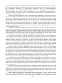
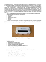
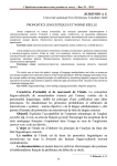
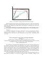
1' I max 4 1 I 3 2 3' 2' 4' 7 6 5 0 U Umax Figure 2 - CVC and PVC forming with parallel PCR and LCR load Model test results showed satisfied static and dynamic characteristics. Pulse current amplitude is within 2%, PCR build-up time within input peak voltage alteration is less than 3 ms. At the 1 kW model load power leakage is less than 100 W. Power increase is obtained by the several cells parallel connection. LCR and PCR collaboration was tested during design the anode-cathode discharge circuit of the plasma generator. In this design we applied structural diagram (Fig. 1) as current regulator cell. References: 1. Способ стабилизации выходного напряжения системы электропитания КА/А.И. Чернышев, Ю.М. Казанцев, С.А. Поляков [и др.] // 3-й Сибирский междунар. авиакосмический салон САКС 2004: Сб. докл. Междунар. науч.-практ. конф. – Красноярск, 2004. – C. 43–48. Electrocardiograph as a Space and Ground Support Equipment Starchak A.S. Scientific advisor: Goldshtein A.E., Doctor of Technical Science, Professor Linguistic advisor: Troitsky O.Yu., Doctor of Physics and Mathematical Sciences, Professor Tomsk Polytechnic University, 30, Lenin Avenue, Tomsk, 634050, Russia E-mail: [email protected] The monitoring of vital physiological signals has proven to be one of the most efficient ways for continuous and remote tracking of the health status of the patients. Electrocardiographs are often used in many medical service centers and hospitals to diagnose and monitor person’s health status by measuring their cardiac activity. Electrocardiography (ECG) is a non-invasive method, which can be applied to evaluate the heart electrical activity, to identify any heart damage, the position of the chambers, to measure the rate and regularity of heartbeats and investigate the effect of drugs and devices used to regulate the heart activity. This procedure is very useful for monitoring people with (or susceptible to) impairments in their cardiac activity [1]. One of the invariable risks, associated with space missions, is the threats posed by the very harsh spatial environments to the physical and mental health of astronauts. Changes in the 386 environment (for example, reduced gravity, high temperature, cosmic dust, sunrays, etc.), exert a long-term negative effect on the heart, muscle and nerve function of the astronauts [2]. Fortunately, the continuous monitoring of physiological parameters such as electrocardiography, electromyography, electroencephalography, oxygen saturation in hemoglobin and variability of cardiac frequency can provide crucial information for a rapid diagnosis of medical state of astronauts health; preventing the growth of any bad health state which can cause mission failure or even death of a crew members. In order to provide monitoring of the several physiological parameters, using only one device, various biomonitors, introduced several years ago and used so far in hospitals and research centers, were created. Although, these devices are commonly used to detect, treatment and record several physiological signals simultaneously, they are not used in the conventional telemetry system because they limit the freedom of movement of the subjects whose biopotentials are being measured. First, most of these systems receive power from an electrical outlet and they are heavy, which makes casual movements very difficult [3]. Moreover, besides limiting the freedom of mobility of the subjects, the wires used to connect the electrodes are frequently a source of noise in a data acquisition system. Although, such stationary conditions might be acceptable in the case of a bed-ridden patient, it cannot be used for astronauts who are required to constantly move by their activities during space flights. The alternative is the commercially available biomonitoring devices, that can be used under severe non-stationary conditions such as athletic and scientific studies, but their application is very limited and prove to be costly. These devices are usually uncomfortable due to their heavy weight, vigorous structure and network of lead cables running from control subject. The goal of our project is to design and fabricate a wireless portable electrocardiograph to help eliminate the restrictions caused by lead wires in conventional systems used in hospitals or research centers. It will allow you to record and transmit the signal from the sensor to the device through a wireless connection. In hospitals, our device would readily be adaptable to any system used to monitor physiological parameters, particularly with bedside ECG monitors, wireless capable computers and portable ECG monitors. It will allow tracking patient’s vital signs of at all times despite his/her proximity to a bedside monitor. This device can also be used in space research centers as telemetric ECG monitoring systems. It allows you to monitor vital signs of astronauts. Together with its wireless capabilities, this system will have to meet several design specifications including technical standards for portable bioinstrumentation devices to ensure safety and functionality. Hence, this device will be battery powered, double insulated and isolated from the ground to keep it from constituting an electrical hazard to the astronaut or patients. The device susceptibility to electromagnetic interference will be minimized to produce more accurate signals. In addition, the device will be implemented audio and visual alarms to alert the user when the measured ratio reaches a critical level. Furthermore, the plastic case will also make this device robust enough to be worn during physical activities such as exercise or research tasks. Currently, there are two different types of ECG systems that are used. The first one is the standard ECG that generally involves twelve or fifteen leads that are connected to the patient’s chest, arms and right leg through electrodes. The device records the ECG signal for almost thirty seconds [2]. Possible ailments can be discovered from reading the ECG signal. However, due to the short time for measuring, sporadic abnormalities that mostly occur in the intensive care unit (ICU) can’t be discovered. In order to solve problem mentioned above, continuous electrocardiogram telemetry is used by many hospitals to monitor patients in the ICU. This device has three electrodes that acquire ECG signal for an extended period and then the signal is displayed on a screen or printed on an ECG graph paper. The Wireless ECG Monitoring System get into second category of ECG devices, and it will be used to monitor the cardiac activity of subjects. Both standard and continuous electrocardiographs are marketed as “portable”, but they are not necessarily small and lightweight. In addition, most such appliances receive power from an electrical outlet and are sufficiently heavy, so they must be mounted on a cart in order to move from 387 one location to another. Wireless devices have the potential to significantly improve this situation by reducing the weight and size of these devices and eliminating the necessity of using lead cables. The purpose of this project is to develop a working prototype of the electrocardiograph, which can be used by astronauts or hospital patients and send measurement data over a wireless connection via GPRS to a server for calculation, transmission and keeping by the doctor. This device will save time and effort for the nurses who are constantly following the patients’ conditions and help them to operate more efficiently. At the same time, the goal of this project is to minimize the cost of the device, so hospitals can afford each one for every patient, especially the ones that are in the ICU. Thus, a design of the handheld electrocardiograph for individual diagnosis was developed. Electrocardiograph must follow the requirements: convenience; reliability; the information content; efficiency; visibility. To ensure the requirements, the device has a simplified registration procedure first standard ECG leads from your fingers and subsequent signal processing in the device and in the PC (Figure 1). Figure 1 - Handheld electrocardiograph The device provides: registration of ECG on the fingers; display the ECG in real time on the screen; saving the information on the SD card; reading and automatic processing from SD card to PC; definition of diagnostic indicators; accumulation of data for further consultation with the doctor. The measuring system follows all the requirements for electrocardiographs. Device specifications: Sampling frequency of 500 Hz; 24-bit resolution of the ADC; The leakage current through the patient 200 PA; Input resistance 500 Megohms; Rejection ratio common mode noise 105dB; 388 Power consumption of 1 mW. The main goal of this project is to create a device capable of measuring the ECG by eliminating the necessity to use [4]. This means that the system will be required to have specifications which can encompass such ECG monitoring systems. The integrity of the signal, which is wirelessly transmitted to the receiver or relayed to an electrocardiograph via input cables, is a very important aspect of this system. It is important that a doctor or astronaut, who uses continuous ECG monitoring, will be able to objectively notice variations in a subject health status based on the trends of the waveform of the signal. Moreover, a distorted signal can cause erroneous medical diagnostic or inaccuracy of measured values (e.g. heart rate). For this reason, the wireless biomonitoring system must have a signal to noise ratio (SNR) deemed acceptable for ECG signals and possibly for other types of signals. In order to optimize the performance of the signal acquisition system, the desired SNR will be set at 60 dB. It will allow our system to receive useful and cut off unwanted signals. We strive to make the system as little as possible, with weight about 100 grams and dimensions about 5x10x2 sm. Now, the dimensions are slightly different from the planned (weight is about 200 grams and dimensions are about 7,5x12x2,75sm). Now, work on the project continued. References: 1. Delsys Inc. (2004). Myomonitor III User Manual. EKG Monitoring System. http://instruct1.cit.cornell.edu/courses/ee476/FinalProjects/s2001/jl175/EE476.htm. 2. Carsten W., Kevin M., Usen U., ValerieB., Guillaume T.,Arnaud T., Robert R., Robert B., Yvonne C., Nathalie C., Stephen R., Judith S., John H., Gregory K. (2005)A Multiparameter Wearable Physiologic Monitoring System for Space and Terrestrial Applications. Institute of Electrical and Electronic Engineers. 3. Montgomery K., Mundt C., Thonier G., Tellier A., Udoh U., Barker V., Ricks R., Giovangrandi L., DaviesP., Cagle Y., Swain J., Hines J., Kovacs G. Lifeguard- A Personal Physiological Monitor For Extreme Environments. Institute of Electrical and Electronic Engineers 4. Ambulatory Data Acquisition System (ADAS) https://hrf.jsc.nasa.gov/hardware/adas.asp. 3D Modeling Elements of Devices for Space Application Stasevsky V.I. Scientific advisor: T.G Kostyuchenko Tomsk Polytechnic University, 30, Lenin Avenue, Tomsk, Russia, 634050 E-mail: [email protected] Complex technical objects design is executed via 3D-modelling. 3D- modelling is creation, visualization, editing of 3-dimentional object in any 3D-modelling program. Design’s key problem is to create and set off design documentation (blueprints, specifications) to the manufacturer. Any ware’s design works take a lot of time. Engineers of various specializations are involved into process. Applying modern high-power calculating equipment and specialized software leads to significant reduction of time spent on design works and, consequently, reduces laboriousness. Computer-aided design (CAD) usage allows to run virtual check outs of designed objects declining outlays for prototypes production. At present offered on the market large numbers Cad-systems that can be used in the design elements of devices space use. It SolidWorks, Autodesk Inventor, AutoCAD, КОМПАС and others. Among all these systems is located Russian CAD-system T-Flex CAD is containing more effective means of parametrization as compared to other mentioned systems. 3D-functionality of system T-FLEX CAD based of graphic core called Parasolid, which at present is one of the best for 389