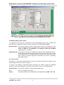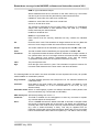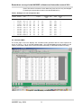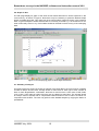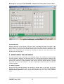
1
BREAST DOSE SURVEYS IN
THE NHSBSP:
SOFTWARE AND
INSTRUCTION MANUAL
Version 2.0
Report 04/
July 2004
K C Young
National Co-ordinating Centre
for the Physics of
Mammography
Guildford
Breast dose surveys in the NHSBSP: software and instruction manual V2.0
CONTENTS
1.
INTRODUCTION................................................................................................................................ 2
2.
DOSIMETRY PROTOCOL USED ................................................................................................... 2
3.
NEW FEATURES IN THIS VERSION............................................................................................. 2
3.1
New standard breast.................................................................................................................. 2
3.2
Diagnostic reference level......................................................................................................... 3
3.3
Correcting for age....................................................................................................................... 3
3.4
Calculation of exposure times .................................................................................................. 3
3.5
Record of compression force.................................................................................................... 4
3.6
Change to s-factors.................................................................................................................... 4
4.
SURVEY METHODOLOGY.............................................................................................................. 4
5.
USING THE SOFTWARE TO CALCULATE DOSES IN A BREAST DOSE SURVEY ........... 4
6.
7.
5.1
Introduction.................................................................................................................................. 4
5.2
Mammography system details.................................................................................................. 5
5.3
X-ray set data.............................................................................................................................. 5
5.4
Exposure data............................................................................................................................. 7
5.5
Calculated MGD ......................................................................................................................... 9
5.6
Graph of data ............................................................................................................................ 10
5.7
Summary of analysis................................................................................................................ 10
5.8
Printed report ............................................................................................................................ 11
MAINTAINING THE DATABASE ................................................................................................... 11
6.1
Backing up data ........................................................................................................................ 11
6.2
Deleting dose surveys ............................................................................................................. 12
6.3
Incorporating survey data from previous version................................................................. 12
REFERENCES ................................................................................................................................. 12
APPENDIX 1: NHSBSP PATIENT DOSE DATA RECORDING FORM........................................ 13
APPENDIX 2: METHOD OF DOSE CALCULATION ....................................................................... 15
APPENDIX 3: TABLES AND FIELDS USED TO CALCULATE DOSE......................................... 16
APPENDIX 4: MODIFICATION OF DOSE CALCULATION TO ADJUST FOR AGE ................. 17
NHSBSP July 2004
1
Breast dose surveys in the NHSBSP: software and instruction manual V2.0
1. INTRODUCTION
Measuring the mean glandular dose to the standard breast1 has for many years been the main
means of assessing the doses from mammographic systems. Although this is a valuable and
simple approach to dose measurement it provides limited information on the doses received by
individual women. As a result the periodic measurement of doses for samples of women
undergoing mammographic examinations is recommended.2,3 This manual describes the use of
Version 2.0 of a software programme that calculates and stores the results of such dose surveys.
(The latest version of the previously distributed software was known as Version 16 i.e. the 16th
variation of Version 1.) It is intended that the use of this software will help to standardise dose
measurements and simplify procedures for national reviews of radiation doses in the NHS Breast
Screening Programme. The software incorporates the changes in the UK dosimetry protocol
outlined by Dance et al.4
2. DOSIMETRY PROTOCOL USED
The UK and European protocols for mammographic dosimetry use conversion factors that relate
incident air kerma to the mean glandular dose (MGD) within the breast. The conversion factors
published in IPSM 59/2 were obtained by computer simulation of a model breast with composition
50% adipose and 50% glandular tissues by weight (50% glandularity). Dance et al 4 have
published additional conversion factors which allow the extension of the protocols to breasts of
varying glandularity and for a wider range of mammographic x-ray spectra. The data have also
been extended to breasts of compressed thickness 11 cm. To facilitate the calculation of MGD in
patient surveys, typical breast glandularities are tabulated for women in the age ranges 40-49 and
50-64 and for breasts in the thickness range 2-11 cm. The software provided with this publication
facilitates the implementation of a breast dose survey using these procedures using equation 1.
D=Kgcs
(1)
where the factor g is unchanged from that given in IPSM 59/2 and corresponds to a glandularity of
50%. The factor c corrects for any difference in breast composition from 50% glandularity. cfactors have been calculated by Dance et al 4 for typical breast compositions at different
compressed breast thicknesses in the age ranges 50 to 64 and 40 to 49. The factor s makes a
correction for the use of an X-ray spectrum other than Mo/Mo.
3. NEW FEATURES IN THIS VERSION
3.1 New standard breast
The Quality Assurance Standards for the NHSBSP were revised on 1st April 2004. In these the
dose standard is based on the new standard breast model introduced in IPEM Report 89. The data
published in Dance et al (2000) for the equivalence between Perspex and typical compressed
breasts has been used to establish a new standard breast simulated with a 45mm thick Perspex
phantom instead of the 40mm thick phantom used previously. The new standard breast model can
be expected to result in doses that are approximately 25% greater than those found with the old
model if the same beam quality (28 kV Mo/Mo) is used. However the exposures should be at the
clinical settings. The minimum standard for the mean glandular dose for the new standard breast
at clinical settings is less than or equal to 2.5 mGy/film.
It is expected that all new dose surveys be recorded using the dose to the standard breast using
45mm Perspex. However some old surveys may have been collected using a 40mm thick
NHSBSP July 2004
2
Breast dose surveys in the NHSBSP: software and instruction manual V2.0
phantom. To accommodate this the software has a new field to record the PMMA thickness used
to measure the standard breast. Old surveys should have 40mm entered and new surveys 45mm.
No calculations depend on this field.
3.2 Diagnostic reference level
The Ionising Radiation (Medical Exposure) Regulations 2000 require employers to establish DRLs
for radiodiagnostic examinations. DRLs are defined in the regulations as dose levels for typical
medical x-ray examinations for groups of standard-sized patients or standard phantoms and for
broadly defined types of equipment. The employer is required to establish DRLs and to ensure that
procedures are in place for using them on the understanding that they are not expected to be
exceeded for standard procedures when good and normal practice is being followed. The
employer is, moreover, obliged to undertake appropriate reviews whenever DRLs are consistently
exceeded and to ensure that corrective action is taken when the excessive exposures cannot be
clinically justified. The Institute of Physics and Engineering in Medicine (IPEM) along with the
National Radiological Protection Board (NRPB), the College of Radiographers (CoR), the Royal
College of Radiologists (RCR) and the British Institute of Radiology (BIR) have established a
Working Party to provide guidance on the implementation of DRLs for diagnostic x-ray
examinations. The Working Party have proposed a dose audit measure for mammography which
is the average MGD for medio-lateral oblique mammograms for breasts with a compressed
thickness of 55 ± 5mm. A minimum of 10 women should be included in the dose sample. The
proposed National DRL for this dose audit measure is 3.5 mGy. In this version of the software the
average MGD for all oblique mammograms (main films only) with a compressed thickness of 50 to
60 mm is calculated for each dose survey. For this calculation the number of films, the statistical
error in the mean and the average thickness are also shown. The statistical error is shown as 2
standard errors in the mean (2 s.e.m.). For a few surveys the number of OB mammograms in this
thickness range may be less than 20 (i.e. equivalent to less than 10 women) and would therefore
be insufficient for accurately assessing compliance with the DRL.
3.3 Correcting for age
The software has composition correction factors (c-factors) for younger women (age 40 to 49) and
women in the usual screening age (aged 50 to 64). In the previous version older and younger
women were entered as separate surveys even if exposed on the same X-ray set. The whole
survey was then identified as either “routine screening” i.e. over 49 or “age trial” i.e. age 40 to 49.
In this new version of the software the appropriate factor is determined by the age information
entered with the exposure data. If no age information is provided (as is usually the case) the
software assumes all the women are aged 50 or over. If an age is entered for an exposure record,
the appropriate factor will be used. Note however that it is no longer sufficient to just enter “age
trial” in the field “routine/age trial” as this no longer has any effect on the dose calculation. Further
details on how this change was introduced are shown in Appendix 4.
3.4 Calculation of exposure times
The software can now calculate exposure times as well as dose for each exposure. To do this an
appropriate mA must be entered in the X-ray set data. The exposure time is then calculated as the
mAs divided by the mA. (Note: On some systems the mA may vary during the exposure, in which
case the calculated time based on a fixed mA will not be correct e.g. Siemens Mammomat 3000
can be set to use a lower mA at the start of the exposure to reduce grid lines on short exposures.
Also if the mAs is selected manually, some systems (e.g. Lorad mark IV) may use a different mA
for small mAs values.)
NHSBSP July 2004
3
Breast dose surveys in the NHSBSP: software and instruction manual V2.0
3.5 Record of compression force
The compression force may be included in a new field along with the exposure data. This has no
effect on the dose calculation but does provide a more complete record of radiographic
procedures.
3.6 Change to s-factors
Some earlier versions of the software used s-factors that were slightly different from those
published by Dance et al.4 In this version the factors are identical to those in the published paper.
Overall the effect of this change is very small. There is no change to doses where a Mo/Mo target
filter combination is used but doses are reduced by about 1% where a Mo/Rh target/filter
combination is used. Although these changes are rather smaller than the errors in the overall
methodology, users may notice small differences in the calculated doses when entering old data in
to this new software version.
4. SURVEY METHODOLOGY
A breast dose survey involves the measurement of the mean glandular doses for a series of breast
examinations (e.g. for 50 to 100 women) on a single mammographic system. For each exposure
the X-ray factors are recorded (including target material, filter material, tube voltage and mAs). The
radiographer determines the compressed breast thickness by reading the display on the X-ray set.
The accuracy of the displayed thickness should be verified before starting the survey. This can
simply be done by compressing a rigid material (e.g. Perspex) of known thickness to a typical force
of 100N. If significant errors (>2mm) are encountered, correction factors should be applied or the
unit re-calibrated.
From a knowledge of the output of the X-ray set for the kV and target and filter material used, the
mAs value may be used to estimate the entrance air kerma at the surface of the breast (K) and
hence the mean glandular dose.
5. USING THE SOFTWARE TO CALCULATE DOSES IN A
BREAST DOSE SURVEY
5.1 Introduction
The software provided comprises a Microsoft Access Database which uses a multi-page form for
the entry and display of data for each survey as shown in Figure 1. The following sections discuss
the use of each page of the form in turn. The first page of the form contains descriptive data for the
mammography system. The second page of the form is used to enter the radiation characteristics
of the X-ray set. Information on each clinical exposure is entered on the third page of the form.
Subsequent pages display the calculated doses and summary data for the current survey. Sample
data for a few dose surveys are included to illustrate how the software works. Once the user is
familiar with the package this demonstration data should be deleted.
NHSBSP July 2004
4
Breast dose surveys in the NHSBSP: software and instruction manual V2.0
Figure 1: Multi-page form used for entering and reviewing dose survey data.
5.2 Mammography system details
It is important to record all the characteristics of the mammography system used. However the
only field that is essential and used in the dose calculation is the field survey number.
Survey Number: The survey number is used to identify all the various sets of data from one
survey and takes the form of an integer, which can be incremented each time a
survey is added to the database. It is simplest if these are numbered
sequentially from 1 upwards.
Routine/age trial: In the previous version of the software this field was used to determine which cfactors should be used for a whole survey. However in this version the c-factors
for younger women are selected whenever the age field is less than 50. The
field is retained to provide background information only, and for compatibility
with earlier versions.
5.3 X-ray set data
Information on the physical characteristics of the X-ray set being surveyed are entered on the
second page of the form as shown in Figure 2.
In order for the software to calculate entrance air kerma one needs to supply the following
parameters for each kV/target/filter combination used clinically on the X-ray set being tested.
Survey Number: This number must be the same as that entered above.
kV:
Set kV and therefore an integer.
Anode:
The anode material must be identified by the appropriate code Mo or Rh or W.
NHSBSP July 2004
5
Breast dose surveys in the NHSBSP: software and instruction manual V2.0
Filter:
The filter material must be identified by the appropriate code Mo or Rh.
Output:
Output in µGy per mAs at 1 metre from the focus of the X-ray tube on the usual
measurement axis (i.e. one that passes through a point 4 cm from the chest
wall edge). It is assumed here that this is measured at a distance of about
50cm and then recalculated to a distance of 1 metre. It is further assumed that
attenuation by a compression paddle is not included. If you wish to use an
output measurement through a compression plate this can be done if the
transmission factor is set to 1.0.
HVL:
This is the half value layer in mm of Aluminium measured as described in
IPSM59/2 and should include attenuation by the compression plate. It should
be quoted to two significant figures.
Distance:
This is the distance from the focus to the table top in mm.
Transmission:
Measured fraction of air kerma transmitted through the compression plate.
mA:
This field is optional and has been added to version 2.0 to allow the calculation
of exposure times. It should be completed for each kV, target filter combination
used for which exposure times are to be calculated. It may also be left blank
and has no effect on the dose calculations. (The mA can be determined from
manufacturer’s literature or exposure time measurements.)
Figure 2: Form for entering physical characteristics of X-ray set.
Figure 2 is based on a suitable dataset for an IGE DMR. It is not necessary to directly measure all
of these parameters as interpolation can easily be applied. It may be convenient to assemble this
NHSBSP July 2004
6
Breast dose surveys in the NHSBSP: software and instruction manual V2.0
data in a spreadsheet before “paste appending” it to the appropriate table in the database (i.e. Xray set characteristics).
5.4 Exposure data
A record of the exposure factors for each mammogram is required. Appendix 1 shows a paper
form that can be used by radiographers to collect this data. An example of such a dataset is shown
in Table 2. The form on the database for entering this data is shown in Figure 3. The types of data
requested, and how they should be entered into the database are discussed below. It is important
that the data entered are in the right format. Where numerical values are requested do not enter
the unit e.g. type in 28 rather than 28 kV.
Figure 3: Form for entering exposure data
Survey Number: Each exposure in the series must be identified with the same survey number
provided in sections above.
Patient Number: Each patient must be identified by a code, which is unique to this survey. This
can be any combination of letters and numbers.
View code:
Each exposure requires a view code that is unique to that patient. The codes
for main films are as follows.
LCC for left cranio-caudal
RCC for right cranio-caudal
LOB for left mediolateral oblique
NHSBSP July 2004
7
Breast dose surveys in the NHSBSP: software and instruction manual V2.0
ROB for right mediolateral oblique
Where additional films have to be taken for the same views (e.g. if the breast
was too big to fit on the first film) the following codes should be used.
LCC2 for a second film of the left cranio-caudal view
LCC3 for a second film of the left cranio-caudal view
and so on for the other views.
The software is designed for the two views used in screening i.e. mediolateral
oblique and cranio-caudal. If some different views are used (e.g. a lateral)
further codes may be used as follows.
LLAT for a left lateral view
RLAT for a right lateral view
Note however that the summary datasheet will only consider the standard
views.
kV:
Enter the set kV used. This should be an integer between 25 and 35. (Note that
the use of a non-integer number will cause the dose calculation to fail.)
Anode:
The anode material must be identified by the appropriate code Mo or Rh or W.
Filter:
The filter material must be identified by the appropriate code Mo or Rh.
Thickness:
The compressed breast thickness should be entered as an integer number of
mm. (Note that the use of a non-integer number will cause the dose calculation
to fail.) Although most modern mammographic X-ray sets will display
compressed breast thickness it is important to check the accuracy.
mAs:
The post exposure mAs.
Age:
Enter the age of the woman in years. This information is optional. However if it
is entered it will determine the c-factor used in the dose calculation.
The following fields are not used in the dose calculation but are requested since they can provide
useful feedback on examination practice.
large cassette:
If a large cassette (24x30cm was used) enter Yes. If a standard cassette was
used leave blank.
auto/man:
Enter man if the beam quality was selected manually, and auto if it was
selected automatically or on the recommendation of the X-ray set (e.g.
Siemens 3000).
Automatic mode: Where a mammography system has different automatic modes please enter
the one used here (e.g. Contrast with an IGE DMR).
AEC density set: AEC density setting such as 0 or +1.
Comment:
This field is available for the radiographer to enter any comments or explain
any unusual aspects of the exposure.
Error:
This is a Yes/No field with the default value No. If this field is changed to Yes
this record of exposure data will not be used in the display and calculation of
summary data. This could be done if the user suspects that the underlying data
(e.g. mAs or breast thickness) is faulty. This may be apparent from an
inspection of the graph of dose versus thickness. Where this occurs it is usually
best to eliminate all the dose data for this patient (i.e. set Yes for each record).
NHSBSP July 2004
8
Breast dose surveys in the NHSBSP: software and instruction manual V2.0
(Note: This field is not shown on the data entry form and can only be changed
by altering the appropriate records in the table DOSEDATA.)
Table 1: Example of a set of exposure data
Survey
Number
Patient
Number
View
code
kV Anode
1
1
1
Filter Thickne mAs
large
Auto
ss (mm)
cassette /man
used
Mo
49
130
auto
Automatic AEC
mode
density
set
CNT
0
AA10615
LCC
25
Mo
AA10615
RCC
25
Mo
Mo
42
101
auto
CNT
0
AA1292
LCC
26
Mo
Rh
72
262
auto
CNT
0
1
AA1292
RCC
26
Mo
Rh
78
318
auto
CNT
0
1
AA14120
LCC
27
Mo
Rh
61
125
auto
STD
0
1
AA14120
LOB
27
Mo
Rh
60
126
auto
STD
0
1
AA14608
LOB
31
Rh
Rh
80
142
auto
STD
0
1
AA14608
LOB2 27
Mo
Rh
72
168
auto
STD
0
1
AA14608
ROB
28
Rh
Rh
80
161
auto
STD
0
1
AA14608
ROB2 27
Rh
Rh
80
162
auto
STD
0
Age
Comment
5.5 Calculated MGD
The fourth page of the form displays the calculated mean glandular dose for each exposure as
shown in Figure 4. This is the field labelled dose. (The field old dose has been deleted in this
version.) If this field is blank some of the information necessary to calculate the dose is missing.
Figure 4: Form displaying calculated doses
NHSBSP July 2004
9
Breast dose surveys in the NHSBSP: software and instruction manual V2.0
5.6 Graph of data
The fifth page displays a graph of the dose versus breast thickness for all the exposures in the
current survey, as shown in Figure 5. Note that it may be necessary to press the Refresh Graph
button to update this graph. This graph may show some outliers caused by errors in the exposure
data. A typical example is where the thickness is incorrectly recorded e.g. it is entered as 10 mm
when it was really 100 mm. Any such outliers should be checked for the accuracy of the underlying
data.
Figure 5: Form displaying graph of doses against thickness.
5.7 Summary of analysis
This page of the form (Figure 6) shows an analysis of the dose data in the current survey including
number of oblique and CC films; average doses for main films for each type of view; number of
films for each target/filter/kV combination. Note that a main film has a view code of LOB, ROB,
LCC or RCC. View codes of LOB2, RCC3 etc are not classed as main films. The average doses
for oblique views of breasts with a compressed breast thickness of 50 to 60mm are shown for
comparison with the DRL. The user may perform more detailed analysis by copying the data to a
spreadsheet.
NHSBSP July 2004
10
Breast dose surveys in the NHSBSP: software and instruction manual V2.0
Figure 6: Form displaying summary data
5.8 Printed report
Pressing the button “Print summary data form” prints a one-page summary of the data in the
current survey. The printer used is the default printer set on the host computer. In addition to the
data in section 3.7 this printout shows the average doses for one and two view examinations. The
doses for an examination are calculated by summing the doses for all the films used in an
examination and dividing by 2.
6. MAINTAINING THE DATABASE
The user has free access to all aspect of the database design. This will allow the user to trouble
shoot any problems. However care should be taken not to corrupt features that are essential to the
correct working of the database. The original version of the database software used is Microsoft
Access 1997. Version 2.0 is also available in later versions of Access. Earlier versions of Access
will not be able to run the database. In the event of difficulties contact the author for help and
software upgrades.
6.1 Backing up data
As with any other software package it is important to regularly back up your data. This can be
achieved by making a backup copy of the database using the command Tools/DatabaseUtilities/Compact Database to both compress the database and save a copy using a different
name.
NHSBSP July 2004
11
Breast dose surveys in the NHSBSP: software and instruction manual V2.0
6.2 Deleting dose surveys
When deleting a survey from the database (e.g. the sample data provided) it is necessary to delete
all the records from the three main tables (SURVHEAD, DOSEDATA and X-ray set
characteristics) with the appropriate survey number. The records from the DOSEDATA and Xray set characteristics tables should be deleted before those in the SURVHEAD table. Any
deleted survey numbers may then be reused for new surveys.
6.3 Incorporating survey data from previous version
It is likely that most users of the software have survey data in a previous version of the software
that they wish to transfer into this new version. The appropriate way to do this is to select and
copy all the records from each of the following three tables in turn. The records should then be
paste appended to the corresponding tables in the new version of the software. Delete the
demonstration data included with the software as described above.
•
•
•
SURVHEAD
X-ray set characteristics
DOSEDATA
This will leave a few new fields added at the end of the new tables blank e.g. PMMA thickness,
mA. These can be completed later if required.
7. REFERENCES
1. Law J et al. Commissioning and routine testing of mammographic X-ray systems, York,
Institute of Physics and Engineering in Medicine, IPEM Report No. 59/2, (1994)
2. Recommended standards for the routine performance testing of diagnostic X-ray
imaging systems. York, Institute of Physics and Engineering in Medicine, 1997 (IPEM Report
No. 77).
3. Guidelines on patient dose to promote the optimisation of protection for diagnostic
medical exposures. NRPB Report Volume 10 No 1 (1999).
4. Dance DR, Skinner CL, Young KC, Beckett JR and Kotre CJ. Additional factors for the
estimation of mean glandular breast dose using the UK mammography dosimetry
protocol. Phys. Med Biol. 45;3225-3240 (2000)
5. Young KC, Burch A. Radiation doses received in the UK breast screening programme in
1997 and 1998. British Journal of Radiology 73;278-287 (2000).
NHSBSP July 2004
12
APPENDIX 1: NHSBSP PATIENT DOSE DATA RECORDING FORM
X-ray Unit:___________________________________________________
Sequential
Patient
Number
View code
LOB,
ROB,
LCC,
RCC
kV
Anode
(Mo, Rh
or W)
Filter
(Mo/Rh)
Compressed
Breast
Thickness
(mm)
Date:________________________
mAs
Large
cassette
used
(Yes/No)
Mode of kV
selection
(auto/man)
Screening Centre:________________________________________
Automatic
mode (e.g.
CONT with
DMR)
AEC
density
set
Age
Comment
Breast dose surveys in the NHSBSP: software and instruction manual
APPENDIX 2: METHOD OF DOSE CALCULATION
The mean glandular dose is calculated for each exposure by means of look-up tables in the database
as illustrated in Appendix 3. (Note that the interconnections between tables have been somewhat
simplified to illustrate the underlying method.) The arrows indicate where a field is used in the dose
calculation. The links between tables used in the dose calculation query are shown as connecting
lines between tables. The formula applied is that shown in equation 1. The g-factor, s-factor and cfactor are taken from three look-up tables depending on the HVL, age group, and anode and filter
material used. Linear interpolation was used to generate additional values for these tables. The
entrance air kerma (K) is calculated from the output per mAs corrected for paddle transmission and
the inverse square law. The equation (2) is used in the database to calculate the mean glandular dose
per exposure, where square brackets denote a field name.
[dose] = [s factor]*[c factor]*[g-factor]*[transmission]*[mAs]*[output]*1000/([distance]-[thickness (mm)])2
NHSBSP July 2004
15
(2)
Breast dose surveys in the NHSBSP: software and instruction manual
APPENDIX 3: TABLES AND FIELDS USED TO CALCULATE DOSE
The tables and fields used to calculate the dose are shown in Figure 7. The straight lines between
tables represent linked fields, and the bold arrows indicate the fields used in the calculation. The
queries that are used in the calculation are discussed in Appendix 4
SURVHEAD
survey number
Region
code
Centre
Date of First Exam
Date of Last Exam
X-Ray Make
Model
Sub model
Local id
Installation
kV mode
Processor model
Processor id
Developer
Fixer
Proc time
Dev Temp
Cassette type
Film make
Film type
Film subtype
Screen make
Screen type
Screen resolution
Block mAs
Block density
24x30 cassettes
MGD auto/manual
MGD AEC setting
MGD kv/target/filter
MGD mAs
MGD HVL
MGD
MGD density
Physics centre
Physicist
tel no
Routine/age trial
MGD kV set
MGD target
MGD filter
24x30 bucky
Processor make
Cassette make
standard kV
Dose data
plus HVL
Survey Number
Patient Number
View c ode
kV
Anode
Filter
Thic kness (mm)
mAs
large cassette used
auto/man
Automatic mode
AEC density set
Age
Comment
Error
Age group
X-ray set
charactersistics
Survey Number
kV
Anode
Filter
HVL
Output
Distance
transmission
C factors
Age group
HVL
thickness
C factor
g values
HVL
Thickness
Gvalue
spectral factors
Anode
Filter
Spectral factor
Figure 7: Tables and fields used to calculate dose
NHSBSP July 2004
16
Breast dose surveys in the NHSBSP: software and instruction manual
APPENDIX 4: MODIFICATION OF DOSE CALCULATION TO ADJUST
FOR AGE
This appendix explains the use of two queries used to calculate doses corrected for the patient’s age.
Query: Dose data plus HVL:
The first query is shown in figure 8. It adds an extra field called Age group.
Age Group: This new field is calculated using an If statement. If the patient is under 50 years old the
age group is set to ‘Age trial’. If the patient is over 50 years old (or the patient age is left blank) the
age group is set to ‘Routine Screening’.
Figure 8: Dose data plus HVL query
Query: MGD calculation:
The second query is shown in figure 9. This query uses the data in Dose data plus HVL to calculate
the dose - which is now corrected for the age of the patient by using the appropriate c factor.
c factors! Age Group: This field is now related to the field Dose data plus HVL! Age group, instead
of the field Dose data plus HVL! Routine/age trial. This means that an appropriate c- factor will
automatically selected based on the age of the patient. If no age information is provided the woman is
assumed to be in the usual screening age.
NHSBSP July 2004
17
Breast dose surveys in the NHSBSP: software and instruction manual
Figure 9: Query used to calculate dose
NHSBSP July 2004
18