1
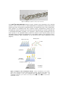
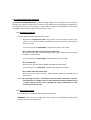
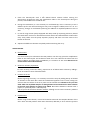
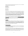
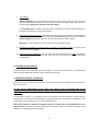
CLART® CMA KRAS·BRAF·PI3K DETECTION AND GENETIC IDENTIFICATION OF POINT MUTATIONS IN 3 OF THE GENES BELONGING TO THE EGFR PATHWAY ASSOCIATED TO COLORECTAL CANCER -KRAS, BRAF AND PI3KFOR IN VITRO DIAGNOSIS 1 CLART® CMA KRAS·BRAF·PI3K Amplification 24 KRAS tests Ref: CS-0412-24 CS-0412-8 24 BRAF tests Ref: CS-0512-24 CS-0512-8 24 PI3K* test Ref: CS-0612-24 CS-0612-8 CLART® CMA KBP Array Detection 24 CMA KBP Array tests Ref: CS-0712-24 CS-0712-8 ® CLART CMA is under protection of patent family corresponding to European Patent Application Nº 11382397.5. ® ® CLART and CLART-Strip are registered Trademarks of GENOMICA. For more information, please refer to the web site: * PI3K analysis is for research use only. GENOMICA, S.A.U. Alcarria, 7, 28823 Coslada, Madrid, Spain Tel: +34 91 674 89 90, Fax: +34 91 674 89 91 Version 3 May 2013 2 TABLE OF CONTENTS: 1. GLOSSARY 2. PROTOCOL DESCRIPTION 3. KIT COMPONENTS AND STORAGE 3.1. Amplification reagents 3.2. Visualization reagents 3.3. Other components 4. MATERIALS REQUIRED BUT NOT PROVIDED 4.1. Reagents and materials 4.2. Equipment 5. RECOMMENDATIONS AND HANDLING PROCEDURES 5.1. General recommendations 5.2. Precautions for visualization 6. SAMPLES 7. WORKING PROTOCOL 7.1. Sample pre-treatment 7.2. Material extracted 7.3. Amplification reaction 7.4. Visualization of the amplified product 8. READING OF THE RESULTS 9. INTERPRETATION OF THE RESULTS 10. TECHNICAL AND OPERATIONAL SPECIFICATIONS 11. REFERENCES 3 1. GLOSSARY Attention, see instructions for use Expiration date In vitro diagnostic medical device Lot 25 ºC Store at room temperature 20 ºC 8ºC Store at 4ºC to 8ºC 4ºC -18ºC Store at –30ºC to –18ºC - 30ºC 4 2.PROTOCOL DESCRIPTION CLARTCMA KRAS·BRAF·PI3K detects the presence of the most prevalent point mutations in the Epidermal Growth Factor Receptor (EGFR) pathway associated to colorectal cancer. The point mutations detected by the kit are the following: KRAS: G12A G12C G12D G12R G12S G12V G13D Q61H Q61L BRAF: V600E V600K PI3K: E542K E545D E545K H1047R Detection is carried out by the specific amplification of the mutation in the sample, originating a variable fragment for each mutation of between 100-200 base pairs. The amplification is performed in a variable number of PCR tubes, depending on the gene of interest: -The detection of point mutations for KRAS requires 3 amplification tubes that are differentiated by colour: white, green and red. -The detection of point mutations for BRAF requires 1 yellow amplification tube. -The detection of point mutations for PI3K requires 2, blue and white, amplification tubes. The detection of the product amplified by PCR is carried out by means of a low-density microarray platform: CLART® (Clinical Array Technology). The platform is based on a principle that is very simple, but at the same time economical and effective It consists in a microarray printed at the bottom of a microtiter plate well (CLART® Strip-CS) (Figure 1), which simplifies the entire hybridization and visualization process when compared to classic microarray systems. 5 Figure 1. CLART Strip® (CS) platform in the form of an 8-well strip. ® The CLART CMA KRAS·BRAF·PI3K detection system is based on the precipitation of an insoluble product in those microarray areas in which hybridization of amplification products with specific probes takes place. During PCR, amplified products are labelled with biotin. After amplification, these products are hybridized with their respective specific complementary probes that are immobilised in specific and well-known microarray areas. Afterwards they are then incubated with a streptavidine-peroxidase conjugate. The conjugate is bound through streptavidine with the biotin present in the amplified products (which are bound to their specific probes) and the peroxidase activity prompts the appearance of a non-soluble product in the presence of the odianisidine substrate, which precipitates on the microarray areas where hybridization occurs (Figure 2). Labelled product Probes on the array Biotin Hybridization Conjugate Incubation with conjugate Development reaction Precipitation of substrate Regardless of the number amplified genes,method. the detection willimmobilized be carried on outthe Figure 2: Diagram of of the visualization Probes, surface, capture their complementary biotin-labelled amplified products. With the help of biotin, they bind to the conjugate, in this case streptavidine-HRP (HorseRadish Peroxidase). The o-dianisidine substrate, by the action of the HRP, produces a precipitate on the area where hybridization occurs. 6 3.-KIT COMPONENTS AND STORAGE The CLART CMA KRAS·BRAF·PI3K kit contains enough reagents for the analysis of 8 or 24 clinical samples. The reagents included in the kit have been grouped into various packages, depending on the temperature at which they should be stored. When storage recommendations are observed, all reagents should remain stable until kit’s expiration date. 3.1. Amplification reagents They are shipped and should be stored at -20ºC. • Ready-to-use amplification tubes. They contain 45 µL of reaction mixture. Only thaw on ice the exact number of amplification tubes that will be used and keep the rest at -20ºC. For the analysis of the KRAS GENE, 3 amplification tubes are provided: Mix 1: White tube; Mix 2: Green tube; Mix 3: Red tube. With these three tubes, mutations G12A, G12C, G12D, G12R, G12S, G12V, Q61H and Q61L are determined. For the analysis of the BRAF GENE, 1 amplification tube is provided: Mix 1: Yellow tube. With this tube, point mutations V600E and V600K are determined. For the analysis of the PI3K GENE, 2 amplification tubes are provided: Mix 1: White tube; Mix 2: Blue tube. With these tubes, point mutations E542K, E545D, E545K and H1047R will be determined. Note: The kit package includes a self-adhesive and irreversible temperature indicator; the appearance of a reddish colour on the visualization window indicates that, at a certain moment, products have exceeded the storage temperature of –20oC and they should not be used. 3.2. Visualization reagents The visualization kit is shipped and should be stored at 4ºC. WARNING!: Upon arrival, the CLART-Strip® (CS), as well as the hybridization solution (SH) must be kept at room temperature. 7 • • • • • • • CS strips (including all specific probes). They are provided in a sealed thermal envelope. Store tore it closed, at room temperature, protected from direct light and high temperatures. SH (Hybridization Solution). Store at room temperature. DC (Conjugate Diluent). Store at 4ºC. CJ (Conjugate). Store at 4ºC. Centrifuge once before use. RE (Development Solution). Store at 4ºC and protected from light light. TL (Wash Buffer). Buffer Store at 4ºC. Adaptor and lid for 8-well 8 strips (microtiter plate). 3.3 Other components For the capture and subsequent processing of the image, a reader unit, running a tailor made software and, and the plate adaptor, are required: • CAR®(Clinical Array Reader): it allows for the automatic reading and interpretation of up to 12 CS, i.e., up to a maximum of 96 samples samples. It is distributed by GENOMICA, to be used exclusively with its diagnostic kits. • An adaptorr support that should be placed on the CAR tray before the scanning. • KRAS software, developed and validated by GENOMICA. SAICLART® CMA KRAS·BRAF·PI3K CAR® (Clinical Array Reader) 4.-MATERIALS REQUIRED BUT NOT PROVIDED 8 Below you can find a list of all materials required but not provided. 4.1. Reagents and materials - Distilled water. - Disposable gloves. - Filter tips or positive displacement pipettes. - Crushed ice container. - 1.5 mL autoclaved Eppendorf tubes. - 1.5 mL tube grids. - 0.5 mL/0.2 mL tube holder. - Saline solution 0.9% NaCl 4.2. Equipment - Microcentrifuge. - UV-visible spectrophotometer - Thermal cycler. - Laminar flow chamber for the extraction laboratory. - Three adjustable micropipettes ranging from 1-20 μL, 20-200 μL, and 2001000 μL for the extraction laboratory. - One adjustable micropipette ranging from 1-20 μL, to add the genetic material to the amplification tubes. - Three adjustable micropipettes ranging from 1-20 μL, 20-200 μL, and 2001000 μL for the visualization laboratory. - Thermoblock (Thermomixer) compatible with 96-well plates and adjustable shaking at 20°C, 25°C and 50ºC. - Vortex. - Vacuum system (desiderable). 5.-RECOMMENDATIONS AND HANDLING PROCEDURES Read carefully before starting the assay in order to avoid contamination!. 5.1. General recommendations 1. This assay should be performed in two physically separated areas, in order to avoid sample contamination with the previously amplified product. Separate working materials should be available in each area (pipettes, tips, tubes, grids, gloves, etc.) which should never be used outside these areas. 1. Pre-PCR area: DNA extraction and sample preparation are performed in this area. Sample manipulation must be carried out within a biosafety cabinet (BSC). 2. Post-PCR area: Amplification and visualization of the amplified product are 9 carried out in this area. The material of this area should never come into contact with the material of the extraction area. Avoid entering the pre-PCR area after having worked in the visualization area. 2. Always use gloves. It is recommended to change gloves quite frequently, and it is mandatory to change gloves before start working in each of the aforementioned areas. New gloves must always be used when DNA is added to the amplification tubes. 3. Clean working areas (laboratory cabinets, hoods, grids, pipettes) thoroughly with a 10% diluted bleach solution after every sample batch processing; it is mandatory to disinfect all working areas in case of contamination. For thermocyclers and thermomixers, it is advised to clean them before and after used, in these same conditions. 4. Always use filter tips and positive displacement pipettes to avoid contamination due to micropipettes. Different sets of pipettes should be used in each area. 5. Use disposable and autoclaved laboratory material. 6. Never mix reagents from two different vials, even if they belong to the same lot. 7. Close reagent tubes immediately after use in order to avoid contamination. 8. Discard the micropipette tip after pipetting. 9. GENOMICA is not responsible for the results obtained with the kit if other samples different to the ones indicated are used. 5.2. Precautions for the extraction and addition of extracted material to the amplification tube 1. Always wear gloves 2. Clean working surfaces of cabinets with a 10% diluted bleach solution. 3. Turn on the laminar flow and UV light at least 20 minutes before extraction. Turn off the UV light when it is working inside the cabinet. 4. The preparation of the samples before extraction must be made inside the cabinet. 5.3 Precautions for visualization 1. Before starting the assay is recommended to verify the THERMOMIXER measuring the temperatures which will be used during the assay: 20°C, 25°C and 50ºC. 2. Avoid the pipette tip or the vacuum system touching the bottom of the well, since this could damage the probes printed at the well’s bottom. 3. It is recommended to add all solutions to the wall of the CS well; never directly at the bottom. 10 4. It is convenient not to add the SH solution (hybridization solution) until the denatured products of PCR are ready. 5. The array must not remain dry. 6. Following incubation with the CJ solution, it is very important to wash the microarray thoroughly in order to avoid any residues that could react with the RE solution, resulting in a non-specific precipitation that could lead to false interpretations of the result. 7. Avoid foaming when adding any reagent. 8. When visualizing the image in the reader, ensure that position markers appear and that there are no bubbles, fibers or spots interfering with the reading. Otherwise, clean the outer face of the well with cellulose paper. 6.-SAMPLES The CLART® CMA KRAS·BRAF·PI3K kit has been designed and validated to be used with DNA extracted from colorectal cancer biopsies. GENOMICA is not responsible for the results obtained if other types of samples are used. 7.-WORKING PROTOCOL CLART® CMA KRAS·BRAF·PI3K has been validated using two different protocols for sample pretreatment. 7.1. Sample pre-treatment: Preprocessing FFPE sections (formalin fixed paraffin embedded tissue) should be placed on a glass slide for the examination by the pathologist. Each sample must be processed using a new sterile scalpel. The pathologist will perform a study of each cut by staining it with hematoxylin and eosin (H&E). The staining will help to define and verify the tumor area, being indicated as a percentage (%) of tumor cells. Afterwards, follow the instructions below: • • • • By a percentage of tumoral cells <50% and a small area: take 6 cuts of 10 µm each. By a percentage of tumoral cells >50% and greater area: take 2-4 cuts of 10 µm each. In case of larger tissue amount, regardless of the percentage: take 1 cut of 10 µm. In the event of endoscopic biopsies (very small): take 10 cuts of 10 µm each. Place all the obtained cuts in a 1,5 mL tube. 11 Select between these two protocols the one that best suits: PROTOCOL 1. 1. 2. 3. 4. 5. 6. 7. 8. Add 1 ml Xilen to the tube containing the sample and vortex it during 5 sec. Incubate at RT during 10 min. Centrifuge the tube at 13.200 rpm during 5 min. Discard supernatant. Add 1ml 96-99%Ethanol and vortex it for 5 sec. Centrifuge the tube at 13.200rpm for 2 min. Discard supernatant. Incubate aprox. 15 min at 56ºC in order to allow the remaining supernatant evaporates (dried pellet). 9. From this step in advance, please follow the protocol outlined in the QIAamp DNA FFPE Tissue Kit user’s manual (Qiagen). The elution volume must be set at 50 µl. PROTOCOL 2. Day 1. 1. Centrifuge the tube containing the sample for 1 minute at 13.200 rpm, in order to get all FFPE cuts at the bottom of the tube. 2. Heat the tube (containing the sample) in the thermomixer for 5 min at 75ºC without shaking. 3. Add 190µl of G2 lysis buffer (EZ1 DNA Tissue Kit, Qiagen) to each sample. 4. Incubate 5 minutes at 75ºC, shaking at 1400 rpm. 5. Set the thermomixer temperature at 56ºC. 6. Add 25 µl Proteinase K, previously prepared according user’s manual of the above mentioned kit (20 mg/ml). 7. Incubate overnight at 56ºC and shaking at 1.400 rpm. Day 2. 1. Add 10µl Proteinase K and vortex briefly. 2. Incubate overnight at 56ºC shaking at 1.400 rpm. Day 3. 1. Centrifuge 1 minute at 13.200 rpm. 2. Add 2 µl glycogen at 20 mg/ml (Optional! It’s indicated for samples with very little prior extracted material). 3. Collect supernatant. 4. Homogenize the sample with the pipette tip in case of remaining tissue. 5. Move the supernatant to a tube provided by the extraction kit (Qiagen) specifically for extracting FFPE samples. 6. Place the specific tube at the BIO-ROBOT EZ1, in order to proceed with the extraction. Card must be “paraffin DNA”, and the elution volume must be set at 50 µl. 12 7.2. Extracted Material The extraction method chosen should be one that ensures the following concentration and purity parameters: 1. Total amount of extracted DNA add in each PCR amplification tube must be 150ng. An excess or a default of DNA can lead to misdiagnosis. Not exceed 150 ng of amount of DNA and 10 μl of volume add to each PCR amplification tube. The following table shows all the possibilities: Extracted DNA (ng/μl) 30 25 ng/μl 21.5 ng/μl 19 ng/μl 17 ng/μl 15 ng/μl Volume (μl) of extracted DNA add to each PCR tube 5 μl 6 μl 7 μl 8 μl 9 μl 10 μl Total amount (ng) of DNA add to each PCR tube 150 ng 150 ng 150 ng 150 ng 150 ng 150 ng If the concentration is below 15ng/μl, the sample should be re-extracted. 2. The extracted DNA should have the following purity enough to avoid misdiagnosis. The ratio between the absorbance at 260nm and the absorbance at 280 nm should be as close to 2 as possible. If the purity is not adequate, sample should be re-extracted. 3. The extracted material should be stored at 4 °C when processing it immediately after extraction, otherwise keep it at -20 º C until its analysis. It is important to include a negative control in every run, to verify that the samples have not been contaminated during the extraction, amplification and visualization processes, which might lead to a false positive result. 7.3. Amplification reaction Amplification-specific recommendations: • Work in the pre-PCR area, always using a cabinet and following the recommendations mentioned in section 5.1. • During the process, keep tubes separate and refrigerated. 1. Thaw the necessary number of amplification tubes according to the number of samples and gene/s to analyse. Do not use temperatures above 25ºC for thawing. 13 2. Centrifuge the amplification tubes for a few seconds, so that all liquid can get to the bottom of the tubes (in case you don't have microcentrifuge adaptors available for the tubes, you can use larger tubes after having cut their cap off). 3. Add 5-10 μL (see point 7.2) of the extracted DNA to each amplification tube, just after having check for DNA concentration and purity and mix several times with the micropipette. Keep the tubes refrigerated at any time. 4. Program the following temperature cycles on the thermocycler: 1 cycle 95ºC 15 min 40 cycles 94ºC 15 sec 62ºC 60 sec 1 cycle 72ºC 10 min 4ºC continuously until tube collection (optional) 5. Start the program and place the tubes in the thermocycler when the block has exceeded 90ºC. This way, possible unspecified amplifications due to incubation below hybridization temperature are minimized. The duration of the amplification ranges between 90-150 minutes depending on the thermocycler. The amplified product must be visualized within a maximum of 5 days to avoid its degradation, and kept at 4ºC if it is to be used immediately or at -20ºC if it will not be used for several days. 7.4. Visualization of the product amplified in CLART-Strip® (CS) Specific recommendations before starting visualization: THE PROTOCOL DESCRIBED BELOW SHOULD ALWAYS BE USED IN THE POST-PCR AREA. DO NOT TAKE THE AMPLIFIED PRODUCT IN THE PRE-PCR AREA. ® 1. Turn on the CAR (Clinical Array Reader) before starting the whole procedure. The selfcalibration of the equipment takes a few minutes, and it is also necessary to introduce the name of the sample in the program before the reading. The device must be ready at the moment of reading to avoid unnecessary waiting that would produce an excessive exposure to developer. 2. Make sure that, before the hybridization begins, the thermomixer temperature has been 50ºC for at least 30 minutes. 3. Warm up the SH (hybridization solution) in the thermomixer. 4. PREPARE THE WASH SOLUTION BEFORE EACH ASSAY; DO NOT REUSE PREVIOUSLY PREPARED SOLUTIONS OR RESIDUES. 14 5. Clean the thermocycler with a 10% diluted bleach solution before starting the denaturation programme. Place the amplification tubes in the thermocycler during the process, that should never exceed 10 min.. 6. During the visualization, it is not necessary to use filtered tips, but it is necessary to use a different tip for each well and change it every time a reagent is added, even if it is TL. It is necessary, though, to use filtered tips during the addition of amplified products to the CS well. 7. In case of using vacuum pumps equipped with 8-tip comb for aspirating solutions, discard the combs after each use or decontaminate them with a 10% diluted bleach solution after every assay. Make sure the pump aspirates properly and does not leave traces at the bottom of the well. 8. Aspirate the different solutions completely without touching the array. VISUALIZATION: 1. Denaturation: Use the thermocycler to denature the PCR products. For this step, place the amplification tubes in the thermocycler and incubate at 95ºC for 8 min. Remove the tubes from the 95ºC incubation and place them immediately in a container at 4ºC. It is advised not to exceed 10 min time of denaturation. 2. Diluted TL solution preparation: For each CS strip (a total of 8 wells), prepare 10 mL of diluted wash solution by adding 1 mL of TL solution to 9 mL of distilled water. 3. Prewash of the CS: Before beginning the assay, it is necessary to wash the strips by adding 200 µL of diluted TL solution to each well. Mix it with the multichannel pipette 10 to 15 times, taking into account that the surface of the array must not be touched. It is advised to carry out this wash while the amplified samples are being denatured and maintain the wash solution in the well until samples are going to be added. Discard the diluted TL solution with a pipette, or preferably with a vacuum pump. The array must be free from solution residues, although it must never remain dry. Add the next solution immediately. 4. Hybridization: Before using the SH solution, it must be heated at 50ºC until the complete dilution of the salts. Once the PCR products have been denatured, add 100 µL of SH solution (prevent 15 foaming) to each CS well. Next, add the denatured PCR product from each mix to the same array well according to the following volumes: Mix 1 KRAS: 5 µL Mix 2 KRAS: 5 µL Mix 3 KRAS: 10 µL Mix 1 BRAF: 5 µL Mix 1 PI3K: 5 µL Mix 2 PI3K: 5 µL Use one array per sample/patient. Mix it several times, being careful not to touch the bottom of the well. It is recommended to load each strip independently and separately from the rest to avoid contaminations. Cover the microtiter plate with the plastic lid provided and incubate in the thermomixer for 1 hour at 50º C shaking at 550 rpm (previously this termomixer has to be prewarm at 50ºC at least for 30 minutes and make sure that the thermomixer reaches 50ºC correctly previously to start the assay, see point 5.3). For the correct interpretation of the results, it is mandatory to visualise all the tubes of the same sample in the same well, even if they are different genes. After this incubation, remove the plate from thermomixer and aspirate the SH solution of the CS with a pipette or, preferably, with a vacuum pump: The array must be free from solution residues, although it must never remain dry. Add the next solution immediately. After incubation set the thermomixer at 20ºC, and in motion, so it may be used later in step 6. 5. Double Wash: Add 200 µL of diluted TL solution to each CS well, resuspend 10 to 15 times with the multichannel pipette. Discard the diluted TL solution with a pipette, or preferably with a multichannel vacuum pump without leaving any residues. Repeat the procedure. This step must be carried out with different tips for each well in both washes. If having arrived at this step, the thermomixer has not reached 20ºC, the wells are left with TL solution until the thermomixer reaches the temperature. 6. Blocking and conjugate: It is recommended to centrifuge the high-affinity CJ solution for 10 seconds before use. Then, prepare the diluted CJ solution as follows: for each CS strip, mix 1 mL of DC solution and 15 μL of high-affinity CJ solution. Discard the diluted TL Solution without leaving any residues of the solution and add 100 µL of diluted CJ solution to each CS well. Incubate for 30 exact minutes in the thermomixer at 20ºC, shaking at 550 rpm. After this incubation, remove the plate and discard the solution rapidly with a pipette or a multichannel vacuum pump. Once finished the incubation, set the thermomixer at 25ºC, and in motion, so it may be used later in step 8. 16 7. Triple Wash: Add immediately 200 µL of diluted TL solution to each CS well, mixing it 10 to 15 times with the multichannel pipette and discard the solution completely with the pipette or vacuum pump. Repeat the procedure two more times. It is very important to avoid any residues of the CJ solution, since they could react with the RE Solution generating an unspecified signal. 8. Development with RE solution: Remove the diluted TL solution completely and add 100 µL of RE solution to each CS well and incubate for 10 minutes at 25 º C in the thermomixer without shaking (previously make sure that the thermomixer reaches 25ºC). Warning! It is very important to use the thermomixer without shaking. 9. Discard the complete TL solution using a pipette or a vacuum system. The array must remain dry for the reading. ® 10. CAR (Clinical Array Reader): Carry out the steps described in point 7.3. Place the adaptor in the CAR tray and the plate on top. Once the tray is closed, the reading is performed automatically. 8. READING OF THE RESULTS The processing of data obtained from each analysis is carried out automatically. The reading and analysis system (CAR) will provide a report indicating the results. 9. INTERPRETATION OF THE RESULTS Each gene has its own extraction control to make sure that there is enough genomic material to carry out the test. For the correct interpretation of the results, the sample must be processed with all the amplification tubes corresponding to each gene to be analysed and visualised, placed in the same array. As described in point 7.1., a negative control of extraction must be included to verify that the samples have not suffered contaminations during the processes of extraction, amplification and visualization, which would give rise to a false positive result. The internal control of extraction of genomic DNA is necessary for the confirmation of a true negative result, as it informs us of the presence of the patient’s DNA in the sample, although there has been no amplification of any mutation. 17 The internal control of amplification will allow us to distinguish between cases of PCR reaction inhibition and those in which no DNA was found in the sample. There are three possibilities that give rise to a “NOT ANALYSED” result : • Non-valid extraction: The presence of inhibitors or a mechanic failure in the extraction of the sample does not allow the amplification of mutations and/or of the controls of amplification and extraction; to solve this problem the entire process must be repeated. • Non-valid amplification: The absence of amplification in one of the tubes, and the presence of amplification in other tubes will indicate that a correct extraction has been carried out, but that there has been a failure in the amplification of one of the tubes; to solve this, the corresponding tubes must be amplified again, before continuing with the process. • Non-inclusion of the gene in the analysis: When any of the three genes is not included in the analysis, it appears in the report as "not analyzed". There is a possibility that gives rise to an “UNCERTAIN” result: • In cases in which the readings of absorbance of the replicate probes of an array are very different from each other. 10. TECHNICAL AND OPERATIONAL SPECIFICATIONS 10.1 Control of known interferences: False negatives are one of the drawbacks in the detection by genomic amplification due to either, an inadequate quality of the extracted DNA (due to insufficient sample quantity, DNA degradation, inadequate storage or DNA loss during extraction) or to the presence of DNA polymerase inhibitors in the samples that are to be processed (alcohol, salts, etc.). With the CLART CMA KRAS·BRAF·PI3K kit, these false negatives have been eliminated by adding the different internal controls that are indicative of the efficiency of the amplification reaction of the different tubes. However, to avoid these interferences, the indications appearing in the sections 5, 6 and 7 of this manual must be followed. 10.2 Technical specifications: Processing parameters: Analytical sensitivity. Analytical sensitivity has been determined by the amplification of serial dilutions of recombinant plasmids for each one of the 18 mutations detected by the kit. Each one of them has the amplified product inserted (including the part that is complementary to the specific detection probes). This sensitivity has also been determined by means of the amplification of serial dilutions of commercial cell lines that contain the mutation to be determined. The visualisation was done in CS, giving rise to the following results (Table 1): Point mutation No. of copies of recombinant plasmid per PCR reaction Amount of DNA from cell lines G12A G12C G12D G12R G12S G12V G13D Q61H V600E V600K E542K E545D E545K H1047R 1000 1 ng Table 1. Relationship of the number of copies of recombinant plasmid/cell line nanograms necessary to obtain a sensitivity of 100% in the detection of each one of the microorganisms. Analytical specificity. Specificity experiments were carried out with 13 recombinant plasmids and cell lines, observing that an unspecific detection of other mutations different to what is sought to be determined is not produced. Therefore, it is considered that the technique reaches an analytical specificity of 100%. Diagnostic utility parameters: In order to determine the diagnostic parameters of the kit, a comparative assessment of the CLART CMA KRAS·BRAF·PI3K kit was carried against the reference technique (TheraScreen®). For this evaluation, we collaborated with the following laboratories: • Department of Pathological Anatomy, Vall de Hebrón University Hospital, Barcelona, Spain. 19 • • Department of Pathology, Clinical Research Department, Copenhagen University Hospital, Hvidore, Denmark. Department of Pathological Anatomy, “12 de Octubre” University Hospital, Madrid, Spain. The presence of each one of the mutations that were detected with the kit was analyzed in 408 biopsy extracts. For each sample, the result was considered true if it there was concordance between the reference technique and the CLART CMA KRAS·BRAF·PI3K. In case of discordances between both techniques, the result offered by sequencing was considered as valid. 20 Mutation G12A G12C G12D G12V G12R G12S G13D Q61H Q61L V600E V600K Wild-type N=408 25 23 68 52 7 25 39 3 2 30 2 132 Sensitivity 96.00 86.96 98.53 92.31 100.00 96.00 92.31 100.00 100.00 100.00 100.00 NA Specificity 99.48 100.00 99.71 100.00 99.75 100.00 100.00 100.00 100.00 99.47 100.00 98.51 PPV 92.31 100.00 98.53 100.00 87.50 100.00 100.00 100.00 100.00 93.75 100.00 NA NPV 99.74 99.23 99.71 98.89 100.00 99.74 99.19 100.00 100.00 100.00 100.00 97.06 Table 2. Diagnostic sensitivity and specificity of the CLART® CMA- KRAS-BRAF.PI3K technique for each mutation. PPV: Positive predictive value. NPV: Negative predictive value. Diagnostic specificity: The technique has been validated with a number of 132 negative samples, giving rise to a specificity close to 100% for all point mutations. Reproducibility and diagnostic repeatability. The data obtained are the following: Reproducibility: 94.4% (n=67 samples) and repeatability: 94.4% (n=51 samples). The reproducibility and repeatability series have been set starting with the extraction of the biopsy up to visualization in the array of the amplified material. 21 11. REFERENCES Molecular Mechanisms of resistance to Cetuximab and Panitumumab in Colorectal Cancer. A Bardelli & S Siena. 2010, J Clin Oncol, 28: 1254-61. Biomarkers Predicting Clinical Outcome of Epidermal Growth Factor Receptor-Targeted Therapy in Metastatic Colorectal Cancer. S Siena, A Sartore-Bianchi, F Di Nicolantonio, J Balfour, A Bardelli. 2009, J Natl Cancer Inst, 101: 1308-24. Effects of KRAS, BRAF, NRAS, and PI3KCA mutations on the efficacy of cetuximab plus chemotherapy in chemotherapy-refractory metastatic colorectal cancer: a retrospective consortium analysis. W De Roock, B Claes, D Bernasconi, J De Schutter, B Biesmans, G Fountzilas et al. 2010, Lancet Oncol, 11: 753-62. KRAS, BRAF, PI3KCA, and PTEN mutations: implications for targeted therapies in metastatic colorectal cancer. W De Roock, V De Vriendt, N Normanno, F Ciardiello, S Tejpar. 2011, Lancet Oncol, 12: 594-603. 22