1
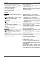
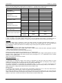
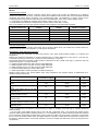
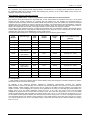
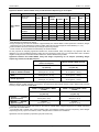
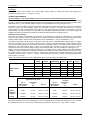
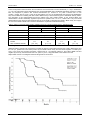
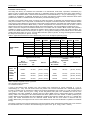
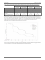
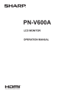
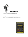
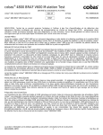
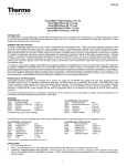
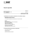
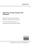
410697
16464 C - en - 2013/09
THxID™-BRAF
The THxID™-BRAF kit is an In Vitro Diagnostic device intended for the qualitative detection of the BRAF V600E and
V600K mutations in DNA samples extracted from formalin-fixed paraffin-embedded (FFPE) human melanoma tissue.
The THxID™-BRAF kit is a real-time PCR test on the ABI 7500 Fast Dx system and is intended to be used as an aid in
selecting melanoma patients whose tumors carry the BRAF V600E mutation for treatment with dabrafenib [Tafinlar®]
and as an aid in selecting melanoma patients whose tumors carry the BRAF V600E or V600K mutation for treatment
with trametinib [Mekinist™].
bioMérieux SA
English - 1
THxID™-BRAF
16464 C - en - 2013/09
SUMMARY AND EXPLANATION
The RAS/RAF/MEK/ERK pathway is a critical proliferation
pathway in many human cancers. This pathway can be
constitutively activated by alterations in specific proteins,
including BRAF, which phosphorylates MEK on 2
regulatory serine residues. Over 45 cancer-associated
mutations have been identified in BRAF (1). BRAF
mutations have been identified at a high frequency in
specific cancers, including approximately 50 to 60% of
melanoma (2), Approximately 90% of all identified BRAF
mutations that occur in human cancer are a T1799A
transversion mutation in exon 15, which results in a
V600E amino acid substitution (3). This mutation appears
to mimic regulatory phosphorylation, locks the BRAF
kinase in its active status, and increases BRAF activity
approximately 10-fold compared to wild type (2).
T1799A alteration (V600E mutation) accounts for 70 to
90% of BRAF mutant melanoma patients. In addition, the
T1799A alteration could be associated with a second
nucleotide mutation (G1798A) and leads to a V600K
mutation in an additional ~6% to 29% of patients with a
BRAF mutation (4).
Dabrafenib is a selective inhibitor of BRAF kinase activity
and trametinib is a selective inhibitor of MEK activity for
tumors that carry T1799A and/or GT1798-1799AA
alterations in the BRAF gene. The clinical utility of this kit
is
to
evaluate
the
BRAF
(V600E
/
K)
(T1799A / GT1798-1799AA) mutation status in order to
screen patients for treatment with the dabrafenib or
trametinib.
bioMérieux SA
PRINCIPLE
The THxID™-BRAF kit allows detection of the V600E and
V600K mutations of the BRAF gene from FFPE sections.
For this, the THxID™-BRAF kit makes use of 2 major
processes:
• Nucleic acid isolation from FFPE sections through
extraction / purification steps:
The paraffin is removed. The sample is lysed then
heated to reverse formalin crosslinking. The DNA is
bound to a membrane. After washing, concentrated
DNA is eluted from the membrane.
• Real time PCR amplification and detection of target
DNA (BRAF gene) present in the total nucleic acids:
Amplification Refractory Mutation Specific System
(ARMS) (5) PCR technology is used.
In the PCR reaction, primers specific for the BRAF gene
allow the amplification of a non-polymorphic gene area,
which is used as an internal control. The primers
specific for the mutations V600E and V600K allow the
amplification of mutated fragments leading to the
identification of BRAF mutations. Target specific probes
bind instantaneously to the newly synthesized
complementary DNA.
In the THxID™-BRAF kit, 2 different probes labeled with
2 different dyes allow the simultaneous detection of the
BRAF internal control and a BRAF mutation. Kinetic
analysis of the fluorescent signals and delta Ct
(Crossing threshold) calculation reveal the presence of
potential BRAF mutations.
English - 2
THxID™-BRAF
16464 C - en - 2013/09
CONTENT OF THE KIT (48 tests)
THxID™-BRAF PUR
Contents
Components
THxID™-BRAF Columns with Wash
Tubes (50)
COL
Columns in tubes
THxID™-BRAF Wash Tubes
3 x 50 (2 mL)
WT
2 mL tubes
THxID™-BRAF Elution Tubes
1 x 50 (1.5 mL)
ET
1.5 mL tubes
THxID™-BRAF Lysis Tubes
1 x 50 (2 mL)
LT
2 mL tubes
THxID™-BRAF Tissue Lysis Buffer
(10 mL)
Composition
ATL
Edetic acid
Sodium dodecyl sulphate
AL
Guanidine salt (GuHCl)*
25-50%
THxID™-BRAF Wash Buffer 1
(concentrate)
(19 mL)
AW1
Guanidine salt (GuHCl)*
50 – 100%
THxID™-BRAF Wash Buffer 2
(concentrate)
(13 mL)
AW2
Sodium azide (NaN3)
< 1%
THxID™-BRAF Elution Buffer
(14 mL)
ATE
Sodium azide (NaN3)
< 1%
THxID™-BRAF Proteinase K
(1.25 mL)
PK
THxID™-BRAF Lysis Buffer
(12 mL)
Proteinase K**
* HARMFUL reagent:
- R22: Harmful if swallowed.
- R36/38: Irritating to eyes and skin.
- S13: Keep away from food, drink and animal feeding stuffs.
- S26: In case of contact with eyes, rinse immediately with plenty of water and seek medical advice.
- S36: Wear suitable protective clothing
- S46: If swallowed, seek medical advice immediately and show this container or label.
** HARMFUL reagent:
- R36/37/38: Irritating to eyes, respiratory system and skin.
- R42: May cause sensitization by inhalation.
- S22: Do not breathe dust.
- S24: Avoid contact with skin
- S26: In case of contact with eyes, rinse immediately with plenty of water and seek medical advice.
- S36/37: Wear suitable protective clothing and gloves.
For further information, refer to the Material Safety Data Sheet available on request.
bioMérieux SA
English - 3
THxID™-BRAF
16464 C - en - 2013/09
THxID™-BRAF AMP
Contents
Components
Cap colors
Composition
V600E Primers and Probes
6 x 5.2 mg (lyophilized)
PRM V600E
Blue
Sphere containing:
• synthetic primers,
• synthetic fluorescent-labeled probes.
Each tube packed in a foil pack with silica
gel desiccant.
V600K Primers and Probes
6 x 5.2 mg (lyophilized)
PRM V600K
Lilac
Sphere containing:
• synthetic primers,
• synthetic fluorescent-labeled probes.
Each tube packed in a foil pack with silica
gel desiccant.
MM
Red
Mix containing:
• Tris Buffer,
• PCR enzyme,
• UNG amperase,
• Glycerol,
• Nucleotides,
• Sodium Azide (NaN3),
• Passive reference dye.
Master Mix*
6 x 0.24 mL (liquid)
Primers Diluent
6 x 0.5 mL (liquid)
PRMdil
Blue
Diluent containing:
• RNase/DNase-free water,
• preservative.
< 1%
Positive Control Diluent
6 x 0.5 mL (liquid)
CONT+dil
Yellow
Diluent containing:
• RNase/DNase-free water,
• preservative.
Positive Control
6 x 6.4 mg (lyophilized)
CONT+
Yellow
Sphere containing: synthetic DNA.
Each tube packed in a foil pack with silica
gel desiccant.
*: contains products of animal origin
MATERIAL REQUIRED BUT NOT PROVIDED
For information, please contact the local bioMérieux
representative.
General
• Calibrated micropipettes with variable settings for 1 to
1000 µL delivery volumes
• Timer
• Vortex-type mixer
• Sterile, disposable, aerosol resistant tips
• Waste container with cap or sealable
• Powder free gloves
Sample preparation
• DNase and RNase-free microtubes
• Scalpel
• Xylene 99%
• Ethanol (96 – 100%) molecular biology grade
• 37°C, 56°C and 90°C dry bath for incubation of 1.5 mL
or 2 mL microtubes
• Microcentrifuge 20000 g for 2 mL microtubes
Amplification with Applied Biosystems 7500 Fast Dx
Real-Time PCR instrument
• 7500 Fast Dx Instrument including Sequence Detection
Software v1.4 Security, Auditing, and E-Signature
module (Ref. Applied Biosystems 4406984 or 4406985).
bioMérieux SA
For 8-tube strips
• Mini-Strip Centrifuge (Ref. bioMérieux 285056 or
equivalent)
• MicroAmp Fast 8-Tube Strip, 0.1 mL (Ref. Applied
Biosystems 4358293)
• MicroAmp Optical 8-Cap Strip (Ref. Applied Biosystems
4323032)
• MicroAmp Cap Installing Tool (Handle) (Ref. Applied
Biosystems 4330015)
• Precision Plate Holder for 0.1 mL tube strips for 7500
Fast System (Ref. Applied Biosystems 4388506)
For 96-well plates
• Centrifuge for 96-well plates
• MicroAmp 96- and 384-well Optical Adhesive Film (Ref.
Applied Biosystems 4311971)
• MicroAmp Fast Optical 96-Well Reaction Plate with
Barcode 0.1 mL (Ref. Applied Biosystems 4346906)
• MicroAmp Adhesive Film Applicator (Ref. Applied
Biosystems 4333183)
Interpretation of results with the THxID™-BRAF
software
• Workstation with THxID™-BRAF software
• THxID™-BRAF templates
• Printer (optional)
English - 4
THxID™-BRAF
16464 C - en - 2013/09
WARNINGS AND PRECAUTIONS
• For In Vitro Diagnostic use only.
• For professional use only.
• This kit contains products of animal origin. Certified
knowledge of the origin and/or sanitary state of the
animals does not totally guarantee the absence of
transmissible pathogenic agents. It is therefore
recommended that these products be treated as
potentially infectious and handled observing the usual
safety precautions (do not ingest or inhale).
• When working with chemicals (for example Xylene),
consult the appropriate Material Safety Data Sheet
available from the product supplier.
• The Buffer AL and Buffer AW1 contain a harmful and
irritant agent (guanidine hydrochloride). Refer to the risk
phrases “R” and the precautions “S” above.
Guanidine hydrochloride can form highly reactive
compounds when combined with bleach. If liquids
containing these buffers are spilt, clean with liquid
detergent and water. If the spilt liquid contains
potentially infectious agents, clean the affected area first
with liquid detergent and water, and then with 1%
sodium hypochlorite.
• The Proteinase K reagent is a harmful agent. Refer to
the risk phrases “R” and the precautions “S” above.
• Kit reagents contain sodium azide which can react with
lead or copper plumbing to form explosive metal azides.
If any liquid containing sodium azide is disposed of in
the plumbing system, drains should be flushed with
water to avoid build-up.
• Do not use reagents or Columns after the expiration
date indicated on the label.
• Do not mix reagents (or disposables) from different lots.
• Check that the reagents are intact before use.
• Make sure reagents and samples are at 18-25°C before
use.
• All centrifugation steps should be carried out at
18-25°C.
• Make sure that the lyophilized material is at the bottom
of the tube before opening the tube.
• Ensure sample-to-result traceability by appropriate
labeling of the container at each transfer step
(e.g. lysate transfer, etc.) and at the level of the strips in
the PCR instrument.
• Avoid contamination or sample-to-sample carry-over:
- Never open more than one tube at a time to minimize
the risk of cross-contamination.
- Perform FFPE extraction and amplification in separate
dedicated laboratory areas (preferably a self-contained
area or laminar flow hood) using dedicated material.
- Use a fresh aerosol-resistant pipette tip (or equivalent)
for each pipetting action.
- Wear disposable gloves when working with amplified
material. Change gloves after contact with nucleic
acids. Wash hands thoroughly after completion of the
test procedure.
- Resuspend the V600E and V600K spheres before the
control sphere.
- To avoid amplicon contamination and to reduce the
risk of evaporation ensure amplification strips remain
closed.
• Use powder free gloves as powder may inhibit the PCR
reaction and cause invalid results.
bioMérieux SA
• Handling of Columns:
- Carefully apply the sample or solution to the Column.
Pipet the sample into the Column without wetting the
rim.
- Avoid touching the Column membrane with the pipet
tip.
- After all pulse-vortexing steps, briefly centrifuge the
microcentrifuge tubes to remove drops from the inside
of the lids.
- Open only one Column at a time and avoid generating
aerosols.
• Collect used disposable materials in a sealable
container. Close and remove the container after each
test run.
• Soak tube racks in a suitable detergent after each test
run for at least one hour.
• All materials and instruments should be regularly
cleaned and decontaminated. Immediately clean up any
spillage containing tissue lysate with liquid detergent or
a solution of household bleach containing at least 1%
sodium hypochlorite. For cleaning spills on or in the
7500 Fast Dx Real-Time PCR instrument, refer to the
User’s Manual.
LIMITATIONS OF THE METHOD
• THxID™-BRAF kit is to be used by professionals trained
and skilled in molecular biology techniques.
• The product must be used in strict accordance with the
instructions for use. Any deviation from the procedure
should be validated by the end-user.
• Melanin is a known inhibitor of PCR reactions. Melanin
may interfere with the THxID™-BRAF assay in highly
pigmented samples. If melanin inhibition is suspected,
repeat testing using a 1:4 dilution as suggested in the
troubleshooting table.
2
• The claimed tumor area for the assay is 20 mm to
2
250 mm (for a 10 µm section). Smaller tissue
2
(i.e., < 20 mm ) areas cannot ensure reliable results.
• The assay has been validated for a DNA input range of
10-350 ng/µL
• Users should ensure thermocycler temperature is
correctly calibrated as incorrect temperatures can lead
to invalid results.
• The test is designed to detect the BRAF V600E and
V600K mutations. Samples with results reported as
BRAF mutation negative may harbor BRAF mutations
not detected by the assay (e.g., V600R).
• Results that suggest E/K heterogeneity of the sample
may potentially come from a homozygous sample (see
accuracy section).
• The V600E PCR in the THxID™-BRAF assay crossreacts with the V600D. The assay has not been
validated to reliably detect V600D. Refer to the section
on performance studies below.
• The presence of AL Buffer in the eluate (tested by
adding 5 µL AL Buffer in the eluate) may lead to a false
result for a mutant sample which may then be reported
as “BRAF mutation negative”.
• The THxID™-BRAF kit is validated for FFPE skin and
lymph node melanoma tissue.
• The THxID™-BRAF kit is only validated for use with the
THxID™-BRAF PUR kit and the ABI 7500 Fast Dx Real
Time PCR instrument.
• The THxID™-BRAF kit is not to be used for diagnosis.
English - 5
THxID™-BRAF
16464 C - en - 2013/09
STORAGE CONDITIONS
THxID™-BRAF PUR
Stability
Kit stability
THxID™-BRAF PUR kit: until the labeled expiration date at 2-8°C or 18-25°C.
The Columns must be stored at 2-8°C.
Reconstituted Buffer AW1
6 months at 18-25°C or until the expiration date of the kit.
Reconstituted Buffer AW2
6 months at 18-25°C or until the expiration date of the kit.
THxID™-BRAF AMP
Stability
Kit stability
THxID™-BRAF AMP kit: until the labeled expiration date at 2-8°C.
PRM V600E solution
• 30 minutes at 18-25°C
• 1 month at -19°C to -31°C
Re-use frozen solution a maximum of 2 times. After each thawing, mix and spin
down briefly before use.
PRM V600K solution
• 30 minutes at 18-25°C
• 1 month at -19°C to -31°C
Re-use frozen solution a maximum of 2 times. After each thawing, mix and spin
down briefly before use.
Master Mix
30 minutes at 18-25°C
Re-use solution a maximum of 2 times.
Reagent Mix solution (primer /
probe solution + Master Mix)
30 minutes at 18-25°C
CONT+ solution
• 30 minutes at 18-25°C
• 1 month at -19°C to -31°C
Re-use frozen solution a maximum of 2 times. After each thawing, mix and spin
down briefly before use.
Samples
Stability
5 and 10 µm thick FFPE
section in tube or mounted on
slides
• 3 months at 18-25°C
• 3 months at 2-8°C
• 3 months at -19°C to -31°C
FFPE specimens can be transported at 18-25°C.
Eluate
• 2 hours at 18-25°C
• 48 hours at 2-8°C
• 7 months at -19°C to -31°C
• 7 months at ≤ - 60°C
Re-use frozen solution a maximum of 4 times. After each thawing, mix and spin
down briefly before use.
bioMérieux SA
English - 6
THxID™-BRAF
16464 C - en - 2013/09
INSTRUCTIONS FOR USE
Sample requirements
• Standard formalin-fixation and paraffin-embedding
procedures should be followed. To limit the extent of
DNA fragmentation:
- Fix tissue samples in 10% formalin as quickly as
possible after surgical removal.
- Use a fixation time of 14–24 hours (longer fixation
times lead to more severe DNA fragmentation,
resulting in poor performance in THxID™-BRAF
assay).
- Thoroughly dehydrate samples prior to embedding
(residual formalin can inhibit the Proteinase K
digestion).
• Sections will be processed according to the pathologist’s
indications:
1. If the sample section contains more than 80% of
tumor cells and does not contain a distinct area of
necrotic tissue, fatty tissue, hemorrhagic tissue or
non-tumoral melanin-rich area, then the entire
section can be placed in a tube, or if the sample is
on a slide, it can be entirely scraped with a scalpel.
2. If the sample section contains less than 80% of
tumor cells, then the section must be manually
macro-dissected in order to reach a final content of
at least 80% tumor cells. Use a dedicated sterile
scalpel to select the tissue part in order to enrich the
sample in tumoral cells.
3. If the sample section contains necrotic tissue, fatty
tissue, hemorrhagic tissue or non-tumoral melaninrich area, then the section should be manually
macro-dissected. Use a dedicated sterile scalpel to
select the tissue part in order to avoid the
undesirable portion.
• The minimum surface of tissue required for a 10 µm
2
section is 20 mm , not counting the necrotic / fatty /
hemorrhagic / non-tumoral melanin-rich area if it is
deemed dissectible (see above). If 5 µm sections are
2
prepared, the minimum is then 40 mm . Therefore a
sufficient number of sections should be included to meet
this requirement, while not exceeding 8 x 10 µm
sections (or 16 x 5 µm sections) to stay within the
recommended limit of the purification column.
2
• The total surface of tissue should not exceed 250 mm if
2
10 µm sections are prepared or 500 mm if 5 µm
sections are prepared.
Note: Use a single scalpel per sample.
Preparation of purification reagents
• Equilibrate all buffers at 18-25°C.
• Before starting the procedure, check whether precipitate
has formed in Buffer AL or Buffer ATL. If necessary,
dissolve by heating to 70°C with gentle agitation.
Preparing Buffer AW1
Add 25 ml ethanol (96-100%) to the bottle containing
19 ml Buffer AW1 concentrate.
Write down the current date on the label after ethanol
addition.
Note: Before starting the procedure, mix reconstituted
Buffer AW1 by shaking.
bioMérieux SA
Preparing Buffer AW2
Add 30 ml ethanol (96-100%) to the bottle containing
13 ml Buffer AW2 concentrate.
Write down the current date on the label after ethanol
addition.
Note: Before starting the procedure, mix reconstituted
Buffer AW2 by shaking.
Sample preparation
• Immediately place the dissected sections in a Lysis
Tube (1.5 mL microtube (not provided) or 2 mL Lysis
Tube (LT)).
Note: electrostatic effect can be observed for dissected
sample. Transfer the whole dissected area inside the
tube and briefly centrifuge.
• Prepare an empty Lysis Tube for the Negative Control
which will undergo the whole process.
• Add 1 mL xylene to all microtubes (samples and
Negative Control).
• Close the tube and mix at full speed using a vortex-type
mixer for at least 10 seconds.
• Centrifuge at full speed (approximately 20000 g) for
2 minutes ± 30 seconds at 18-25°C.
• Remove the supernatant by pipetting. Make sure to
leave 50-100 µL of supernatant in order not to remove
any of the pellet.
• Add 1 mL ethanol (96-100%) to the pellet, and mix
briefly at full speed using a vortex-type mixer.
• Centrifuge at full speed (approximately 20000 g) for
2 minutes ± 30 seconds at 18-25°C.
• Remove the supernatant by pipetting. Do not remove
any of the pellet. Carefully remove any residual ethanol
using a fine pipet tip.
• Open the tube and dry at 37 ± 2°C for 10 ± 1 minutes or
until all residual ethanol has evaporated.
• Add 180 μL Buffer ATL to the dry pellet.
• Add 20 μL proteinase K.
• Mix briefly at full speed using a vortex-type mixer.
• Incubate at 56 ± 3°C for 1 hour ± 5 minutes.
• Incubate at 90 ± 5°C for 1 hour ± 5 minutes.
If using only one heating block, leave the sample at
18-25°C after the 56°C incubation until the heating block
has reached 90°C.
• Briefly centrifuge the Lysis Tube to remove drops from
inside the lid.
• Add 200 μL Buffer AL to the sample, and mix at full
speed using a vortex-type mixer.
• Add 200 μL ethanol (96-100%) and mix again at full
speed using a vortex-type mixer.
A white precipitate may form upon addition of Buffer AL
and ethanol. This precipitate does not interfere with the
extraction procedure.
• Briefly centrifuge the Lysis Tube to remove drops from
inside the lid.
• Carefully transfer the entire lysate (with the white
precipitate if any) to the Column (in a 2 mL Wash Tube)
without wetting the rim.
• Close the lid.
• Centrifuge at approximately 6000 g for 1 minute at
18-25°C.
If the lysate has not completely passed through the
membrane after centrifugation, centrifuge again at a
higher speed until the Column is empty.
• Place the Column in a clean 2 mL Wash Tube (WT).
• Discard the Wash Tube containing the flow-through.
• Carefully open the Column and add 500 μL of Buffer
AW1 without wetting the rim.
English - 7
THxID™-BRAF
16464 C - en - 2013/09
• Close the lid.
• Centrifuge at approximately 6000 g for 1 minute at
18-25°C.
• Place the Column in a clean 2 mL Wash Tube.
• Discard the Wash Tube containing the flow-through.
• Carefully open the Column and add 500 μL of Buffer
AW2 without wetting the rim.
• Close the lid.
• Centrifuge at approximately 6000 g for 1 minute at
18-25°C.
• Place the Column in a clean 2 mL Wash Tube.
• Discard the Wash Tube containing the flow-through.
• Centrifuge at full speed (approximately 20000 g) for
3 minutes at 18-25°C to dry the membrane completely.
Note: Avoid contact between the Column and the flowthrough. Take care when removing the Column and
Wash Tube from the rotor so that flow-through does not
come into contact with the Column.
• Place the Column in a clean 1.5 mL Elution Tube (ET).
• Discard the Wash Tube containing the flow-through.
• Carefully open the lid of the Column and apply 60 μL of
Buffer ATE to the center of the membrane.
• Close the lid.
• Incubate at 18-25°C for at least 1 minute.
• Centrifuge at full speed (approximately 20000 g) for
1 minute at 18-25°C.
Note: The volume of eluate will be up to 5 μL less than
the volume of elution solution applied to the Column.
• Transfer the tubes with nucleic acid extracts to the
amplification laboratory area or store (see “Storage
Conditions”).
Amplification
Switch on the 7500 Fast Dx Real-Time PCR instrument.
Preparation of the SDS run file on the SDS software
Refer to the section "Perform a run on the 7500 Fast Dx
Real-Time PCR instrument".
• Create a new run selecting the appropriate template.
• Adapt the created run to the plate layout.
Preparation of PCR reagents
Equilibrate reagents and samples at 18-25°C before use.
Preparation of primer and probe solution
• Add 85 µL of PRM diluent to the V600E and V600K
primer sphere.
When more than 8 reactions are required, spheres with
the same lot number must be pooled in one tube and
diluent volume must be adapted.
Maximum number
of samples and
controls to be run
Number of
spheres
Volume of primer
diluent (µL)
8
1
85
16
2
170
24
3
255
48
6
510
• Immediately mix at full speed using a vortex-type mixer
until a clear solution has been obtained.
• Centrifuge briefly.
bioMérieux SA
Notes:
• With one sphere it is possible to do 3 runs of one
sample (including the 2 controls).
• As long as the remaining volume of V600E and V600K
solutions is sufficient for 3 reactions, it can be stored
(see “Storage Conditions”).
• Do not pool the remaining volume of primer and probe
solutions from different tubes.
Preparation of reagent mix
• Briefly centrifuge the Master Mix.
• Mix the V600E or V600K solution with Master Mix
according to the following table:
Number of
samples and
controls to be run
V600E or V600K
solution
(µL)
Master Mix
(µL)
3
27
36
8
85
110
16
170
220
24
255
330
48
510
660
If another number of samples is to be run, make sure to
use 8 µL of V600E or V600K solution per reaction and to
leave a certain margin to calculate the volumes needed. It
is important to maintain a volume of Master Mix that is 1.3
times higher than the volume of primer solution (rounding
up to the nearest whole number).
• Mix at full speed briefly using a vortex-type mixer.
• Centrifuge briefly.
Warning:
The remaining volume of reagent mix cannot be stored
and should be used within 30 minutes.
Preparation of Positive Control solution
• Add 150 µL of Positive Control Diluent to the Positive
Control sphere.
• Mix at full speed using a vortex-type mixer until a clear
solution has been obtained.
• Centrifuge briefly.
Notes:
The remaining volume of Positive Control can be stored
(see “Storage conditions”).
Do not pool the remaining volumes of Positive Control
solutions from different tubes.
Repartition of reagents in 8-tube strips or
96-well plates
Per single sample, the DNA amplification is carried out in
2 wells each containing a duplex PCR reaction. The
V600E duplex PCR contains the internal control PCR and
the V600E mutation-specific PCR. The V600K duplex
PCR contains the internal control PCR and the V600K
mutation-specific PCR. A Negative Control (starting from
the extraction process) and a Positive Control for
amplification will be tested per run for each duplex
reaction.
For dispensing of the reagent mix and eluate, respect the
plate layout from the created run.
Note: Do not invert the V600E and V600K reagent mixes.
English - 8
THxID™-BRAF
16464 C - en - 2013/09
In each tube of an 8-tube strip or of a 96-well plate:
• Following the plate layout, transfer 18 µL of the V600E
or V600K reagent mix to the bottom of a single tube.
• Add 2 µL of Positive Control or Negative Control or
sample eluate to the dedicated tube.
Mix by aspiration and dispensing.
Note: After adding the reagent mix and controls or
sample eluate(s) to all tubes, visually check that the
volume in each tube is identical.
Generate an SDS result file
• At the end of the run, start the analysis on the
instrument using "Auto Ct".
Note: Do not modify any other items. The
THxID™-BRAF software checks the instrument
configuration. If configuration is incorrect, the software
will not be able to interpret.
• At the end of the analysis, save the SDS result file
generated.
Run amplification on the 7500 Fast Dx Real-Time PCR
instrument
Generate a BRAF mutation test report
• Transfer a copy of the SDS result file to the dedicated
THxID™-BRAF computer using for instance a USB stick
and the menu "File\Save As" of the SDS software.
Warning: If modifications need to be made to the SDS
result file after it has been copied, do the following
steps:
- Update the SDS run file on the SDS software.
- Destroy all existing copies of the previous versions of
the SDS result file (recommended).
- Destroy all the BRAF mutation test reports
corresponding to the previous version of the SDS
result file (recommended).
- Resume the instructions for the modified SDS result
file from the section “Generate an SDS result file”.
• Generate a BRAF mutation test report for the SDS result
file using the THxID™-BRAF software.
For 8-tube strips
• Close the strips using the MicroAmp Cap Installing Tool
(Handle).
• Properly identify the strips on the extremity of the cap
strip according to the position in the plate layout.
Note: Do not write directly on the caps or tubes as
ink interferes with fluorescence readings.
• Briefly centrifuge the strips.
• Place the strips on the Precision Plate Holder according
to the plate layout.
Notes :
- A minimum of 2 tube strips and a maximum of 6 tube
strips can be run on the Precision Plate Holder at the
same time.
- Do not invert the V600E and V600K strips.
• Insert 2 fully capped empty MicroAmp Fast 8-Tube
st
Strips in the 1 left and last columns of the Precision
Plate Holder.
• Start amplification on the instrument.
For 96-well plates
• Cover the 96-well plates with the adhesive film using the
Adhesive Film Applicator.
Note: Do not write directly on the plate as ink
interferes with fluorescence readings; if necessary, use
the sides of the plate.
• Centrifuge the 96-well plates at 200 g for 3 minutes at
18-25°C.
• Transfer the plates to the Applied Biosystems 7500 Fast
Dx Real-Time PCR instrument.
• Start amplification on the instrument.
For complete instructions, refer to the 7500 Fast Dx RealTime PCR instrument User's Manual or / and to the
THxID™-BRAF software User's Manual.
RESULTS AND INTERPRETATION
The THxID™-BRAF software interprets the results
automatically and highlights the presence of valid or
invalid results in the generated report.
The 2 possible outcomes for Positive and Negative
Controls are "valid" or "invalid".
The result validity of clinical specimens is determined first
by the internal control Ct (Crossing threshold) values that
should fall within pre-specified limits.
A result is invalid if one of the delta Ct or internal control
Ct values falls outside the expected limits.
If a specific amplification is detected for the mutant target,
the result of each reaction (V600E or V600K) is based on
the delta Ct value (Ct mutant – Ct IC):
• If the delta Ct value is below a threshold value then a
V600E or V600K BRAF mutation is present,
• If the delta Ct value is above a threshold value then no
V600E or V600K BRAF mutation is present or it is below
the limit of detection.
If no amplification is detected for the mutant targets
(V600E and V600K not detected), the sample will be
characterized as BRAF mutation-negative.
The final result is determined based on results obtained
for both multiplexes.
bioMérieux SA
English - 9
THxID™-BRAF
16464 C - en - 2013/09
Valid results of clinical specimens
Reported result
Description
BRAF mutation negative
V600E and V600K mutations not detected
V600E mutation
V600E mutation detected
V600K mutation
V600K mutation detected
V600E and V600K mutations
V600E and V600K mutations detected
Invalid results
Invalid control
An invalid Positive or Negative Control invalidates the complete run.
If one or more controls is invalid, the results of the clinical specimen obtained in the run are not reported.
If an invalid result is obtained for a control, refer to the troubleshooting table below:
Sample type
Negative Control
Message
Description
Amplification detected
in Negative Control well
for IC
Contamination
with
internal control DNA
was detected in the
Negative Control well.
Amplification detected
in Negative Control well
for V600E
Contamination
with
V600E mutation DNA
was detected in the
Negative Control well.
Amplification detected
in Negative Control well
for V600K
Contamination
with
V600K mutation DNA
was detected in the
Negative Control well.
Appropriate corrective measure
The complete run must be repeated using
frozen sample eluates, frozen Negative
Control eluate and either frozen Positive
Control if available or a new preparation.
If the Negative Control is still invalid
repeat the complete procedure starting
from the extraction.
Amplification for IC out
of bounds in Positive
Control well
Positive Control
Amplification for V600E
out of bounds
in
Positive Control well
Amplification for V600K
out of bounds
in
Positive Control well
This indicates that an
error occurred during
the process or that
reagents failure was
detected.
Delta Ct out of bounds
in Positive Control well
bioMérieux SA
English - 10
The complete run must be repeated using
frozen sample eluates, frozen Negative
Control eluate and a new preparation of
Positive Control.
THxID™-BRAF
16464 C - en - 2013/09
Invalid clinical specimen
If an invalid result is observed for a clinical specimen in a valid run, refer to the troubleshooting table below.
Sample type
Message
Description
Appropriate corrective measure
Too much DNA in the
reaction, possible PCR
overloading.
The eluate has to be diluted 1:4 in Buffer
ATE before amplification by adding 10 µL
of eluate to 30 µL of Buffer ATE. If the
result is still invalid, repeat the test
starting from the extraction using lower
tissue area.
Sample IC amplification
above maximum
threshold in well XX.
Too low DNA in the
reaction
and/or
PCR inhibition.
If the tested sample has been
characterized as containing no or a low
level of melanin (≤ 10%) by the
pathologist, repeat the test, starting from
the eluate. If the result is still invalid,
repeat the test for the invalid sample,
starting from the extraction using higher
tissue area.
If the tested sample has been
characterized as containing a medium to
high level of melanin (> 10%) by the
pathologist, the eluate has to be diluted
1:4 in Buffer ATE before amplification by
adding 10 µL of eluate to 30 µL of Buffer
ATE. If the result is still invalid, repeat the
test starting from the extraction using
higher tissue area.
Sample delta Ct too low
in well XX.
An unexpected ratio
between IC and mutant
PCR is detected.
Repeat the test starting from the eluate.
No available result due
to control failure.
No result is reported for
this sample due to an
invalid Positive and/or
Negative Control.
Please refer to the table for invalid control
troubleshooting.
Sample IC amplification
below minimum
threshold in well XX.
Clinical specimen
PERFORM A RUN ON THE 7500 Fast Dx Real-Time PCR INSTRUMENT
(Refer to the 7500 Fast Dx Real-Time PCR instrument User's Manual).
Templates for the 7500 Fast Dx Real-Time PCR instrument
bioMérieux provides 4 templates for the 7500 Fast Dx Real-Time PCR instrument and its associated Sequence Detection
Systems (SDS) Software. The templates configure the 7500 Fast Dx Real-Time PCR instrument for a BRAF run.
Note: On the Precision Plate Holder displayed on the screen, numbers indicate the position of the Plate's columns (from
1 to 12), letters indicate the position of the Plate's lines (from A to H starting from the top).
The proper template is chosen according to the number of clinical samples to be tested in the run:
Number of clinical samples
Position of the strips on the plate
Position of the controls
on the plate
1-6 THxID™-BRAF
template .sdt (2 strips)
From 1 to 6
Columns 6 and 7
Negative: A6 and A7
Positive: B6 and B7
7-14 THxID™-BRAF
template.sdt (4 strips)
From 7 to 14
Columns 5 to 8
Negative: A5 and A6
Positive: B5 and B6
15-22 THxID™-BRAF
template.sdt (6 strips)
From 15 to 22
Columns 4 to 9
Negative: A4 and A5
Positive: B4 and B5
23-46 THxID™-BRAF
template.sdt (plate)
From 23 to 46
Non applicable
Negative: A1 and A2
Positive: B1 and B2
The templates also include positions and settings for the Positive Control and Negative Control.
Strips cannot be used for the 23-46 layout configuration. If 23 or more samples are tested, use a plate.
bioMérieux SA
English - 11
THxID™-BRAF
16464 C - en - 2013/09
Run layout
Creation of a new SDS run file for the 7500 Fast Dx
Real-Time PCR instrument
• Update the names of the clinical samples to be amplified
using the Well Inspector icon from the top menu. See
figure below:
• Open the Sequence Detection Systems (SDS) Software.
• Log in as a routine user.
• Create a new document (Select "New…" in the file
menu or "Create New Document…" in the "Quick
Startup" dialog).
A "New Document Wizard" dialog box is displayed:
• Check the value of the following items:
Item
Mandatory values
Assay
Standard curve (Absolute Quantitation)
Container
96-Well Clear
Template
Browse to one of the THxID™-BRAF
templates provided (1-6, 7-14, 15-22 or
23-46) and open the selected template.
Run Mode
Standard 7500
• Fill in the “Comments” field with the lot numbers of the
THxID™-BRAF PUR kit and the THxID™-BRAF AMP kit
used in order to comply with the reagent tracking.
• Change the name of the plate in the "Plate Name" field.
This field is used to suggest the name of the created
SDS run file.
• Click on "Finish". The SDS run file pre-configured for a
THxID™-BRAF run is created and automatically
opened. See figure below:
•
•
•
•
Ensure names of V600E and V600K reactions for the
same sample are strictly identical. Make sure that
each sample has a unique name.
Do not modify the selection of the detectors for the
clinical samples.
Do not modify the name and the selection of the
detectors for the Positive Control and Negative
Control.
Delete unused clinical sample wells from the run layout
if any: delete sample name and uncheck selected
detectors.
Note: Unused wells and detectors not deleted may
create background noise, which would alter results.
Do not change other run layout settings.
Save the modified SDS run file. Select a name for the
SDS run file that clearly identifies the run.
The run layout of the SDS run file can be printed and
used as a guide for the dispensing of the reagent mix
and eluates.
The run is ready to start.
For complete instructions, refer to the 7500 Fast Dx RealTime PCR instrument User's Manual.
QUALITY CONTROL
A Positive Control is included in each THxID™-BRAF kit.
A Negative Control must be performed at the same time
as the samples, starting from sample preparation.
These controls must be performed in each run to ensure
that the reagents have not been altered and to check the
absence of contamination.
The instrument will be able to check the control value or
the validity only if their positioning corresponds to the
plate layout.
Moreover, the integrity of each individual result can be
monitored by reference to the performance of the internal
control PCR.
Results cannot be validated if the control values are not
valid.
bioMérieux SA
English - 12
THxID™-BRAF
16464 C - en - 2013/09
WASTE DISPOSAL
• Unused AL, AW1 and PK reagents must be disposed of
following procedures for hazardous chemical waste.
• Do not add bleach or acid solutions directly to the
sample preparation waste containing Buffer AL and
Buffer AW1.
• Dispose of used or unused Master Mix reagents as well
as any other contaminated disposable materials
following procedures for infectious or potentially
infectious products.
• The other unused reagents may be considered as non
hazardous waste and disposed of accordingly.
It is the responsibility of each laboratory to handle waste
and effluents produced according to their nature and
degree of hazardousness and to treat and dispose of
them (or have them treated and disposed of) in
accordance with any applicable regulations.
NON-CLINICAL PERFORMANCE
Sample characterization
All FFPE samples used in the non-clinical studies underwent a pathology review in order to determine the tumor content
(% tumor cells), the melanin content, the presence of necrotic, lipidic or hemorrhagic tissue in the stained sections.
The genetic status of the samples on the V600 locus was determined by bi-directional Sanger sequencing.
Genomic input range
The THxID™-BRAF assay was validated for a DNA input range of 10-350 ng/µL (i.e. 20-700 ng / PCR reaction). This
range was used for most performance evaluation studies.
Among the 891 clinical samples included in the accuracy study (see “Accuracy study” section) with a Sanger sequencing
result and a THxID™-BRAF result, 94.6% DNA values were within [10-350] ng/µL. DNA values ranged from
2 to 1764 ng/µL.
Figure 1: Distribution of the DNA concentrations observed for the 891 clinical specimens measured. The x axis
represents the concentration (in ng/μL, 2 μL input are used per reaction) and the y axis the number of samples per
concentration. One sample which has a concentration of 1764 ng/µL is not represented in this plot to allow the reduction
of the scale.
Analytical sensitivity - Limit of Blank
Potential background amplification (Ct value) was assessed on 6 clinical procured specimens at high DNA input (target
of 350 ng/μL, skin V600K at 150 ng/μL) covering all testing conditions (i.e.: Wild Type, V600E and V600K for skin and
lymph node) and tested over 3 runs total of 60 replicates (20 for skin V600K samples). Background amplification was
also evaluated on DNA extracted from 3 cell lines (Wild Type, V600E homozygous, V600K heterozygous) at high DNA
input (350 ng/µL). The THxID™-BRAF assay did not show any background amplification in all tested conditions.
Analytical sensitivity - Limit of Detection for V600E or V600K mutations
The Limit of Detection (LoD) for the THxID™-BRAF assay is defined as the lowest mutation level in a specimen for which
the assay yields a positive result in 95% of the tests. DNA was extracted from FFPE melanoma skin and lymph node
specimens with either the V600E or V600K mutation and blended with wild-type FFPE DNA from the same clinical
specimen type. DNA input concentrations spanned the claimed input range (high, medium and low) and a total of
24 replicates (12 replicates per lot) for each condition were evaluated. LoD was determined by Probit analysis (the
calculation was based on the assumption that the starting material extracted from the mutant specimens contained
100% mutant DNA). The data support a claimed LoD of 5% mutant DNA in a background of wild-type DNA for V600E
and V600K positive FFPE skin and lymph node specimens across the DNA input range.
bioMérieux SA
English - 13
THxID™-BRAF
16464 C - en - 2013/09
This 5% LoD was subsequently confirmed with 20 replicates on a sample panel including FFPE skin and lymph node
specimens with high melanin, and FFPE cell lines on a third lot as described in the following table:
Condition
10 ng/reaction
(below DNA range)
20 ng/reaction
(lower limit of DNA range)
High melanin content*
700 ng/reaction
(higher limit of DNA range)
1000 ng/reaction
(above DNA range)
Melanoma cell line FFPE blocks
Sample
Mutant allele at 5%
Mutation positive / tested
Lymph node
V600E
20/20
Skin
V600K
20/20
Lymph node
V600E
20/20
Skin
V600E
20/20
Lymph node
V600K
20/20
Skin
V600K
20/20
Skin
V600E
20/20
Lymph node
V600K
20/20**
Lymph node
V600E
20/20
Skin
V600E
20/20
Lymph node
V600K
20/20
Skin
V600K
20/20
Lymph node
V600E
20/20
Lymph node
V600K
20/20
Melanoma
cell line
V600E
20/20
Melanoma
cell line
V600K
20/20
* DNA input for high melanin samples were at 60 ng/reaction for the V600E sample and 526 ng/reaction for the V600K
sample and melanin content were respectively 80% and 75%.
** After 1:4 dilution of the eluate. Before dilution each of the 20 replicates was invalid. In accordance with the
troubleshooting table, the eluate was diluted 1:4 in Buffer ATE and re-tested with results as described in the table.
Inclusivity
The THxID™-BRAF assay is designed to detect the V600E (T1799A) and V600K (GT1798/1799AA) mutations. In
addition, the THxID™-BRAF assay was shown to detect a rare form of the V600E mutation (i.e., rare codon GAA) and
the V600E/K601E mutation (also referred to as V600E2) using 2 FFPE lymph node specimens and plasmids.
Cross-reactivity
Cross-reactivity of the THxID™-BRAF assay was assessed by testing:
• plasmids representing Wild Type, V600D, V600R, V600L, V600M, V600G, V600A and BRAF pseudogene plasmids
(BRAF homologue present on chromosome X),
• procured clinical specimens representing V600R specimens.
5
Plasmids were tested in triplicate at a concentration of 2 x 10 copies/reaction.
No cross-reaction with V600E nor V600K PCR was reported on Wild Type, V600E, V600K, V600R (plasmid and clinical
5
samples), V600L, V600M, V600G and V600A mutants and the pseudogene at 2 x 10 copies/reaction and on V600G
5
with up to 3 x 10 copies/reaction.
V600E cross-reaction was reported for V600D.
Interfering substances
Hemoglobin and triglycerides
2 concentrations of hemoglobin (4 mg/mL and 2 mg/mL) or triglycerides (74 mM and 37 mM) were added to 11 FFPE
samples during the lysis step (i.e. directly in the lysis buffer between deparaffinization and extraction). Each condition
was tested in 3 replicates (from 3 extractions) with one lot of THxID™-BRAF assay.
®
The tested concentrations of hemoglobin and triglycerides reflect 2 x and 1 x the CLSI recommended high
concentration respectively. The same FFPE specimens were also tested without interfering substance, as a reference.
Neither hemoglobin nor triglycerides interfered with the THxID™-BRAF assay.
Necrotic tissue
21 melanoma FFPE specimens with necrotic tissue concentrations ranging from 15% to 60% were tested with the
THxID™-BRAF assay. Necrotic tissue content was determined by pathologist review.
Each sample was tested in 3 replicates (from 3 extractions) with one lot of THxID™-BRAF assay. The correct allele was
called in all instances including one sample with high melanin content following the instruction to dilute, showing that the
presence of up to 60% of necrotic tissue does not interfere with the THxID™-BRAF assay.
bioMérieux SA
English - 14
THxID™-BRAF
16464 C - en - 2013/09
Melanin
Melanoma FFPE samples
A population of 56 FFPE samples containing melanin levels ranging from 50-100% (as determined by a pathology
review) were selected for the study. A total of 16 specimens resulted in invalid results. Nine of these samples remained
unresolved after further dilution according to the troubleshooting table. The distribution of melanin content for the
9 invalid specimens (4 lymph node and 5 skin) is shown in the table below. There were no false negative results.
• 1 invalid result was obtained for samples with a melanin content < 80% (1/25 = 4.0%),
• 8 invalid results were obtained on samples with a melanin content ≥ 80% (8/31 = 25.8%).
Thus, in the presence of a very high melanin content the risk of obtaining an invalid result is elevated.
Melanin content (%)
50
60
70
80
90
100
Number of final invalid
samples
1
0
0
2
5
1
Number of final valid
samples
11
5
8
12
10
1
Number of samples
12
5
8
14
15
2
Total: 56
Dark skin FFPE samples
5 non-melanoma dark skin samples were tested with the THxID™-BRAF assay. The results were conform with the bidirectional Sanger status of the samples, i.e. Wild Type in all instances.
Repeatability: within-laboratory precision
An internal study was conducted to evaluate the precision of the entire THxID™-BRAF workflow, i.e. extraction and
amplification.
Results were obtained in duplicates on 8 panel members using 2 THxID™-BRAF lots, 4 days per lot, 2 runs per day,
2 instruments (2 days per instrument), 2 operators, each performing 1 run per day.
The precision panel was comprised of FFPE melanoma specimens of skin or lymph node origin. (1 specimen was from
subcutaneous tissue.). The panel included:
• 2 negative samples (Wild Type) with a low and a high DNA input,
• 2 V600E samples close to LoD, with a low and a high DNA input,
• 2 V600K samples close to LoD, with a low and a high DNA input,
• 1 moderate V600K sample at a medium DNA input,
• 1 V600E high melanin containing sample at a medium DNA input.
Mutation status results of the THxID™-BRAF assay were compared to the expected results, as determined by bidirectional Sanger sequencing.
Panel member
Skin Wild Type with low DNA input
Lymph node Wild Type with high DNA
input*
Skin V600E close to LoD with low DNA
input
Lymph node V600E close to LoD with
high DNA input
Skin V600E with medium DNA input
and high melanin content
Lymph node V600K close to LoD with
low DNA input
Lymph node V600K with medium DNA
input and moderate mutation content
Lymph node V600K close to LoD with
high DNA input**
Mean DNA
concentration (ng/µL)
12
32/32
% Correct
Call
100%
174
32/32
100%
43
32/32
100%
579
32/32
100%
140
32/32
100%
44
32/32
100%
85
32/32
100%
317
16/16
100%
N°. correct calls / N°. replicates
*: had a moderate DNA concentration during the study (174 ng/µL on average), rather than high as originally determined
before the study.
**: for the Lymph node V600K sample close to the LoD with a high DNA input, a first sample was tested and the
observed results agreed with the expected result for 17 of 32 replicates (53.1%), while a Wild Type result was observed
for the remaining 15 replicates.The presumptive cause for the low positive rate of this panel is a mutation content below
the 5% LoD of the THxID™-BRAF assay, and slight variations in mutation content (around the positivity cut-off) that can
be present in different sections of this FFPE specimen. The second sample was tested with one run per day (hence the
16 replicates).
bioMérieux SA
English - 15
THxID™-BRAF
16464 C - en - 2013/09
As a conclusion, 100% agreement with expected results was obtained for 7 of 8 original panel members as well as for
the additional V600K panel member tested, demonstrating the precision of the THxID™-BRAF assay across runs,
operators, instruments, days and lots.
Reproducibility: between-laboratory precision
Precision using DNA eluates: analysis of quantitative results obtained for all sites/instruments
This precision study was evaluated on all pooled data of each panel member and positive control (N = 72 per panel
member and per control), performed at 3 external sites via multiple runs over multiple non-consecutive days by
2 operators at each site using 3 lots of the THxID™-BRAF assay total (only 2 lots were used at any one site and all panel
members were run in duplicate for each run including the controls). The panel members are representative of the main
possible sample types that can be analyzed with the THxID™-BRAF assay: skin and lymph node, Wild Type, V600E or
V600K, low to high DNA concentration, low to high mutation content, presence or absence of melanin.
The results from the qualitative analysis, coupled with information from the quantitative analysis, demonstrate that the
THxID™-BRAF assay shows a high degree of reproducibility at each study site, across 3 instruments, and with different
operators with near 100% correct identification of each panel member.
The following table describes the overall agreement estimate across the sites by panel member:
Panel
Member
Specimen type
DNA
input
Percent
mutant
Dilution
step*
No. of valid tests all three
sites / Total number of tests
(95% CI)
Wild-Type
Skin
Low
n/a
n/a
72/72 [94.9% ; 100%]
Wild-Type
Skin
High
n/a
n/a
72/72 [94.9% ; 100%]
Wild-Type
Lymph node
Low
n/a
n/a
72/72 [94.9% ; 100%]
Wild-Type
Lymph node
High
n/a
n/a
72/72 [94.9% ; 100%]
Wild-Type
Skin –
high melanin
Med
n/a
Diluted
72/72 [94.9% ; 100%]
V600E
Skin –
high melanin
Med
Med
Diluted
72/72 [94.9% ; 100%]
V600E
Skin
Low
Close to LoD
n/a
60/72 [73.1% ; 90.2%]
V600E
Lymph node
High
Close to LoD
n/a
72/72 [94.9% ; 100%]
V600E
Skin
Med
Med- high
n/a
72/72 [94.9% ; 100%]
V600E
Lymph node
Med
Med- high
n/a
72/72 [94.9% ; 100%]
V600K
Lymph node –
high melanin
Med
Med
Diluted
72/72 [94.9% ; 100%]
V600K
Skin
Low
Close to LoD
n/a
72/72 [94.9% ; 100%]
V600K
Lymph node
High
Close to LoD
n/a
72/72 [94.9% ; 100%]
V600K
Skin
Med
Med-high
n/a
72/72 [94.9% ; 100%]
V600K
Lymph node
Med
Med-high
n/a
72/72 [94.9% ; 100%]
CI: Confidence Interval
n/a: not applicable
*: a high melanin content may interfere with the THxID™-BRAF assay (see paragraph on melanin interference). Dilution
per the instructions restored the PCR signal.
An estimate of the within-run precision, between-run (operators), between-days, between lots, between
sites/instruments, and the total precision was conducted. The standard deviation and %CV for Ct and ΔCt results for
V600E samples, V600K samples, internal control (IC) Ct values for the Wild Type (WT) samples, and positive and
negative controls, were investigated as a measure of the variability of the assay. For the Wild-type panel samples, the
internal control Ct for the V600E multiplexes ranged from 24.4 to 28.7 with %CV range 0-3.3% and the V600K
multiplexes IC ranged from 24.2 to 28.5 with %CV range 0-2.8%. The mean ΔCt ranged from 1.9 to 6.1 for the V600E
mutation positive samples with associated %CV values ranging from 0 to 16.7%. The mean ΔCt ranged from 1.2 to 4.9
for the V600K mutation positive samples with associated %CV values ranging from 0 to 25.6%. The higher imprecision
was associated with the high melanin content sample. For the V600E positive control the mean ΔCt was 3.7 and the
%CV values ranged from 0 to 12.8%. The V600K positive control mean ΔCt value was 3.5 with associated %CV ranging
from 0 to 15.1%.
bioMérieux SA
English - 16
THxID™-BRAF
16464 C - en - 2013/09
Sample handling variability
3 specimens for 3 different samples were studied. Serial sections from a single block were used. Three sections of each
sample type were forwarded to each of the 3 external sites, alternating the sections. For example, section 1, 4, 7 were
sent to Site 1. Sections 2, 5, 8 were sent to Site 2, and similarly for Site 3. One specimen contained lymph node tissue,
the second contained skin tissue. One of these specimens was positive for V600E and the other was positive for V600K.
The third specimen was Wild Type with a high melanin content.
A breakdown of the specimens provided is in the table below.
Site 1
Site 2
Site 3
Wild Type with high melanin (lymph nodes)
3 sections
3 sections
3 sections
V600E (skin)
3 sections
3 sections
3 sections
V600K (lymph nodes)
3 sections
3 sections
3 sections
1, 4, 7
2, 5, 8
3, 6, 9
Section numbers
The table below presents the results obtained for each site for each of the 3 specimens tested for each of the 3 FFPE
tissue samples provided.
Site
Site 1
Sample
Sample type
Results
Sample 1
Curls
3/3 Wild type
Sample 2
Curls
3/3 V600E
Sample 3
Sections on slides
3/3 V600K
Subtotal
9/9 (100%)
Sample 1
Site 2
Curls
3/3 Wild type
Sample 2
Curls
3/3 V600E
Sample 3
Sections on slides
3/3 V600K
Subtotal
9/9 (100%)
Sample 1
Site 3
Curls
3/3 Wild type
Sample 2
Curls
3/3 V600E
Sample 3
Sections on slides
3/3 V600K
Subtotal
9/9 (100%)
Total
27/27 (100%)
The table above shows 100% agreement with the expected results for all sites. This high agreement rate indicates the
assay’s ability to yield accurate, consistent results even when there is variability in sample preparation due to multiple
users.
Accuracy: correlation to Reference Method for Clinical Samples
The accuracy of the THxID™-BRAF assay was assessed relative to an analytical reference standard based on PCR
amplification followed by bi-directional Sanger DNA sequencing method on a representative sampling of clinical trial
samples, consecutively taken from the pool of clinical samples until a statistically significant sample size was reached for
each allele, in particular V600K. There were 898 samples available for testing. Excluding all invalids and QNS samples (total
43) there were 35 discordant cases [35/(898-43) = 4.1%]. Two samples determined to be V600D were detected by the
THxID™-BRAF assay as V600E.
The overall results are shown in the tables below.
bioMérieux SA
English - 17
THxID™-BRAF
16464 C - en - 2013/09
Agreement between THxID™-BRAF assay and Bi-directional Sequencing for all samples
Bi-directional Sequencing
BRAF V600
mutations
V600E
5
V600E
341
V600K
THxID™-BRAF
result
1
V600E and K
4
E and K mutation
negative
Invalid
QNS
V600K
E and K
detected
1
2
Total
mutations
not
Invalid
5
1
QNS
2
Total
V600D
V600R
2
0
21
7
0
373
0
2
3
0
0
60
3
0
0
4
2
57
0
WT
3
0
2
0
0
2
6
2
0
11
406
5
0
430
6
1
0
0
20
2
0
29
0
0
0
0
2
0
1
3
354
64
2
11
453
14
1
899
1
No result was obtained.
QNS: Quantity Not Sufficient for testing.
3
Sanger sequencing has a limit of detection of approximately 20% mutant alleles in FFPE specimens. Therefore, Sanger
sequencing may not be adequate to confirm mutation status at lower percentages of mutant alleles (6, 7, 8, 9).
4
Double mutants cannot be confirmed by the Sanger sequencing method.
5
These variants are not intended to be detected by the THxID™-BRAF.
2
For the purposes of analyzing agreement between the THxID™-BRAF assay and Sanger, any specimen that was
deemed E or K was considered mutation positive and any sequencing result not E or K was deemed E and K mutation
negative. Analyses were conducted with and without the THxID™-BRAF assay invalids.
Agreement between the THxID™-BRAF assay and Sanger sequencing for all samples (excluding Sanger
sequencing invalids and all QNS samples)
Including THxID™-BRAF invalids
(total of 27 samples)
Without THxID™-BRAF invalids
No. of concordance / No. of tests
(%) [95% CI]
No. of concordance / No. of tests
(%) [95% CI]
Positive Percent Agreement
(PPA) for V600E and V600K
403/418
96.4% [94.2% ; 97.8%]
403/411
98.1% [96.2% ; 99.0%]
Negative Percent Agreement
(NPA)
417/464
89.9% [86.8% ; 92.3%]
417/444
93.9% [91.3% ; 95.8%]
Overall Agreement
820/882
92.3% [91.1% ; 94.5%]
820/855
95.9% [94.4% ; 97.0%]
The accuracy of the V600E and V600K was individually assessed. THxID™-BRAF invalids were included in this analysis
(QNS and Sanger invalids excluded). The results in the table below demonstrate that the THxID™-BRAF assay has high
accuracy for the V600E and V600K allele.
V600E – including THxID™-BRAF
invalids
V600K – including THxID™-BRAF
invalids
No. of concordance / No. of tests
(%) [95% CI]
No. of concordance / No. of tests
(%) [95% CI]
Positive
Percent
Agreement (PPA)
341/354
96.3% [93.8 ; 97.8]
59 /64
92.2 [79.7 ; 94.7]
Negative
Percent1
Agreement (NPA)
503/528
99.2% [93.1 ; 96.8]
813/817
99.5 [98.8 ; 99.8]
2
1
Negative agreement for V600E was based on the total non-V600E alleles. Negative agreement for V600K was based
on the total non-V600K alleles.
2
Two samples with a V600E and K THxID™-BRAF status were detected V600K by Sanger sequencing. Since Sanger
sequencing can only report one mutation, these 2 samples were included in the calculation.
Agreement was not impacted by specimen type (data not shown).
bioMérieux SA
English - 18
THxID™-BRAF
16464 C - en - 2013/09
Invalid rate
Out of 896 tested clinical samples, the THxID™-BRAF assay yielded 29 invalid final results after applying the
troubleshooting instructions, i.e. 3.2% [2.7% ; 5.2%].
CLINICAL PERFORMANCE
Clinical validation of the THxID™-BRAF assay to select patients for dabrafenib and trametinib treatment
Clinical validation of the THxID™-BRAF assay was applied retrospectively to the available samples used for selecting
patients for treatment with dabrafenib (BRAF inhibitor) in GlaxoSmithKline (GSK) sponsored clinical trial BRF113683
(dabrafenib trial 1), or with trametinib (MEK inhibitor) in clinical trial MEK114267 study (trametinib trial 1).
Enrollment in the clinical trials was limited to patients whose melanoma tissue tested positive by the clinical trial assay
(CTA) and if they met other eligibility criteria. The therapeutic outcome of the trials was linked to the result of
THxID™-BRAF in order to establish the clinical efficacy of the drugs when using this test for the selection of patients
retrospectively. The primary endpoint for both trials was investigator-assessed Progression Free Survival (PFS). All
available specimens from patients screened for the GSK trials were evaluated in a blinded manner.
Dabrafenib clinical efficacy
The safety and efficacy of dabrafenib was evaluated in an international, multi-center, randomized (3:1) open-label,
phase III study in patients with advanced (Stage III) or metastatic (Stage IV) melanoma whose melanoma tissue harbors
a BRAF V600E mutation. Patients were randomized to receive dabrafenib (n = 187) or dacarbazine (n = 63).
The ability of the THxID™-BRAF assay to support the safety and efficacy of dabrafenib was demonstrated in a bridging
study that consisted of two components: an assessment of analytical concordance between results obtained with the
CTA and the THxID™-BRAF kit, and analysis of the primary endpoint based on the V600E mutation positive subset
identified by the THxID™-BRAF assay. A total of 734 patients were screened for the trial. Of these, a total of
584 specimens had CTA results (including invalids). Of the 584 specimens, 565 were available for retesting (96.7%).
Table 1 shows the analytical concordance between the CTA results and the THxID™-BRAF results with specimens
available for retesting. Dabrafenib is indicated for patients whose melanoma harbor V600E mutations. The agreement for
the V600E mutation was 96.7% (95% CI: [93.6% ; 98.3%]) when including the test invalids. Agreement for non-V600E
mutations was 95% (95% CI: [92.7% ; 97.0%]). Overall agreement between the assays was approximately 95%
(95% CI: [92.7% ; 96.4%]).
Table 1: agreement between the THxID™-BRAF assay and CTA for all subjects (all testing sites)
Clinical Trial Assay (CTA)
THxID™-BRAF
assay
V600E
V600K
V600E and K
WT
Invalid
Total
V600E
232
1
0
8
1
242
V600K
0
45
0
1
0
46
V600 E and K
0
0
0
0
0
0
WT
4
2
0
245
2
253
Invalid
4
2
0
4
14
24
Total
240
50
0
258
17
565
Agreements (without THxID™-BRAF and CTA
invalids)
Agreements (all, 5x5)
No. of
concordance /
No. of tests
Agreement
rate
(%)
95% CI
No. of
concordance /
No. of tests
Agreement
rate
(%)
95% CI
V600E
232/240
96.70%
[93.6% ; 98.3%]
232/236
98.30%
[95.7% ; 99.3%]
V600K
45/50
90.00%
[78.6% ; 95.7%]
45/48
93.80%
[83.2% ; 97.9%]
Mutationnegative
245/258
95.00%
[91.6% ; 97.0%]
245/254
96.50%
[93.4% ; 98.1%]
Overall
536/565
94.90%
[92.7% ; 96.4%]
522/538
97.00%
[95.2% ; 98.2%]
CI: Confidence Interval.
The primary endpoint of the trial was Progression Free Survival (PFS) which demonstrated median PFS of 5.1 months in
the dabrafenib arm and 2.7 months in the chemotherapy arm (HR 0.33. 95% CI [0.20 ; 0.54], p-value < 0.0001).
bioMérieux SA
English - 19
THxID™-BRAF
16464 C - en - 2013/09
A total of 250 patients were enrolled in the trial. Patients were randomized to receive dabrafenib (n = 187) or dacarbazine
(n = 63). The trial results shows that patients who received dabrafenib had a statistically significant increase in median
PFS as compared to patients who received dacarbazine (5.1 months vs 2.7) with a hazard ratio of 0.33
(p-value < 0.0001, 95% CI: [0.20 ; 0.54]). Of the 250 patients, 237 (177 in the dabrafenib arm and 55 in the dacarbazine
arm) were available for retesting with the THxID™-BRAF assay to demonstrate support of dabrafenib efficacy claims.
The reanalysis of PFS demonstrated that those patients who were V600E positive by the THxID™-BRAF test
demonstrated a similar statistically significant improvement (median PFS 5.0 vs 2.7; hazard ratio 0.34) The results
demonstrate that the observed clinical benefit with the CTA results is observed with THxID™-BRAF assay and support
the use of the THxID™-BRAF test to aid in the identification of patients for dabrafenib therapy.
Table 2: efficacy in subjects testing positive (V600E) with CTA vs THxID™-BRAF assay in dabrafenib trial 1
TM
THxID -BRAF
Number of subjects
CTA
Dabrafenib
Dacarbazine
Dabrafenib
Dacarbazine
177
55
187
63
Hazard Ratio Estimate
95% Confidence Interval
p-value
0.34
0.33
[0.20 ; 0.57]
[0.20 ; 0.54]
< 0.0001
< 0.0001
Estimates for PFS (months)
Median
95% Confidence Interval
5.0
2.7
5.1
2.7
[4.9 ; 6.8]
[1.5 ; 3.2]
[4.9 ; 6.9]
[1.5 ; 3.2]
PFS: Progression-free Survival
Additional efficacy analysis was conducted to consider the impact of discordance between the THxID™-BRAF assay and
the CTA (i.e., patients who were tested positive by the THxID™-BRAF assay but were tested negative or invalid by the
CTA). In the worst case scenario (assuming a hazard ratio of 1 for patients positive by the THxID™-BRAF test and
negative by the CTA), the hazard ratio was 0.34, (95% CI: [0.23 ; 0.50]) and similar to the results in the trial.
Kaplan-Meier Curves of Investigator-Assessed Progression-Free Survival
bioMérieux SA
English - 20
THxID™-BRAF
16464 C - en - 2013/09
Trametinib clinical efficacy
The safety and efficacy of trametinib were evaluated in an international, multi-center, open-label, randomized (2:1)
phase III study in patients with advanced (Stage III) or metastatic (Stage IV) melanoma whose tumor tissue harbor a
BRAF V600E or V600K mutation. Patients were randomized to receive trametinib (n = 214) or chemotherapy (n = 108)
consisting of dacarbazine or paclitaxel. Enrollment in the study was limited to patients whose melanoma tissue tested
positive for the V600E or V600K mutation as detected by a clinical trial assay (CTA).
The ability of the THxID™-BRAF assay to support the safety and efficacy of trametinib was demonstrated in a bridging
study that consisted of two components: an assessment of analytical concordance between results obtained with the
CTA and the THxID™-BRAF kit, and analysis of the primary endpoint based on the V600E and V600K mutation positive
subset identified by the THxID™-BRAF assay. A total of 1108 patients were screened for the trial. Of these, a total of
808 specimens had CTA results (including invalids). Of the 808 specimens, 766 were available for retesting (94.8%).
Table 3 shows the analytical concordance between the CTA results and the THxID™-BRAF results with specimens
available for retesting. Trametinib is indicated for patients whose melanoma harbor V600E and V600K mutations. The
PPA for the V600E mutation was 93.5% (95% CI: [90.1% ; 95.7%]) when including the test invalids. PPA for the V600K
mutation was 86.8% (95% CI: [72.7% ; 94.2%]). MNPA was 96% (95% CI: [93.8% ; 97.5%]). Overall agreement between
the assays was 95% (95% CI: [93.3% ; 96.4%]).
Table 3: Agreement between the THxID™-BRAF assay and CTA for all subjects (all testing sites)
Clinical Trial Assay (CTA)
V600E
THxID™-BRAF
assay
V600K
V600E and K
WT
Invalid
Total
V600E
252
1
0
11
0
264
V600K
0
33
0
0
0
33
V600 E and K
0
2
1
0
0
3
WT
5
0
0
434
0
439
Invalid
10
2
0
7
8
27
Total
267
38
1
452
8
766
Agreements (without THxID™-BRAF and CTA
invalids)
Agreements (all, 5x5)
No. of
concordance /
No. of tests
Agreement
rate
(%)
95% CI
No. of
concordance /
No. of tests
Agreement
rate
(%)
95% CI
PPA
286/306
93.50%
[90.1% ; 95.7%]
286/294
97.30%
[94.7% ; 98.6%]
PPA for
V600E
252/267
94.40%
[90.9% ; 96.6%]
252/257
98.10%
[95.5% ; 99.2%]
PPA for
V600K
33/38
86.80%
[72.7% ; 94.2%]
33/36
91.70%
[78.2% ; 97.1%]
MNPA
434/452
96.00%
[93.8% ; 97.5%]
434/445
97.50%
[95.6% ; 98.6%]
OPA
728/766
95.00%
[93.3% ; 96.4%]
720/739
97.40%
[96.0% ; 98.3%]
PPA: Positive Percent Agreement
MNPA: Mutation Negative Percent Agreement
OPA: Overall Percent Agreement
CI: Confidence Interval.
A total of 322 patients were enrolled in the trial. Patients were randomized to receive trametinib (n = 214) or
chemotherapy (n = 108). The trial results shows that patients who received trametinib had a statistically significant
increase in median PFS as compared to patients who received dacarbazine (4.8 months vs 1.5) with a hazard ratio of
0.47 (p-value < 0.0001, 95% CI: [0.34 ; 0.65]). Of the 322 patients, 289 (196 in the trametinib arm and 93 in the
chemotherapy arm) were available for retesting with the THxID™-BRAF assay to demonstrate support of trametinib
efficacy claims. The reanalysis of PFS demonstrated that those patients who were V600E or V600K positive by the
THxID™-BRAF test demonstrated the same statistically significant improvement (median PFS 4.8 vs 1.5 hazard ratio
(0.48)). The results demonstrate that the observed clinical benefit with the CTA results is observed with the
THxID™-BRAF assay and support the use of the THxID™-BRAF test to aid in the identification of patients for trametinib
therapy.
The primary endpoint of the trial was Progression Free Survival (PFS) which demonstrated median PFS of 4.8 months in
the trametinib arm and 1.5 months in the chemotherapy arm (HR 0.47 CI: [0.34 ; 0.65], p-value < 0.0001).
bioMérieux SA
English - 21
THxID™-BRAF
16464 C - en - 2013/09
Table 4: efficacy in subjects testing positive (V600E or V600K) with CTA vs THxID™-BRAF assay in trametinib trial 1
TM
THxID -BRAF
Number of subjects
CTA
Trametinib
Chemotherapy
Trametinib
Chemotherapy
196
93
214
108
Hazard Ratio Estimate
95% Confidence Interval
p-value (log rank test)
0.48
0.47
[0.34 ; 0.68]
[0.34 ; 0.65]
< 0.0001
< 0.0001
Estimates for PFS (months)
Median
95% Confidence Interval
4.8
1.5
4.8
1.5
[4.2 ; 4.9]
[1.4 ; 2.7]
[4.3 ; 4.9]
[1.4 ; 2.7]
PFS: Progression-free Survival
Additional efficacy analysis was conducted to consider the impact of discordance between the THxID™-BRAF assay and
the CTA (i.e., patients who were tested positive by the THxID™-BRAF assay but were tested negative or invalid by the
CTA). In the worst-case scenario (assuming a hazard ratio of 1 for patients positive by the THxID™-BRAF test and
negative by the CTA), the hazard ratio was 0.48, (95% CI: [0.35 ; 0.63]), and similar to the results in the trial.
Kaplan-Meier Curves of Investigator-Assessed Progression-Free Survival
Refer to the most recent Mekinist™ (trametinib) and Tafinlar® (dabrafenib) drug labels available at Drugs@FDA available
on the FDA website for more information regarding dabrafenib and trametinib indications.
bioMérieux SA
English - 22
THxID™-BRAF
16464 C - en - 2013/09
LITERATURE REFERENCES
INDEX OF SYMBOLS
1. WELLBROCK C., KARASARIDES M., MARAIS R. - The RAF
proteins take center stage - Nature Reviews Molecular Cell
Biology – 2004, vol. 5, p. 875-885.
Symbol
Meaning
Catalog number
2. DAVIES H., BIGNELL G. R., COX C., et al. - Mutations of the
BRAF gene in human cancer - Nature – 2002, vol. 417,
p. 949-954.
In Vitro Diagnostic Medical Device
3. WAN P. T., GARNETT M. J., ROE S. M., et al. - Mechanism
of activation of the RAF-ERK signaling pathway by oncogenic
mutations of B-RAF - Cell – 2004, vol. 116, p. 855-867.
Manufacturer
4. RUBINSTEIN J. C., SZNOL M., PAVLICK A. C., et al. Incidence of the V600K mutation among melanoma patients
with BRAF mutations, and potential therapeutic response to
the specific BRAF inhibitor PLX4032 - Journal of translational
medicine – 2010, vol. 8, p. 67
Temperature limit
Use by
5. NEWTON C. R., GRAHAM A., HEPTINSTALL L. E., et al. Analysis of any point mutation in DNA. The amplification
refractory mutation system (ARMS). - Nucleic Acids
Research – 1989, vol. 17, p. 2503-2516.
Batch code
Consult Instructions for Use
6. QUERINGS S., ALTMÜLEER J., ANSÉN S. et al. Benchmarking of Mutation Diagnostics in Clinical Lung
Cancer
Specimens
PLoS
ONE
DOI:
10.1371/journal.pone.0019601 - 2011, vol. 6, n° 5.
Contains sufficient for <n> tests
7. PICHLER M., BALIC M., STADELMEYER E., et al. Evaluation of High-Resolution Melting Analysis as a
Diagnostic Tool to Detect the BRAF V600E Mutation in
Colorectal Tumors - Journal of Molecular Diagnostics - March
2009, vol. 11, n°2, p. 140-147.
MAT
CONT
8. TSIATIS A. C., NORRIS-KIRBY A., RICH R. G., et al. Comparison of Sanger Sequencing, Pyrosequencing and
Melting Curve Analysis for the Detection of KRAS Mutations Journal of Molecular Diagnostics - July 2010, vol. 12, n°4,
p. 425-432.
Material number
Contains
ADD
Adding
EtOH
Ethanol
Write down the current date after
adding ethanol to the bottle
9. OGINO S., KAWASAKI T., BRAHMANDAM M., et al. Sensitive Sequencing Method for KRAS Mutation Detection
by Pyrosequencing - Journal of Molecular Diagnostics August 2005, vol. 7, no. 3,.p. 413-421.
Caution
WARRANTY
bioMérieux disclaims all warranties, express or implied,
including any implied warranties of MERCHANTABILITY
AND FITNESS FOR A PARTICULAR USE. bioMérieux
shall not be liable for any incidental or consequential
damages. IN NO EVENT SHALL BIOMERIEUX’S
LIABILITY TO CUSTOMER UNDER ANY CLAIM
EXCEED A REFUND OF THE AMOUNT PAID TO
BIOMERIEUX FOR THE PRODUCT OR SERVICE
WHICH IS THE SUBJECT OF THE CLAIM.
BIOMERIEUX, the blue logo, THxID are used, pending, and/or registered trademarks belonging to bioMérieux, or one of its subsidiaries, or one of its companies.
Mekinist and Tafinlar are trademarks belonging to GlaxoSmithKline LLC.
CLSI is a trademark belonging to Clinical and Laboratory Standards Institute Inc.
Any other name or trademark is the property of its respective owner.
The purchase of this product grants the purchaser rights under certain Roche patents to use it solely for providing human in vitro diagnostic services. No general
patent or other license of any kind other than this specific right of use from purchase is granted hereby.
bioMérieux SA
Chemin de l’Orme
69280 Marcy-l'Etoile – France
RCS LYON 673 620 399
Tel. 33 (0)4 78 87 20 00
Fax 33 (0)4 78 87 20 90
www.biomerieux.com
Distributed by
bioMérieux, Inc.
100 Rodolphe Street
Durham, North Carolina 27712 - USA
www.biomerieux.com