1
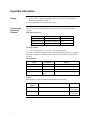
PL Expression System A Prokaryotic Expression System Catalog no. K450-01 Version H 7 September 2010 25-0080 Corporate Headquarters Invitrogen Corporation 1600 Faraday Avenue Carlsbad, CA 92008 T: 1 760 603 7200 F: 1 760 602 6500 E: [email protected] For country-specific contact information visit our web site at www.invitrogen.com User Manual ii Table of Contents Table of Contents.............................................................................................................................iii Important Information......................................................................................................................iv Introduction ................................................................................................................... 1 Overview...........................................................................................................................................1 Vectors ..............................................................................................................................................3 Methods ......................................................................................................................... 5 Overview...........................................................................................................................................5 Cloning into pLEX............................................................................................................................7 Transformation................................................................................................................................10 Expression.......................................................................................................................................13 Appendix...................................................................................................................... 17 Recipes............................................................................................................................................17 Protocol for Chemically Competent Cells ......................................................................................23 Protocol for Electrocompetent Cells ...............................................................................................25 β-Galactosidase Assay ....................................................................................................................27 Analysis by SDS-PAGE Gels .........................................................................................................29 Technical Service............................................................................................................................30 References.......................................................................................................................................32 iii Important Information Storage PL Expression System Kit Contents • Store cell stabs, 10 mg/ml tryptophan, and pre-mixed media at room temperature. Protect the tryptophan from light. • Store lyophilized vectors and primers at -20°C. The PL Expression System includes vectors, primers, an E. coli strain, and media reagents. Stab (cells) and Vectors. Name pLEX, 20 µg Supplied as Storage supercoiled, lyophilized -20°C pLEX/LacZ, 20 µg supercoiled, lyophilized -20°C GI724 room temperature a stab Genotype of GI724 F-, λ-, lacIq, lacPL8, ampC::Ptrp cI, mcrA, mcrB, INV(rnnD-rnnE). This strain is included for growth of pLEX and optimal expression from the PL promoter. This strain contains the cI repressor under control of the trp promoter (Mieschendahl et al., 1986). Media Reagents Name Amount Storage RM Base Media 2 pouches* room temperature, away from moisture RM Agar Base Media 1 pouch room temperature, away from moisture Induction Base Media 2 pouches room temperature, away from moisture Tryptophan Solution 5 ml (10 mg/ml), room temperature, protect from light sterile *each pouch contains reagents to prepare 1 liter of medium. Primers Both primers are supplied lyophilized and should be stored at -20°C. Sequencing Primer iv Sequence Amount pLEX Forward 5´-GGTGACGCTCTTAAAAATTAAGCC-3´ 2 µg (~0.25 nmole) AspA Reverse 5´-TGTAAAACGACGGCCAGTGC-3´ 2 µg (~0.30 nmoles) Important Information, continued Location of Primers The table shows where the two primers bind on the pLEX vector supplied in this kit. To see where these primers bind relative to the multiple cloning site, please see page 8. Primer Binding site on pLEX pLEX Forward 2222-2245 AspA Reverse 2507-2488 Reagents and Chemicals Supplied by User The following reagents and chemicals must be supplied or made by the user. Please check the Recipes section for instructions on how to make solutions. Use reagent or analytical grade chemicals. • Media (see Recipes, page 17-22) • 100 mg/ml ampicillin stock solution (see Recipes, page 17) • FSB Solution (see Recipes, page 21) • Dimethyl sulfoxide (DMSO) (See Recipes, page 21) • Restriction enzymes and buffers • T4 DNA ligase and buffer • Solutions for SDS-PAGE gels. (The size of your recombinant protein will determine what sort of SDS-PAGE gel to use. Please see Ausubel, et al., 1990; Bollag and Edelstein, 1991; Sambrook, et al., 1989; or Schägger and von Jagow, 1987 in the Reference section, page 32, for suggestions.) • Solutions for ONPG assay (see Recipes, page 22) • Dry Ice • Technical grade ethanol for dry ice baths • Reagent grade ethanol for use with DNA Equipment Supplied by User The user should have access to the following equipment: • 1.5 ml microcentrifuge tubes • 15 ml snap-cap polyethylene tubes (Falcon 2059 or equivalent) • Microbiological equipment to plate cells and grow cultures • Shaking incubator, 25-37°C • Incubators: 15°C, 30°C, 37°C • Autoclave • Vacuum pump (for filter sterilization) • Electroporation device (optional) • Centrifuge, refrigerated, low-speed, 50-500 ml volumes • Autoclavable or sterile 50 ml centrifuge tubes • Autoclavable or sterile 250 or 500 ml centrifuge bottles • UV-Vis Spectrophotometer • Polyacrylamide gel apparatus • Sonicator with microtip v Important Information, continued Product Qualification The pLEX and pLEX/LacZ vectors are qualified by restriction digest. Restriction digests must demonstrate the correct banding pattern when electrophoresed on an agarose gel. The table below lists the restriction enzymes used to digest the vector and the expected fragments. Vector vi Restriction Enzyme Expected Fragments (bp) pLEX BamH I Cla I Not I Sac I Bgl I 2886 uncut 2886 2886 1120, 1766 pLEX/LacZ BamH I Cla I Not I Sac I Bgl I 5869 5869 uncut 1948, 3921 1020, 1120, 1608, 2121 Introduction Overview Description The prokaryotic PL Expression System (Catalog No. K450-01) allows expression of heterologous proteins in E. coli. Foreign genes inserted into the multiple cloning site of the vector, pLEX, are tightly regulated by a tryptophan-inducible expression system utilizing the strong PL promoter from bacteriophage lambda (Buell and Panayotos, 1986; LaVallie et al., 1993). This system is especially useful for the expression of potentially toxic proteins in E. coli. In addition, the PL promoter provides high-level expression of recombinant proteins. This means that even if the recombinant protein in question is toxic, significant amounts may be produced before the host cell dies. Regulation of Expression To drive expression of recombinant proteins, pLEX uses the major leftward promoter (PL) from bacteriophage lambda. This promoter is one of the most efficient promoters for bacterial expression (Buell and Panayotos, 1986). It is also tightly regulated. Expression from the PL promoter is controlled by the cI repressor protein. The cI repressor gene was engineered into the bacterial chromosome under the control of the trp promoter (Mieschendahl et al., 1986). Expression is induced by the addition of tryptophan. Transcription 1. In the absence of tryptophan, expression of the cI repressor is driven by the trp promoter. 2. The cI repressor protein binds to the operator region up-stream of the PL promoter and prevents transcription of the gene of interest. 3. Tryptophan is added to the medium and a tryptophan-trp repressor complex is formed. This complex binds tightly to the trp operator blocking expression of the cI repressor. Poo P The cI repressor falls off of the PL operator, allowing transcription of the gene of interest. Po 4. Po Ptrp lcI Repressor Bacterial Chromosome No Transcription Po PL ATG Gene of Interest pLEX Vector cI Repressor Tryptophan No Transcription Ptrp lcI Repressor Bacterial Chromosome Gene of Interest pLEX Vector trp Repressor Transcription PL ATG cI Repressor Continued on next page 1 Overview, continued Contents of the PL Expression System Kit Features of pLEX This kit contains all the necessary reagents for tightly regulated, high-level expression in E. coli: • The pLEX expression vector for high-level production of recombinant protein in E. coli (see below and page 3 for more details). • The specially constructed strain, GI724, for proper regulation of expression. • Specially formulated media for plasmid growth and proper regulation of expression. • The control vector, pLEX/LacZ, to help you evaluate your expression conditions. • Forward and reverse sequencing primers to confirm that your insert is in frame with the initiation ATG provided in the vector. This table describes the features and benefits of the pLEX vector. Feature 2 Benefit PL promoter High-level expression of recombinant protein Lambda cII ribosome binding site and initiation ATG Efficient translation of recombinant protein E. coli aspA transcription terminator Efficient transcription termination of mRNA Ampicillin resistance Selection and maintenance in E. coli pUC origin Maintenance in bacteria and high copy number Polylinker region Cloning of the desired gene into the pLEX vector Vectors The map below describes the pLEX vector. The multiple cloning site (see page 8) allows in-frame fusion with the lambda cII ribosome binding site and the initiation ATG. Note that if you do not want non-native amino acids fused to your protein, you can insert your gene into the Nde I site. The complete sequence of pLEX is available for downloading from our Web site (www.invitrogen.com) or by contacting Technical Service (page 30). Nde I Kpn I Sac I BamH I Spe I Eag I* BstX I* EcoR I Pst I* EcoR V BstX I* Not I Eag I* Xho I Sph I Nsi I Xba I Sal I Pst I* Map of pLEX PL aspA Transcription Termination pLEX ici l ori lin pU C 2.9 kb p Am Comments for pLEX: 2886 nucleotides * These enzymes have two recognition sites both of which are found in the multiple cloning site. Ampicillin Resistance Gene: bases 201-1061 pUC origin: bases 1206 - 1879 -35 Region of PL Promoter: bases 2159-2164 -10 Region of PL Promoter: bases 2182-2187 pLEX Forward Primer Binding Site: bases 2222-2245 Lambda cII Ribosome Binding Site: bases 2291-2294 Lambda cII Initiation ATG: bases 2306-2308 Multiple Cloning Site: bases 2303-2413 aspA Transcription Terminator: bases 2414-2480 AspA Reverse Primer Binding Site: bases 2507-2488 Continued on next page 3 Vectors, continued Map of pLEX/LacZ The gene for β-galactosidase (lacZ) was cloned into pLEX to make the pLEX/LacZ control vector. It expresses β-galactosidase (~117 kD) from the PL promoter and serves as a positive control for expression. The lacZ gene was cloned as a BamH I-Pst I fragment in frame with the lambda cII initiation ATG. β-galactosidase is expressed as a fusion protein to a seven amino acid N-terminal leader peptide. This does not appear to affect activity of β-galactosidase. The complete sequence of pLEX/LacZ is available for downloading from our Web site (www.invitrogen.com) or by contacting Technical Service (page 30). La c Z pLEX/LacZ PL 5.9 kb p U C Comments for pLEX/LacZ: 5869 nucleotides or i aspA BamH I Sac I Kpn I Nde I Pst I Transcription Termination n illi c i Amp Ampicillin Resistance Gene: bases 201-1061 pUC origin: bases 1206- 1879 -35 Region of PL Promoter: bases 2159-2164 -10 Region of PL Promoter: bases 2182-2187 pLEX Forward Primer Binding Site: bases 2222-2245 Lambda cII Ribosome Binding Site: bases 2291-2294 Lambda cII Initiation ATG: bases 2306-2308 b-galactosidase Gene: bases 2327-5386 aspA Transcription Terminator: bases 5397-5463 AspA Reverse Primer Binding Site: bases 5490-5471 Use as a Control 4 The pLEX/LacZ vector is used to evaluate growth and expression conditions in the PL Expression System. Successful expression of β-galactosidase from the pLEX/LacZ vector demonstrates that your cells, medium, and induction conditions are functioning properly. If your gene of interest is not expressed it may suggest that either the recombinant construct is not in frame with the initiation ATG or the expression conditions are not optimal. If β-galactosidase does not express from pLEX/LacZ, it suggests there is a problem with either the medium or the host strain. You must use GI724 as your host strain; otherwise, regulation of gene expression with tryptophan will not occur. Methods Overview Important Because of the nature of transcription regulation with this system, cells containing plasmids CANNOT be grown in LB medium or at temperatures above 30°C. LB medium contains large amounts of tryptophan which allows transcription from the PL promoter. Also, growing above 30°C, except when inducing, will cause some low-level transcription through the PL promoter. This may cause plasmid rearrangements or deletions. Cells without plasmid can be grown using LB medium at 37°C. Description of Process The table below outlines the general steps of constructing and expressing a recombinant protein using the PL Expression System. Step Flow Chart of Experimental Process Action 1 Ligate the gene of interest in frame with the initiation ATG in pLEX, and transform the ligation product into GI724 competent cells. 2 Select ampicillin-resistant transformants and analyze the plasmid DNA by restriction mapping or sequencing for the presence, orientation, and reading frame of the gene of interest. 3 Grow the desired clones and induce for expression of the recombinant protein. Analyze expression of the recombinant protein by SDS-PAGE gels. 4 Optimize growth and induction conditions to maximize the amount of your recombinant protein. Ligate Gene of Interest into pLEX Optimize Growth of Cells and Expression of Recombinant Protein Transform Desired Clone GI724 Cells Perform Restriction Analysis or Sequence AmpR Transformants Continued on next page 5 Overview, continued Before You Start It is important to read through the entire manual to familiarize yourself with the procedures. Some general things you need to check are listed below: Step General Knowledge 6 Action 1 Prepare media and plates for expression and transformation based on the amounts required for your particular project. 2 Prepare solutions you need. 3 Have suitable restriction enzymes, T4 DNA ligase, and modifying enzymes on hand to ligate your gene of interest into pLEX. 4 Prepare competent cells in advance and store at -70°C. Users should be familiar with sterile technique, molecular biology techniques, and standard microbiological practices. For information on DNA ligations, E. coli transformations, restriction enzyme analysis, purification of single-stranded DNA, DNA sequencing, and DNA biochemistry, please refer to Molecular Cloning: A Laboratory Manual (Sambrook et al., 1989) or Current Protocols in Molecular Biology (Ausubel et al., 1994). Cloning into pLEX Before You Start The table below outlines the general steps to consider when cloning your gene of interest into pLEX. Step Action 1 Prepare either electrocompetent or chemically competent GI724 cells. 2 Develop a cloning strategy to fuse your protein in frame with the initiation ATG in the vector pLEX. 3 Prepare RM medium and RMG-Amp plates. Plan on two plates per each ligation-transformation reaction. Remember to have additional plates on hand to purify your desired clone. Preparing Competent Cells Use either of the protocols in the Appendix to make competent cells. For the procedure to prepare chemically competent cells, see page 23; for electrocompetent cells, see page 25. Store the cells at -70°C until ready for use. Chemically competent cells appear to be stable and maintain their efficiency for at least three months. Before Cloning Resuspend the lyophilized vectors in 20 µl of sterile water, such that the final concentration is 1 µg/µl. Store the resuspended vectors at -20°C. General Guidelines for Control Reactions The table below gives some suggestions for possible control reactions for the experiments presented in this manual. It is useful to have control data available when contacting Invitrogen Technical Service for assistance (page 30). Experiment Ligation and Transformation Expression Control Reason No DNA Checks for contamination of ligation reagents. Linearized pLEX (may be dephosphorylated) Checks for uncut or self-ligated vector. Insert only Checks for plasmid contamination of insert. Cells only Checks for the presence of antibiotic in the plates and contamination of competent cells and SOC medium. pLEX or pLEX/LacZ, supercoiled Checks the efficiency of the competent cells. See Transformation experiment, page 10. pLEX/LacZ, supercoiled Positive control for expression. Production of β-galactosidase in the Expression experiment (page 13) shows the cells and the media are working properly. Continued on the next page 7 Cloning into pLEX, continued Since all constructs will be unique, please consult general molecular biology references (Ausubel et al., 1994; Sambrook et al., 1989) for recommendations on restriction digests, dephosphorylation of vector, ligations, and plasmid preparations. General Considerations Follow the instructions of the manufacturer for restriction enzymes, T4 DNA polymerase, calf intestinal alkaline phosphatase, and T4 DNA ligase in order to clone your gene of interest into pLEX. Remember that resulting ligations will be transformed into GI724. You cannot use DH5α, JM109, or other common E. coli strains. Cloning Considerations Your gene will need to be in-frame with the lambda cII initiation ATG in order to utilize the PL promoter and ribosome binding site for high-level expression. If you do not want non-native amino acids fused to your protein, you may clone into the Nde I site which contains the initiation ATG. If you clone in-frame with the initiation ATG using any of the other sites, your protein will be produced as a fusion protein with an N-terminal leader peptide. Multiple Cloning Site of pLEX The pLEX multiple cloning site is shown below. Choose carefully when selecting a cloning site. Your gene must be in-frame with the initiation ATG in order to achieve maximal expression. The complete sequence of pLEX is available for downloading from our Web site (www.invitrogen.com) or by contacting Technical Service (see page 30). 2133 -35 -10 GCGGTGATAA ATTATCTCTG GCGGTGTTGA CATAAATACC ACTGGCGGTG ATACTGAGCA PL 2193 2253 2312 pLEX Forward Primer CATCAGCAGG ACGCACTGAC CACCATGAAG GTGACGCTCT TAAAAATTAA GCCCTGAAGA RBS Nde I AGGGCTTTAT TTGCATACAT TCAATCAATT GTTATCTAAG GAAATACTTA CAT ATG GTA 1 Met Val Kpn I Sac I BamH I Spe I Eag I* BstX I* EcoR I Pst I* CCG AGC TCG GAT CCA CTA GTA ACG GCC GCC AGT GTG CTG GAA TTC TGC AGA Pro Ser Ser Asp Pro Leu Val Thr Ala Ala Ser Val Leu Glu Phe Cys Arg EcoR V BstX I* Not I Eag I* Xho I Nsi I Sph I Xba I Sal I Pst I* TAT CCA TCA CAC TGG CGG CCG CTC GAG CAT GCA TCT AGA GTC GAC CTG CAG Tyr Pro Ser His Trp Arg Pro Leu Glu His Ala Ser Arg Val Asp Leu Gln aspA Transcriptional Terminator 2414 TAA TCGTACAGGG TAGTACAAAT AAAAAAGGCA CGTCAGATGA CGTGCCTTTT TTCTTGTGAG Stop (1) Stop (2) Stop (3) AspA Reverse Primer 2477 CAGTAAGCTT GGCACTGGCC GTCGTTTTAC AACGTCGTGA CTGGGAAAAC CCTGGCGTTA 2363 2537 CCCAACTTAA TCGCCTTGCA - 2556 *These enzymes have two recognition sites both of which are found in the multiple cloning site. Continued on next page 8 Cloning into pLEX, continued Calculation of Molar Ratios To ligate your gene of interest into pLEX, you will need to know the concentration of each DNA solution. This may be determined by OD260, agarose gel electrophoresis, fluorescence, or using the DNA Dipstick™ Kit from Invitrogen (Catalog no. K5632-01). The concentration is needed to calculate the volume required to achieve a particular molar ratio of vector to insert. 1. Determine the concentration of insert and linearized pLEX in µg/ml. 2. Use the following formula to calculate the amount of insert needed to give an equimolar (1:1) ratio between insert and linearized pLEX: x ng insert = (bp insert) (ng linearized pLEX) (2886 bp pLEX) Amount of pLEX can range from 50-200 ng. Ligation Reaction 3. Based on fs the calculation above, compute the volumes needed for the ligation reaction. 4. You may wish to consider other ratios of vector to insert, i.e. 1:2 or 1:3 to increase the chances of obtaining the correct clone. The following protocol gives a general ligation reaction: Linearized pLEX vector (50-200 ng) x µl Linearized insert (5-200 ng) y µl 10X Ligation Buffer (should have ATP) 1 µl Sterile Water T4 DNA Ligase (0.5 Weiss Units) Total Volume to 9 µl 1 µl 10 µl 1. Incubate for 4 hours at room temperature or 12-16 hours at 15°C. 2. At the end of the reaction time, transform into competent GI724 E. coli cells or store at -20°C. 9 Transformation Introduction At this point you have ligation reactions which you will transform by chemical means or electroporation into competent GI724. For procedures to prepare chemically competent cells, see page 23 and for electrocompetent cells, see page 25. Plate transformed cells onto RMG-Amp plates (see Recipes, page 19) and select ampicillin resistant colonies. Screen these ampicillin resistant colonies by restriction mapping and/or sequencing to find the desired clones for the expression and purification experiments. Controls Please see the section on Control Reactions (page 7) to determine which controls you wish to include. GI724 is wild-type for restriction and modification of DNA; therefore your Transformation Efficiency of GI724 transformation efficiencies will be much lower than for strains like DH5α, JM109, or HB101. Typical transformation efficiencies are 5 x 10⁵ cfu/µg DNA for chemically competent cells and 1 x 10⁶ cfu/µg DNA for electrocompetent cells. You may have to transform with more DNA (1-10 ng), plate a larger volume of cells (100-300 µl), or increase the number of plates (2-5 plates) to obtain a reasonable number of transformants. Before You Start 1. 2. 3. 4. Chemical Transformation 1. 2. 3. 4. 5. 6. If transforming by electroporation, be sure to have on hand sterile, glass transfer pipettes, electroporation cuvettes, and sterile, 15 ml, polypropylene snap-cap tubes. Determine the total number of transformations, including controls. Since you will be plating two different volumes from these reactions, you will need two RMGAmp plates per ligation/transformation. Be sure the lyophilized vectors are resuspended in 20 µl sterile water such that the final concentration is 1 µg/µl. This will be your stock solution. To transform supercoiled pLEX and pLEX/LacZ as controls, prepare 10 µl of a 10 ng/µl solution in water for each vector. Keep on ice. Make sure the SOC medium is at room temperature. Equilibrate a water bath or heat block to 42°C. Remove the appropriate number of tubes of frozen GI724 chemically competent cells (100 µl/tube) and thaw on ice. Add 3-5 µl of each ligation reaction to a separate tube of competent cells. Mix gently with the pipette tip. DO NOT PIPETTE UP AND DOWN. Repeat for all ligations. For control reactions, add 1 µl (10 ng) of each supercoiled plasmid (pLEX and/or pLEX/LacZ) to a separate tube of cells. Incubate tubes on ice 30 minutes. Transfer all tubes to 42°C heat block or water bath and incubate for exactly 90 seconds, then place on ice for 1-2 minutes. Add 800 µl of room temperature SOC medium to each tube and shake at 225 rpm for 45 minutes at 37°C. Incubate the tubes horizontally and secure with tape to maximize aeration. Incubation at 37°C does not cause rearrangements or deletions in the plasmid for this short amount of time. Plate 100 µl and 300 µl of each transformation mix on the RMG-Amp transformation plates. It may be necessary to increase the number of plates and plate out more of the transformation mix. Let all the liquid absorb, invert, and incubate at 30°C overnight. Proceed to Analysis of Transformants, page 11. Continued on next page 10 Transformation, continued Electroporation Transformation MEND ION AT RECOM Analysis of Transformants 1. Remove the appropriate number of microcentrifuge tubes of GI724 electrocompetent cells from the -70°C freezer and thaw on ice. Chill electroporation cuvettes on ice. 2. Set up your electroporation device for electroporation of bacteria using the manufacturer's instructions. 3. Add 1-2 µl of a ligation reaction to a tube containing 50 µl competent cells. Repeat for all ligation reactions. 4. For the control reactions, add 1 µl (10 ng) of each supercoiled plasmid (pLEX and/or pLEX/LacZ) to a separate tube of 50 µl competent cells. Incubate all tubes on ice for 1-2 minutes. 5. Take one sample at a time and transfer the cell/DNA mix to an electroporation cuvette. Place the cuvette in the chamber and discharge the electrical pulse. 6. Remove cuvette and immediately add 800 µl room temperature SOC medium and transfer using a sterile glass pipette to a 15 ml snap-cap polypropylene tube (Falcon 2059 or similar). Place on ice. Repeat steps 4-6 until all samples have been transferred to 15 ml tubes. 7. Incubate all tubes with shaking (200-225 rpm) at 37°C for 45 minutes. Incubation at 37°C does not cause rearrangements or deletions in the plasmid for this short amount of time. 8. Plate 25 µl and 100 µl of the transformation mix on the RMG-Amp transformation plates. After the liquid is absorbed, invert and incubate at 30°C overnight. 1. Remove plates from the incubator. There may be a slight background haze, but true ampicillin resistant transformants will appear as distinct colonies. Pick 10 transformants and inoculate into 2-5 ml RM medium with 100 µg/ml ampicillin. Grow overnight at 30°C. 2. Isolate plasmid DNA by miniprep for restriction analysis and sequencing (see below). Remember to purify the desired clone by streaking for singles on RMGAmp plates before making a glycerol stock. 3. Make a glycerol stock of your desired clone for safekeeping by combining 0.85 ml of a overnight bacterial culture with 0.15 ml of sterile glycerol. Mix by vortexing and transfer to a labeled storage tube. Freeze the tube in liquid nitrogen or a dry ice/ethanol bath and store at -70°C. 4. Once the desired clone is isolated, proceed to the Expression section, page 13. Since it is particularly important to make sure your protein is in frame with the initiation ATG, it is strongly recommended that you sequence your construct. To sequence pLEX, use the pLEX Forward Sequencing Primer and the AspA Reverse Sequencing Primer. Continued on next page 11 Transformation, continued Estimating Nmoles In order to determine how much oligonucleotide to use in a sequencing reaction, you will need to estimate the number of nmoles. We have provided this information for each of of the oligonucleotides in this kit (see page iv). However, for your convenience, we have Oligonucleotides included a method for estimating the molecular weight of any oligonucleotide and determining the number of nmoles. To estimate the number of nmoles of an oligonucleotide, assume that each nucleotide in the oligo has an average molecular weight of 330 g/mole. Multiply by the number of nucleotides in the oligonucleotide. This will give you the approximate molecular weight of the oligonucleotide. To determine the number of nmoles, take the weight (in grams) and divide by the molecular weight. For example, the pLEX Forward Primer is 24 nucleotides and we supply 2 µg. (24)(330 g/mole) = 7,920 g/mole Then-- 2 x 10⁻⁶ g = 0.25 nmoles 7920 g/mole Important Troubleshooting 12 Constructs based on pLEX must always be propagated in GI724 in RM medium plus 100 µg/ml ampicillin and grown at 30°C. Other E. coli strains are not suitable for use with pLEX. Do not use LB medium with plasmid-containing GI724 . If plasmid yield is low, try growing the cells longer to increase cell density. If you get plasmid deletions or rearrangements, make sure you are transforming into GI724. Remember to always grow at 30°C and use RM medium with ampicillin. If isolating plasmid by alkaline lysis, be sure to extract with phenol/chloroform to remove exonucleases. This will increase the stability of the plasmid preparation. Expression Introduction At this point, you should have GI724 containing pLEX with the gene encoding your protein ligated in frame to the initiation ATG. In this section, you will determine if this construct expresses and then optimize expression conditions by varying the time of induction. Stage 1 Action Grow cells containing your construct to mid-log at 30°C in Induction Medium. 2 Induce by adding tryptophan and transfer to 37°C. 3 Take time points from the cell culture and lyse the cells by sonication and freeze-thaw. 4 Fractionate cell lysate by centrifugation. 5 Analyze pellets and supernatant for recombinant protein by SDS-PAGE. 6 Vary time of induction to increase yield of recombinant protein. To analyze expression and eventually purification, it is necessary to use SDS-PAGE technology. There are many different types of SDS-PAGE systems. Because all proteins are different, it is difficult to recommend a particular system for your individual application. Please see the following references: Ausubel, et al., 1990, Unit 10; Bollag and Edelstein, 1991; Sambrook, et al., 1989, Chapter 18; and Schägger and von Jagow, 1987. Optimizing Expression To achieve maximum levels, expression may be optimized. The time of induction is the major variable manipulated to achieve optimal expression. We recommend using the pLEX/LacZ expression control to evaluate growth and Expression of induction conditions. You may assay cell lysates from GI724/pLEX/LacZ for expression pLEX/LacZ Control one of two ways: SDS-PAGE or enzymatic assay. For analysis by SDS-PAGE, GI724/pLEX/LacZ can be grown using the same conditions as your construct in pLEX. Note that the approximate molecular weight of β-galactosidase is 117 kD. MEND ION AT RECOM To enzymatically assay for β-galactosidase activity, there is a slight modification to the growth procedure for GI724/pLEX/LacZ. Please turn to page 27 for this information and the ONPG assay for β-galactosidase activity. For testing and optimizing expression, Invitrogen recommends using small volumes (5-50 ml), then scaling up for large-scale expression and purification of the recombinant protein (1 liter) once optimal conditions have been determined. The volume of cell culture you use depends on how much you need to read the absorbance of the cultures and the number of 1 ml time points that will be taken. The instructions that follow are for 10 ml cultures and may be scaled up or down to meet your individual needs. Continued on next page 13 Expression, continued Preparation See Recipes, page 17-22 for the following media and plates. You will need: 1. 2. 3. 4. One RMG-Amp plate 1 ml RM medium with 100 µg/ml ampicillin in a 5 ml sterile culture tube 10 ml Induction Medium with 100 µg/ml ampicillin in a sterile 25 ml small flask Five microcentrifuge tubes labeled: "t=0", "t=1", "t=2", "t=3", and "t=4". Make sure you have extra tubes if you wish to extend the time course. Growth of Cells: Day 1 Streak out the clone of interest on a RMG-Amp plate with 100 µg/ml ampicillin and grow at 30°C until single colonies are visible (12-16 hours). Growth of Cells: Day 2 Using a single colony from the plate from Day 1, inoculate 1 ml of RM medium containing 100 µg/ml ampicillin and incubate at 30°C at 200-225 rpm in a shaking incubator overnight. Growth and Induction of Cells: Day 3 1. 2. 3. 4. 5. 6. Lysing Cell Samples: Day 4 1. 2. 3. Inoculate 10 ml of fresh Induction Medium in a 25 ml culture flask to an OD550 of 0.1 using the overnight culture. Grow this culture at 30°C to an OD550 of 0.5 (approximately 2-3 hours). When an OD550 of 0.5 is reached, transfer a 1.0 ml sample to the microcentrifuge tube labeled "t=0", and centrifuge the tube for 2-3 minutes at maximum speed to pellet the cells. This is the zero time sample. Decant the supernatant and freeze the cell pellet at -20°C until ready to assay. Return the cell culture to the incubator. To your cell culture, add tryptophan to a final concentration of 100 µg/ml using the 10 mg/ml stock solution included in the kit. Transfer the culture to 37°C and incubate with shaking (200-225 rpm). At time t=1 hour, read the OD550 and record. Then take a 1 ml sample and place into the microcentrifuge tube labeled "t=1". Return the cell culture to the incubator to continue growing. Centrifuge the 1 ml sample at maximum speed for 2-3 minutes, decant the supernatant, and freeze the cell pellet at -20°C. Repeat at t=2, 3, and 4 hours. The cell pellets can be kept frozen until ready for analysis by SDS-PAGE. See the section Analysis by SDS-PAGE Gels in the Appendix. Before preparing whole cell lysates: • Prepare an SDS-PAGE gel • Prepare a dry ice/ethanol bath • Equilibrate a 37°C bath • Prepare and chill TE Buffer, pH 7.5 to +4°C Remove the five samples from Growth and Induction of Cells, (above) from the freezer and keep samples on ice. Resuspend each cell pellet in 500 µl cold (+4°C) TE Buffer, pH 7.5 and keep on ice. Take a hand-held sonicator with a micro-tip and sonicate each sample one at a time with two or three 10 second bursts. Flash freeze the lysate in a dry ice/ethanol bath. Continued on next page 14 Expression, continued Lysing Cell Samples: Day 4, continued 4. 5. 6. Analysis of Samples 1. 2. 3. Quick Alternative Method of Sample Lysis What You Should See Quickly thaw the lysates at 37°C and perform two more rapid sonication-freezethaw cycles. Repeat steps 3 and 4 until three sonication-freeze-thaw cycles are completed for all samples. After the last thaw, centrifuge all tubes at maximum speed for 5-10 minutes at +4°C to pellet cell debris and insoluble matter. Decant the supernatants into fresh microcentrifuge tubes. Keep all samples (supernatants and cell pellets) on ice. Resuspend the pellets from Step 5 in 500 µl TE Buffer, pH 7.5. Keep on ice. You now have two samples per time point--a soluble fraction (supernatant) and an insoluble fraction (resuspended pellet). In a fresh microcentrifuge tube, take 10 µl from the supernatant fraction and mix with 10 µl of any SDS-PAGE sample buffer. Likewise, take 10 µl of the resuspended pellet and mix with 10 µl SDS-PAGE sample buffer. Boil samples for 5 minutes and load onto an SDS-PAGE gel with molecular weight standards. Process and develop the gel as described for the procedure you are using. Compare pellets with supernatants for each time point to determine the extent of expression and the solubility of the recombinant protein. This method will allow you to quickly determine if your protein is expressed; however, you will not be able to determine if the protein is soluble or insoluble. Resuspend cell pellets (from Growth and Induction of Cells, page 14) in 1X SDSPAGE Sample Buffer, boil for 5 minutes, and load 10-20 µl onto an SDS-PAGE gel. Be sure to resuspend the cell pellets in a volume of sample buffer which is proportional to the OD550. This will insure that approximately the same amount of protein will be loaded for each sample. At the zero time point of induction, there should be very little protein expressed. At successive time points postinduction, you should see increasing amounts of protein produced. If your protein is soluble, it will appear in the supernatant fraction of each time point after induction and reach a maximum. If you do not see the protein expressed, either in the supernatant or the pellet, check the pLEX/LacZ control. GI724/pLEX/LacZ can be grown and expressed as a positive control. The zero time point from the pLEX/LacZ control should show no expression of wild-type β-galactosidase (117 kDa) or very little activity by ONPG assay. When induced with tryptophan, β-galactosidase should be expressed and detected in the crude cell lysate or soluble (supernatant) fraction. If you do not get expression of β-galactosidase, be sure pLEX/LacZ is transformed into GI724 and that you are using the correct medium for expression. Optimization of Expression The time of induction is the major variable to optimize when using the PL Expression System. Increase or decrease the time to achieve maximum levels of recombinant protein. Continued on next page 15 Expression, continued Troubleshooting If your recombinant protein does not express at all, it may be out-of-frame with the initiation ATG. Use the pLEX Forward and the AspA Reverse Sequencing Primers to confirm that your insert is in frame and in the correct orientation. In some cases, heterologous proteins expressed in E. coli will fail to fold properly and will aggregate into inclusion bodies and precipitate out of solution. The insoluble protein will pellet with the unbroken cells and cell debris during the low-speed centrifugation of your cell lysate. If you do not detect soluble recombinant protein in the supernatant, check the pellet for the presence of your protein. If your protein is insoluble, try the following: Scale-up and Purification 16 1. Solubilize the inclusion bodies with guanidinium chloride or urea and then slowly dialyze against a low ionic strength buffer to refold the protein. Techniques for solubilization and refolding of insoluble proteins are described in Chapter 20 of Deutscher, 1990. 2. Use the His-Patch ThioFusion™ Expression System (Catalog no. K360-01) from Invitrogen to produce soluble recombinant protein. The His-Patch ThioFusion™ Expression System allows fusion of the heterologous protein to the highly soluble protein thioredoxin. Fusion to thioredoxin increases the likelihood of isolating a soluble form of your heterologous protein. Digestion with enterokinase will release your native protein from thioredoxin. Note that the His-Patch ThioFusion™ Expression System uses the PTrc promoter which may not be as tightly regulated as the PL promoter. Once you have optimized the time of induction for your protein, you can scale-up your culture conditions for purification of your protein. Bollag and Edelstein, 1991 and Deutscher, 1990 are very good references for protein purification techniques. Appendix Recipes Introduction Special medium is required for growth and expression of the pLEX and pLEX/LacZ plasmids. Instructions are provided on pages 17-20 for making media and agar plates. Medium Stock Solutions for RM Medium and RMG-Amp Plates Purpose RM Base Medium Plasmid propagation Induction Base Medium Expression If you are using the pre-mixed media included in the kit, follow instructions on the product label for preparation. Note: If you use the pre-mixed media, there’s no need to prepare the 10X M9 Salts solution or the 1 M MgCl2 solution listed below. 10X M9 Salts (for 1 liter) 60 g 30 g 5g 10 g 900 ml Na2HPO4 KH2PO4 NaCl NH4Cl deionized water 1. Dissolve chemicals in the water and pH to 7.4 with 10 M NaOH. 2. Add water to 1 liter and autoclave for 20 minutes on liquid cycle. 3. Store at room temperature. 1 M MgCl2 1. Dissolve 20.33 g MgCl2 in 100 ml deionized water. 2. Autoclave for 20 minutes on liquid cycle. 3. Store at room temperature. 100 mg/ml ampicillin 1. Dissolve 1 g ampicillin in 10 ml deionized water 2. Filter-sterilize and store at -20°C. 50% glycerol (v/v) 1. Dissolve 50 ml glycerol in 100 ml deionized water. 2. Filter sterilize and store at room temperature. 20% glucose (w/v) Important 1. Dissolve 20 g glucose (dextrose) in 100 ml deionized water. 2. Filter sterilize and store at room temperature. Pre-mixed media from Invitrogen are tested to ensure they contain low levels of tryptophan. Continued on next page 17 Recipes, continued Vendors If you are not using the pre-mixed media included in the kit, you will need to purchase Casamino Acids. Please note that Casamino Acids need to be low in tryptophan. We recommend the following vendors: Vendor RM Medium Chemical Difco (1-800-521-0851) Casamino Acids Sigma-Aldrich (1-800-325-3010) Casamino Acids (Amicase) Amount Catalog number 100 g 0230-15-5 500 g 0230-17-3 250 g to 5 kg A 2427 Use the pre-mixed RM Base medium included in the kit. You will need a 50% sterile glycerol solution and a 100 mg/ml ampicillin solution. Follow the directions on the package to prepare RM Medium. Composition 1X 2% 1% 1 mM 100 µg/ml M9 Salts Casamino Acids glycerol MgCl2 ampicillin 1. For 1 liter of RM medium mix 20 g Casamino Acids, 20 ml 50% sterile glycerol, and 880 ml water and autoclave 20 minutes on liquid cycle. 2. After the autoclaved solution has cooled, add aseptically: 100 ml 10X M9 Salts (see recipe, previous page) 1 ml 1 M MgCl2 (see recipe, previous page) 1 ml 100 mg/ml ampicillin 3. Mix well and store medium containing ampicillin at +4°C. Medium is good for 1 month at +4°C. Continued on next page 18 Recipes, continued RMG-Amp Plates Use the pre-mixed RMG Agar Base medium included in the kit. You will need a 20% sterile glucose (dextrose) solution and a 100 mg/ml ampicillin solution. Follow the directions on the package to prepare RMG-Amp plates. Composition 1X 2% 0.5% 1 mM 100 µg/ml 1.5% M9 Salts Casamino Acids glucose MgCl2 ampicillin agar 1. For 1 liter of plating medium, mix 20 g Casamino Acids, 15 g agar, and 875 ml water and autoclave for 20 minutes on liquid cycle. 2. Cool autoclaved solution to ~55°C and add: 100 ml 1 ml 25 ml 1 ml Induction Medium 10X M9 Salts (see recipe, previous page) 1 M MgCl2 (see recipe, previous page) 20% glucose 100 mg/ml ampicillin 3. Mix well and pour into 10 cm petri plates. Yield: ~40 plates. 4. Let agar harden, invert, and store plates in the dark at +4°C. Plates are good for one month at +4°C. Use the pre-mixed Induction Base medium included in the kit. You will need a 20% sterile glucose (dextrose) solution and a 100 mg/ml ampicillin solution. Follow the directions on the package to prepare RMG-Amp plates. Composition 1X 0.2% 0.5% 1 mM 100 µg/ml M9 Salts Casamino Acids glucose MgCl2 ampicillin 1. For 1 liter of induction medium mix 2 g Casamino Acids with 875 ml water and autoclave for 20 minutes on liquid cycle. 2. After the autoclaved solution has cooled, add: 100 ml 1 ml 25 ml 1 ml 3. 10X M9 Salts (see recipe, page 17) 1 M MgCl2 (see recipe, page 17) 20% glucose 100 mg/ml ampicillin Mix well and store medium containing ampicillin at +4°C. Medium is good for 1 month if stored at +4°C. Continued on next page 19 Recipes, continued Low Salt LB Medium Low Salt LB Agar Plates SOB and SOC Medium Composition 1% 0.5% 0.5% Tryptone Yeast Extract NaCl pH 7.0 1. For 1 liter, dissolve 10 g tryptone, 5 g yeast extract, and 5 g NaCl in 950 ml deionized water 2. Adjust the pH of the solution to 7.0 with 5 M NaOH and bring the volume to 1 liter. 3. Autoclave for 20 minutes on liquid cycle. 4. Store at room temperature or at +4°C. 1. Make LB Medium above and add 15 g/liter agar before autoclaving. 2. Autoclave for 20 minutes on liquid cycle. 3. Let agar cool to ~55°C. Pour into 10 cm petri plates. Let the plates harden, then invert and store at +4°C. SOB (per liter) 2% 0.5% 0.05% 2.5 mM 10 mM Tryptone Yeast Extract NaCl KCl MgCl2 1. Dissolve 20 g tryptone, 5 g yeast extract, and 0.5 g NaCl in 950 ml deionized water. 2. Make a 250 mM KCl solution by dissolving 1.86 g of KCl in 100 ml of deionized water. Add 10 ml of this stock KCl solution to the solution in Step 1. 3. Adjust pH to 7.0 with 5 M NaOH and add deionized water to 1 liter. 4. Autoclave this solution, cool to ~55°C, and add 10 ml of sterile 1 M MgCl2. 5. Store at room temperature or +4°C. SOC (per liter) SOB 20 mM glucose 1. After making SOB medium above, add 7.2 ml of 50% glucose. 2. Store at room temperature or +4°C. Continued on next page 20 Recipes, continued FSB Transformation Solution Composition 10 mM 45 mM 10 mM 100 mM 3 mM 10% Potassium acetate, pH 7.5 MnCl2-4H2O CaCl2-2H2O KCl Hexaamminecobalt chloride (Sigma-Aldrich Catalog no. 20309-2) glycerol 1. Make 100 ml of 1 M potassium acetate by dissolving 9.82 g in 90 ml deionized water. Adjust pH to 7.5 with 2 M acetic acid. Bring the volume up to 100 ml. 2. For 100 ml of FSB transformation solution combine the following ingredients: 1 ml 890 mg 150 mg 750 mg 80 mg 10 ml 80 ml DMSO 1 M Potassium acetate, pH 7.5 MnCl2-4H2O CaCl2-2H2O KCl Hexaamminecobalt chloride 100% glycerol deionized water 3. Carefully adjust pH to 6.4 with 0.1 N HCl. If you go past the correct pH, remake solution. Do not readjust pH with base. 4. Adjust the final volume to 100 ml with deionized water and filter sterilize. Store at +4°C. It is very important to use fresh, analytical grade DMSO. If you routinely transform cells by chemical means using the method of Hanahan, 1983, you probably have frozen aliquots of DMSO in your laboratory. If you do not use this method, then follow this procedure: 1. Order the smallest amount of analytical grade DMSO you can. 2. When the DMSO arrives, take about 5-10 ml and aliquot 200-500 µl per microcentrifuge tube. You may use the rest of the DMSO for other applications or you may aliquot the remainder for competent cells. It depends on whether you plan to use the method described in this manual on a routine basis. 3. Freeze these tubes at -20°C and use one tube per preparation of competent cells. Discard any remaining DMSO in the tube. Use a fresh tube for every preparation of competent cells. Continued on next page 21 Recipes, continued Z Buffer Composition 60 mM 40 mM 10 mM 1 mM 50 mM 1. Na2HPO4-7H2O NaH2PO4-H2O KCl MgSO4-7H2O β-mercaptoethanol pH 7.0 Dissolve the following: 16.1 g 5.5 g 0.75 g 0.246 g 2.7 ml Na2HPO4-7H2O NaH2PO4-H2O KCl MgSO4-7H2O β-mercaptoethanol in 950 ml deionized water. ONPG Solution 2. The pH should be 7.0 if made correctly. Bring the volume up to 1 liter with water. 3. Do not autoclave! Store at +4°C. 4 mg/ml in 100 mM phosphate buffer, pH 7.0 1. Dissolve the following: 1.61 g 0.55 g Na2HPO4-7H2O NaH2PO4-H2O in 90 ml deionized water. 1 M Sodium Carbonate 22 2. The pH should be 7.0 if made correctly. 3. Add 400 mg of ONPG. Stir to dissolve and bring the volume up to 100 ml with water. 4. Store at -20°C away from light. Dissolve 12.4 g sodium carbonate in 100 ml of deionized water. Store at room temperature. Protocol for Chemically Competent Cells Introduction This protocol is used to make chemically competent cells for transformation with plasmid DNA. These cells will not substitute for electrocompetent cells for electroporation. The cells are grown to mid-log phase, then washed with FSB solution, and treated with DMSO. The cells are frozen in a dry ice/ethanol bath and stored at -70°C. Yield This protocol will yield enough cells for about 30 transformations. The expected efficiency of chemically competent GI724 cells is ~5 x 10⁵ cfu/µg supercoiled DNA. The efficiency is low because these strains are essentially wild-type for restriction and modification of DNA. Some of your plasmid will be degraded before it is modified. Important Sterile technique is absolutely essential to avoid contamination of the competent cells. Remember to use sterile solutions, medium, and supplies. Preparation For each cell preparation, prepare the following solutions (see Recipes, pages 20-22): 5 ml SOB medium in a sterile culture tube 250 ml SOB in a sterile 500 ml or 1 liter culture flask FSB solution (~25 ml) Fresh, reagent grade DMSO Growth of Cells: Day 1 Streak GI724 on an LB plate, invert the plate, and incubate at 37°C overnight. LB plates and 37°C incubators are only used for cells without plasmid. Growth of Cells: Day 2 • • Inoculate 5 ml of SOB medium in a sterile culture tube with one colony from the LB plate. Grow overnight (12-16 hours) in a shaking incubator (200-225 rpm) at 37°C. 1. For each cell preparation, place the following items on ice or at +4°C. Growth of Logphase Cells: Day 3 Two 250 ml sterile centrifuge bottles Two 50 ml sterile centrifuge tubes Two 5 ml sterile pipettes 2. Inoculate 250 ml of fresh SOB medium in a 500 ml or 1 liter culture flask with 2.5 ml of the overnight culture. 3. Grow the culture at 37°C at 200-225 rpm in a shaking incubator until the OD550 reaches between 0.55-0.65 (2-3 hours). 4. Divide the culture between the two cold (0-4°C), sterile 250 ml centrifuge bottles and place on ice for 30 minutes. Continued on next page 23 Protocol for Chemically Competent Cells, continued Preparing the Cells: Day 3 Aliquoting and Storage of Cells Day 3 24 1. Centrifuge the 250 ml bottles at 2000 x g for 10-15 minutes at 0-4°C. 2. Decant the medium and resuspend each pellet in 10 ml cold (0-4°C) FSB solution and transfer to two cold, sterile 50 ml centrifuge tubes. Incubate on ice for 15 minutes. 3. Centrifuge the tubes at 2000 x g for 10-15 minutes at 0-4°C. 4. Decant the buffer and resuspend each pellet in 1.8 ml cold FSB solution using a sterile 5 ml pipette. 5. While gently swirling the tubes, slowly add 65 µl of DMSO drop by drop to each tube. Incubate on ice for 15 minutes. 6. While gently swirling the tubes, slowly add an additional 65 µl of DMSO drop by drop to each tube. 7. Combine the cell suspensions from both tubes into one and incubate on ice for 15 minutes. Keep on ice. 1. Prepare a dry ice/ethanol bath. 2. For each cell preparation, place approximately thirty 1.5 ml microcentrifuge tubes on ice. Keep cell suspension on ice. 3. Pipette 110 µl of cell suspension into each tube. 4. As soon as all of the cell suspension is aliquoted, quick-freeze the tubes in the dry ice/ethanol bath and store at -70°C. Protocol for Electrocompetent Cells Introduction The purpose of this procedure is to prepare cells for transformation with plasmid DNA by electroporation. The procedure describes the growth of cells and subsequent washing and concentrating steps. The washing is necessary to ensure that salts are removed to reduce the conductivity of the cell solution. High conductivity may result in arcing during electroporation. These cells are only to be used for electroporation. Do not use them for any other transformation protocol. Yield The following procedure will yield enough electrocompetent cells for about 30 transformations. Remember to use sterile solutions, medium, and supplies. The expected efficiency of the electrocompetent GI724 cells is ~1 x 10⁶ cfu/µg supercoiled DNA. The efficiency is low because these strains are essentially wild-type for restriction and modification of DNA. Some of your plasmid will be degraded before it is modified. Important Sterile technique is absolutely essential to avoid contamination of the electrocompetent cells. Growing the Cells: Streak GI724 on an LB plate, invert the plate, and incubate at 37°C overnight. LB plates and 37°C incubators are only used for cells without plasmid. Day 1 Growing the Cells: Day 2 1. For each cell preparation, prepare the following a day in advance: 50 ml LB medium in a 250 ml sterile culture flask 1 liter of LB medium in a 2 liter or 4 liter sterile culture flask Store at room temperature 50 ml of sterile 10% glycerol 1.5 liter of sterile water Store at +4°C 2. Inoculate the 50 ml of LB medium in a 250 ml culture flask with a single colony from the LB plate and incubate at 37°C with shaking (200-225 rpm) for 12-16 hours (overnight). Continued on next page 25 Electrocompetent Cells, continued Growing the Cells: Day 3 1. For each cell preparation, prechill on ice or at +4°C: Two sterile 500 ml centrifuge bottles Two sterile 50 ml centrifuge tubes Two sterile 25 ml pipettes One sterile 5 ml pipette 2. Inoculate 1 liter of LB medium in a 2 liter or 4 liter flask with the 50 ml overnight culture. Grow the 1 liter culture in shaking incubator (200-225 rpm) at 37°C until the OD550 is between 0.5 and 0.6 (approximately 2-3 hours). 3. Transfer the 1 liter culture to the two chilled, sterile, 500 ml centrifuge bottles and incubate on ice for 30 minutes. 1. Harvesting and Washing the Cells: Day 3 2. Aliquoting and Storage of Cells: Day 3 26 Centrifuge the cultures at 2000 x g for 15 minutes at 0-4°C. Keep the cell pellet and decant the broth. Place bottles back on ice. Resuspend the cell pellet in each bottle in approximately 500 ml of cold (0-4°C), sterile water. 3. Centrifuge cells at 2000 x g for 15 minutes at 0-4°C. Keep the pellet and decant the water. Place bottles back on ice. 4. Resuspend the cells in each bottle in approximately 250 ml of cold (0-4°C), sterile water. 5. Centrifuge cells at 2000 x g for 15 minutes at 0-4°C. Decant the water and place bottles back on ice. 6. Using a prechilled, sterile 25 ml pipette, resuspend cells in each bottle in 20 ml cold (0-4°C), sterile, 10% glycerol and transfer each cell suspension to a chilled, sterile, 50 ml centrifuge tube. 7. Centrifuge cells at 4000 x g for 15 minutes at 0-4°C. Decant the 10% glycerol and place tubes on ice. 8. Resuspend each cell pellet in 1 ml cold (0-4°C), sterile, 10% glycerol. Using a prechilled 5 ml pipette, pool the cells into one of the 50 ml tubes. Keep on ice. 1. Prepare a dry ice/ethanol bath. 2. For each cell preparation, place thirty-five 1.5 ml microcentrifuge tubes on ice and pipette 55 µl of the cell suspension into each tube. Keep cell suspension and tubes on ice until all of the cell solution is aliquoted. 3. After all of the cell suspension is aliquoted, quick-freeze tubes in the dry ice/ethanol bath and store at -70°C until ready for use. β-Galactosidase Assay Introduction This protocol is used to assay the activity of β-galactosidase in GI724 cells transformed with pLEX/LacZ (Sambrook, et al., 1989). Cells are grown in the Induction Medium, then induced with tryptophan. Small aliquots of cells are lysed with chloroform and SDS, then assayed for β-galactosidase using o-nitrophenyl-β-D-galactopyranoside (ONPG). Preparation See Recipes, page 17-22 for the following media and plates. You will need: 1. One RMG-Amp plate Important Growth and Induction of Cells 2. 1 ml RM medium with 100 µg/ml ampicillin in a 5 ml sterile culture tube 3. 10 ml Induction Medium with 100 µg/ml ampicillin in a sterile 25 ml small flask 4. Five microcentrifuge tubes labeled: "t=0", "t=1", "t=2", and "t=3". 5. Make sure you have Z buffer, the ONPG solution, and the 1 M sodium carbonate solution made (see page 22). Read the culture density at 600 nm. This is important for the calculation on the next page. Using a single colony of GI724/pLEX/LacZ from a RMG-Amp plate, inoculate 1 ml of RM medium with 100 µg/ml ampicillin and incubate at 30°C at 200-225 rpm in a shaking incubator overnight. 1. Inoculate 10 ml of fresh Induction Medium in a 25 ml culture flask to an OD600 of 0.1 using the overnight culture. Grow this culture at 30°C to an OD600 of 0.5 (approximately 2-3 hours). 2. When an OD600 of 0.5 is reached, transfer 1 ml of the cell culture to the microcentrifuge tube labeled "t=0" and place on ice. This is the zero time sample. 3. To your cell culture, add tryptophan to a final concentration of 100 µg/ml using the 10 mg/ml stock solution included in the kit. 4. Transfer the culture to 37°C and incubate with shaking (200-225 rpm). 5. At time t=1 hour, read the OD600 and record. Then take a 1 ml sample and place into the microcentrifuge tube labeled "t=1". Place on ice. Repeat at t=2 and t=3 hours. 6. After all the time points are collected, take a 30 µl aliquot from each sample and mix with 0.97 ml of Z buffer, 20 µl of chloroform, and 20 µl of 0.1% SDS. Vortex for 10 seconds, and then equilibrate to 28°C. Make a blank reaction with 30 µl of Induction Medium. 7. Add 0.2 ml of ONPG to the lysed culture prepared in Step 6 and to the blank. After 1 minute, stop the reaction by adding 0.5 ml of 1 M sodium carbonate. Continued on next page 27 β-Galactosidase Assay, continued Calculation Read the OD550 and OD420 of the reaction samples. Units of β-galactosidase activity are calculated as follows: 1000 x [OD420 - (1.75 x OD550)] (t) (v) OD600 where t = time in minutes (1 minute) and v = the volume of culture assayed (0.030 ml). Units are typically: Cells only < 5 x 10³ Units Uninduced pLEX/LacZ < 5 x 10⁴ Units Induced pLEX/LacZ > 1 x 10⁶ Units Time and volume may be varied if the absorbance is too high or too low to be read accurately by the spectrophotometer. 28 Analysis by SDS-PAGE Gels Introduction This section provides references and suggestions for analysis of recombinant protein expression by SDS-PAGE (sodium dodecyl sulfate polyacrlyamide gel electrophoresis). SDS-PAGE will allow you to analyze the solubility, purity, and yield of the recombinant protein. We recommend using 10% Tricine gels to analyze cell lysates if the protein is less than 20 kDa. This gel system resolves differences between low molecular weight proteins. Types of SDSPAGE Gels A variety of Tricine Gels are available from Invitrogen. For details, visit www.invitrogen.com. There are many types of SDS-PAGE gels. Protein Methods by Bollag and Edelstein (1991) describe the basic types. The Tricine gels used at Invitrogen are described by Schägger and von Jagow, 1987. Citations for these publications are found in the Reference section, page 32. There are also sections in Ausubel, et al., 1990 and Sambrook, et al., 1989. General Procedure 1. for Sample 2. Preparation Before lysing the cells and preparing your samples, assemble the SDS-PAGE gel. Lyse the cells and fractionate if necessary. Take 10 µl aliquots from fractionated cell lysates and mix with 10 µl of SDS-PAGE sample buffer. 3. Boil the samples 5 minutes and load all 20 µl onto the SDS-PAGE gel. If aggregation forms after boiling, remake the sample and load without boiling. Be sure to include molecular weight standards. 4. Electrophorese the gel and process according to your protocol of choice. 5. Analyze for the extent of solubility, purity, and yield. 29 Technical Service World Wide Web Contact Us Visit the Invitrogen Web site at www.invitrogen.com for: • Technical resources, including manuals, vector maps and sequences, application notes, MSDSs, FAQs, formulations, citations, handbooks, etc. • Complete technical service contact information • Access to the Invitrogen Online Catalog • Additional product information and special offers For more information or technical assistance, please call, write, fax, or email. Additional international offices are listed on our Web page (www.invitrogen.com). Corporate Headquarters: Invitrogen Corporation 1600 Faraday Avenue Carlsbad, CA 92008 USA Tel: 1 760 603 7200 Tel (Toll Free): 1 800 955 6288 Fax: 1 760 602 6500 E-mail: [email protected] Material Data Safety Sheets (MSDSs) Japanese Headquarters: Invitrogen Japan LOOP-X Bldg. 6F 3-9-15, Kaigan Minato-ku, Tokyo 108-0022 Tel: 81 3 5730 6509 Fax: 81 3 5730 6519 E-mail: [email protected] European Headquarters: Invitrogen Ltd Inchinnan Business Park 3 Fountain Drive Paisley PA4 9RF, UK Tel: +44 (0) 141 814 6100 Tech Fax: +44 (0) 141 814 6117 E-mail: [email protected] MSDSs are available on our Web site at www.invitrogen.com. On the home page, click on Technical Resources and follow instructions on the page to download the MSDS for your product. Continued on next page 30 Technical Service, continued Limited Warranty Invitrogen is committed to providing our customers with high-quality goods and services. Our goal is to ensure that every customer is 100% satisfied with our products and our service. If you should have any questions or concerns about an Invitrogen product or service, please contact our Technical Service Representatives. Invitrogen warrants that all of its products will perform according to the specifications stated on the certificate of analysis. The company will replace, free of charge, any product that does not meet those specifications. This warranty limits Invitrogen Corporation’s liability only to the cost of the product. No warranty is granted for products beyond their listed expiration date. No warranty is applicable unless all product components are stored in accordance with instructions. Invitrogen reserves the right to select the method(s) used to analyze a product unless Invitrogen agrees to a specified method in writing prior to acceptance of the order. Invitrogen makes every effort to ensure the accuracy of its publications, but realizes that the occasional typographical or other error is inevitable. Therefore Invitrogen makes no warranty of any kind regarding the contents of any publications or documentation. If you discover an error in any of our publications, please report it to our Technical Service Representatives. Invitrogen assumes no responsibility or liability for any special, incidental, indirect or consequential loss or damage whatsoever. The above limited warranty is sole and exclusive. No other warranty is made, whether expressed or implied, including any warranty of merchantability or fitness for a particular purpose. 31 References Ausubel, F. M., Brent, R., Kingston, R. E., Moore, D. D., Seidman, J. G., Smith, J. A., and Struhl, K. (1994). Current Protocols in Molecular Biology (New York: Greene Publishing Associates and Wiley-Interscience). Bollag, D. M., and Edelstein, S. J. (1991). Protein Methods (New York: Wiley-Liss). Buell, G., and Panayotos, N. (1986) Mechanism and Practice. In Maximizing Gene Expression, W. Reznikoff and L. Gold, eds. (Boston, MA.: Butterworth Publishers). Deutscher, M. P. (1990) Guide to Protein Purification. In Methods in Enzymology, Vol. 182. (J. N. Abelson and M. I. Simon, eds.) Academic Press, San Diego, CA. Hanahan, D. (1983). Studies on Transformation of Escherichia coli with Plasmids. J. Mol. Biol. 166, 557-580. LaVallie, E. R., DiBlasio, E. A., Kovacic, S., Grant, K. L., Schendel, P. F., and McCoy, J. M. (1993). A Thioredoxin Gene Fusion Expression System That Circumvents Inclusion Body Formation in the E. coli Cytoplasm. Bio/Technology 11, 187-193. Mieschendahl, M., Petri, T., and Hänggi, U. (1986). A Novel Prophage Independent trp Regulated Lambda PL Expression System. Bio/Technology 4, 802-808. Sambrook, J., Fritsch, E. F., and Maniatis, T. (1989). Molecular Cloning: A Laboratory Manual, Second Edition (Plainview, New York: Cold Spring Harbor Laboratory Press). Schagger, H., and Jagow, G. v. (1987). Tricine-Sodium dodecyl sulfate-Polyacrylamide Gel Electrophoresis for the Separation of Proteins in the Range from 1 to 100 kDa. Anal. Biochem. 166, 368-379. ©1998-2005 Invitrogen Corporation. All rights reserved. 32 Corporate Headquarters Invitrogen Corporation 1600 Faraday Avenue Carlsbad, CA 92008 T: 1 760 603 7200 F: 1 760 602 6500 E: [email protected] For country-specific contact information visit our web site at www.invitrogen.com User Manual