1
COBAS® AmpliScreen HIV-1 Test, version 1.5
FOR IN VITRO DIAGNOSTIC USE.
HIV.I
96 Tests
PIN: 03322114 018
IMUl1lPREP/CfL I
96 Tests
PIN: 03302555 018
500 Tests
PIN: 20759899 123
ART: 07 5989 9
US: 83314
COBAS@ AmpliScreen HIV-1 Test. version 1.5
COBAS@ AmpliScreen Multiprep Specimen Preparation and Control Kit
COBAS@ AMPLlCOR@ Wash Buffer
WB
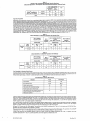
INTENDED USE
The COBAS® AmpliScreen HIV-1 Test, version 1.5 (v1.5) is a qualitatlve in vitro test for the direct detection of Human Immunodeficiency Virus Type 1
(HIV-1) RNA in human plasma.
The COBAS® AmpliScreen HIV-1 Test, v1.5 is intended to be used for detection of HIV-1 RNA in conjunction with licensed tests for detecting antibodies to HIV-1. This product is intended for use as a donor screening test to detect HIV-1 RNA in plasma specimens from individual human donors,
including donors of whole blood and blood components, rource plasma and other living donors. It is also intended for use to screen organ donors
when specimens are obtained while the donor's heart is still beating and to detect HIV-1 RNA in blood specimens from cadaveric (non-hearl-beating)
organ and tissue donors. This test is not intended for use on samples of cord blood. This test is not intended for use as an aid in diagnosis.
Plasma from all donors may be screened as individual specimens. For donations of whole blood and blood components, plasma may be tested in
pools comprised of equal aliquots of not more than 24 individual donations. For donations of hematopoietic stem/progenitor cells (HPCs) sourced
from bone marrow, peripheral blood or cord blood, and donor lymphocytes for infusion (DLQ, plasma may be tested in pools comprised of equal
aliquots of not more than 24 individual donor specimens. For donations of Source Plasma; plasma may be tested in pools comprised of equal aliquots
of not more than 96 individual donations.
This assay may be used as an alternative to licensed HIV-1 p24 antigen tests for screening human plasma from donors.
The COBAS® ArripliScreen HIV-1 Test, v1.5 can be considered a supplemental test that confirms HIV-1 infection for specimens that are repeatedly reactive on a licensed donor screening test for antibodies to HIV-1, and reactive on the coBAS® AmpliScreen HIV-1 Test, v1.5.
SUMMARY AND EXPLANATION OF THE TEST
Human Immunodeficiency Virus (H1V-1)is the etiologic Bgent of Acquired Immunodeficiency Syndrome (AlDS)1-3. HIV-1 infection can be transmitted
by sexual contact, exposure to infected blood or blood products, or by an infected mother to the fetus'. Within three to six weeks of exposure to
HIV-1, infected individuals generally develop a brief, acute syndrome characterized by flu-like symptoms and associated with high levels of viremia
in the peripheral blood5-8. In most infected individuals this is followed by an HIV-1-specific immune response and a decline of plasma viremia; usually within four to six weeks of the onset of symptoms9,10. The prevalence of HIV-1 infection is 1.1 % overall in the world, 0.56% in North America
and 0.25% in West Europe11.
Serol~ical screening assays have greatly reduced, but not completely eliminated, the risk of transmitting viral infections by transfusion of blood products12-15. HIV-1 p24 antigen is the principal core protein of HIV-1 and is found in serum or plasma either free or bound by anti-p24 antibody.
HIV-1 p24 antigen can be measured with commercially available enzyme immunoassays (EIA), which reduce the seroconversion window period, Le.,
the time between infection and the rise of antibodies to the virus 16, by approximately 5 to 6 days17.18. Recent studies indicate that nucleic acid based
amplification tests for HIV-1 RNA will further reduce the residual transmission risk by detecting HIV-1 RNA in donations made during the seroconversion window period. Nucleic acid based tests can detect viremic units donated by carriers who do not seroconvert or who lack antibodies to serological markers normally detected by immunological assays16.19.20.
HIV-l RNA in plasma can be detected by nucleic acid amplification technologies, such as the Polymerase Chain Reaction (PCR)21-23.The COBAse
f'.mpliScreen HIV-1 Test, v1.5 uses peR technology to achieve maximum sensitivity for the detection of HIV-1 RNA in plasma samples24
A number of proposals have been made for performing nucleic acid tests on mini-pools comprised of small aliquots from many individual samples.
The high sensitivity of PCR has demonstrated that potentially infectious donations contained within mini-pools can be detected even if the mini-pool
contains a single viremic donor13.25.26.
The assay incorporates an Intemal Control for monitoring assay performance in each individual test as well as the AmpErase® enzyme (uracil-N-glycosylase) to reduce potential contamination by previously amplified material (amplicon).
PRINCIPLES OF THE PROCEDURE
The COBAS® AmpliScreen HIV-1 Test. vl.5 is based on five major processes:
1. Sample Processing
2. Reverse transcription of target RNA to generate complementary DNA (cDNA)27
3. PCR amplification27 of target cDNA using HIV-specific complementary primers
4. Hybridization of the amplified products to oligonucleotide probes specific to the target(s)
5. Detection of the probe-bound amplified products by colorimetric determination.
Sample Processing
Two specimen processing procedures are used with the COBAS@ AmpliScreen HIV-1 Test, v1.5 as follows:
•
Multiprep Specimen Processing Procedure for preparation of mini-pool specimens and individual cadaveric specimens
•
Standard Sample Processing for preparation of individual donor samples
NOTE: For testing of cadaveric specimens, the specimen should be first diluted
processing using the Multiprep Specimen Processing Procedure.
1:5 in Multlprep
Specimen
Diluent
(MP OIL) prior
to
In the Standard Specimen Processing Procedure. HIV-1 RNA Is isolated directly from plasma by lysis of the virus particles with Multiprep Lysis Reagent
followed by precipitation of the RNA with alcohol. In the Multiprep Specimen Processing Procedure, HIV-l viral particles are first pelleted from the
plasma sample by high speed centrifugation, followed by lysis of the pelleted virus with a chaotropic agent (Mu~iprep Lysis Reagent) and precipitation of the RNA with alcohol.
The Multiprep Internal Control (MP Ie). containing the HIV-1 Intemal Control. is introduced into each sample with the Multiprep Lysis Reagent and
serves as an extraction and amplification control for each processed specimen and control. The HIV-l Internal Control is an RNA transcript with primer
binding regions identical to those of the HIV-1 target sequence, a randomized internal sequence of similar length and base composition as the
HIV-1 target sequence, and a unique probe binding region that differentiates the HiV-1 Intemal Control ampllcon from target amplicon. These features
were selected to ensure equivalent amplification of the HIV-1 Internal Control and the HIV-1 target RNA
Reverse Transcription
The reverse transcription and amplification reactions are performed with the thermostable recombinant enzyme Thermus thermophilus DNA Polymerase
(rTth poij. In the fresence of manganese (Mn2"'j and under the appropriate buffer conditions, rTth pol has both reverse transcriptase and DNA polymerase activity2 . This allows both reverse transcription and PCR amplification to occur in the same reaction mixture. Reverse transcription using
rTth pol produces a cDNA copy of the HIV-1 target and the HIV-1 Intemal Control RNA
PCR Amplification
Following reverse transcription using rTth pol, a second DNA strand is produced from the cDNA copy, thereby yielding a double-stranded DNA copy
of the HIV-1 target and HIV-1 Internal Control RNA. The reaction mixture is heated to separate the resulting double-stranded DNA As the mixture
cools, primers anneal to the target DNA, in the presence of Mn2+ and excess deoxynucleotide triphosphates (dNTPs), the rnh pol extends the annealed
primers along the target templates to produce a double-stranded DNA molecule termed an amplicon. The COBAse AMPucoR® Analyzer automatically repeats this process for a designated number of cycles, each cycle effectively doubling the amount of amplicon DNA The required number of
cycles is preprogrammed in the COBAS4i>AMPLlCoR® Analyzer.
The Document Revision Information section is located at the end of this document.
0512070S001-02EN
Doc Rev. 2.0
Selective Amplification
To ensure selective amplification of nucleic acid target in the sample and prevent amplification of pre-exi~ing amplicon, the AmpErase (umcil-N-glycosylase) enzyme is added to the COBAS@ AmpliScreen HIV-1 Test, ~1:5. The AmP,E~s~ enzyme reco~ntzes and catalyzes the destruction of DNA
eirance containing deoxyuridine2&, but not DNA contalntng deoxythymidlne. Deoxyundlne ISnot present In naturally occumng DNA, .but IS always pr't
sent in amp/icon because of the use of deoxyuridine triphosphate in place of deoxylhymidine triphosphate as one of the dNTPs In the Master MIX
reagent; therefore, only amplicon contain deoxyuridine. Deoxyuridine renders contaminating amplicon susceptible to destruction by the AmpErase
enzyme before amplification of the target DNA. The AmpErase enzyme, which is included in the Master Mix reagent, catalyzes the cleavage of DNA,
thereby rendering the DNA non-amplifiable. The AmpErase enzyme is inactive at temperatures above 55·C, i.e., throughout the thennal cycling steps,
and therefore does not destroy target amplicon. Following amplification, any residual enzyme is denatured by the addltion of the Denaturation Solution,
thereby preventing the degradation of any target amplicon.
Hybridization
Reaction
FOllowing PCR amplification, the COBAS<l!>AMPUCOFl® Analyzer automatically adds Denaturation Solution to the A-tubes to chemically denature the
HIV-l target amplicon and the HIV-1 Internal Control amplicon to form single-stranded DNA A1iquots of denatured amplicon are then transferred to
two detection cups (O-cups). A suspension of magnetic particles coated with an oligonucleotide probe specific for HIV-1 target amplicon or HIV-1
Internal Control amplicon is added to the individual o-cups. The biotin-labeled HIV-1 target and HIV-1 Internal Control amplicon are hybridized to the
target-speciflC oligonucleotide probes bound to the magnetic particles. This hybridization of amplicon to the target-specific probe increases the overall
specificny of the COBAS® AmpIiScreen HIV-1 Test, v1.5.
Detection
Reaction
FoRowing the hybridization reaction, the COBAS0 AMPUCOFl® Analyzer washes the magnetic particles in the O-cups to remove unbound material, and
then adds avidin-horseradish peroxidase conjugate. The avidin-horseradish peroxidase conjugate binds to the hybridized biotin-labeled amplicon. The
COBAS® AMPucoFl® Analyzer removes unbound conjugate by washing the magnetic particles and then adds a substrate solution containing hydrogen
peroxide and 3,3',5,5'-tetramethylbenzidine (TMB) to each D-cup. In the presence of hydrogen peroxide, the particle-bound horseradish peroxidase catalyzes the oxidation of 1MB to fonn a colored complex. The absorbance is measured by the COBAS® AMPucoFl® Analyzer at a wavelength of 660 nm.
MATERIAlS
PROYIDED SY ROCHE
COBAS® AmpliScreen Multiprep Specimen Preparation and Control Kit and the COBAS® AMPUcOFl® Wash Buffer kit are provided as standalone kits to be used in conjunction with the COBAs<!!>AmpllScreen HIV-1 Test, v1.5, as well as the COBAS® AmpliScreen HCV Test. v2.0, and the
COB~AmpliScreen
.HBV Test.
The
cosAS<I!> AmpliScreen
(PIN: 03302555 018)
Multiprep
Specimen Preparation
and Control KIt
I MULTIPREPICTL I
96 Tests
MP (-) C
[Multiprep Negative (-) Control]
MP(+)C
[Multiprep Positive (+) Controij
MPLYS
(Multiprep Lysis Reagent)
MPDIL
(Multiprep Specimen Diluent)
MPIC
(Multiprep Internal ControQ
NHP
[Negative Plasma (Human)]
COBAse AmpliSereen
(PIN: 03322114 018)
HIV-1 Test, version 1.5
COBAS<I!>AmpliScreen
HIY-1 Amplification
HIV·I
Reagents, version 1.5
HIV
96 Tests
AMP
HIV-1 MMX, v1.5
(HIV-1 Master Mix. version 1.5)
HIV-1 Mn2+, v1.5
(HIV-1 Manganese Solution, version 1.5)
COBASfI AmpliScreen
HIV-1 Detection
HIVDK
Reagents, version 1.5
IH PS1, vr.s
(HIV-1 Probe Suspension 1, version 1.5)
1H4, v1.5
(HIV-l Probe Suspension 2, version 1.5)
II PS1, v1.5
QC Probe Suspension 1)
114,v1.5
(IC Probe Suspension 2)
DN4
(Denaturation Solution)
CN4
(Avidin-Horseradish Peroxidase Conjugate)
SB3
(substrate A)
SB
(SUbstrate B)
WB
COBAse AMPLlCOR<I!>Wash Buffer
(PIN: 20759899 123; ART: 07 5989 9; US: 83314)
500 Tests
WB
(10X-Wash Concentrate)
OTHER MATERIALS REQUIRED BUT SOLD SEPARATELY (MAY BE PURCHASED FROM ROCHE)
•
COBAS® AMPUCOFl® Analyzer wnh software version 0022B, Printer, and Operator's Manual for the COBAS® AMPucoFl®
•
COBAS® AMPucoFl®
Analyzer
•
COBAS'" AMPUCOR® D-cups
•
AMPUUNK Software. version 1.4 and Operator's Manual for the AMPUUNK software
•
Hamilton MICROLAB® AT plus 2 Pipettor (with Hamilton SUNPLUS and RUNENDE Software, and the Roche Pooling Methods Software,
version 1.3), the COBAS® AmpliScreen Pooling System Guide (Roche POOlingMethods Software, version 1.3 and the COBAS® AmpliScreen
Pooling System Guide are validated to prepare pools of equal a1iquots of not more than 24 individual plasma donations using Harnljton
MICROLAB AT plus pipettor with Hamilton SUN PLUS and RUNENDE Software)
•
Additional MP OIL from the coBAS0
mens
A-rings
AmpiiScreen Multiprep Specimen Preparation and Control Kit is required for testing of cadaveric speci-
NOTE: The user must validate a/l pooling algorithms and eqUipment other than those supplied by Roche.
•
Sarstedt 1.5-mL tube Barcode Labels
•
Hamilton Archive and Intermediate Plate Barcode Labels
•
Refrigerated high speed centrifuge with fixed angla rotor (45 degrees, capacity for at least 24 x 1.5-mL tubes) with an ReF of 23,600 x g
(Heraeus Centrifuge 17RS or Biofuge 28RS with HFA 22.1 rotor, Heraeus Biotuge Stratos with the 3331 rotor or equlvelent).
05120705001-02EN
.2
Doc Rev. 2.0
MATERIALS REQUIRED BUT NOT PROVIDED BY ROCHE
•
Mlcrocentrifuge, (max. ReF 16,000 x g, min. ReF 12,500 x g) (Eppendorf® 5415C, HERMLE Z230M, or equivalent)
•
Eppendorf 1.25 mL Combitip'" Reservoir (sterile) or equivalent
•
Eppendorf Mu~ipette<!!lpipette or equivalent
•
Ethanol, 90% or 95%, reagent grade for Molecular Biology or Histology use
•
Distilled or deionized water
•
Powder1ess, disposable gloves
•
Isopropyl alcohol, reagent grade
•
Disposable, Sterile, Polystyrene pipettes (5 mL, 10 mL and 25 ml.)
•
Sterile, RNase-free, fine-tip transfer pipettes
•
Pipettors (capacity 20 pl, to 1000 Ill, capable of providing ± 3% accuracy and precision S 5%) with aerosol barrier or positive displacement RNase-free tips
•
Tube racks (Sarstedt PIN 93.1428 or equivalent)
•
1.5 mL sterile, non-siliconized, conical polypropylene screw-cap tubes, (Sarstedt 72.692.105 or equivalent)
•
Vortex mixer
•
Hamilton Slotted Deepwell Archive Plate, 2.2 mL and Sealing Capmat
•
Hamilton Slotted Intermediate Plate
REAGENTS
COBASQ!)AmpliScreen MuHiprep Specimen
Preparation and Co!!trol Kit
I MULTlPREP/CfL I
96 Tests
8 xO.l mL
MPHC
[Muniprep Negative (-) Control]
< 0.005% Poly rA RNA (synthetic)
EDTA
0.05% Sodium azide
MP(+)C
[Multiprep Positive (+) Cornrof
8 x 0.1 mL
Tris-Hel buffer
< 0.001 % Non-infectious linearized plasmid DNA (microbial) containing HBV sequences
< 0.001 % Non-infectious in vitro transcribed RNA (microbiaO containing HCV sequences
< 0.001 % Non-infectious in vitro transcribed RNA (microbiaO containing HIV-1 sequences
< 0.005% Poly rA RNA (synthetic)
EDTA
0.05% Sodium azide
MPLYS
(Multiprep lysis Reagent)
8 x 9.0 ml
Tris-Hel buffer
60% Guanidine thiocyanate
3 % Dithiothreitol
< 1%Glycogen
Xn
60% (w/w) Guanidine thiocyanate
Harmful
MPDlL
(Multiprep Specimen Diluent)
8 x 4.8 mL
Tris-HCI buffer
< 0.005% Poly rA RNA (synthetic)
EDTA
0.05% Sodium azide
MPIC
(Multiprep Internal Control)
8 xO.l mL
Tris-HCI buffer
< 0.001 % Non-infectious plasmid DNA containing HBV primer binding sequences and a
unique probe binding region
.
< 0.001 % Non-infectious in vitro transcribed RNA (microbiaO containing HCV primer binding
sequences and a unique probe binding region
< 0.001% Non-infectious in vitro transcribed RNA (microbiaQ containing HIV-l primer binding
sequences and a unique probe binding region
< 0.005% Poly rA RNA (synthetic)
EDTA
< 0.1% Amaranth dye
0.05% Sodium azide
NHP
[Negative Plasma (Human)]
16 x 1.6 mL
Human plasma, non-reactive by US FDA licensed tests for antibody to HCV, antibody to HIV-1/2,
HIV p24 antigen and HBsAg
0.1 % ProClin® 300 preservative
COBAS® AmpliScreen
HIV-1 Test, version 1.5
COBAS® AmpiiScreen
HIV-1 Amplification
HIV-I
Reagents, version 1.5
96 Tests
HIV AMP
HIV-1 MMlC, v1.5
(HIV-l Master Mix, version 1.5)
8 x 0.7 mL
Bicine buffer
Glycerol
< 0.01% rTth DNA Polymerase (rTth pol, microbiaQ
Potassium acetate
< 0.07% dATP, dCTp, dGTP, dUTP, dlTP
< 0.001% SKCC1B and SK145 biotinylated primers
< 0.01 % AmpErase (uracil-N-glycosylase) enzyme (microbial)
0.05% Sodium azide
HIV-1 Mn2+ v1.5
(H1V-1M~ganese
8xO.lmL
Solution, version 1.5)
< 2% Manganese
Acetic acid
Amaranth dye
0.05% Sodium azide
05120705001-02EN
3
Doc Rev. 2.0
COBAs«' AmpllScreen HIV·1 Detection Reagents, version 1.5
HI" DK
1 x 100 Tests
IH PS1, 111.5
(HIV.1 Probe Suspension 1, version 1.5)
MESbuffer
0.Q194 Suspension of DynabeadsQ!l(paramagnetic particles) coated with HIV- 1-specific
oligonucleotide capture probe SK102
0.09% Sodium azide
-c
1 x 100 Tests
1H4.II1.5
(HIV-1 Probe Suspension 2, version 1.5)
Sodium phosphate buffer
24.9% Sodium thiocyanate
0.2% Solubilizer
1 x 100 Tests
" PS1, 111.5
(IC Probe Suspension 1)
MES buffer
< 0.01 % Suspension of Dynabeads (paramagnetic particles) coaled with HIV-l
IC-specific oligonucleotide
0.0994 Sodium azide
capture probe CP35
1 x 100 Tests
114,111.5
(IC Probe Suspension 2)
Sodium phosphate buffer
24.9% Sodium thiocyanate
< 02% Solubilizer
1 x 100 Tests
DN4
(Denaturation Solution)
1.6% Sodium hydroxide
EDTA
Thymol blue
I
Xi
X
11.6%
(w/w) Sodium hydroxide
Irritant
2 x 100 Tests
CN4
(Avidin-Horseradish Peroxidase Conjugate)
Tris-HCI buffer
< 0.001 % Avidin-horseradish peroxidase conjugate
Bovine serum albumin (mammalian)
Emulsit 25 (Dai-lchl Kogyo Seiyaku Co., Ltd.)
0.1% Phenol
1% ProClin® 150 preservative
10 x 75 Tests
SB3
(Substrate I\)
Citrate solution
0.01 % Hydroaen peroxide
0.1 % ProClinm>150 preservative
10 x 75 Tests
(10 x5 ml..)
S8
(Substrate B)
1*'
0.1 % 3,3',5,5'-Tetramethylbenzidine
40% Dimethylformamide (DMF)
T
(TMB)
40% (w/w) Dimethylformamide (OMF)
Toxic
R: 61-20/21 -36
May cause hann to the unborn child. Harmful by inhalation
and in contact with skin. Irritating to eyes.
S: 53-45
Avoid exposure - obtain special instructions betore use. In
case of accident or if you feel unwell, seek medical advice
immediately (show the label where possible).
COBAS«> AMPUCO~
Wash Buffer
WB
we
500 Tests
2 x 250 Tests
(10X-Wash Concentrate)
'" 294 Phosphate buffer
-c 9% Sodium chloride
EDTA
< 2% Detergent
0.5% ProClin® 300 preservative
STORAGE INSmUCTlONS
A.
Room Temperature is defined as 15-30"C.
B.
Do not freeze reagents.
C.
Store the following reagents at 2-8°C. Unopened, these reagents are stable until the expiration date Indicated.
MP LYS, MP IC, MP (+) C, MP
H C,
MP OIL and NHP
HIV-1 MMX, 111.5and HIV·1 Mn2+, 111.5
IH PS1, 111.5,1H4, vl.5.11 PSl, 111.5and 114,v1.5
CN4, SB3 and SB
we at 2-30·C.
D.
Store DN4 at 2-25·C. Store
E
Do not expose SB3, SB or Working Substrate to metals, OXidizing agents or direct sunlight
F.
The following reagents are one time use. Discard any unused portion.
MP IC, MP (+) C, MP
H C,
ON4 and WB are stable until the expiration dates indicated.
MP DlL and NHP
HIV-1 Mn2+. 111.5.and 58
PRECAUTIONS
FOR IN VITRO DIAGNOsnC
A
USE
SpecimenS may be infectious. Use Universal Precautions when performing the assay.30-31 Only personnel proficient in the use of the COB~
AmpliScreen System and lmined in handling infectious materials should perform this procedure. Thoroughly clean and disinfect all work surfaces
05120705001-02EN
4
Doc Rev. 2.0
'-
._-'.
with a freshly prepared solution of 0.5% 50dium hypochlorite in distilled or deionized water. Follow by wiping down the surface w~h 70% ethanol.
B.
CAUTION: The Negative Human Plasma (NHP) of this kit contains human blood products non-reactive by US FDA licensed tests for
anb"bodyto HIV-112, antibody to HCV, HIV-1 p24 antigen and HBsAg. No known test method can offer complete assurance that products derived from human blood will not _""mit
infoetio"" a9onts, All human blood-:oourced metenab 5hould be considered potentially infectious and should be handled with Universal Precautions. If spillage occurs. immediately disinfect. then wipe up wRh a 0.5% (final
concentration) sodium hypochlorite solution (diluted bleach) or follow appropriate stte procedures.
C.
Use routine laboratory precautions. Do not pipette by mouth. Do not eat, drink or smoke in designated work areas. Wear disposable gloves. laboratory coats and eye protection when handling specimens and klt reagents. Wash hands thoroughly after handling specimens and kit reagents.
D.
This product contains sodium azide as a preservative. Do not use metal tubing for reagent transfer. If solutions containing azide compounds
are disposed of in a plumbing system. they should be diluted and flushed wRh generous amounts of fUnning water. These precautions are recommended to avoid accumulation of deposits in metal piping in which explosive conditions could develop.
E.
F.
Heparin has been shown to inhibit PCR. Do not use heparinized plasma
G.
Screw-cap tubes must be used for specimen and control preparation to prevent splashing and potential cross-contamination
and controls. Do not use snap cap tubes.
.
H.
I.
Adequately vortex. where specified, to ensure optimal assay performance.
J.
Before use. visually inspect each reagent bottle to ensure that there are no signs of leakage and/or abnormal color. II there is any evidence of
leakage and/or abnormal color. do not use that boUle for testing.
with this procedure.
Use only supplied or specified required disposables to ensure optimal assay perfonnance.
of specimens
Handle all materials containing specimens or controls according to Good Laboratory Practices in order to prevent cross-contamination
irnens or controls.
of spec-
K.
Dispose of all materials that have come in contact with specimens and reagents in accordance with country. federal. state and local regulations.
L
Do not use a kit alter its expiration date. DO NOT interchange. mix, OJ' combine reagents lrom krts with different master lot numbers. Do not
use expired reagents.
M.
Material Safety Data Sheets (MSDS) are available on request
N.
Supplies and equipment must be dedicated to each pre-amplification activity and should not be used for other activities or moved between
areas. Fresh, clean gloves must be worn in each area and must be changed before leaving that area. Equipment and supplies used for
reagent preparation must not be used for specimen preparation activlties or for pipetting or processing amplified DNA or other sources of target
DNA. Post-amplification supplies and equipment must remain in the Post-Amplification lVea at all times.
o.
Avoid contact of MP LYS, HIV-1 MMX, v1.5, HIV-1 Mn2+, v1.5, IH4, v1.5, 114,v1.5, ON4, CN4, SB3, SB and Working Substrate (mixed SB3
and SB reagent) with the skin. eyes or mucous membranes. If contact does occur, immediately wash with large amounts of water, otherwise bums can occur. If these reagents are spilled. dilute with water before wiping dry. 00 not allow MP LY5, which contains guanidine
thiocyanate, or IH4. v1.5 and 114,v1.5. which contain sodium thiocyanate, to contact sodium hypochlorite (bleach) solution. This mixture can produce a highly toxic gas.
p.
SB and WorIdng Substrate contain dimelhylfonnamide,
which has been reported to be toxic in high oral doses and may be hannful
to the unborn child. Skin contact, inhalation of fumes and ingest/on should be avoided. If skin contact occurs, wash thoroughly with
soap and water and seek medical advice immediately.
.
Q.
Refer to 'Precautions" in the package inserts accompanying other COBASi!!>AmpliScreen products, the COBAS® AmpliScreen Pooling System
Guide, and the Operator's Manuals for the AMPLIUNK Software and COBAS<B>AMPLlCOR® Analyzer.
R.
Closely follow procedures and guidelines provided to ensure that the specimen and control preparation is periorrned correctly. Any deviation
from the given procedures and guidelines may affect optimal assay performance.
S.
The use of excessively hemolyzed cadaveric specimens should be avoided.
REAGENT PREPARATION
A
MP IC, MP (+) C. MP
H C.
MP OIL and NHP
1. Wann MP /C. MP (+) C. MP
B.
H C. MP OIL and NHP to room temperature
before use by using a 37'C incubator or on the laboratory bench top.
Working Lysis Reagent
1. Warm MP LYS to 25-37'C to dissolve precipitate (maximum 30 minutes). Mix thoroughly until the crystals are dissolved. Prior to use,
examine each bottle of MP LYS against a white background for appearance of a yellow color or signs of leakage. If there is any yellow
color or signs of leakage, do not use that bottle for testing. Contact your local Roche office for replacement.
2. Vortex MP IC briefly before use. Tap vial to collect the solution in the base. Pipette 100 flL MP IC into 1 bottle MP LYS. Cap the
MP LYS bottle and vortex briefly. The pink color confinns that the MP IC has been added to the MP LYS. Discard the remaining MP IC.
3. Store Working Lysis Reagent at room temperature. Use within 4 hours of preparation.
c.
Wor1<ing Amp6fication
Master Mix
1. Prepare Working Master Mix in a template-free area (e.g., in a dead air box). Aeagent preparation area must be Glean and disinfected
in accordance with methods outlined in Precautions (Item A). Failure to do so may result in reagent contamination.
2. Pipette 100 flL HIV-1 Mn2+. vt.5 into 1 bottle HIV-1 MMX. v1.5. Recap HIV-1 MMX, v1.5 bottle and mix well by inverting 10-15 times. The
pink color confirms that the HIV-1 Mn2 ". v1.S has been added to the HIV-1 MMX. v1.S. Discard the remaining HIV-1 Mn2+. v1.5. Do not
vortex the Working Master Mix. These reagents do not need to be at room temperature before use.
3. Store at 2-8'C and use within 4 hours of preparation.
D.
Working Probe Suspension
Detection Reagents
1. Prepare Working HIV-1 Probe Suspension: Mix IH PS1, v1.5 well by vortexing briefly to suspend the microparticles.
IH PS1. v1.5 into one IH4. v1.5 cassette.
Pipette 2_5 mL
2. Prepare Working IC Probe Suspension: Mix II PS1, v1.5 well by vortexing briefly to suspend the micropar1icles. Pipette 2.5 mL II PS1, v1.5
into one 114,v1.S cassette.
3. Both Working Probe Suspension Detection Reagents are stable for 30 days at 2-8°C. Working ReaQents can be used for a maximum of
ten instrument cycles (12 hours per cycle). Mixing occurs automatically on the COBAS<il>AMPUCOR~ Analyzer.
4. Store Working Probe Suspension Detection Reagents at 2-8'C between instrument cycles. Remove from refrigerator 30 minutes before use
on the COBAS® AMPUCOR<ll>Analyzer.
E.
DN4 - Denaturation
Reagent and CN4 Conjugate
Reagent
1. Once opened. 0N4 and CN4 are stable for 30 days at 2-8'C, or until the expiration date, whichever comes first. Both ON4 and CN4 can
be used for a maximum of ten instrument cycles (12 hours per cycle).
2. Store oN4 and CN4 at 2-8"C between instrument cycles. Remove from refrigerator 30 minutes before use on the COBAS® AMPLlCOfl®
Analyzer.
F.
Working Substrate Reagent
1. Working Substrate must be prepared each day by pi petting 5 mL 58 into one SB3 cassette. Pipette up and down at least 5 times to mix.
2. Working Substrate is stable on the COBAs<B>AMPUcOfl®
Analyzer for a maximum of 16 hours.
3. Do not expose SB3. SB or Working Substrate to metals, oxidizing agents, or direct light.
G.
Wash Buffer Reagent
1. Examine WB before dilution and if necessary, warm at 3O-37°C to dissolve any precipitate. Add 1 volume of WB to 9 volumes of distilled
or deionized water. Mix well. Keep a minimum of 3-4 liters of Working Wash Buffer (1X) in the Wash Bulfer Reservoir of the COBAS®
AMPUCOfl®Analyzer at all times.
~L Working Wash Buffer {I Xl should be stored at 2-2S"C in the COBAs® AMPUCOR<B>Wash Buffer Reservoir and is stable for 2 weeks from
the date of preparation.
H.
70"10 Ethanol
1. Prepare 70% ethanol Iresh daily.
05120705001-02EN
5
Doc Rev. 2.0
2. One mL 70% ethanol is needed for each specimen and control processed. For example, mix 11.7 mL 90% ethanol and 3,3 mL of distilled
or deionized water for every 12 specimens and controls to be processed.
SPECIMEN COLLECTION, STORAGE AND POOUNG
NOTE: Handle all specimens as if they aro potentially infectious agents,
Living Donor Specimens
A.
EOTA, CPO, CPOA-l, CP20, ACO-A and 4% Sodium Oltrats may be used with the COBAS® AmpliScreen HIV-l Test. v1.S. Follow sample
tube manufacturer's instructions.
B.
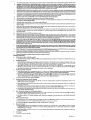
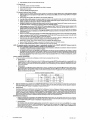
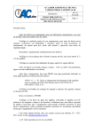
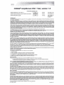
Blood collected in EOTA may be stored at 2-30°C for up to 72 hours from time of draw, followed by an additional two days at 2-SoC. For
storage longer than five days, remove the plasma from the red blood cells by centrifugation at eOO-l600 x g for 20 minutes. FollOWing removal,
plasma may be stored at 2-SoC for an additional seven days, Alternatively, plasma may be stored at 5 -lSoC for up to one month.
30
0
~
~
::l
+-'
•...
C1l
(l)
a.
E
~
1------
2to BOC
8
Plasma
2
0
2
345
6
7
8
9
10 11 12 13 14 15
Time (Days Post Collection)
C.
Blood collected in CPD, CPDA-l, or CP2D may be stored for up to 72 hours at 1-24°C. Following centrifugation of the CPD, CPDA-l, or CP2D
samples at SOO-1600 x 9 for 20 minutes, plasma may be stored at 1-6°C for an additional 7 days from the date the plasma WaS removed from
the red blood cells. Plasma separated from the cells may be stored at s -lS C for up to one month.
D.
ACD-A or 4 % sodium citrate anticoagulated
at 5 -18DC for up to one month.
D
apheresis plasma can be stored at 1-6°C for up to 6 hours, followed by subsequent storage
E.
Do not freeze whole blood.
F.
Heparin has been shown to inhibit PCR. Use of heparinized specimens is not recommended.
G.
Warm pooled or individual donor specimens to room temperature before using.
H.
Covered Archive Plates may be stored at 2-8'C for up to 7 days from the date the plasma was removed from the red blood cells.
I.
No adverse effect on assay performance was observed when plasma specimens were subjected to three freeze-thaw cycles.
J.
Thaw frozen specimens at room temperature before using.
K.
The user should validate other collection and storage conditions. If specimens are to be shipped, they should be packaged and labeled in
compliance with applicable federal and intemational regulations covering the transport of clinical specimens and etiologic agents.32
L
False positive results may occur if cross contamination of specimens Is not adequately controlled during specimen handling and processing.
M.
SPECIMEN POOUNG:
NOTE: Pooling of specimens should only be performed on Individual whole blood and source plasma donations, or on plasma specimens from donors of hematopoiefic
progenitor cells or donor lymphocytes for infusion. Cadaveric specimens must be tested
Individually and not as part of a pool.
1.
The COBAS® AmpliScreen Pooling System performs barcode scanning and pooling operations that combine aliquots from 24 individual
samples Into a single Primary Pool that is used for testing. The pooling algorithm requires preparation of Secondary Pools as well as Individual specimens for follow-up testing in the event a Primary Pool tests posltrve. If less than 24 specimens are available, testing Is performed using the individual specimens.
2.
For Source Plasma, the Hammon pertorms barcode scanning and pooling operations that combine aliquots from 96 individual samples
into a single Primary Pool that is used for testing. Positlve Primary pools are traced to the positive individual using an overlapping pool
testing matrix. Minipools are prepared from the eight individual donations for columns 1 • 12 and from the 12 individual donations for rows
1 - 8. The posltive unit is identified by the intersection of the positive column and positive row. Confirmatory testing is conducted on the
implicated unit using Standard Specimen Processing Procedure. (Hamilton MICROLAS@ AT plus 2 Pipettor with SUNRISE PLUS v3.3 software was used to prepare pools of up to 96 equal aliquots of plasma during clinical trials).
NOTE: The user must validate other pooling algorithms
Cadaveric
N.
and equipment
other than those supplied by Roche,
Blood Specimens
Cadaveric blood specimens can be collected in serum or EDTA anticoagulant tubes.
NOTE: A serum or plasma specimen collected from a donor prior to death may be tested instead of a cadaveric blood specimen using
either the instructions for cadaveric donor specimens or the instructions for living donor blood specimens,
O.
For collection, storage and handling of specimens from deceased donors, follow general standards and/or regulations. Cadaveric samples may
be stored for up to 72 hours at refrigerated conditions (2-S'C), or for up to 48 hours at ambient temperature (lS-3O"C). Other storage and handling conditions must be validated by the user.
NOTE: Cadaveric samples should be placed at 2-8°C as soon as possible after cottection. The use of excessively hemolyzed cadaveric
specimens should be avoided.
PROCEDURAL NOTES
A.
Run Size
1.
Each kit contains reaients sufficient for eight 12-specimen runs, which may be performed separately or simultaneously. At least one preparation of the COSAS AmpliScreen Multiprep Negative (-) Control and one preparation of the COBAS® AmpliScreen Multiprep Positive (+)
Control must be included in each A·ring (see "Quality Control' section).
2.
The Specimen Preparation and Amplification Reagents are packaged in eight single-use bottles. The Mulliprep Negative (-) and Multiprep
Posltiva (+) Controls are packaged in single-use vials. For the most efficient use of reagents, specimens and controls should be processed
in batches that are multiples of 12.
3. The use of sterile gauze, when uncapping sample tubes may reduce the potential for cross contamination between specimens.
S.
Equipment
1.
Prepam the COBAS® AMPUCOfl'lil Analyzer and the Data Station for the AMPUUNK Software for use according to instructions in the
Operator'S Manual for the AMPUUNK software and the Operator's Manual for the COBAS@ AMPLlCOR® Analyzer.
2,
Prepare the Hamilton MICROLAB AT plus 2 System and SUN PLUS Data Station for use according to instructions in the Operator's Manuals.
3.
Pre-cool the high-speed centrifuge and rotor to 2-8°C. See operating instructions for the high speed centrifuge for details.
4.
Perform manufacturer recommended maintenance and calibration on all Instruments, including pipattors, to ensure proper functioning.
0512070S001-02EN
6
Doc Rev. 2.0
C.
Reagents
1. All reagents, except HIV-l MMX, vi.S and HIV·i Mn2 •., vi.S, must be at room temperature before use. Visually examine .~gents
fICient volume before beginning the test procedure. See section '1=1''''gent Preparation' for specific reagent GloragG conditIons.
for suf-
2.
Add all reagents using a pipettor capable of delivering specified volume with ± 3% accuracy and a precision of ,; 5% CV. Check pipettor
functionality and calibrate as recommended by pipettor manufacturer.
3.
Prepare Working Master Mix in a template-free area (e.g., in a dead air box). Reagent preparation area must be clean and disinfected
in accordance with methods outlined in "Precautions" (Item A). Failure to do so may result in reagent contamination.
4.
Prepare 70% ethanol fresh each day.
5.
Check expiration date of opened or Working Reagents before loading on the C08ASi!!> AMPucOR®
6.
Check to ensure that all reagents used are of the same master lot of kit reagents.
Analyzer.
D.
Workftow
1. To minimize the possibility of laboratory areas becoming contaminated with amplicon, the laboratory area should be separated into several distinct areas organized around Pre-Amplification and Post-Amplification. Personnel should use proper anti-contamination safeguards
when moving between areas.
2. The Pre-Amplificalion Area should have a template-free area for preparation of Working Master Mix and an amplicon free area for specimen and control preparation.
3. The Post-Amplification Area should have a coBASl!!> AMPUCOR® Analyzer(s) and AMPUUNK Data Station(s) with additional area for
preparing Working Amplification and Detection Reagents.
4. Pipettors and other supplies should be dedicated to a specific area. Samples, equipment and reagents should not be returned to the area
where a previous step was performed.
E.
Temperature
F.
Room temperature is defined as 15 to 30°C.
Vortexing
0
Proper vortexing during sample preparation is important to ensure homogeneous mixture after additions of reagents.
G.
Plpetting
1.
2.
H.
Pooled or individual plasma specimens must be at room temperature before pipetting.
Use a clean pipette tip or disposable transter pipette with each specimen or control. Use aerosol barrier or positive displacement RNasefree tips.
3. Confirm that all pipettors are correctly set to dispense the specified volumes in accordance with the specimen preparation procedures and
guidelines.
Specimen Processing
1. Screw-<:ap tubes must be used for specimen and control preparation to prevent splashing and potential cross-contamination of specimens and controls. Do not use snap cap tubes.
2. Avoid contaminating gloves when manipulating specimens.
3.
Specimens and controls should be prepared in a laminar flow hood. Failure to do so may result in sample contamination. Specimen
and control preparation area must be cleaned and disinfected in accordance with methods outlined in 'Precautions' (Item A).
Decontamination
Thoroughly clean and disinfect all work surfaces with a freshly prepared solution of 0.5% sodium hypochlorite in distilled or deionized water.
Follow by wiping down the surface with 70% ethanol.
INSTRUCTIONS FOR USE
The Multiprep Specimen Processing Procedure is used for extracting nucleic acid from pooled specimens and from individual cadaveric specimens.
The Standard Specimen Processing Procedure is used for extracting nucleic acid from individual specimens. The Standard Specimen Processing
Procedure may also be used for testing of Source Plasma minipools.
1he Multiprep and the Standard Specimen Processing Procedures are generic nucleic acid extraction procedures and can be used for the extraction
of HIV-l RNA, HCV RNA, and/or H8V DNA. A single extraction is suffICient for multiple assays. Workfiow can be performed on the same day or over
multiple days under the following conditions:
Amplification.
Hybridization
and Detection of Stored Processed Specimens
Amplification. hybridization and detection can occur on the same day as specimen processing or on a subsequent day. If amplification. hybridization
and detection are to be done on a subsequent day. perform the Multiprep Specimen Processing Procedure described in steps 81 through 821 or
the Standard Specimen Processing Procedure described in steps 922 through 938. Store the processed specimens and controls as indicated. On
the subsequent day, begin with Step A (Reagent Preparation - Working Master Mix), thaw processed specimens and controls at room temperature,
and continue with Step 839.
.
.
Hybridization
and Detection of Stored Denatured Amplicon
Hybridization and detection of the denatured amplicon may occur on the same day as amplification or on a subsequent day. If hybridization and
detection are to be done on a subsequent day. the denatured amplicon may be left on-board the COBASi!!>AMPUCO~
Analyzer for not more than
24 hours before starting the hybridization and detection steps. Mematively. the denatured amplicon may be stored at 2-8°C for not more than five
days before starting the hybridization and detection steps.
A.
Reagent Preparation - Working Master Mix
Perlonned
in: Pre-Amplification
- Reagent Preparation Area (e.g., dead air box)
A1. Detennine the appropriate number of A-ring(s) needed for specimen and control testing.
A2. Place the A-ring(s) on the A-ring holder(s).
A3. For each A-ring, prepare one Working Master Mix.
M. Pipette 50 I'lWorking Master Mix into each A-tube. Discard unused Working Master Mix. Do not close the covers of the A-tubes at this time.
AS. Place the A·ring containing Working Master Mix in a sealable bag and seal the plastic bag. Record the assay name (HIV-l) and the time
the Working Master Mix was prepared.
A6. Store the A-ring(s) containing 'Norking Master Mix at 2-8°C until specimen and control preparation is completed. The A-rings with Working
Master Mix must be used within 4 hours of preparation.
A7. Decontaminate area See 'Procedural Notes'. Item I.
B.
Specimen and Control Preparation
Perlonnad
in Pre-Amplification
Multiprep
Specimen Processing
- Specimen and Control Preparation Area
Procedure (pOOled SpecImens and Individual
Cadaveric Specimens)
81. For pooled specimens. pipette 1000 ilL of each pool into an appropriately labeled screw-cap tube using the COBASi!!>AmpliScreen
Pooling System, a hand-held pipettor or other user-validated method. Cap the tubes. Proceed to Step B2.
For individu.af cadaveric specimens, pipette 200 ul, into an appropriately labeled screw-cap tube and add 800 ilL Multiprep Diluent
(MP OIL) uSing a hand-held pipettor or other user-validated method. Cap the tubes. Vortex each specimen tube briefly. Proceed to
Step B2.
82. Vortex NHP briefly.
83. For each Negative and Positive COntrol pipette 1000 ilL NHP into an appropriately labeled screw-cap tube. Cap the tubes.
For cad:>1I9ric. tm;:tJng. plp<>tte200 ilL NHP into an appropriately labeled screw-cap tube and add 800 IJL MUltiprep Diluent (MP OIL) using
a hand-held pipettor or other user-validated method. Cap the tubes. Vortex each specimen tube briefly.
84. Use a permanent marker to make an orientation mark on each tube.
85. Place the specimen and c~
tubes into the pre-cooled high-speed centrifuge with the orientation marks facing outward, SO that the orientatIon marks WJII a"9O with the pellets formed during centrifugation.
86. Centrifuge specimens and control tubes at 23,000 - 24,000 x g for 60 ± 4 minutes at 2-8°C. The pellet will form on the outer wall as indi05120705001-02EN
7
Doc Rev. 2.0
caled by the orientation mark.
NOTE: The 60 :I: 4 minutes begins when the centrifuge
reaches 23,000 - 24,000 x g.
87 Remove the tubes from the centrifuge and remove the caps. Slowly aspirate 900 III of the supematant from each centrifuged tube leaving
. approximately 100 ilL of supematant. Avoid contact with the pellet. Discard the supernatant and pipette tip appropriately. Use a fresh
pipette tip for each tube.
B8. Prepare a WorKing Lysis Reagent bottle for every batch of 12 specimens and controls to be processed.
89. Pipette 600 ilL WorKing lysis Reagent into each specimen and control tube. Cap and vortex tubes briefly.
810.
Prepare Controls as follows:
a.
Negative Control
Vortex MP H Cbriefly. Tap vial to collect the solution in the base. Pipette 20 III MP
Wor1Iing lysis Reagent and NHP. Cap the tube and vortex briefly.
b.
H Cto the tube
labeled 'MP (-) C' containing
Positive Control
Vortex MP (+J C briefly. Tap vial to collect the solution in the base. Pipette 20 "l MP {+J C to the tube labeled 'MP (+) C' containing
Working Lysis Reagent and NHP. Cap the tube and vortex briefly.
Bll.
Incubate all tubes for 10 to 15 minutes at room temperature after adding Working lysis Reagent to the last tube, After the incubation
period. briefly vortex all tubes.
B12. Pipette 700 ul, of isopropanol into each tube. Cap the tubes and vortex briefly.
B 13. Place the tubes into a mlcrocentrifuqe w~h the orientation marks facing outward to align with the pellets that will form. Centrifuge at
14,250 ±'1750 X g for 15-20 minutes at room temperature.
B14. Slowly aspirate the supernatant from each tube. Remove as much liquid as possible wnhout disturbing the pellet.
B15. Pipette 1.0 mL of 70% ethanol into each tube, Cap the tubes and vortex briefly.
B16. PlaCe the tubes into a microcentrifuge with the orientation marks facing outward to align with the pellets that will fonn. Centrifuge at
14,250 ± 1750 x g for 5-10 minutes at room temperature.
617. Slowly aspirate the supennatsnt from each tube using a fine-tip disposable transfer pipette. Remove as much liquid as possible without
disturbing the pellet. Use a new transfer pipette for each tube.
B18. Using a new transfer pipette for each tube, repeat Step B17 to remove as much of the remaining supernatant as possible without disturbing the pellet. Residual ethanol can inhibit ampllffcation.
B19. Pipette 200 III MP OIL into each tube. Use a pipette tip to break apart the pellet. This can be done by aspirating 30-40 ilL of the diluent
in the tip and scraping the sides and base of the tube in an up/down motion for at least 10 seconds and dispensing 30-40 I'L Cap the
tubes and vortex briefly to resuspend the extracted RNA. Note that some insoluble material may remain.
820. At this point amplification of the processed specimens and controls must be started within 2 hours. If not, the processed specimens and
controls can be stored at -70·C or colder for up to one month. Thawing should be completed within one hour at room temperature.
621. Proceed to step 839, Loading the A-ring.
Standard Specimen Processing
Procedure [Individual Specimens (Non-Cadaveric)
and Source Plasma Minipools]
B22.
Pipette 200 III of each specimen into an appropriately labeled screw-cap tube using the COBAS@ AmpliScreen Pooling System, a handheld pipettor or other user-validated method. Cap the tubes.
B23.
Vortex NHP briefly.
B24.
For each Negative and Posrtive Control pipette 200 pt, NHP into appropriately labeled screw-cap tubes. Cap the tubes.
825.
Use a pennanent marker to make an orientation mark on each tube.
626.
Prepare a WorKing lysis Reagent bottle for every 12 specimens and controls to be processed.
627.
Pipette 600 III Working Lysis Reagent into each tube. Cap and vortex tubes briefly.
B28.
Prepare Controls as follows:
a.
Negative Control
Vortex MP H C briefly. Tap vial to collect the solution in the base. Pipette 20 pl, MP
taining Working Lysis Reagent and NHP. Cap the tube and vortex briefly.
b.
HC
into the tube labeled 'MP (-) C' con-
Positive Control
Vortex MP (+/ C briefly. Tap vial to collect the solution in the base. Pipette 20 ilL MP (+/ C into the tube labeled 'MP (+) C' containing Working lysis Reagent and NHP. Cap the tube and vortex briefly.
B29.
Incubate all tubes for 10-15 minutes at room temperature after adding WorKing lysis Reagent to the last tube, After the incubation period,
briefty vortex all tubes.
630.
Pipette 800 ut, of isopropanol into each tube. Cap the tubes and vortex briefly.
B31.
Place the tubes into a microcentrifuge with the orientation marks facing outward to align with the pellets that will fonn. Centrifuge at
14,250 ± 1750 x g for 15-20 minutes at room temperature.
B32.
Slowly aspirate the supematant from each tube. Remove as much liquid as possible without disturbing the pellet.
B33.
Pipette 1.0 mL of 70% ethanol into each tube. Cap the tubes and vortex briefly.
834.
Place the tubes into a mlcrocentrifuge wrth the orientation marks facing outward to align with the pellets that will fonn. Centrifuge at
14,250 ± 1750 x g for 5-10 minutes at room temperature.
635.
Slowly aspirate the supematant from each tube using a fine-tip disposable transfer pipette. Remove as much liquid as possible without
disturbing the pellet. Use a new transfer pipette for each tube.
636.
Using a new transfer pipette for each tube. repeat Step B35 to remove as much of the remaining supematant as possible without disturbing the pellet. Residual ethanol can inhibit ampliffcation.
B37.
Pipette 200 III MP OIL into each tube. Use a pipette tip to break apart the pellet. This can be done by aspirating 30-40 ut, of the diluent
in the tip and scraping the sides and base of the tube in an up/down motion for at least 10 seconds and dispensing 30-40 ~L Cap the
tubes and vortex briefly to resuspend the extracted RNA. Note that some insoluble material may remain.
838.
At this point amplification of the processed specimens and controls must be started within 2 hours. If not, the processed specimens.and
controls can be stored at -70·C or colder for up to one month. Thawing should be completed within one hour at room temperature.
Loading the A-ring
B39. Create an A-ring work/ist record for each A-ring to identify the A-tube with the appropriate control or specimen to be pipetted.
840.
If processed specimens and controls wet'e stored frozen, thaw at room temperature before proceeding. Briefly vortex the processed
specimens and controls.
641.
Pipette 50 ilL of each processed specimen and control into the appropriate A-tube ccntaining HIV-1 Working Master Mix. Immediately
cap the A-tube and repeat this step for all the 12 A-tubes to complete the A-ring loading. Use the A-ring work/ist record to ensure tne
appropriate specimen or control is added to the correct A-tube position for each A-ring.
B42. Transfer the A-ring with sealed tubes containing
Amplification/Detectlon Area. Proceed to Part C.
NOTE: Ampllffcation
Master Mix..
C.
must begin
Reverse Transcription.
Performed
within
Amplification
in Post-Amplification
45 minutes
the processed
specimens
from when the first specimen
and
or control
controls
in Working
in the A-ring
;s
Master
added
Mix to the
to the Working
and Detection
- Amplification/Detection
Area
Cl. Perform Daily Instrument Maintenance as outlined in the Operaror's Manual for the COBAS@ AMPUCOR'i!' Analyzer including:
a.
Wipe D-cup handler tip with a lint-free moist cloth and dry.
05120705001-02EN
8
Doc Rev. 2.0
b.
Wipe initialization post wtth a lint-free moist cloth and dry.
C2. Before eaCh run:
a.
Check waste container and empty if necessary.
b.
Check Wash Buffer Reservoir and add prepared Wash Buffer il necessary.
c.
Replace used D-cup racks,
d.
Prime the COBAS@ AMPUCOROl!Analyzer.
C3. Instrument Loading and System Operation
a.
Prepare enough 01 the lollowing detection reagent cassettes to complete the workload: Working HIV-1 Probe Suspension Reagent
(IH4, v1.5), Working IC Probe Suspension Reagent (II PS1, v1.5), Working Substrate (583), Denaturation Reagent (DN4), and Conjugate
Reagent (CN4).
b.
Place the IH4, v1.5 and II P51, v1.5 cassettes in the test-specific reagent rack.
c.
Place DN4. CN4 and S83 cassettes in the generic reagent rack. Record on the cassette the date when each cassette was opened.
d.
Identify the reagent racks as generic or test specific using the COBAS~ AMPLlCOR® Analyzer barcode scanner for the AMPUUNK
software, as descri~
in the Operator'S Manual for AMPULINK SOftware.
e.
Configure the reagent racks by entering the reagent positions and lots using the COBAs<!!>AMPUCOR@ Analyzer barcode scanner lor
the AMPUUNK software, as described in the Operator's Manual for AMPLlLlNK software.
f.
Load the reagent racks onto the COSAS<!!>
AMPUCO~ Analyzer using the COSAS<!!>AMPLlCoR® Analyzer barcode scanner.fo~ the
AMPUUNK software, as described in the Operator's Manual lor AMPULINK software. Make sure that eaCh reagent cassette rs m itS
assigned posltion and that each cassette fits tightly into its rack.
g.
Place the D-cup rack on the D-cup platform. Two D-cups are required lor each A-tube and two D-cups are required for each Working
Substrate cassette to allow lor blanking by the COSAS"" AMPLlCOR<!!>Analyzerr as described in the Operator's Manual for the COBAS®
AMPUCOR<!!>Analyzer.
h.
Place the A-ring into the thermal cycler segment of the COBAS® AMPucoH®
Analyzer and close the cover on the thennal cycler segment.
;-.,-
Load the A-ring into the COSAS® AMPUcoH® Analyzer using the COBAS® AMPLlCOH® Analyzer barcode scanner for the AMPULINK
software, as described in the Operator's Manual for AMPLIUNK software.
j.
Create an A-ring order, using the AMPLlLlNK software, as described in the Operator's Manual for AMPLIUNK
A-ring worklist record created for specimen processing to assist in entering the A-ring order.
k.
Repeat steps h. through j. above to load a second A-ring on the COSAS® AMPLlCOR® Analyier.
I.
Start the COBAS® AMPLlCOR® Analyzer as described in the Operator's Manual for AMPLIUNK software.
-":-
software. Use the
m. Watt for the COSAS® AMPUCOR@ Analyzer to indicate that the load check has passed.
NOTE: The required quantity of each detection reagent is automatically calculated by the COBA,s4D AMPUCOR'f'J Analyzer during
Load Check to detennine if sufficient reagents are available for the requested tesls.
the
n.
The COBAs® AMPLlCOH® Analyzer automatically performs reverse transcription, amplification and detection. Hesults are expressed
as absorbance values at 660 nm and as positive or negative.
o.
As a Quality Control measure, the AMPUUNK A-ring Results Report and the Run Log may be printed (e.g. daily, weekly or monthly)
and retained along with the respective A-ring worklist. A selection of A-ring worklist records should be periodically compared wtth the
AMPULINK A-ring Results Report to verify that the A-ring 10, Instrument serial number, and specimen IDs are identical. Reconcile the
Run Log with the selected A-ring worklist to account for all A-ring IDs associated with the run. If there are discrepancies, perform
follow-up investigation.
QUAUTV CONTROL PROCEDURES
1.
At least one Multiprep H Control and one Multiprep (+) Control must be processed with each A-ring.
a.
'.
Negative Control
The absorbance lor the MP H C should be less than 0.2 at 660 nm and its associated MP .Ie should be greater than or equal to 0.2 for the
Negative Control to be valid. If the absorbance value for the MP H C is greater than or equal to 0.2 andlor lts associated MP IC is less
than 0.2. the entire A·ring is invalid, and the entire test procedure for that A-ring (sample and control preparation, amplification and detection) must be repeated.
b.
Positive Control
The absorbance for the MP (+) e should be greater than or equal to 1.0 at 660 nm and its associated MP Ie should be greater than or
equal to 0.2 at 660 nm for the Positlve Control to be valid. If the absorbance value for the MP (+) C is less than 1.0 and/or its associated
MP IC is less than 0.2, the entire A-ring is invalid, and the entire test procedure for that A-ring (specimen and control preparation, amplification and detection) must be repeated.
Summary of Control Acceptance Criteria
HIV-1 Result
IC Result
Aeoo
Comment
Aeoo
Negative Control
< 0.2
Negative
~02
Valid
Positive Control
~ 1.0
Positive
~ 0.2
Valid
Comment
2.
Flags and comments may be generated by the COBAS~ AMPUcoH® Analyzer during a run. The Operator must Check the run printout(s} for
flags and comments to verify that the run is valid. Refer to the Operator's Manual for the AMPUUNK software and the Operator's Manual for
the cosAS<!!> AMPUCOR<!!>Analyzer for interpretation of flags and comments.
3.
Extemal Control
If an Extemal Control Q.e., an additional run control other than the Muttiprep (+) Control or Multiprep H Control) is required by the laboratory,
the External Control should meet regulatory requirements for suCh controls. The absorbance of the HIV-1 External Control should be equal to
or greater than 0.2 at 660 nm, irrespective of the MP Ie absorbance. If the absorbance of the HIV-1 External Control does not meet the above
criterion, the negative results for specimens in the associated run may be invalidated. However, positive results for specimens in such a run
should llil1 be invalidated solely on the basis of the results obtained for an External Control; those positive results should remain the test of
record. The laboratory should follow its established Standard Operating Procedure for the appropriate action.
INTERPRETATION OF RESULTS
1.
2.
Rags and comments may be generated by the COBAs<!!>AMPLlCOR® Analyzer during a run. The Operator must check the run printout(s)
for flags and comments to verify that the run is valid. Refer to the Operator's Manual for the AMPLIUNK software and the Operator's Manual
for the COSASI!>AMPLlCOR<!!>Analyzer for Interpretation Of fiags and comments.
Soecimen Resutts
T,:",oabsorbance values are obtained for each specimen: one for the HIV-1 target and one for the internal control (MP Ie). For a sample
wtth an absorbance less than 02, the MP IC absorbance for that specimen must be greater than or equal to 0.2 at 660 nrn for a valid
negative specimen test result If the absorbance for the HIV-1 target is greater than or equal to 0.2, the MP Ie resutt is disregarded and
the test result is valid and positive.
3.
For a valid run, results are interpreted as follows:
05120705001-02EN
9
Doc Rev. 2.0
r
:,.-
Ie Re~ult
HIV-1 Result
A.."
Comment
A.."
Comment
<0.2
NEGATNE
2:0.2
VAUD
<02
NEGATIVE
< 0.2
INVAUD
;'0.2
POSITIVE
PNY
VAUO
Interpretation
Specimen is negative for HIV-1 RNA
Invalid result Repeat entire test procedure for invalid specimen.
Specimen is positive for HIV-l RNA
Invalid Test Runs
When invalid Positive or Negative Control resu~s ere obtained on an A-ling, that A-ring is invalid. Repeat the entire test procedure f~ the associated .specimens Qncludingspecimen and control preparation, amplification and detection) in the A-ring by processing another auquot of the original plasma specimens,
With the exception of instrument failures subsequent to denaturation of ampllcon, an instrument failure during a test run, as indicated by syst~f!l er;or
messages, also constitutes an invalid test run. In such instances, repeat the test procedure for the associated controls and specimens (amplification
and detection) in the run by processing another aliquot of the processed specimen.
For instrument failures subsequent to successful denaturation of amplicon, it is not necessary to repeat the entire test procedure for th';' associated
specimens. In such instances, the denatured ampllcon may be redetected by the COBAS® AMPUCOA® Analyzer. The denatured ampilco~ may be
left on the COBAS® AMPUCoA® Analyzer for not more than 24 hours before continuing with the hybridization and detection steps. Alternatively, the
denatured amplicon may be stored at 2-8'C for not more than five days before continuing with the hybridization and detection steps.
Invalid Specimen Results
For plasma specimen(s) that are invalid, perform repeat testing in single on the remaining replicate tube(s). The test result f?r th.e pool Of indivi.du~1
coner specimen is based only on the repeat valid test result If the last available replicate of a pooled specimen gIVes an lnvalld result, each lndlvidual donor specimen in that pool should be tested. If an individual donor specimen gives an invalid result, the test result for that individual donor
specimen should be considered invalid for HIV-1 RNA
For cadaveric specimens that are invalid, additional cadaveric specimen is diluted 1:5 with MP OIL reagent and retested in duplicate using the
Multiprep Specimen Processing Procedure. The test result for the cadaveric specimen is based on the repeat valid test results.
Results of Pooled Donor Specimens
(Pools of up to 24 Individual Donations)
1he testing algorithm for testing of pooled samples for the COBAS® AmpliScreen HIV-1 Test, v1.S requires a single level of testing for Primary Pools
that are negative for HIV-1 RNA and three levels of testing (Primary Pool, Secondary Pool and tertiary resolution) for Primary Pools that are positive
for HIV-1 RNA
Negative Primary Pools
When the Primary Pool is negative, report the results for all associated individual donor specimens in that Primary Pool as 'HIV-1 RNA Negative".
Positive Primary Pools·
Secondary
Pool Testing
When the Primary Pool is positive, prepare four Secondary Pools containing the associated donor specimens. The Secondary Pools must be processed
using the Muniprep Specimen Processing Procedure.
•
If one or more of the Secondary Pools tests positive, report the results for the donor specimens in the negative Secondary Pools as
'HIV-1 RNA Negative'. For positive Secondary Pools, proceed to the section entitled 'Positive Primary Pool, Positive Secondary Pools
- Tertiary Resolution Testing.·
•
If all four Secondary Pools are negative, the individual donor specimens in that Primary Pool may be reported as 'HIV-l RNA Negative.'
•
As part of an overall Quality Assurance proqrarn, you may wish to conduct additional testing to determine the cause of the initial positivity
of the Primary Pool.
Positive Primary Pool, Positive Secondary
Pools - Tertiary Resolution
Testing
For a positive Secondary Pool, test each of the individual donor specimens in that Secondary Pool. The individual donor specimens must be processed
using the Standard Specimen ProceSSing procedure.
•
If one or more of the individual donor specimens is positive, the positive donor specimen(s) is (are) reported as 'HIV-1 RNA Positive' and
the remaining negative donor specimens associated with the positive Secondary Pool are reported as "HIV-1 RNA Negative."
•
If all of the individual donor specimens in that Secondary Pool test negative, the donor specimens in the Secondary Pool may be reported
as "HIV-1 RNA Negative.'
•
As part of an overall Quality Assurance program, you may wish to conduct additional testing to determine the cause of the positivity of the
Primary and Secondary Pools.
Results of Individual Donor Samples
If an individual donor specimen is positive, the positive donor specimen is reported as 'HIV-1 RNA Positive.'
If an individual donor specimen is negative, the negative donor specimen is reported as "HIV-1 RNA Negative.'
Results of Pooled Source Plasma SpeCimens (Pools of up to 96 Individual
Donations)
The testing algorithm for testing of pooled samples for the COBAS® AmpliScreen HIV-1 Test, v1.5 requires a single level of testing for Primary Pools
that are negative for HIV-1 RNA and three levels of testing (primary Pool, Minipool and confirmatory testing) for Primary Pools that are positive for
HIV-1 RNA
Negative
Primary Pools
When the Primary Pool is negative, report the results for all associated individual donor specimens in that Primary Pool as "HIV-1 RNA Negative. '
Positive
Primary Pools - Minipool
Testing
Positive Primary pools are traced to the positive individual using an overlapping pool testing matrix. Minipools are prepared from the eight individual
donations for columns 1 - 12 and from the 12 individual donations for rows 1 - B. The 20 minipools are tested using the Standard Specimen Processing
Procedure. The positive unit is identified by the intersection of the positive column and positive row. Confirmatory testing is conducted on the implicated unit using Standard Specimen Processing Procedure.
Results of Individual
Cadaveric
Specimens
If an individual cadaveric specimen is positive, the positive cadaveric specimen is reported as 'HIV-l RNA Positive.'
If an individual cadaveric specimen is negative, the negative cadaveric specimen is reported as 'HIV-l RNA Negative. '
For cadaveric specimens that hed an initial invalid result and were repeated in duplicate, if either or both the duplicate samples are positive, the specimen is reported as 'HIV-1 RNA Positive.' If both duplicate specimens are negative, or if one duplicate is negative and one is invalid, the specimen
IS reported as "HIV-1 RNA Negative." If both replicates are invalid, it is most likely due to inhibitory substances in the specimen, and the results should
be marked as invalid or unresolved.
PROCEDURAL LIMITATIONS
1.
This test has been evaluated only for use in combination with the COBAS® AmpliScreen Muaiprep Specimen Preparation and Control Kit,
COBAS® AMPLlCOR® Analyzer, and the Hamilton MICROlAB AT plus 2 Pipettor for the automated preparation of plasma pools.
2.
Eight Group 0 culture specimens were only evaluated as diluted samples dus to limited specimen volume. All HIV-l Group 0 specimens tested
were round to be HN-l p24 antigen positive, however, only five (63%) were detected by the COBAS® AmpliScreen HIV-1 Test, vl.5. These
data indicate that the COBAS@ AmpliScreen HIV-l Test, v1.5 will not consistently detect HIV-l RNA in all Group 0 specimens.
3.
This COBAS® AmpliScreen HIV-l Test. vl.5 is intended to be used in conjunction with licensed tests for detecting antibodies to HIV-1. The
GOBAS® AmpliScreen HIV-1 Test. v1.S may not be used to replace HIV-1 antibody detection tests such as EIA or Westem Blot (See
Performance Characteriotics section. Tables 12 and 13).
4.
Heparin inhibits PCR; specimens collected using heparin as the anticoagulant
HIV-1 Test, v1.5.
5.
Reliable results are dependent on adequate specimen collection and proper transport procedures.
05120705001-02EN
10
should not be used with the COBAS® AmpliScl'gen
Doc Rev. 2.0
6.
Detection of HIV-1 RNA is dependent on the number of virus particles present in the specimen and may be affected by specimen collection
methods, patient factors ~.e., age, presence of symptoms), and/or stage of infection and pool size.
7.
Only the Hamitton MICROLAB AT plus 2 Pipettorhas been validated for use wtththe COBAS@AmpIiScreenHIV-1 Test, vl.5 for the automated
preparation of plasma pools. Adhere to the hardware instructions and safety precautions outlined in the User Manual for the Hamilton
MICROLAB AT plus 2 Pipettor.
Though rare, mutations within the highly conserved region of the viral genome covered by the COBASi!!l AmpliScreen HIV-l Test, vl.5 primers
and/or probe may resuK in the failure to detect the virus.
8.
9.
Due to inherent differences between technologies, it is mcommended that, prior to switching from one technology to the next, users perform
method correlation studies In their laboratory to qualify technology differences.
PERFORMANCE
CHARACTERISTICS
ReproducibUity
The reproducibility of the C08AS® AmpliScreen HIV-1 Test, vi .5 was established by testing two six-member EDTA plasma panels with known concentrations of HIV-1. Panel One was tested using the Multiprep Specimen Processing Procedure. Panel One was comprised of HIV-1 RNA positive
samples at concentrations of 10, 25, 50, 75, and 25,000 copies/mL and one HIV-l-negative sample. Panel Two was tested using the Standard
Specimen Processing Procedure. Panel Two was comprised of HIV-l positive samples at concentrations of 50,100,150,250,
and 25,000 copieslmL
and one HIV-l negative sample.
Testing was performed at three sites wtth two operators at each site using five C08ASiI!> AmpliScreen HIV-l Test, vl.5 kit lots. Each operator used a
dedicated COBASOl>AMPUCORO!>Analyzer throughout the study. Each operator was provided panel sets that had been randomized and labeled in
blinded fashion.
All valid reproducibility data were evaluated by calculating the percentage of correct results for each panel member. The data were analyzed by site,
lot, testing day, run, and operator for each Specimen Processing Procedure (Multiprep and Standard).
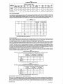
The reproducibility study for the COflAS® AmpliScreen HIV-l Test, version 1.5 demonstrated consistency by lot and site for both the MuKiprep and
Standard Specimen Processing Procedures as seen in Table 1 and 2 below:
•
Table 1
Reproducibility Results - Mu/tipI'ep Specimen
Processing Procedure
Results By Lot Iii Positive I it Tested)
10 clmL
25c1mL
SOclmL
75c1mL
Lot ill
1188
51190
77/90
86/90
89/89
90190
(%)
(1%)
(57%)
(86%)
(96%)
(100%)
(100%)
Negative
25,OOOc/mL
Lot #2
0/89
47190
72190
83/90
88/90
9019O
(%)
(0%)
(52%)
(60%)
(92%)
(98%)
(100%)
Lot #3
2190
50189
60/89
88/89
88/90
90/90
(%)
(2%)
(56%)
(90%)
(99%)
(98%)
(100%)
Lot #4
0190
45190
78/90
84/90
90/90
90/90
(%)
(0%)
(50%)
(87%)
(93%)
(100%)
(100%)
Lot #5
0/89
51/89
73/89
83/90
(%)
(0%)
(57%)
(82%)
.(92%)
Site #1
31150
721150
1331150
(%)
(2%)
(48%)
(89%)
Site #2
01147
821148
1081148
90/90
90/90
(100%)
(100%)
1421150
149/150
1501150
(95%)
(99%)
1361149
146/149
Results By SIte (# Positive 1 # Tested)
(%)
(0%)
(55%)
Site #3
01149
901150
(%)
(0%)
(60%)
(100%)
150/150
(73%)
(91%)
(98%)
139/150
1461150
150/150
150/150
(1oo%)
(93%)
(97%)
(100%)
(100%)
Table 2
Reproducibility Results - Standard Specimen Processing Procedure
Resutts By Lot (il Positive I # Tested)
Negative
SOc/mL
100 c/ml
1SOc/ml
250c/mL
25,000 c/ml
Lot #1
0190
44190
75/89
83189
85/88
90190
(%)
(0%)
(49%)
(84%)
(93%)
(97%)
(100%)
Lot #2
0/89
49/88
72188
83/89
86/89
90/90
(%)
(0%)
(56%)
(82%)
(93%)
(97%)
(100%)
Lot #3
0189
39/88
72189
74/87
86//90
90/90
(%)
(0%)
(44%)
(81%)
(85%)
(96%)
(100%)
Lot #4
1/87
49190
59/88
71/89
85190
9Of9O
(%)
(1%)
(54 %)
(67%)
(80%)
(94%)
(100%)
Lot #5
0/89
37190
65/89
76188
85/89
89/89
(%)
(0%)
(41%)
(73%)
(86%)
(96%)
(100%)
Results By SIte (il Positive / # Tested)
S~e #1
0/150
731149
117/150
1341150
1451150
(%)
(0%)
(49%)
(78%)
(89%)
(97%)
(100%)
Site #2
0/144
631147
1091144
1161142
1381146
1501150
(%)
(0%)
(43%)
Site #3
1/150
821150
(%)
(1%)
(55%)
(76%)
117/149
(79%)
(83%)
1351150
(90%)
(95%)
144/150
(96%)
150/150
(100%)
149/149
(100%)
Analytical Sensltivity - Dilutlonal Panels
The analytical sensitivity of ~e COBAS® AmpliScreen HIV-1 Test vl.S was determined by testing 10 HIV-l seropositive clinical specimens. The titer
of each specimen was quantitated wtth a commercially available assay using a secondary standard calibrated against the WHO International Standard.
These specimens were dIluted In normal human plasma to 150, 50, and 16.7 copieslmL for the Muniprep Specimen Processing Procedure and 300
100. and 33.3 copleslmL for the Standard Specimen Processing Procedure.
.
The COBAS® AmpliScreen HIV-1 Test, v1.S detected 50 copieslmL HIV-1 RNA at a frequency greater than 98% with a lower 95% confidence limit
of 96.5% using the Multiprep Specimen Processing Procedure. The assay detected 100 copieslmL HIV-1 RNA at a frequency greater than 98% with
05120705001-02EN
11
Doc Rev. 2.0
a lower 95% confidence limn of 96.5% using the Standard Specimen Processing Procedure. The data are presented in Tables 3 and 4.
When evaluated using PROBIT analysis, the combined data for all samples processed by the MullipreJ) SpeCimen Processing Procedure indicate an
average 95% Limit of Detection (LOO) of 39.2 coples/ml; with the lower and upper 95% confidence hm~s of 34.0 coples/rnl, and 48.3 copies/rnt;
respectively, The LOD of 39.2 copieslmL corresponds to approximately 61.25 IU/mL
When evaluated using PROBIT analysis, the combined data for all samples processed by the Standard Specimen Processing Procedure indicate an
average 95% LOD of 96.2 copiesJmL with the lower and upper 95% confidence limit of 83.3 copiesJmL and 116.7 copiesJmL, respectively. The LOD
of 96.2 copiesJmL corresponds to approximately 150.3 IUlmL
Table 3
Multiprep Procedure Testing Summary for All Clinical Samples
Combined Input Values with 95% One-tailed Lower Confidence Umit
Multiprep Sample Processing Procedure
Number of
Positives
Number of
Individual Tests
% Positive
95% Lower
Confidence Limit One-Tailed
150
220
220
100.0%
98.6%
SO
214
217
98.6%
96.5%
16.7
116
219
53.0%
47.2%
HIV-1 RNA
Concentration
(c/mL)
Table 4
Standard Procedure Testing Summary for All Clinical Samples
Combined Input Values with 95% One-tailed Lower Confidence Umit
Standard Sample Processing Procedure
Number of
Individual Tests
% Positive
95% Lower
Confidence Umit One-Tailed
216
218
99.1%
97.1%
216
219
98.6%
96.5%
97
217
44.7%
39.0%
HIV-l RNA
Concentration
(c/mL)
Number of
Positives
300
100
33.3
Analytical Sensitivity - WHO HIV-l International Standard
The analytical sensitivity of the COBAS@ AmpliScreen HIV-1 Test, v1.5 was also detennined using the WHO HIV-l International Standard (97/656).
The WHO HIV-1 Intemational Standard was serially diluted in HIV-1-negatlve plasma to final concentrations of 140, 100. 70, 50, 35, and 25 IU/mL for
the Multiprep Specimen Processing Procedure and 800, 560, 400, 280, 200, and 140 IU/mL for the Standard Specimen Processing Procedure. Each
dilution was tested using two lots of COBAS® AmpliScreen HIV-1 Test, v1.S.
When evaluated using PROBIT analysis, the combined data from all samples using the Multiprep Sample Processing Procedure indicate an average
95% LOD of 78.4 IUlmL, wlth lower and upper 95% confidence limits of 68.4 IU/mL and 94.4 IU/mL, respectively.
When evaluated using PROBIT analysis, the combined data from all samples tested using the Standard Sample Processing Procedure indicate an
average 95% LCD of 323.4 IUlmL, with lower and upper 95% confidence limits of 284.9 IUlmL and 387.3 IUlmL, respectively.
Tables 5 and 6 summarize the overall results for the Mu~iprep and Standard Specimen Processing Procedures, respectively.
Table 5
Serial D,lution Testing Summary for MultJprep Method with HIV·1 RNA WHO International
Combined Input Values with 'Lower 95% Confidence Limit (One-Sided)
HIV-1 RNA
Concentration
(IUlmL)
Standard
Number of
Positives
Number of
Individual Tests
% Positive
140
128
130
98.5%
95.2%
100
115
120
95.8%
91.4%
95.2%
95% Lower
Confidence Umit
(One-sided)
70
128
130
98.5%
50
103
120
8S.8%
79.5%
35
79
118
66.9%
59.1%
25
70
120
58.3%
50.4%
Table 6
Serial D,1ution Testing Summary for Standard Method with HIV-1 RNA WHO International
Combined Input Values with Lower 95% Confidence Umit (One-Sided)
HIV-1 RNA
Concentration
(iU/mL)
Number of
Positives
(971656)
Standard
(971656)
Number of
Individual Tests
% Positive
95% Lower
Confidence Limit
(One-sided)
96.1%
800
119
120
99.2%
560
119
120
99.2%
96.1%
400
118
119
99.2%
96.1%
87.1%
280
126
137
92.0%
200
100
119
84.0%
77.5%
140
82
120
68.3%
60.6%
Analytical Sensitivity - CBER HIV-l Panel
The FD~ CBER HIV-1 Panel Members were processed using the Mu~iprep and Standard Specimen Processing Procedures. The Mu~iprep Specimen
Processing Procedure detected 100% of all positive members ranging from 10 - 250,000 copies/mL The Standard Specimen Processing Procedure
detected 100% of all positive members ranging from 100 - 250,000 copies/mL The data are shown in Table 7.
05120705001-02EN
12
Doc Rev. 2.0
Table 7
FDA CBER HIV-1 RNA Panel Results
CBER HIV-1 Panel Test Results
CBER HIV-1
B7
A:3
1,000
A4
100
AS
0
61
2,500
B2
10
B3
250,000
B4
0
85.
100
B6
50
25,000
B8
0
(CopieslmL)
Ai
250,000
A2.
25,000
Multiprep
Method
100%
100%
100%
100%
0%
100%
100%
100%
0%
100%
100%
100%
0%
Standard
Prep Method
100%
100%
100%
100%
0%
100%
0%
100%
0%
100%
75%
100%
0%
Group I Subtype Delectability
One hundred culture specimens representing 20 each of HIV-1 Group M, subtypes A through E. 3 culture specimens of Subtype F, 4 culture specimens of Subtype G, 8 culture specimens of Group 0, and 1 culture specimen of Group N were tested. The Group M specimens were tested at 400
copieslmL using the Standard Specimen Processing Procedure, and at 200 copieslmL usi~g the Multiprep Specimen Processing Procedure. The
Group 0 and N specimens were diluted 5-, 25-, 125-, 625-, and 3125-fold and teste? usmq the Multlprep an? Standa.rd Specimen Processing
Procedures. Data are provided in Table 8. Group 0 specimens were only evaluated as diluted samples due to hmited specimen volume.
Table 8
HIV-1 Group/Subtype
Tested
Group
Subtype
Quantity
Reactive Total
(Multiprep)
'Reactive Total
(Standard Prep)
M
A
20
20/20
20/20
B
20
20/20
20/20
C
20
20/20
20/20
0
20
20/20
20/20
E
20
20/20
20120
F
3
3/3
3/3
G
4
4/4
4/4
0'
N/A
8
5/8
5/8
N"
NfA
1
III
111
, Due to limited volume, specimens were only tested diluted and the actual HIV-1 RNA Group 0 and
Group N copy numbers were not determined.
Seroconversion
Panels
Forty-one commercially available anli-HIV seroconversion panels were tested undiluted using the Standard Specimen Processing Procedure and
diluted 1:24 using the Muitiprep Specimen Processing Procedure. The COBAS® AmpliScreen HIV-l Test, vl.5 detected HIV-l RNA earlier than Abbott
HIV-1/2 antibody test in 39 of the 41 panels, using both the Multiprep and Standard Specimen Processing Procedures.
The COBAS® AmpliScreen HIV-l Test, vl.5 detected HIV-l RNA a mean of 12.8 days (median 11 days, minimum 0 days and maximum of 89 days)
before HIV-1/2 antibody using the Multiprep Specimen Processing procedure and a mean of 14.2 days (median 12 days, minimum 0 days and maximum of 89 days) before HIV-lf2 antibody when using the Standard Specimen Processing Procedure. The data are presented in Tables 9 and 10.
The COBAS® AmpliScreen HIV-l Test, vl.5 was also compared to the licensed HlV-l p24 antigen assays (Abbott and Coulter). Forty of the 41 panels
contained specimens collected before the antigenemia 'ramp up' phase. and were used to assess the effectiveness of the COBAS@ AmpliScreen
HIV-l Test, vi.s in closing the pre-seroconversion window period, as compared to licensed Abbott HIV-l p24 antigen assays (due to limited volume,
only 38 panels were tested with the licensed Coulter HIV-l p24 antigen test). In every instance- where HIV-l p24 antigen is detected. HIV-1 RNA was
also detected in the same specimen time point. In some panels, HIV-l RNA was detected before HIV-l p24 antigen.
COBAS@AmpIiScreen HIV-l Test, vl_5 detected HIV-l RNA a mean of 4.4 to 6.8 days before the licensed HIV-l p24 antigen tests using the Mu/tiprep
Specimen Processing procedure and a mean of 5.8 to 8.3 days before the licensed HIV-l p24 antigen tests when using the Standard Specimen
Processing Procedure. The data are presented in Tables 9 and 10.
Table 9
Summary of the Pre-Seroconversion Detection of
HIV-1 RNA vs. HIV-112 Antibody and HIV-1 p24 Antigen Assays - Multiprep Specimen Processing
Days Before HIV-1/2 Days Before Abbott
Antibody
p24 Antigen
(41 Panels Tested)
(40 Panels Tested)
Procedure
Days Before Coulter
p24 Antigen
(38 Panels Tested)
Mean
12.8
6.8
4.4
Median
11
5
3.5
Maximum
89'
32
28
Minimum
0
0
0
, For one panel, the time interval between sampling was 80 days.
Table 10
Summary of the Pre-Seroconversion Detection of
HIV-1 RNA vs. HIV-112 Antibody and HIV-1 p24 Antigen Assays - Standard Specimen Processing
Days Before HIV-lf2
Antibody
(41 Panels Tested)
Days Before Abbott
p24 Antigen
(40 Panels Tested)
Days Before Coulter
p24 Antigen
(38 Panels Tested)
5.8
Mean
14.2
8.3
Median
12
7
5
Maximum
89·
32
28
Minimum
0
0
0
Procedure
, For one panel, the time interval between sampling was 80 days.
Dilutional
Sensitivity
with Wealdy Reactive HIV-1 p24 Antigen Samples
Twenty-five HIV-1 p24 antigen weakly positive (S/CO 1.00 to 3.7 using a licensed HIV-1 p24 ElA) samples were evaluated. These were diluted with
HIV-l negative plasma to 5,000 copiesl':1L and further diluted 1;24 to represent the Primary Pool. The HIV-l RNA copy numbers were determined by
a commerCially available HIV-l quantitative assay (Roche's AMPUCOR'H' HIV-l MONITOR Test). The final viral concentration was approximately 206
coptes/mt., In addrtl~n, another set was diluted to 100 coptes/rnl; All 25 samples tested at 5,000 copieslmL were negative for HIV-1 p24 antigen. All
25 samples tested with COBAS® AmphScreen HIV-l Test, vl.5 at the 1:24 dilution of the 5.000 coples/mL (208 copieslmL) and all 25 samples tested
05120705001-02EN
13
Doc Rev. 2.0
at 100 copieslmL were positive for HIV-1 RNA.
Diluti.onal Sensitivity with Weakly Reactive HIV-1 Antibody Positive Samples
Twenty-five known HIV-1 seropositive specimens were diluted to SignaVeutoff (S/CO) levels between 1 and 5 and tested using a licensed HIV-1 antibody assay (Abbott HIVAB HIV-11H1V-2(rDNA) EIA). These weakly reactive, seropositive samples were then singly introduced into pools wit~ 2~ negative plasma samples in random fashion. An additional 144 negative plasma tubes were used to make SIXnegatIve pools and randomly dlstrlbutsd
as discrete sets among the 25 positive pools for testing. A total of 744 samples were tested accordinq to the eOBAS® AmphScreen test algOrithm.
NAT-positive specimens were deconstructed and resolved to the individual sample. Of the 25 weakly-reactive serologically positive samples, a total
of 19 were concordant positive and six were discordant negative in the CQBAS® AmpliSc_n
HIV-l Test. v1.5.
Each of the six discordant NAT-negative samples was subject to viral load determination by Roche's quantitative peR assay, AMPUeofl®
HIV-1
MONITOR Test, v1.5. FIVe of the six discordant NAT negative samples were observed to have less than 100 copieslmL HIV-1 RNA. and one had a
mean titer of 100 copieslmL Because each of these samples, when diluted 24-fold, would not be exp~ed to be reliably detected in 24-membered
mini-pools, they were removed from the sensitivity calculation. Therefore, the overall observed sensitlvity of the COBASGIlAmphScreen HIV-l Test,
v1.5, in this study was 100.0%.
Analytical Specificity
- Potentially Cross Reactive and Interfering
Microorganisms
The analytical specifiCity of the COBASiIPAmpliScreen HIV-1 Test, v1.5 was evaluated by testing a panel of microorganisms and other disease states,
including 21 viral isolates, five bacterial strains and one yeast isolate. No-cross reactivity was observed with the COBAS® AmpliScreen HIV-1 Test,
v1.5. Table 11, below summarizes the microorganisms studied.
Analytical
Specificity
Table 11
- Microorganisms
and Disease States Tested
Adenovirus type 2
Epstein Barr Virus
Adenovirus type 3
Hepatitis A Virus
Human Papilloma Virus, Type 16
Human Papilloma Virus, Type 18
Adenovirus type 7
Hepatitis B Virus
HTLV-I
Autoimmune samples
Hepatitis C VIrus
HTLV-ll
Burkitt's lymphoma
Herpes Simplex type 1
Neisseria gonorrhoeae
Candida aibicans
Herpes Simplex type 2
Propionibacterium
Chlamydia trachomatis
HIV-2
Staphylococcus
aureus
epidermidis
Coxsackievirus B1
Human Herpes Virus 6
Staphylococcus
Cytomegalovirus
Human Herpes Virus 7
Varicella-Zoster
Echovirus 1, 5
Human Papilloma ViruS, Type 6a
acnes
Up to 25 individual patient plasma specimens from each of the following disease categories were spiked with low levels of HIV-l positive plasma:
HAV, HBV, HCV, HIV-2, autoimmune disease, EBV, CMV, and Candida albicans. No false negative test results were observed.
Analytical
Speclficity
- Non-HIV-1 Samples
Up to 25 individual patient plasma specimens (all HIV-l negative) from each of the following disease categories: HAV, HBV, HCV, HIV-2, autoimmune
disease, EBV, CMV, and Candida albicans, were tested with COBAS@ AmpliScreen HIV-1 Test, v1.5 by using both Multipt"ep and Standard Specimen
Processing Procedures. All samples were found to be negative. No false positive test results were observed.
Potentially
Endogenous
Interfering
Interfering
Substances
Substances
HIV-1 spiked and non-spiked plasma samples derived from whole blood containing abnormally high concentrations of bilirubin (up to 20 mg/mL),
triglycerides (up to 3000 mg/dL), hemoglobin (up to 1.0 g/dL), and albumin (uP to 6 g/dL) were tested. These endogenous substances did not interfere with the sensitivity or specificity of the COBAS® AmpliScreen HIV-l Test, v1.5 using either the Multiprep or Standard Specimen Processing
Procedures.
Exogenous Interfering
Substances
HIV-l spiked and non-spiked plasma samples derived from whole blood containing abnormally high concentrations of aspirin (up to 50 mg/mL), pseudoephedrine-HCI (up to 3 mg/dL), ascorbic acid (up to 20 mg/dL), acetaminophen (up to 40 mg/dL), or ibuprofen (up to 40 mg/dL) were tested. These
exogenous substances did not interfere with the sensitivity or specificity of the COBASiI!>AmpliScreen HIV-1 Test, vl.5 using either the Multiprep or
Standard Specimen Processing Procedures.
CUNICAL PERFORMANCE
AIDS and HIV-1 Asymptomatic:
Populations
HIV-1 antibody EIA Repeatedly Reactive samples from 217 patients diagnosed with AIDS and HIV-l antibody EIA Repeatedly Reactive samples from
784 HIV-l asymptomatic patients were randomly intermixed with 1,399 HIV-1 antibody EIA non-Reactive plasma samples. These 2,400 samples were
used to create 100 Primary Pools that contained on average 10 HIV-l antibody EIA Repeatedly Reactive and 14 HIV-l antibody ElA non-Reactive
samples. In addition, 600 HIV-1 antibody EIA non- Reactive samples were used to create 25 negative Primary Pools. This resulted in 125 panels, each
representing a Primary Pool comprised of 24 sample tubes, (20 panels containing AIDS samples, 80 panels containing asymptomatic samples, and
25 negative panels). These panels were pooled using the Hamilton MICROLAB AT plus and tested with the COBAS® AmpliScreen HIV-1 Test, v1.S.
Primary, Secondary and Tertiary testing was performed at the clinical sites. If discordant results between the Primary Pool test result and either the
Secondary or Tertiary testing were observed at the sites, resolution testing was performed at Roche.
A summary of the testing performed at the clinical sites is provided in Tables 12 and 13. There were a total of 23 HIV-1 antibody EIA Repeatedly
Reactive specimens that resulted in one or more HIV-l RNA positive primary pools. All were found to be negative in either the secondary or tertiary
testing at the clinical sites. Of these 23 specimens, 9 tested negative at the secondary pool level in 5 different secondary pools at a single clinical
site and 14 tested negative by tertiary testing. The results of the resolution testing performed at Roche yielded 21 of 23 specimens that were resolved
as HlV-1 RNA positive with the CO~
AmpliScreen HIV·l Test, vl.S. A summary of the testing after resolution at Roche is provided in Table 13.
Sensitivity and specificity were based upon the final resolution status of all samples. Individual specimens known to contain less than 100 coples/mL
were not included in the sensitivity calculation. The sensitivity of the COBAS® AmpliScreen HIV-1 Test, v1.5 relative to HIV-1 antibody EIA Repeatedly
Reactive status in this study was determined to be 99.7% with the 95% confidence interval ranging from 99.1 % to 100%. The specificity of the
COBAS® AmpliScreen HIV·1 Test, v1.5 relative to HIV-1 antibody ElA non-Reactive status in this study was determined to be 98.9% with the 95%
confidence interval ranging from 98.3% to 99.3%.
Table 12
Results of HN- t Antibody ElA Repeatedly Reactive Specimens Tested at the Clinical Sites
(Discordant Specimens < 100 CopieslmL Removed)
HIV-1 Antibody
Repeatedly
Reactive
COBAS~ AmpliScreen
HIV-1 Test, v1.5 Result
I
r
Total
Positive
733
22
755
Negative
23
1977
2000
756
1,999
2,755
Total
05120705001-02EN
EIA
NonReactive
14
Doc Rev. 2.0
(Discordant
Table 13
Results of HIV-1 Antibody EIA Repeatedly Reactive Specimens
Specimens < 100 Copies/mL Removed) Following Resolution Testing at Roche
HIV-i Antibody EIA
I
COBAS® AmpliScreen
HIV-i Test, vr.s Result
r
Total
NonReactive
Repeatedly
Reactive
Positive
754
22
776
Negative
2
1977
1979
756
1,999
2,755
Total
High Risk Population
Specimens were prospectively collected from a population of patients being evaluated at AIDS clinics. Specimens were tested in a blinded fashion in
order to identify at least 50 HIV-1 RNA positives with the COBAS® AmpliScreen HIV-1 Test, v1.5 using both the Standard Sample Processing Procedure
and the Mu~iprep Sample Processing Procedure. Specimens for the Mu~iprep Procedure were diluted 1:24 with Normal Human Plasma. Samples for
the Standard Procedure were tested without dilution. Of 374 specimens tested, 55 were found positive for HIV-1 RNA when tested using the Standard
procedure and 54 were found positive when tested using the Mu~iprep procedure. One sample was found to be positive when tested using the Standard
Sample Processing Procedure but negative when diluted 1:24 with NHP and tested using the Mu~iprep Sample Processing Procedure. This sample
was negative when tested by both HIV-1 p24 antigen and HIV-1 antibody tests, indicating that this sample may be a window period specimen. Fiftyfour of the 55 specimens were positive either by a quantitative HIV-1 RNA test or p24 Antigen test. Samples were judged to be NAT/serology concordant if the NAT result was: 1} positive and at least one serologic assay is positive; or 2) negative and serologic assays are both negative. A total of 316
of the 374 samples were negative for HIV-1 antibody. There were three antibody-positive specimens that were negative for HIV-1 RNA using the COBAS®
AmpliScreen HIV-1 Test, v1.5. However, these specimens were negative by HIV-l p24 antigen EIA, and when teSfed with a quantitative assay
(AMPUCOR® HIV-1 MONITOR Test, v1.5), the titer was below the assay detectable limit. The data are presented in Tables 14 and 15.
Table 14
Clinical Sensitivity in a High Risk Population
HIV-l Antibody
Reactive by EIA
HIV-i
Antibody
Negative
with Standard Prep
HIV-1 Antibody Reactive
by EIA,
HIV-l RNA ~ 100 c/mL
Standard
Prep
+
-
Neg /
Ind
Pos
NT
Neg /
Ind
Pos
55
54
0
0
1
54
0
0
0
I 319
0
7
3
309
0
0
0
307
I
Table 15
Clinical Sensitivity in a High Risk Population
HIV-l Antibody
Reactive by EIA
HIV-l
Antibody
Negative
with Multi prep
HIV-l Antibody Reactive
by EIA,
HIV-l RNA ~ 100 c/mL
Pos
54
54
0
0
0
- I 320
0
7
3
310
Pool Reactivity
+
I
Negative,
RNA ~ 100 c/mL
Neg I
Ind
Pos
54
0
0
0
0
0
0
307
NT
Neg I
Ind
HIV-1 Antibody
Western Blot
Western Blot
NT
Total
Tested
Multiprep
Negative,
RNA ~ 100 c/mL
Western Blot
Western Blot
NT
Total
Tested
HIV-l Antibody
in Volunteer Blood Donors
A random selection of 10,727 primary pools revealed that 26 primary pools were reactive with the COBAS® AmpliScreen HIV-1 Test, v1.5 for an initially reactive rate of 0.24%. There were 11 reactive pools with at least 1 confirmed anti-HIV positive specimen and 0 pools were positive due to confirmed window period cases. A total of 15 pools were reactive bU1were not confirmed. Results are summarized in Table 16.
Table 16
Pool Reactivity in Volunteer Blood Donors
Pools
Category
Percentage
Pools Tested
10,727
100
Non-Reactive pools
10,701
99.75
Initially reactive pools
26
0.24
Initial pools with concordant serology
11
0.1
Positive pools due to window case
0
0
Initial reactive pools with negative serology and
negative individual donation AmpliScreen
Testing (false positive)
15
0.14
A total of 792,055 specimens were sel.ected from geographically divergent sites. The results from these specimens were used to determine the specif'c:'ty and sensitivity of COBAS® AmphScreen HIV-1 Test, v1.5. Using the antibody and antigen results, the HIV-1 status of each specimen was determined. HIV-1 status-negative included either: 1) anti-HIV-1 EIA negative and HIV-1 p24 antigen negative (EIA nonreactive or neU1ralization negative)
unless the subject was enrolled In the follow-up study and had test results that changed this assessment, or 2) anti-HIV EIA repeatedly reactive
WB/IFA negative and HIV-1 p24 antigen negative or indeterminate.
'
HIV-1. status-positive includ~d either: 1) anti-HIV-1 EIA repeatedly ~eactive, WBIIFA positive, or 2} follow-up study test results of anti-HIV-1 repeatedly
reactlye or ~IV-1 RN.A positive. HIV status unkn0W1! Included antl-Hlv-t EIA repeat reactive, WBIIFA indeterminate or unknown, HIV-1 p24 antigen
negative or indeterminate.There were 791,733 specimens that were determined to be HIV status-negative.
Of these, 791,732 were also HIV-l RNA-negative. The specificity of the COBAS® AmpliScreen HIV-l Test v1.5 in this study was 791 7321791 733 or
99.9999% with 95% confidence limits of 99.99% to 100.00%.
"
,
There were 42 specimens that were determined as HIV-1 status-positive. Of these, 38 were also HIV-1 RNA positive. The sensitivity of the COBAS@
AmphScreen HIV-1 Test, v1.5 in this study was 38/42 or 90.48% with 95% confidence limits of 77.38% to 97.34%.
A total of 463 specimens were repeatedly reactive by EIA and of those, 39 were also COBAS@ AmpliScreen HIV-1 Test, v1.5 positive. Of the 39
COBAS® AmpliScreen HIV-1 Test, v1.S positive specimens, none were negative by Western Blot or IFA.
05120705001-02EN
15
Doc Rev. 2.0
Table 17
COBAS@ AmpliScreen HIV-1 Test, v1.S
Results for EIA Repeatedly Reactive Specimens
RR =463
NA" + ~ 39
Western BlotllFA
NAT - ,424
Western 810tliFA
NO
T
9
I
I
I
+
4
-
I
IND
NO
I
+
321
I
90
1
I
38
I
I
-
IIND
0
I
0
Detection of Window Period Cases
From November 11, 1999 to December 31. 2001, approximately 8 million donations were tested. During this period there were 2 confirmed window
period cases detected. A confirmed window period case is defined as an enrolled individual from whom the index donation was positive in the
COBAS® AmpliScreen HIV-1 Test, v1.5 but non-reactive by EIA for HIV-1/2 and a follow-up specimen was shown to be anti-HIV-1 EIA repe~tedly
reactive and/or HIV-1 RNA positive. The detection rate of such window period cases was 0.0000002% (1 In 4,000,000). There was one additional
specimen that was anti-HIV-1 EIA negative, HIV-l p24 antigen positive and HIV-1 RNA positive; however, this donor was not enrolled in the follow-
up study.
Single Donation Testing Perfonnance
A total of 587 specimens were tested individually in the COBAS® AmpliScreen HIV-1 Test, v1.S clinical trial. The HIV-1 status of these samples was
based upon EIA and supplemental test results as described above.
Of the 587 specimens, 271 specimens had available HIV-1 antibody test data. Of these 271 specimens, 271 were classified as HIV-1 status-neg,ative; there were no HIV-1 status-positive donors. lhe specificity of the COBAS® AmpliScreen HIV-1 Test, v1 ,5 in this study was 100%(271/271) With
95% confidence interval of 98.65% to 100%, There were no HIV-1 RNA positive specimens detected by individual donation testing using the COBAS®
AmpliScreen HIV-1 Test, vl ,5.·
PERFORMANCE
Clinical
CHARACTERISTICS
OF SOURCE PLASMA
Performance
A total of 104,448 donations from 35,905 donors were tested in the 96-member minipool format in 1,088 pools. Two donations from 2 donors were
positive for HIV-l RNA and negative by antibody to HIV-1/2 and HIV-1 p24 antigen. One additional donation from one donor was positive for HIV-1
RNA, and negative by antibody to HIV-1/2 but positive for HIV-1 p24 antigen,
Of the 2 eligible donors, one was lost to follow-up and the other refused enrollment. Additional testing on the index donation was positive by both
an alternate NAT procedure and a commercially available HIV quantitation assay. The quantitation in these two samples were 4,116 and 74,428
copies/mL The data are presented in Table 18.
Table 18
Pool Reactivity in Source Plasma Donors
No. of Pools
Category
Percentage
Pools tested
1088
100
Non-Reactive pools
1082
99.45%
Initially Reactive pools
6
0.55%
Initial pools containing donation with
concordant serology
1
0.09%
Positive pools due to window case
2
0.18%
Initially Reactive pools with negative resolution
COBAS® AmpliScreen Testing (false positive)
3
0.28%
There were 1085 pools that were used to determine the specificity of HIV-l RNA. Of these pools, 1082 were HIV-l RNA-negative. The specificity of
the COBAS® AmpliScreen HIV-1 Test, v1.5 in this study was 108211085 or 99.7235% with 95% confidence level of 99.19% to 99.94%.
NON-CLINICAL
Analysis
Twenty
PERFORMANCE
of HIV-1 p24 Positive, Antibody
samples
were selected
Negative Samples
from commercially
available
seroconversion
panels that met the criterion
of positive
for HIV-1 p24 antigen
and nega-
tive for anti-HIV-1/2 using licensed tests. The selected samples were diluted 1:96 in Normal Human Plasma that was found negative for HIV-l RNA
using the COBAS® AmpliScreen HIV-l Test, v1.5. Each sample was processed diluted 1:96 using the Multiprep Specimen Processing Procedure to
simulate Primary Plasma Pools. The COBAS® AmpliScreen HIV-l Test, vl.5 successfully detected HIV-1 RNA in all 20 samples that were positive by
HIV-l p24 antigen and negative for anti-HIV-1 /2. TI,e results demonstrate that the test has sutficient sensitivity to detect HIV-l yield samples in a 96sample minipool format. Results are summarized in Table 19.
Table 19
Summary of Test Results
US FDA Ucensed HIV-1 p24 Antigen
Positive
I
HIV-l Seroconversion
COBAS@ AmpliSclreen
HIV-l Test, v1.!;
I
I
Negative
Positive
20
0
Negative
0
0
Panels
Ten commercially available HIV-l seroconversion panels were tested using the Multiprep Specimen Processing Procedure, Blinded panel members
were diluted 1:96 with HIV-1 negative human plasma. COBAS® AmpliScreen HIV-1 Test, v1.5 results were compared to HIV 1/2 antibody and HIV-1
p24 antigen resutts.
In two panels, ~he COBAS® AmpliScreen HIV-l Test, vl.5 detected HIV-1 RNA on the same bleed as HIV-l p24 antigen. In the remaining 8 panels
COBAS® AmpliScreen HIV-l Test, vl.5 detected HIV-l RNA 2 to 12 days earlier. Data are presented in Table 20.
Summary of the Pre-Seroconversion
05120705001-02EN
Det"ction
Table 20
of HIV-1 RNA vs. HIV 112 Antibody and HIV-1 p24 Antigen Assays - Multiprep
Specimen Processing Procedure
Days Before
HIV-t/2 Antibody
(10 panels testetl)
Days Before
Abbott HIV-1
p24Antlgen
(10 panels tested)
Days Before
Coulter HIV-l
p24 Antigen
(10 panels tested)
4.3
Mean
12
7.5
Median
12
6
5
Mwlimum
15
21
12
Minimum
8
0
0
16
Doc Rev. 2.0
In 100% ofthe HIV-1 seroconversion panels tested, COSAS@AmpIiScreenHIV-1 Test, vl.5 detected HIV-l RNA prior to anti-HIV-1/2 reactivity (range
9
to
15 days).
Positive
Pooled Deconstruction
This study was performed with 10 pools. Each pool had 1 to 10 positive sample(s) intermixed among the 96 members and was used to evaluate the
ability of the resolution algorithm to correctly identify the positive member or members and to evaluate the pooling dilution effect. The algorithm
includes the following three testing levels: level 1: 96 Member Mini-pool Testing, Level 2: Column and Row Mini-pool Testing, and Level 3: Single
sample testing. The results of this study demonstrate that the 96-sample pooling strategy, in combination with the COSAS@ AmpliScreen HIV-l Test,
V1.5 is capable of detecting and identifying HN-l-positive donations in plasma minipools of 96 samples.
NON-CUNICAL
PERFORMANCE
CHARACTERISTICS
FOR CADAVERIC SPECIMENS
Sensitivity Study
Sixty pre-mortem EDTA plasma and frfty-eight cadaveric EDTA plasma specimens non-reactive for HN-1 were divided into 5 groups. Specimens within
each group were spiked with HN-1 viral target to a concentration of 3X the LaD using a different clinical viral isolate for each group. The spiked specimens were equally divided and tested with three COSAS@ AmpliScreen HN-1 Test, v1.5 kit lots.
The COBAS@'AmpliScreen HN-1 Test, v1.5 using samples diluted 1:5 and the Mu/tiprep Specimen Processing Procedure correctly detected 100%
(60/60) pre-mortem EDTA plasma specimens and 94.8% (55/58) of cadaveric specimens spiked with HIV-l RNA at 3X the LOD. Two post-mortem
specimens negative on the initial test were repeated and found to be positive. One specimen that exhibited inhibition on initial testing was negative
on repeat testing. The summary of the test results of this study is presented in Table 21 below.
Table 21
Summary of Sensitivity
Pre-Mortem EOTA
Plasma Specimen
Post-Mortem EOTA
Plasma Specimen
60
58
Total Specimens Tested
Test Results
+
60
55
-
0
3
Inhib.
0
0
100%
94.8%
Upper
100%
98.9%
Lower
94%
85.6%
Sensitivity
95% Confidence
Interval
Specificity
Test Results
-
Study
Sixty pre-mortem and 60 post-mortem specimens which were negative for HIV-1 RNA were divided into three groups, diluted 1:5 in MP OIL, processed
using the Muitiprep Specimen Processing Procedure, and tested using 3 lots of the COBAS® AmpliScreen HIV-1 Test, v1.5.
The COBAS@ AmpliScreen HIV-1 Test, v1.5 using samples diluted 1:5 and the Multiprep Specimen Processing Procedure yielded negative results on
96.7% (58/60) of the pre-mortem EDTA plasma specimens, and 100% (57/57) of the post-mortem EDTA plasma specimens. One post-mortem EDTA
plasma specimen exhibited inhibition on initial and repeat testing. The summary of the specificity study results is presented in Table 22 below.
Table 22
Summary of Specificity
Pre-Mortem EOTA
Plasma Specimen
Post-Mortem EDTA
Plasma Specimen
60
58
Total Specimens Tested
Test Results
(+)
2'
0
H
58
57
0
1
96.7%
100%
Upper
100%
100%
Lower
94.0%
90.8%
Inhib.
Final Specificity
95% Confidence
Interval
Test Results
• Two pre-mortem specimen found initially reactive were negative upon repeat testing.
Reproducibility
Study
Twenty pr~mortem EDTA plasma and 20 individual cadaveric specimens were spiked with HIV-1 viral target using a secondary standard to a final
concentration of 3X the LaD. Each of the 20 pre- and post-mortem specimens were tested using three different COSAS@ AmpliScreen HIV-1 Test
vl.5 kit. lots at three different testing sites in this study. At each testing site, each specimen was tested singly in two separate runs using each of th~
three different kit lots (total of six valid test results for each specimen at each site). There were a total of 18 valid test results (six results per site x 3
testing sites) for each specimen.
All valid reproducibility data for post-mortem and pre-mortem specimens were evaluated by calculating the percentage of correct results for each
assay. The data were analyzed by lot and by testing site. The summary of results of the reproducibility study test is presented in Table 23 below.
Table 23
Summary of Reproducibility
Study Test Results Post-Mortem versus Pre-Mortem
Post-Mortem
Pre-Mortem
Results by Lot (# Positive 1 # Tested, Percent Hit Rate)
Lot #1
118/120
98.3%
119/120
99.2%
Lot #2
120/120
100%
1191119
100%
Lot #3
116/120
96.7%
119/120
99.2%
Results by Site (II Positive III Tested, Percent Hit Rate)
05120705001-02EN
Site #1
114/120
95%
118/120
98.3%
Site #2
120/120
100%
120/120
100%
Site #3
120/120
100%
119/119
100%
17
Doc Rev. 2.0
REFERENCES
1.
Barre-Sinoussi, F., Chermann, J.C., Rey, F. 1983. Isolation Of a T-Iymphotropic retrovirus from a patient at risk for acquired immunodeficiency
syndrome. Science. 220:866-671.
2.
Popovic. M., Samgadharan, M.G., Read, E., et al. 1964. Detection,
(HTLV-1I0 from patients with AIDS and pre-AIDS. Science. 224:497-500.
3.
Gallo, R.C., Salahuddin, S.l., Popovic, M., et al. 1984. Frequent detection and isolation of cytopathiC retroviruses (HTlV-III) from patients with
AIDS and at-risk for AIDS. Science. 224:500-502.
4.
Curran, J.w., Jaffe, H.W, Hardy, A.M., et al, 1988. Epidemiology of HIV-l Infection and AIDS in the United States, Science. 239:610-616.
isolation and continuous
production
of cytopathic
retroviruses
5.
Gaines, H., von Sydow, M.A., von Stedingk, LV. 1990. Immunological changes in primary HIV-l infection. AIDS 4:995-999.
6.
Tindall, B., and Cooper. DA
7.
Daar, E.S., Moudgil, T., Meyer. R.D., et al. 1991. Transient high levels of viremia in patients with primary human immunodeficiency virus type 1
Infection. New England Joumal of Medicine 324:961-964.
8.
Clark, S,J., Saag, M.S., Decker, WD. 1991. High titers of cytopathic virus in plasma of patients with symptomatic primary HIV-l infection. New
England Joumal of Medicine 324:954-960.
9.
Albert, J., Abrahamsson, B.• Nagy, K 1990. Rapid development of isolate-specific neutralizing antibodies after primary HIV-l infection and consequent emergence of virus variants which resist neutralization by autologous sera. AIDS 4:107-112.
10.
Hornsburgh, C.R., Jason, J., longini, I.M., et al. 1989. Duration of human immunodeficiency virus infection before detection of antibody. lancet
26:637-640.
1991. Primary HIV-1 infection: host responses and intervention strategies. AIDS 5:1-14.
11.
AIDS epidemic update-December 1998. UNAIDS Joint United Nations Programme on HIV-l/AIDS.
12.
Dodd, R.Y. 1994 Adverse consequences of blood transfusion: quantitative risk estimates. In: Nance ST, ed. Blood supply: risks, perceptions
and prospects for the future. Bethesda: American Association of Blood Banks 1-24.
13.
Schreiber GB, Busch MP, Klein~an SH, Korelitz JJ. 1996 The risk of transfusion-transmitted
Study. New England Joumal of Medicine 334:1685-90.
viral infec1ions. The Retrovirus Epidemiology Donor
14.
Holland PV. 1996 Viral infections and the blood supply (editoriall, New England Joumal of Medicine 334:1734-35.
15.
Kleinman SH, Busch MP. General overview of transfusion transmitted infections. In: Petz lD, Swisher S, Kleinman SH, Spence R, Strauss RG,
eds. 1995 Clinical practice of transfusion medicine, 3rd ed. New York: Churchill Livingstone 809-21.
16.
Busch MP, Stramer SI., Kleinman SH. 1997 Evolving applications of nucleic acid amplification assays for prevention of virus transmission by
blood components and derivatives. In: Garratty G, ed. Applications of molecular biology. Bethesda, American Association of Blood Banks12173. (Presented at a workshop during the 50th Annual Meeting of the AABB, October 1997, Denver, CO).
17.
Busch MP, Lee ll, Satten FA, et aI. 1995 TIme course of detection of viral and serologic markers preceding human immunodeficiency
type 1 seroconversion: implications for screening of blood and tissue donors. Transfusion 35:91-7.
18.
Henrard DR, Phillips J, Windsor I, et al. Detection of human immunodeficiency
terminate serologic test. Transfusion 1994; 34:376-80.
virus
virus type 1 p24 antigen and plasma RNA: relevance to inde-
19.
Soriano V. Oronda F, Gonzalez-lopez
20.
Centers for Disease Control and Prevention. Persistent lack of detectable HIV-l antibody in a person with HIV-l infection - Utah, 1995. MMWR
199645:181-85.
21.
Saiki, R.K. Scharf, S., Faloone, F., et al. 1985. Enzymatic amplification of B-Globin genomic sequences and restriction site analysis for diagnosis of Sickle Cell Anemia Science. 230:1350-1354.
22.
Saiki, R.K., Gelfand, D.H., Stoffel, S., et al. 1988. Primer-directed enzymatic amplification of DNA with a thermostable DNA polymerase. Science.
239:487-491.
23.
Mullis, KB., Faloona, FA 1987. Specific synthesis of DNA in vitro via a polymerase-catalyzed chain reaction. Methods in Enzymology. 155:335350.
24.
Mulder, J. McKinney, N., Christopherson, C., et al. 1994. Rapid and simple PCR assay for quantitation of human immunodeficiency virus type
1 RNA in plasma: Application to acute retroviral infection. Journal of Clinical Microbiology. 32:292-300.
A et aI. 1994 HIV-l causing AIDS and death in a seronegative individual [letter]. Vox Sang 67:410-11.
25.
Mortimer J. 1997 lntersectinq pools and their potential application in testing donated blood for viral genomes. Vox Sanquinis 73:93-6.
26.
Yerly, S., Pedrocchi, M., and Perrin, L 1998. The use of polymerase chain reaction in plasma pools for the concomitant detection of hepatitis
C and HIV-1 type 1 RNA. Transfusion. 10:908-914.
27.
Myers, T.W, and Gelfand, D.H. 1991. Reverse transcription and DNA amplification by a Thermus thermophilus DNA polymerase. Biochemistry.
30:7661-7666
'
28.
Kwok, S., Sninsky, J.J. 1993. PCR detec1ion of human immunodeficiency virus type 1 proviral DNA sequences. In: Diagnostic Molecular Biology:
Principles and Applications. Eds. Persing, D.H., Smith, T.F., Smith, F.C., et al. ASM, Washington, D.C.
29.
longo, M.C., Berninger, M.S., Hartley, J.L 1990. Use of uracil DNA glycosylase to control canry-over contamination in polymerase chain reactions. Gene. 93:125-128.
30.
Richmond, J.Y. and McKinney, R.W (eds.). 1999 Biosafety in Microbiological and Biomedical Laboratories HHS Publication Number (CDC) 938395.
31.
Clinical and laboratory Standards Institute (ClSI). Protection of laboratory
Guideline- Third Edition. ClSI Document M29-A3 Wayne, PA:ClSI, 2005.
32.
Intemational Air Transport Association Dangerous Goods Regulations, 41st Edition. 2000. 704 pp.
Workers from Occupationally
Acquired Infections. Approved
rI
05120705001-02EN
18
Doc Rev. 2.0
Document Revision Information
Doc Rev. 2.0
1012009
The part number of the C08AS@Amp/iScreen
The PROCEDURAL
UMITATIONS
HIV-1 Test, v1.5 package insert has been updated to 05120705001.
section has been modified to include the following statements:
• Though rare, mutations within the highly conserved region of the viral genome covered by the C08AS®
AmpliScreen HIV-1 Test, v1.5 primers and/or probe may result in the failure to detect the virus.
• Due to inherent differences between technologies, it is recommended that, prior to switching from one technology
to the next, users pertorm method correlation studies in their laboratory to qualify technology differences.
The CUN/CAL PERFORMANCE-
Pool Reactivity in Volunteer Blood Donors section has been modified to add the following:
• A total of 463 specimens were repeatedly reactive by EIA and of those, 39 were also COBAS@ AmpliScreen
HIV-1 Test. v1.5 positive. Of the 39 COBAS® AmpliScreen HIV-1 Test, vl.5 positive specimens, none were
negative by Westem Blot or IFA
• Table 17. COBAS® AmpliScreen HIV-1 Test, vl.5 Results for EfA Repeatedly Reactive Specimens
Please contact your local Roche Representative if you have any questions.
05120705001-02EN
19
Doc Rev. 2.0
aMI
Roche Molecular Systems, Inc., Branchburg, NJ 08876 USA
U.S. Ucense No. 1636
A Member of the Roche Group
'Ifl
Roche Diagnostics
9115 Hague Road
Indianapolis, IN 46250-0457 USA
Distributedby (For Technical Assistance call the
Roche Response Center
toll-free: 1-8005261247)
~
Roche Diagnostics
201, boulevard Armand-Frappier
H7V 4A2 Laval, Quebec, Canada
(For Technical Assistance call:
Pour toute assistance technique,
appeler le: 1-a77 273 3433)
ROCHE, AMPUCOR, AMPUCOR HIV-1 MONITOR, COBAS, AMPUSCREEN, AMPUUNK and AMPERASE are trademarks of Roche.
ROCHE RESPONSE CENTER is a service mark of Roche.
Dynabeads<B>
paramagnetic particles are licensed under patents owned by Dynal Biotech ASA, Oslo, Norway. Dynabeads0l is a registered trademark
of Dynal Biotech ASA, Oslo, Norway, licensed to Roche Diagnostics Corporation, Indianapolis, Indiana
Eppendort®, Eppendorf Multipette0l and Eppendorf Combitip0l are registered trademarks of Eppendorf-Netheler-Hinz GmbH, Hamburg, Gennany.
MICROLAB® is a registered trademark of the Hamilton Company.
ProClin® is a registered trademark of Rohm and Haas Company.
Copyright 2009 Roche Molecular Systems, Inc. All rights reserved.
10/2009
Doc Rev 2.0
05120705001-02EN
05120705001-02
20
Doc Rev. 2.0