1
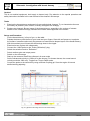
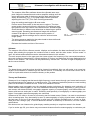
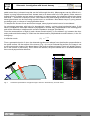
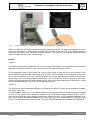
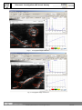
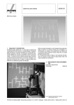
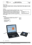
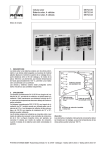
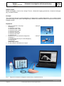
Ultrasonic investigation with breast dummy
TEAS
9.5.03
-00
Related Topics
Breast sonography, tumour size, benign Tumour, ultrasound imaging procedures, ultrasound echography, A-mode, B-mode.
Principle
This experiment shows a typical application of ultrasounds in medical diagnostics. On a realistic breast
dummy a benign tumour has to bee diagnosed, localized and measured with an ultrasound cross section
imaging method.
Equipment
1
1
1
Basic Set Ultrasonic echoscope
consisting of:
1x Ultrasonic echoscope
1x Ultrasonic probe 1 MHz
1x Ultrasonic probe 2 MHz
1x Ultrasonic test block
1x Ultrasonic cylinder set
1x Ultrasonic test plates
1x Ultrasonic gel
Extension set: medical ultrasonic diagnostics
consisting of:
1x simplified heart dummy for echocardiography
1x breast dummy
1x eye dummy
Additionally needed
PC with USB port, Windows XP or higher
Fig. 1:
13921-99
13921-04
Equipment for ultrasonic investigation of breast dummy, experimental set-up
www.phywe.com
P5950300
PHYWE Systeme GmbH & Co. KG © All rights reserved
1
TEAS
9.5.03
-00
Ultrasonic investigation with breast dummy
Caution!
This is not medical equipment, don’t apply to human body. Pay attention to the special operation and
safety instructions included in the user manual of the ultrasonic echoscope.
Tasks
1. Examine the breast dummy and search for any pathological changes. Try to characterize them as
accurately as possible (size, location, mobility, strength of the change).
2. Produce an ultrasonic B-scan image of the breast dummy, especially in the regions of interest.
Based on the ultrasound image, estimate the location and magnitude of the tumour.
Set-up and Procedure
Put the breast dummy, in front of you, on the table.
Palpate the dummy with the flat of your hand and your fingers. Start with soft pressure, to palpate
the surface. Increase continuously the pressure to examine also deeper layers of the breast dummy.
With this method you will locate two thickenings close to the nipple.
Examine he two regions with ultrasounds
Connect the 1 MHz probe to the “Probe (Reflection)” plug.
Switch the selection knob to “Reflection”.
Select medium gain and output power.
Switch on the echoscope
Start up the measure Ultra Echo software in A-scan mode
Set the ultrasound velocity to 1300 m/s for the dummy (For human tissues, the normal sound
velocity would be 1540 m/s). Toggle from Time to Depth scale.
Couple the probe to the test dummy using sufficient coupling gel. Scan the region of interest
determined by palpating.
Fig. 2: Typical A-scan image of tumour in breast dummy.
2
PHYWE Systeme GmbH & Co. KG © All rights reserved
P5950300
Ultrasonic investigation with breast dummy
-
-
-
-
The measure Ultra Echo software shows the reflected wave as a
peak. Adjust the transmitter and receiver amplifier settings until the
upper and bottom side of the tumour and the back wall echo can
clearly be detected. (Fig.: 2) The peak shape can be optimised
adjusting the time-TGC (Time Gain Control) parameters.
Keep the settings and toggle to B-scan mode.
Perform some linear scans in the two tumour regions. The ultrasound probe is scanned uniformly, with plenty of coupling gel and
light pressure over the surface of the breast model. Several images
should be taken in different scan directions to determine an optimal
scanning path. Recording an ultrasound image with sufficient
contrast of all objects of interest requires some practice. If
necessary, the device settings can be varied to achieve better
image quality.
Too strong pressure applied to the probe leads to tissue deformations and induces incorrect results.
Estimate the location and size of the tumour.
TEAS
9.5.03
-00
Fig. 3:
Scan lines for tumour 1
and tumour 2
Software
The measure Ultra Echo software records, displays and evaluates the data transferred from the echoscope. After starting the program the measure mode is active and the main screen “A-Scan mode” is
open. All actions and evaluations can be selected and started in this window.
The main screen shows in the upper part the A-scan signal, the frequency of the connected transducer,
the mode (reflection/ transmission). Actual positions of cursors (red and green line) are displayed at the
bottom of the window. The cursors can be positioned by mouse click. The time of flight is displayed under the cursor buttons.
Note
The breast dummy and the probes should be cleaned immediately after use with water or a normal detergent. Dried residues of ultrasonic gel are hard to remove. If necessary use a soft brush. Never use alcohol or liquids with solvents to clean the dummy or the probes.
Theory and Evaluation
Ultrasound is an imaging test that sends high-frequency sound waves through your breast and converts
the reflected waves into images on a viewing screen. The ultrasound technician places a sound-emitting
probe on the breast to conduct the test. There is no radiation involved.
Mammography and sonography are the standard imaging techniques for detection and evaluation of
breast disease. Mammography is the most established screening modality. Especially in young women
and women with dense breasts, sonography appears superior to mammography, and differentiation between solid tumours and cysts is easier. Sensitivity and specificity of sonography or mammography are
higher if sonography and mammography are combined
Ultrasound is not used on its own as a screening test for breast cancer. Rather, it is used to complement
other screening tests. If an abnormality is seen on mammography or felt by physical exam, ultrasound is
the best way to find out if the abnormality is solid (such as a benign fibroadenoma or cancer) or fluidfilled (such as a benign cyst). It cannot determine whether a solid lump is cancerous, nor can it detect
calcifications.
But doctors can use ultrasound to guide biopsy needles precisely to suspicious areas in the breast.
If you’re under age 30, your doctor may recommend ultrasound before mammography to evaluate a pal-
www.phywe.com
P5950300
PHYWE Systeme GmbH & Co. KG © All rights reserved
3
TEAS
9.5.03
-00
Ultrasonic investigation with breast dummy
pable breast lump (a breast lump that can be felt through the skin). Mammograms can be difficult to interpret in young women because their breasts tend to be dense and full of milk glands. (Older women’s
breasts tend to be fattier and are easier to evaluate.) In mammograms, this glandular tissue looks dense
and white — much like a cancerous tumor. Some doctors say that locating an abnormality in the midst of
dense gland tissue can be like finding a polar bear in a snowstorm. Most breast lumps in young women
are benign cysts, or clumps of normal glandular tissue.
To interpret the A-scan curves and B-scan images, some physics basics have to be considered.
An echoscope generates, with help of a piezoelectric ceramic, a short pulsed mechanical wave. If this
wave is coupled into solid state material it propagates in a linear way, perpendicular to the emitting piezo
and will be reflected on interfaces with acoustic impedance changes (boundaries).
From the measured time of flight (t) and a known sound velocity (c), the distance (s), between the ultrasonic probe and the boundary of a solid can be determined for perpendicular sound incidence, in the following way:
In reflection mode
(1) s =
c *t
2
From a geometrical point of view, the detected signals come always from the direction perpendicular to
the probe surface. As the surface of the dummy (Fig.: 4) is not a horizontal flat surface, the image is not
a real cross section image of the breast dummy but gives a deformed image. Press only slightly the probe on the surface of the dummy. Don’t deform the surface; the probe should look in a different direction
then normal perpendicular direction.
e
ob
r
P
Fig. 4:
4
Schematic representation and typical image of tumour in breast dummy in B-scan mode.
PHYWE Systeme GmbH & Co. KG © All rights reserved
P5950300
Ultrasonic investigation with breast dummy
Fig. 5:
TEAS
9.5.03
-00
A clinical instrument, clinical B-scan and schematic representation of a phase array scan.
Without a scanner, only dept measurements give significant results. For lateral information the beam
must be mechanically or electronically scanned over the area of interest. The most clinical ultrasound
equipment use phase array electronic scanning method. The probe is fixed and the ultrasound beam
scans the object by varying the incident angle.
Results
Tumour 1
The tumour was located by palpation at 2 cm at the right of the nipple. It is located near the surface and
has about a diameter of about 1-3 cm. The tumour can be easily moved in the tissue.
The sonographic image (Fig.6) shows the tumour region along a scan line from top to bottom of the
breast passing at the right side of the nipple (Fig. 3). The tumour is located 10 mm below the surface and
has an axial expansion of 24 mm. The lateral extent can not be accurately determined from the ultrasound image, as it is a hand guided B-mode image. The “continuous” line at the bottom of the screen
comes from the back wall echo of the breast dummy. Due to the higher absorption of the tumour, a clear
acoustic shadow appears in the wall echo of the breast dummy.
Tumour 2:
The 2nd tumour was located approximately 5 cm above the nipple. It shows similar properties to palpation as the 1st tumour.
The sonographic image (Fig. 7) was taken along a line starting just above the nipple and going straight
up (Fig. 3). The tumour is also located at a depth of 10 mm and has a length of about 26 mm. The tumour has an oval shape with a slightly inclined axis. Similar to tumour 1, the increased absorption of tumour tissue, causes an acoustic shadow on the back wall echo. The reflected signal, on the lower right
corner, is a multiple echoes of the back wall. This is an artefact and has non importance for diagnostics.
www.phywe.com
P5950300
PHYWE Systeme GmbH & Co. KG © All rights reserved
5
TEAS
9.5.03
-00
Ultrasonic investigation with breast dummy
Fig. 6:
Sonographic image of tumour 1.
Fig. 7: Sonographic image of tumour 2.
6
PHYWE Systeme GmbH & Co. KG © All rights reserved
P5950300