1
Contact Information
DRX UM InCELL HUNTER EPIGENETIC 1012V1
DiscoveRx Corporation
(World Wide Headquarters)
42501 Albrae Street
Fremont, CA 94538
United States
InCELL Hunter™ Epigenetic
Cell-Based Assay
t | 510.979.1415
f | 510.979.1650
toll-free | 866.448.4864
For chemiluminescent detection of protein levels
User Manual
KINOMEscan
A division of DiscoveRx
11180 Roselle Street, Suite D
San Diego, CA 92121
United States
t | 800.644.5687
f | 858.630.4600
DiscoveRx Corporation Ltd.
(Europe Headquarters)
Faraday Wharf, Holt Street
Aston Science Park
Birmingham, B7 4BB
United Kingdom
t | +44.121.260.6142
f | +44.121.260.6143
www.discoverx.com
© 2012 DiscoveRx Corporation, Fremont, CA 94538
All rights reserved.
Simple Solutions for Complex Biology
CONTENTS
NOTES:
LEGAL SECTION
PAGE 3
INTENDED USE
PAGE 4
TECHNOLOGY PRINCIPLE
PAGE 4
ASSAY OVERVIEW
PAGE 4
MATERIALS PROVIDED
PAGE 4
MATERIALS NOT PROVIDED (REQUIRED)
PAGE 5
STORING & REMOVING CRYOVIALS FROM LIQUID NITROGEN
PAGE 5
CELL THAWING AND PROPAGATION METHODS
PAGE 6
CELL FREEZING PROTOCOL
PAGE 7
TIPS FOR OPTIMAL PERFORMANCE
PAGE 7
ASSAY PROCEDURE — COMPOUND DOSE RESPONSE CURVE
PAGE 8
QUICK-START PROCEDURE: COMPOUND DOSE RESPONSE
PAGE 10
Read the entire product insert before beginning the assay.
For additional information or Technical Support, contact
DiscoveRx or visit www.discoverx.com.
2
11
QUICK-START PROCEDURE: COMPOUND DOSE RESPONSE
LEGAL SECTION
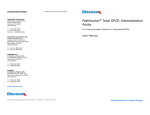
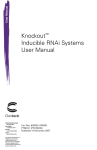
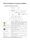
In a white-walled 384-well plate perform the following:
This product and/or its use is currently subject of pending U.S. and/or
foreign patents, patent applications. The right to use or practice the
inventions by using or propagating this product is granted solely in
connection with the use of appropriate Detection Reagents (protected under
trade secret) purchased from DiscoveRx® Corporation or its authorized
distributors.
Seed cells using 20 μL
Cell Plating Reagent
(Refer to target specific data sheet
for the cell numbers per well)
Incubate cells
overnight
@ 37°C
LIMITED USE LICENSE AGREEMENT
The designated cells and reagents purchased from DiscoveRx® are restricted in
their use. DiscoveRx® has developed an assay for Target Engagement ("InCELL
Hunter™ Assay") employing genetically modified cells ("Cells”) and detection
reagents (“Reagents”) (collectively referred to as “Materials”). The Cells and
Reagents are designed and optimized to be used together in the Assay. DiscoveRx
wishes to ensure that these Cells and Reagents are used properly and effectively.
By purchasing the Materials, you recognize and agree to the restrictions:
1)
The Materials cannot be transferred to third parties. Transfer to third parties
will be permitted only upon written request by Purchaser followed by subsequent
written approval by DiscoveRx®.
2)
Purchaser will not analyze the Reagents nor have them analyzed on Purchaser’s
behalf.
3)
Purchaser will use only the Reagents supplied by DiscoveRx® or an authorized
DiscoveRx® distributor for the Assays.
Induce cells with 5 µL compound (5X)
Incubate for
indicated time
and temperature
(refer target
specific data
sheet)
Add 30 µL InCELL Hunter™
Detection Reagent
Incubate for
30 min
@ RT
If the purchaser is not willing to accept the limitations of this limited use statement
and/or has any further questions regarding the rights conferred with purchase of
the Materials, please contact:
DiscoveRx® Corporation
Attn: Licensing Department
42501 Albrae Street
Fremont, CA 94538
tel | 510.979.1415 x104
[email protected]
For some products/cell lines, certain 3rd party gene specific patents may
be required to use the cell line. It is the purchaser's responsibility to
determine if such patents or other intellectual property rights are required.
Read
Chemiluminescent Signal
10
3
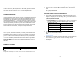
INTENDED USE
InCELL Hunter™ Epigenetic assay cell Line, when used in conjunction with a InCELL
Hunter™ Detection Kit (96-0002, 96-0002L or 96-0002XL), provides a cell based
assay to look at protein stability upon compound binding. The assay described in
this booklet have been validated for use in 384-well microplate formats.
6.
Remove InCELL Hunter™ cells from the incubator (previously plated on day 1).
7.
Transfer 5 µL of the compound (5X) to the plate as shown in the quick start
guide.
8.
Incubate cells with the compounds for the indicated times and temperatures in
the target specific data sheet.
DETECTION REAGENT PREPARATION AND ADDITION
TECHNOLOGY PRINCIPLE
1.
InCELL Hunter™ cell lines feature a novel in vivo application of the Enzyme Fragment Complementation (EFC) technology in which the β-galactosidase enzyme has
been split into two inactive fragments, the enhanced ProLabel (ePL) and the enzyme acceptor (EA). The platform measures compound-protein binding using a
novel β-galactosidase tag, ePL. In this system the protein of interest is tagged to
ePL. The cellular amount of protein is detected by the addition of enzyme acceptor
(EA) which complements with ePL to form a fully active β-galactosidase enzyme,
that can be quantitatively detected using the chemiluminescent substrate. The
amount of enzyme activity obtained is proportional to the amount of ePL tagged
protein present in the well. Cells expressing the designated fusion protein will be
tested for changes in protein levels, in response to compound treatment.
ASSAY OVERVIEW
To perform InCELL Hunter™ Assays, you will also need the InCELL Hunter™ Detection Kit (96-0002, 96-0002L or 96-0002XL) in order to generate the chemilumescent signal. Assays should be run using fresh, low-passage cells that have not
been allowed to reach confluency for more than 24 hours. Ideally cells should be
grown to 70-80% confluence. Following cell treatment, the assay is performed by
adding a working solution of InCELL Hunter™ Detection Reagents to the treated cells
in a no-mix, one-addition protocol. After addition of the detection reagents, the
samples must be read after 30 minutes. The Assay Procedure sections and
Quick Start Guide in this booklet contain detailed information about how to run
the assay.
2.
Prepare InCELL Hunter™ Detection Reagent as described in the InCELL Hunter™
Detection Kit Product Insert (Cat. #96-0002). Add 30 μL of prepared detection
reagent to the appropriate wells. DO NOT pipette up and down in the well
to mix or vortex/shake plates.
a) EA reagent: Ready to use, no preparation necessary.
b) Working Solution: Prepare Working Solution by mixing 1 part EA reagent
with 1 part Lysis Buffer and 4 parts Substrate Reagent. Gently mix the
components prior to use.
Component
Entire Plate (384 wells)
EA Reagent
2 mL
Lysis Buffer
2 mL
Substrate Reagent
8 mL
Incubate for 30 min at room temperature (23°C) in the dark before reading
the plate on a chemiluminescent reader.
NOTE:
For a list of readers and settings, please visit the URL below. http://www.discoverx.com/
instrument_chart.php
3.
Read samples on any standard luminescence plate reader. [Compound potencies
can be derived from a four-parameter nonlinear curve-fitting analysis.]
4)
Use GraphPad Prism® or other comparable program to plot your compound
dose response.
MATERIALS PROVIDED
Description
Storage
Liquid N2 (Vapor phase)
InCELL Hunter™ cells (2 vials)
NOTE:
Please refer to the datasheet of the InCELL Hunter™ cell line for detailed information on the
target that you are testing.
4
9
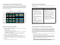
The steps outlined below provide the assay volumes and procedures for performing
assays using the InCELL Hunter™ Cell Lines and InCELL Hunter Detection Reagents
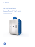
in a 384-well format. Although plate layouts and experimental designs may vary,
we recommend performing a 12-point dose curve for each compound using at least
duplicate wells for each dilution.
The following equipment and additional materials are required to perform InCELL
Hunter™ Assays:
1
Compound 1
Compound 3
Compound 5
Compound 7
Compound 9
Compound 11
Compound 13
Compound 15
3-fold serial
dilutions of
of compound
agonist
Low
2
3
4 5 6
7
8
High
No Compound
No
agonist
MATERIALS NOT PROVIDED (REQUIRED)
No Compound
No
agonist
ASSAY PROCEDURE - COMPOUND DOSE RESPONSE CURVE
Low
3-fold serial
dilutions of agonist
compound
High
9 10 11 12 13 14 15 16 17 18 19 20 21 22 23 24
A
B
C
D
E
F
G
H
I
J
K
L
M
N
O
P
Compound 2
Equipment
Materials
Single- and multichannel micro-pipettors
and pipette tips
InCELL Hunter™ Detection Kit
(DiscoveRx, Cat. #96-0002 series)
Tissue culture disposables and
plasticware (T25 and T75 flasks, etc.)
Revive™ Media
(DiscoveRx, Cat. #92-0016RM Series)
Cryogenic vials for freezing cells
V-bottom 384-well compound dilution
plates (DiscoveRx, Cat. #92-0011 or
similar)
PathHunter®select Cell Culture Kits
(DiscoveRx, Cat. #92-0018G Series)
Preserve™ Freezing Reagent
(DiscoveRx, Cat. #92-0017FR Series)
Disposable Reagent Reservoir (Thermo
Scientific, Cat. #8094 or similar)
Cell Detachment Reagent
(DiscoveRx, Cat. #92-0009)
Hemocytometer
White wall, clear bottom 384–well
microplates
(DiscoveRx, Cat. #92-0013 or similar)
PathHunter® Cell Plating (CP) Reagent
(DiscoveRx, Cat. #93-0563R Series)
Multimode or luminescence plate
reader (LumiLite; DiscoveRx Cat.
#75-0001 or similar)
Compound 4
Compound 6
Compound 8
Compound 10
Compound 12
Compound 14
Compound 16
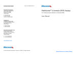
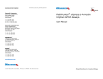
Figure 1. This plate map shows 12-point dose curves with 2 data points at each concentration
for 16 compounds per plate for a total of 160 compound dilutions per 384-well plate.
Control compound
NOTE:
# Refer target specific data sheet for additional details.
PROTOCOL: COMPOUND DOSE RESPONSE
STORING & REMOVING CRYOVIALS FROM LIQUID NITROGEN
1.
Harvest cells as follows from a confluent T25 or T75 flask using Cell Detachment
Reagent (DiscoveRx, Cat. #92-0009).
a) Remove medium.
b) Wash cells with 5 mL PBS and aspirate.
c) Add 0.5 mL Cell Detachment Reagent for a T25 flask, or 1 mL Cell Detachment Reagent for a T75 flask.
d) Place flask in the incubator for 5 minutes, or until cells have detached.
e) Add 3 mL of Cell Plating Reagent and transfer to a conical tube.
Cryovials are shipped in 2 vials on dry ice and contain 1.2 - 2.0 x 10⁶ cells/vial in
1 mL of freezing medium. Upon receipt, the vials can be stored for up to 2 weeks
at -80°C or transferred to the vapor phase of liquid nitrogen. DO NOT store at
–80°C for extended periods as this could result in significant loss in cell viability.
The following procedures are for the safe storage and removal of cryovials from
liquid nitrogen storage. A face shield, gloves, and a lab coat should be worn during
these procedures.
2.
Determine cell density using a hemocytometer.
3.
Using Cell Plating Reagent, adjust the volume of the suspension to achieve a
cell concentration per well indicated in the target specific data sheet.
4.
Transfer 20 μL of the cell suspension to each well of a 384-well white-walled
microplate.
5)
Incubate the plate overnight at 37°C/5% CO2.
8
1.
InCELL Hunter™ cells must arrive in a frozen state on dry ice. If cells arrive
thawed, do not proceed, contact technical support.
2.
Frozen cells must be transferred to either liquid nitrogen storage or a –80°C
freezer immediately upon arrival. If cells will be thawed and used within 24
hours, they can be stored temporarily at –80°C. For longer storage, place
vials in the vapor phase of liquid nitrogen storage.
NOTE:
# CRYOVIALS ARE NOT RATED FOR STORAGE IN THE LIQUID PHASE OF LIQUID
NITROGEN. CRYOVIALS SHOULD BE STORED IN THE VAPOR PHASE.
5
3.
When removing cryovials from liquid N2 storage, use tongs and place
immediately on dry ice in a covered container. Wait at least one minute for
any liquid nitrogen inside the vial to evaporate.
4.
Proceed with the thawing protocol in the following section.
CELL FREEZING PROTOCOL
The following procedures are for freezing cells from confluent T225 flasks. If smaller
flasks are used, adjust the volumes accordingly. Care should be taken in handling
to avoid contamination.
1.
Remove T225 flasks from incubator and place in tissue culture hood. Aspirate
the media from the flasks.
2.
Add 10 mL PBS into each T225 flask and swirl to rinse the cells. Aspirate PBS
from flask.
3.
Add 5 mL of Cell Detachment Reagent to the flask. Rock the flask back and
forth gently to ensure surface of the flask is covered. Return flask to the
incubator for 5 minutes, or until cells have detached.
4.
Remove the flask from the incubator and view under a microscope to confirm
that the cells are detached. Tap the edge of the flask to detach cells from the
surface, if necessary.
5.
Add 8-10 mL of Cell Plating Reagent (refer target specific data sheet) to each
T225 flask. Rinse the cells from the surface of the flask using the added
media. Remove the cells from the flask and transfer to a 50 mL conical tube.
(If necessary, add an additional 5 mL of reagent to the flask and rinse to
collect the remaining cells and transfer the additional volume to the 50 mL
conical tube). Remove 0.5 mL of the resuspended cells and count the cells
using a hemocytometer.
6.
Centrifuge the collected cells at 1500 x g for 5 minutes.
7.
After centrifugation, discard the supernatant. Resuspend the cell pellet in
Preserve™ Freezing Reagent. Based on the cell number obtained from Step 5,
dilute the resuspended cells to a concentration of 1.2– 2.0 x 10⁶ cells/mL.
8.
Transfer 1 mL cells to each 2 mL cryogenic tube. (Keep cells on ice during this
process and transfer to a cryogenic container pre-chilled at 4°C).
9.
Transfer tubes to –80°C and store overnight. Transfer tubes into the vapor
phase of a liquid nitrogen tank for long-term storage.
CELL THAWING AND PROPAGATION METHODS
The following procedures are for thawing cells in cryovials, seeding and expanding
the cells, and maintaining the cultures once the cells are expanded. Cells are free
of contamination prior to shipment and care should be taken in their handling to
avoid contaminating them. Face shield, gloves and a lab coat should be worn
during the thawing procedure.
1.
Pre-warm 5-10 mL Revive™ Medium (RM) in a 37°C water bath.
2.
Place the frozen cell vials briefly in a 37°C water bath under sterile conditions
until only small ice crystals remain and the cell pellet is almost completely
thawed (30 sec - 1 min). Caution: Longer incubation times may result in
cell death.
3.
Transfer thawed cells to a sterile 15 mL conical tube containing the 5-10 mL of
pre-warmed Revive medium. Centrifuge at 300 x g for 4 minutes to pellet
cells. Remove media.
4.
Resuspend cell pellet in 5 mLs of pre-warmed RM. Transfer cells to a T25 flask
and incubate for 24 hours at 37°C/5% CO2.
5.
After 24 hours, gently remove media (being careful not to disturb the cell
monolayer) and replace with 5 mLs of Cell Culture Media (refer to target
specific data sheet for specific Cell Culture Media requirements).
6.
Once the cells become >70% confluent in the T25 flask, trypsinize using Cell
detachment reagent and transfer the cells to a T75 flask containing 10-12 mLs
of cell culture media.
NOTE:
To maintain the logarithmic growth of the cells, cultures should be maintained in a subconfluent monolayer.
7.
8.
Passage the cells every 2–3 days, based on the doubling time of the cell line,
using the Cell Detachment Reagent. For routine passaging, prepare a 1:2
dilution of cells in a total volume of 10 mL of cell culture media. Transfer 5 mL
of the diluted cells to each of two new T75 flasks containing 10-12 mLs of cell
culture media.
The clone has been found to be stable for at least 10 passages with no significant
drop in assay window and EC50.
6
TIPS FOR OPTIMAL PERFORMANCE:
Cells must be maintained exactly as mentioned to maintain expression of
fusion protein.
Ideally cells should be maintained at approximately 70% confluence. Cells
should not be allowed to grow at confluence for more than 24 hours.
Allowing the cells to adhere and grow overnight prior to any assay is
recommended.
7
3.
When removing cryovials from liquid N2 storage, use tongs and place
immediately on dry ice in a covered container. Wait at least one minute for
any liquid nitrogen inside the vial to evaporate.
4.
Proceed with the thawing protocol in the following section.
CELL FREEZING PROTOCOL
The following procedures are for freezing cells from confluent T225 flasks. If smaller
flasks are used, adjust the volumes accordingly. Care should be taken in handling
to avoid contamination.
1.
Remove T225 flasks from incubator and place in tissue culture hood. Aspirate
the media from the flasks.
2.
Add 10 mL PBS into each T225 flask and swirl to rinse the cells. Aspirate PBS
from flask.
3.
Add 5 mL of Cell Detachment Reagent to the flask. Rock the flask back and
forth gently to ensure surface of the flask is covered. Return flask to the
incubator for 5 minutes, or until cells have detached.
4.
Remove the flask from the incubator and view under a microscope to confirm
that the cells are detached. Tap the edge of the flask to detach cells from the
surface, if necessary.
5.
Add 8-10 mL of Cell Plating Reagent (refer target specific data sheet) to each
T225 flask. Rinse the cells from the surface of the flask using the added
media. Remove the cells from the flask and transfer to a 50 mL conical tube.
(If necessary, add an additional 5 mL of reagent to the flask and rinse to
collect the remaining cells and transfer the additional volume to the 50 mL
conical tube). Remove 0.5 mL of the resuspended cells and count the cells
using a hemocytometer.
6.
Centrifuge the collected cells at 1500 x g for 5 minutes.
7.
After centrifugation, discard the supernatant. Resuspend the cell pellet in
Preserve™ Freezing Reagent. Based on the cell number obtained from Step 5,
dilute the resuspended cells to a concentration of 1.2– 2.0 x 10⁶ cells/mL.
8.
Transfer 1 mL cells to each 2 mL cryogenic tube. (Keep cells on ice during this
process and transfer to a cryogenic container pre-chilled at 4°C).
9.
Transfer tubes to –80°C and store overnight. Transfer tubes into the vapor
phase of a liquid nitrogen tank for long-term storage.
CELL THAWING AND PROPAGATION METHODS
The following procedures are for thawing cells in cryovials, seeding and expanding
the cells, and maintaining the cultures once the cells are expanded. Cells are free
of contamination prior to shipment and care should be taken in their handling to
avoid contaminating them. Face shield, gloves and a lab coat should be worn
during the thawing procedure.
1.
Pre-warm 5-10 mL Revive™ Medium (RM) in a 37°C water bath.
2.
Place the frozen cell vials briefly in a 37°C water bath under sterile conditions
until only small ice crystals remain and the cell pellet is almost completely
thawed (30 sec - 1 min). Caution: Longer incubation times may result in
cell death.
3.
Transfer thawed cells to a sterile 15 mL conical tube containing the 5-10 mL of
pre-warmed Revive medium. Centrifuge at 300 x g for 4 minutes to pellet
cells. Remove media.
4.
Resuspend cell pellet in 5 mLs of pre-warmed RM. Transfer cells to a T25 flask
and incubate for 24 hours at 37°C/5% CO2.
5.
After 24 hours, gently remove media (being careful not to disturb the cell
monolayer) and replace with 5 mLs of Cell Culture Media (refer to target
specific data sheet for specific Cell Culture Media requirements).
6.
Once the cells become >70% confluent in the T25 flask, trypsinize using Cell
detachment reagent and transfer the cells to a T75 flask containing 10-12 mLs
of cell culture media.
NOTE:
To maintain the logarithmic growth of the cells, cultures should be maintained in a subconfluent monolayer.
7.
8.
Passage the cells every 2–3 days, based on the doubling time of the cell line,
using the Cell Detachment Reagent. For routine passaging, prepare a 1:2
dilution of cells in a total volume of 10 mL of cell culture media. Transfer 5 mL
of the diluted cells to each of two new T75 flasks containing 10-12 mLs of cell
culture media.
The clone has been found to be stable for at least 10 passages with no significant
drop in assay window and EC50.
6
TIPS FOR OPTIMAL PERFORMANCE:
Cells must be maintained exactly as mentioned to maintain expression of
fusion protein.
Ideally cells should be maintained at approximately 70% confluence. Cells
should not be allowed to grow at confluence for more than 24 hours.
Allowing the cells to adhere and grow overnight prior to any assay is
recommended.
7
The steps outlined below provide the assay volumes and procedures for performing
assays using the InCELL Hunter™ Cell Lines and InCELL Hunter Detection Reagents
in a 384-well format. Although plate layouts and experimental designs may vary,
we recommend performing a 12-point dose curve for each compound using at least
duplicate wells for each dilution.
The following equipment and additional materials are required to perform InCELL
Hunter™ Assays:
1
Compound 1
Compound 3
Compound 5
Compound 7
Compound 9
Compound 11
Compound 13
Compound 15
3-fold serial
dilutions of
of compound
agonist
Low
2
3
4 5 6
7
8
High
No Compound
No
agonist
MATERIALS NOT PROVIDED (REQUIRED)
No Compound
No
agonist
ASSAY PROCEDURE - COMPOUND DOSE RESPONSE CURVE
Low
3-fold serial
dilutions of agonist
compound
High
9 10 11 12 13 14 15 16 17 18 19 20 21 22 23 24
A
B
C
D
E
F
G
H
I
J
K
L
M
N
O
P
Compound 2
Equipment
Materials
Single- and multichannel micro-pipettors
and pipette tips
InCELL Hunter™ Detection Kit
(DiscoveRx, Cat. #96-0002 series)
Tissue culture disposables and
plasticware (T25 and T75 flasks, etc.)
Revive™ Media
(DiscoveRx, Cat. #92-0016RM Series)
Cryogenic vials for freezing cells
V-bottom 384-well compound dilution
plates (DiscoveRx, Cat. #92-0011 or
similar)
PathHunter®select Cell Culture Kits
(DiscoveRx, Cat. #92-0018G Series)
Preserve™ Freezing Reagent
(DiscoveRx, Cat. #92-0017FR Series)
Disposable Reagent Reservoir (Thermo
Scientific, Cat. #8094 or similar)
Cell Detachment Reagent
(DiscoveRx, Cat. #92-0009)
Hemocytometer
White wall, clear bottom 384–well
microplates
(DiscoveRx, Cat. #92-0013 or similar)
PathHunter® Cell Plating (CP) Reagent
(DiscoveRx, Cat. #93-0563R Series)
Multimode or luminescence plate
reader (LumiLite; DiscoveRx Cat.
#75-0001 or similar)
Compound 4
Compound 6
Compound 8
Compound 10
Compound 12
Compound 14
Compound 16
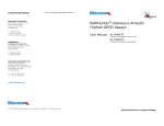
Figure 1. This plate map shows 12-point dose curves with 2 data points at each concentration
for 16 compounds per plate for a total of 160 compound dilutions per 384-well plate.
Control compound
NOTE:
# Refer target specific data sheet for additional details.
PROTOCOL: COMPOUND DOSE RESPONSE
STORING & REMOVING CRYOVIALS FROM LIQUID NITROGEN
1.
Harvest cells as follows from a confluent T25 or T75 flask using Cell Detachment
Reagent (DiscoveRx, Cat. #92-0009).
a) Remove medium.
b) Wash cells with 5 mL PBS and aspirate.
c) Add 0.5 mL Cell Detachment Reagent for a T25 flask, or 1 mL Cell Detachment Reagent for a T75 flask.
d) Place flask in the incubator for 5 minutes, or until cells have detached.
e) Add 3 mL of Cell Plating Reagent and transfer to a conical tube.
Cryovials are shipped in 2 vials on dry ice and contain 1.2 - 2.0 x 10⁶ cells/vial in
1 mL of freezing medium. Upon receipt, the vials can be stored for up to 2 weeks
at -80°C or transferred to the vapor phase of liquid nitrogen. DO NOT store at
–80°C for extended periods as this could result in significant loss in cell viability.
The following procedures are for the safe storage and removal of cryovials from
liquid nitrogen storage. A face shield, gloves, and a lab coat should be worn during
these procedures.
2.
Determine cell density using a hemocytometer.
3.
Using Cell Plating Reagent, adjust the volume of the suspension to achieve a
cell concentration per well indicated in the target specific data sheet.
4.
Transfer 20 μL of the cell suspension to each well of a 384-well white-walled
microplate.
5)
Incubate the plate overnight at 37°C/5% CO2.
8
1.
InCELL Hunter™ cells must arrive in a frozen state on dry ice. If cells arrive
thawed, do not proceed, contact technical support.
2.
Frozen cells must be transferred to either liquid nitrogen storage or a –80°C
freezer immediately upon arrival. If cells will be thawed and used within 24
hours, they can be stored temporarily at –80°C. For longer storage, place
vials in the vapor phase of liquid nitrogen storage.
NOTE:
# CRYOVIALS ARE NOT RATED FOR STORAGE IN THE LIQUID PHASE OF LIQUID
NITROGEN. CRYOVIALS SHOULD BE STORED IN THE VAPOR PHASE.
5
INTENDED USE
InCELL Hunter™ Epigenetic assay cell Line, when used in conjunction with a InCELL
Hunter™ Detection Kit (96-0002, 96-0002L or 96-0002XL), provides a cell based
assay to look at protein stability upon compound binding. The assay described in
this booklet have been validated for use in 384-well microplate formats.
6.
Remove InCELL Hunter™ cells from the incubator (previously plated on day 1).
7.
Transfer 5 µL of the compound (5X) to the plate as shown in the quick start
guide.
8.
Incubate cells with the compounds for the indicated times and temperatures in
the target specific data sheet.
DETECTION REAGENT PREPARATION AND ADDITION
TECHNOLOGY PRINCIPLE
1.
InCELL Hunter™ cell lines feature a novel in vivo application of the Enzyme Fragment Complementation (EFC) technology in which the β-galactosidase enzyme has
been split into two inactive fragments, the enhanced ProLabel (ePL) and the enzyme acceptor (EA). The platform measures compound-protein binding using a
novel β-galactosidase tag, ePL. In this system the protein of interest is tagged to
ePL. The cellular amount of protein is detected by the addition of enzyme acceptor
(EA) which complements with ePL to form a fully active β-galactosidase enzyme,
that can be quantitatively detected using the chemiluminescent substrate. The
amount of enzyme activity obtained is proportional to the amount of ePL tagged
protein present in the well. Cells expressing the designated fusion protein will be
tested for changes in protein levels, in response to compound treatment.
ASSAY OVERVIEW
To perform InCELL Hunter™ Assays, you will also need the InCELL Hunter™ Detection Kit (96-0002, 96-0002L or 96-0002XL) in order to generate the chemilumescent signal. Assays should be run using fresh, low-passage cells that have not
been allowed to reach confluency for more than 24 hours. Ideally cells should be
grown to 70-80% confluence. Following cell treatment, the assay is performed by
adding a working solution of InCELL Hunter™ Detection Reagents to the treated cells
in a no-mix, one-addition protocol. After addition of the detection reagents, the
samples must be read after 30 minutes. The Assay Procedure sections and
Quick Start Guide in this booklet contain detailed information about how to run
the assay.
2.
Prepare InCELL Hunter™ Detection Reagent as described in the InCELL Hunter™
Detection Kit Product Insert (Cat. #96-0002). Add 30 μL of prepared detection
reagent to the appropriate wells. DO NOT pipette up and down in the well
to mix or vortex/shake plates.
a) EA reagent: Ready to use, no preparation necessary.
b) Working Solution: Prepare Working Solution by mixing 1 part EA reagent
with 1 part Lysis Buffer and 4 parts Substrate Reagent. Gently mix the
components prior to use.
Component
Entire Plate (384 wells)
EA Reagent
2 mL
Lysis Buffer
2 mL
Substrate Reagent
8 mL
Incubate for 30 min at room temperature (23°C) in the dark before reading
the plate on a chemiluminescent reader.
NOTE:
For a list of readers and settings, please visit the URL below. http://www.discoverx.com/
instrument_chart.php
3.
Read samples on any standard luminescence plate reader. [Compound potencies
can be derived from a four-parameter nonlinear curve-fitting analysis.]
4)
Use GraphPad Prism® or other comparable program to plot your compound
dose response.
MATERIALS PROVIDED
Description
Storage
Liquid N2 (Vapor phase)
InCELL Hunter™ cells (2 vials)
NOTE:
Please refer to the datasheet of the InCELL Hunter™ cell line for detailed information on the
target that you are testing.
4
9
QUICK-START PROCEDURE: COMPOUND DOSE RESPONSE
LEGAL SECTION
In a white-walled 384-well plate perform the following:
This product and/or its use is currently subject of pending U.S. and/or
foreign patents, patent applications. The right to use or practice the
inventions by using or propagating this product is granted solely in
connection with the use of appropriate Detection Reagents (protected under
trade secret) purchased from DiscoveRx® Corporation or its authorized
distributors.
Seed cells using 20 μL
Cell Plating Reagent
(Refer to target specific data sheet
for the cell numbers per well)
Incubate cells
overnight
@ 37°C
LIMITED USE LICENSE AGREEMENT
The designated cells and reagents purchased from DiscoveRx® are restricted in
their use. DiscoveRx® has developed an assay for Target Engagement ("InCELL
Hunter™ Assay") employing genetically modified cells ("Cells”) and detection
reagents (“Reagents”) (collectively referred to as “Materials”). The Cells and
Reagents are designed and optimized to be used together in the Assay. DiscoveRx
wishes to ensure that these Cells and Reagents are used properly and effectively.
By purchasing the Materials, you recognize and agree to the restrictions:
1)
The Materials cannot be transferred to third parties. Transfer to third parties
will be permitted only upon written request by Purchaser followed by subsequent
written approval by DiscoveRx®.
2)
Purchaser will not analyze the Reagents nor have them analyzed on Purchaser’s
behalf.
3)
Purchaser will use only the Reagents supplied by DiscoveRx® or an authorized
DiscoveRx® distributor for the Assays.
Induce cells with 5 µL compound (5X)
Incubate for
indicated time
and temperature
(refer target
specific data
sheet)
Add 30 µL InCELL Hunter™
Detection Reagent
Incubate for
30 min
@ RT
If the purchaser is not willing to accept the limitations of this limited use statement
and/or has any further questions regarding the rights conferred with purchase of
the Materials, please contact:
DiscoveRx® Corporation
Attn: Licensing Department
42501 Albrae Street
Fremont, CA 94538
tel | 510.979.1415 x104
[email protected]
For some products/cell lines, certain 3rd party gene specific patents may
be required to use the cell line. It is the purchaser's responsibility to
determine if such patents or other intellectual property rights are required.
Read
Chemiluminescent Signal
10
3
CONTENTS
NOTES:
LEGAL SECTION
PAGE 3
INTENDED USE
PAGE 4
TECHNOLOGY PRINCIPLE
PAGE 4
ASSAY OVERVIEW
PAGE 4
MATERIALS PROVIDED
PAGE 4
MATERIALS NOT PROVIDED (REQUIRED)
PAGE 5
STORING & REMOVING CRYOVIALS FROM LIQUID NITROGEN
PAGE 5
CELL THAWING AND PROPAGATION METHODS
PAGE 6
CELL FREEZING PROTOCOL
PAGE 7
TIPS FOR OPTIMAL PERFORMANCE
PAGE 7
ASSAY PROCEDURE — COMPOUND DOSE RESPONSE CURVE
PAGE 8
QUICK-START PROCEDURE: COMPOUND DOSE RESPONSE
PAGE 10
Read the entire product insert before beginning the assay.
For additional information or Technical Support, contact
DiscoveRx or visit www.discoverx.com.
2
11
Contact Information
DRX UM InCELL HUNTER EPIGENETIC 1012V1
DiscoveRx Corporation
(World Wide Headquarters)
42501 Albrae Street
Fremont, CA 94538
United States
InCELL Hunter™ Epigenetic
Cell-Based Assay
t | 510.979.1415
f | 510.979.1650
toll-free | 866.448.4864
For chemiluminescent detection of protein levels
User Manual
KINOMEscan
A division of DiscoveRx
11180 Roselle Street, Suite D
San Diego, CA 92121
United States
t | 800.644.5687
f | 858.630.4600
DiscoveRx Corporation Ltd.
(Europe Headquarters)
Faraday Wharf, Holt Street
Aston Science Park
Birmingham, B7 4BB
United Kingdom
t | +44.121.260.6142
f | +44.121.260.6143
www.discoverx.com
© 2012 DiscoveRx Corporation, Fremont, CA 94538
All rights reserved.
Simple Solutions for Complex Biology