1
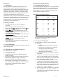
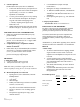
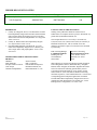
One Lambda, Inc.
21001 Kittridge Street, Canoga Park, CA 91303-2801 Tel: (818) 702-0042 Fax: (818) 702-6904
WEB:www.onelambda.com
P R O D U C T
I N S E R T
SSP Minor Histocompatibility Antigen Primer Sets
Catalog # BSSPMHAG
For Research Use Only. Not for use in diagnostic procedures.
STORE REAGENTS AT TEMPERATURE INDICATED ON PACKAGE. USE BEFORE EXPIRATION DATE.
Warning: Use only the D-mix packaged with this Liquid Bulk Primer Set.
DIRECTIONS FOR USE:
Formulation
Use the following formulation to prepare the reagents for your test:
1. Use 2 µl of primer set per PCR reaction.
2. Use 1 µl of the test sample and 7 µl D-mix per reaction.
3. Use .05 µl at 5 units/µl of recombinant Taq polymerase per reaction.
4. Perform standard PCR with the Micro SSP™ PCR program (OLI-1) as specified below.
INTENDED USE
DNA typing of the Minor Histocompatibility locus HA-1.
SUMMARY AND EXPLANATION
Successful marrow stem cell engraftment requires that bone
marrow (BM) donor-recipient pairs be matched for HLA-A,
-B, -DR, and –DQ. Nevertheless, among HLA genotypeidentical donor recipient pairs, acute graft-versus-host disease
(GvHD) occurs in 15%-35% of cases after allogeneic BM
transplantation, depending on the age of the recipient and the
amount of T-cell depletion of the graft. The occurrence of
acute GvHD has been ascribed to mismatching the minor
histocompatibility antigens (mHags) encoded by the HA-1
locus.1 The HA-1 locus is composed of two alleles, HA-1H
and HA-1R, which differ in two nucleotides, resulting in a
single amino acid substitution.
Disparities between mHags of otherwise HLA-identical bone
marrow donor and recipient pairs can elicit strong major
histocompatibility complex restricted cytotoxic and
proliferative T-cell responses which can be measured in vitro.2
It has been demonstrated that HLA-A*0201 molecules have a
high affinity for the HA-1H peptide, and this mHag
peptide/MHC complex is recognized by HA-1 specific
cytotoxic T-cells.3
PRINCIPLE
The PCR-SSP methodology is based on the principle that
completely matched oligonucleotide primers are more
efficiently used in amplifying a target sequence than a
mismatched oligonucleotide primer by recombinant Taq
polymerase. Primer pairs are designed to have perfect matches
only with a single allele or group of alleles. Under strictly
controlled PCR conditions, perfectly matched primer pairs
Rev 2
BSSPMHAG_PI.DOC
result in the amplification of target sequences (i.e., a positive
result) while mismatched primer pairs do not result in
amplification (i.e., a negative result). After the PCR process,
the amplified DNA fragments are separated by agarose gel
electrophoresis and visualized by staining with ethidium
bromide and exposure to ultraviolet light. Interpretation of
PCR-SSP results is based on the presence or absence of
specific amplified DNA fragment. Since the amplification
during the PCR reaction may be adversely affected by various
factors (pipetting errors, poor DNA quality, presence of
inhibitors, etc.), an internal control primer pair is included in
every PCR reaction. The control primer pair amplifies a
conserved region of the human β-globin gene, which is present
in all DNA samples and is used to verify the integrity of the
PCR reaction. In the presence of a positive typing band
(specific amplification of an HA-1 allele), the product of the
internal control primer pair may be weak or absent due to the
differences in concentration and melting temperatures between
the specific primer pairs and the internal control primer pair.
The amplified DNA fragments of the specific HLA primer
pairs are smaller than the product of the internal control primer
pair, but larger than the diffuse, unincorporated primer band.
Thus, a positive reaction for a specific HA-1 allele is
visualized on the gel as an amplified DNA fragment between
the internal control product band and the unincorporated
primer band (see Results Section).
Page 1 of 4
REAGENTS
A. Identification
The SSP Minor Histocompatibility Antigen Primer Sets
provide sequence-specific oligonucleotide primers for
amplification of alleles and the human β-globin gene by the
polymerase chain reaction (PCR) and specially formulated
dNTP buffer mix (Primer Set D-mix). The internal control
PCR product amplified from the human β-globin gene is the
most likely contaminating PCR product due to its
amplification in every test reaction. The amount of each primer
is adjusted for optimal amplification of 100 ng of sample DNA
when used in conjunction with the Primer Set D-mix, the
prescribed amount of recombinant Taq polymerase, and the
PCR reaction profile detailed below.
B. Warning or Caution
1. For research use only. Not for use in diagnostic
procedures.
2. Limit refreezing of primers no more than twice.
3. Pipettes used for Post-PCR manipulations should not be
used for Pre-PCR manipulations.
4. Biohazard Warning: The ethidium bromide used for
staining of DNA is a potential carcinogen. Always wear
gloves when handling stained gels.
5. Biohazard Warning: All blood products should be treated
as potentially infectious.
6. Caution: Wear UV-blocking eye protection, and do not
view UV light source directly when viewing or
photographing gels.
7. Aliquot D-mix into convenient to use volumes. Do not
refreeze more than twice.
C. Storage Instructions
Store reagents at temperature indicated on package. Use before
printed expiration date.
D. Purification for Treatment Required for Use
See “Directions for Use” below.
E. Instability Indications
1. Any primer set with a vial that is damaged should be
considered unusable.
2. If salts have precipitated out of the solution in the D-mix
aliquots during shipping or storage, re-dissolve by
extended vortexing at room temperature (20-25°C).
3. D-mix aliquots, upon thawing at room temperature (20 25°C), should be pink to light purple in color. Any D-mix
aliquot without the specified coloration should be
considered unusable.
Rev 2
BSSPMHAG_PI.DOC
INSTRUMENT REQUIREMENTS
1. Programming the Thermocycler
The following program is designed for use only on the PerkinElmer 9600 or 9700 thermocyclers. If you have a different
make and/or model thermocycler, you may need to adapt the
programming to your machine. Program your thermocycler
before starting the “Directions for Use.”
Micro SSP™ PCR Program (OLI-1):
# of
Cycles
Step
Temp
(°C)
Time
(sec)
1
1
2
96
63
130
60
9
1
2
96
63
10
60
20
1
2
3
96
59
72
10
50
30
End
1
4
---
For specific thermocycler information, refer to the
manufacturer’s user manual.
2. 2.5% Agarose Gel Preparation
(for Micro SSP™ Gel System, OLI Cat. #MGS108)
a. To set up:
• Slide the locking pin on the base to the open
position.
• Insert the gel box into the base by sliding the
locking pin into the locked position.
• Use the leveling bubble and the three height
adjustable legs to level the base.
b. Orient and fully insert the 14 gel combs into the gel
comb holder.
c. To 100 ml of 1x Tris Borate EDTA buffer (1xTBE)
with 0.5 µg/ml ethidium bromide (in a 500 ml glass
bottle), add 2.5 g electrophoresis grade agarose. Heat
until a homogeneous solution is formed.
d. Add 30 ml of the gel solution to the gel box. Make
sure the agarose covers the entire surface evenly by
tilting the gel box back and forth immediately after
the gel solution is added. Quickly place the gel comb
holder on the filled gel box by matching the color
coding. Allow to set for 15 minutes.
e. Remove the gel combs by lifting the gel comb holder
while holding the base. Add 10 ml of 1X TBE
containing 0.5 µg/ml ethidium bromide evenly across
the gel to fill every well.
Page 2 of 4
3.
Gel Electrophoresis
(for Micro SSP™ Gel System, OLI Cat. #MGS108)
a. Transfer each PCR reaction (10 µl) in sequence to the
2.5% agarose gel. Make sure to transfer all samples in
the proper sequence. (No addition of electrophoresis
dye is necessary.) Use of an 8- or 12-channel
Pipetman® is recommended.
b. Cover the gel box with the gel box cover by matching
the color-coded sides. Electrophorese the samples at
140 - 150 volts until the red tracking dye has
migrated about 0.5 cm into the gel (approximately
3 -5 minutes, depending on the agarose used).
Remove the cover.
c. Slide the locking pin on the base to the open position
and remove the gel box. Transfer the gel box to a UV
transilluminator. Photograph the completed gel.
SPECIMEN COLLECTION AND PREPARATION
1. DNA can be purified from human leukocytes by any
preferred method.
2. The DNA sample to be used for PCR-SSP analysis should
be re-suspended in sterile water or in 10 mM Tris-HCl,
pH 8.0 - 9.0 at a concentration of 25 - 200 ng/µl with the
A260/A280 ratio of 1.65 - 1.80.
3. Samples should not be re-suspended in solutions
containing chelating agents, such as EDTA, above 0.5
mM in concentration.
4. DNA samples may be used immediately after isolation or
stored at -20° C or below for extended periods of time
(over 1 year) with no adverse effects on results.
5. DNA samples should be shipped at 4° C or below to
preserve their integrity during transport.
PROCEDURE
A. Materials Provided
1. 2 vials, 60 µl primer (2µl per test) each
2. 1000 µl Primer Set D-mix (7 µl per test)
Note: The volumes provided are slightly more than the
amount required for testing. This is to account for inadvertent
losses which may result from pipetting.
B. Materials Required, but Not Provided
• Pipetting devices, such as Gilson® P20, Gilson®
P200 Pipetman®
• Disposable pipette tips
• Vortex mixer
• Microcentrifuge
• PCR tray microtube storage rack and cover (Robbins
Scientific, Cat #1044-39-5)
• Thermocycler for PCR with 96-well format and
tray/retainer for 0.2 ml thin-walled reaction tubes
• Hot plate or microwave oven for heating agarose
solution
• Electrophoresis apparatus/power supply (150V
minimum capacity)
Rev 2
BSSPMHAG_PI.DOC
•
•
•
•
UV transilluminator (example: Fotodyne
FOTO/UV®21)
Photographic or image documentation system
1x TBE buffer (89mM Tris Borate; 2 mM disodium
EDTA, pH 8.0) with 0.5 µg/ml ethidium bromide or
5XTBE Buffer with Ethidium bromide (OLI Cat. #
5XTBE100)
Electrophoresis grade agarose (e.g., FMC Seakem®
LE)
C. Step-by-Step Procedures
Test Procedure Refer to “Directions for Use”.
LIMITATIONS OF THE PROCEDURE
PCR-SSP is a dynamic process requiring highly controlled
conditions to insure discriminatory amplification. The
procedure provided this product must be strictly followed.
The extracted sample DNA provides the template for the
specific amplification process and, thus, must have its
concentration and purity within the ranges specified in the
procedure.
All instruments (e.g., PCR machine, pipetting devices) must be
calibrated according to the manufacturer’s recommendations.
RESULTS
A. Data Analysis
1. An internal control band (slower-migrating) should
always be visible in negative wells (except in the
negative control well) as a control for successful
amplification. Non-amplification of any well can void
test results.
2. A faster-migrating, positive typing band will be
observed on the electrophoretic gel if a specific mHag
gene was amplified during PCR. This indicates a
positive test result.
3. The internal control band may be weak or absent in
positive wells.
4. A positive band indicates the presence of the allele
identified in the table on the next page (Primer
Recognition Sites).
B. Gel Interpretation
Positive
Reaction
Negative
Reaction
Nonamplification
Well
Internal Control Band
Positive Typing
Primer Band
Page 3 of 4
PRIMER RECOGNITION SITES*
Alleles
HA-1H (Histidine)
5’ Recognition Site
345ACACT349
3’ Recognition Site
500CTGCA504
Approx. Size Base Pairs
190
HA-1R (Arginine)
345ACACT349
500TTGCG504
190
*Based on the KIAA0223 cDNA Sequence Information, Gene Bank Accession Number D86976.
REFERENCES
1. Goulmy E, Schipper R, Pool J, et al. Mismatches of minor
histocompatibility antigens between HLA-identical donors
and recipients and the development of graft-versus-host
disease after bone marrow transplantation. New Engl Med
1996: 334:281-5.
2. Goulmy E., Human minor histocompatibility antigens.
Curr Opin Immunol 1996: 8:75-81.
3. Den Haan JMM, Meadows LM, Wang W, et al. The
minor histocompatibility antigen HA-1: a diallelic gene
with a single amino acid polymorphism. Science 1998:
279:1054-7.
TRADEMARKS USED IN THIS DOCUMENT/
PRODUCT
ARMS™
Zeneca Limited
FMC SeaKem®
FMC Corporation
Fotodyne FOTO/UVX®21
FOTODYNE Incorporated
Gene-Amp®
Hoffmann-LaRoche, Inc
Gilson® and Pipetman®
Rainin Instrument Co., Inc.
Rev 2
BSSPMHAG_PI.DOC
PATENTS USED IN THIS DOCUMENT
Nothing in this publication should be construed as an
authorization or an implicit license to practice PCR under any
patents held by Hoffmann-LaRoche, Inc.
Gene-Amp PCR Process is covered by U.S. Patent Nos.
4,683,202, 4,683,195, 4,800,159 and 4,965,188 owned by F.
Hoffmann-La Roche, Inc. The use of PCR for in vitro
diagnostic procedures requires a license that can be obtained
by contacting:
PCR Licensing Manager
F. Hoffmann-LaRoche
Building 222/350
CH-4500 Basel
Switzerland
Director of Licensing
Roche Molecular Systems
1145 Atlantic Avenue
Alameda, CA 94501
USA
SSP technology is licensed from Zeneca Limited, through its
Zeneca Diagnostics business, Blacklands Way, Abingdon
Business Park, Abingdon, Oxfordshire, England OX14 IDY
and covered under the following patents held by Zeneca
Corporation: European Patent No. 0 332 435 B1, United
States Patent No. 5,595,890,entitled “Method of detecting
nucleotide sequences,” and Canadian Patent No. 1323592, and
corresponding patents and patent applications worldwide.
The SSP Minor Histocompatibility Antigen Primer Sets are
manufactured and distributed by One Lambda, Inc., 21001
Kittridge Street, Canoga Park, CA 91303, USA.
Page 4 of 4