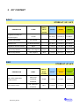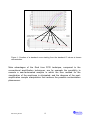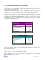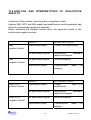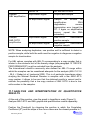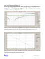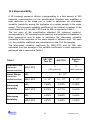
1
User manual REALQUALITY RS-AML1-ETO code RQ-S59 Kit for identification and quantification of the t(8;21)(q22;q22) translocation RQ-S59-48_EN.doc 1. 1.1 PRODUCT INFORMATION 3 Intended Use 3 2. KIT CONTENT 4 3. STORAGE AND STABILITY OF THE REAGENTS 5 4. PRECAUTIONS FOR USE 5 5 SAFETY RULES 5.1 General safety rules 5.2 Safety rules about the kit 6. HATA! YER İŞARETİ TANIMLANMAMIŞ. Hata! Yer işareti tanımlanmamış. 7 MATERIALS REQUIRED, BUT NOT PROVIDED 8 6.1 Reagents 8 6.2 Instruments 8 6.3 Materials 8 7. INTRODUCTION 9 8. TEST PRINCIPLE 11 9. PRODUCT DESCRIPTION 13 10. COLLECTION, MANIPULATION AND PRE-TREATMENT OF THE SAMPLES 14 11. PROTOCOL 144 11.1 RNA EXTRACTION 144 11.2 RETROTRANSCRIPTION (RT) FOR cDNA SYNTHESIS 154 11.3 INSTRUMENT PROGRAMMING 11.3.1 Creation of thermal protocol 11.3.2 Plate Setup 166 166 166 1 RQ-S59-48_EN.doc 11.4 QUALITATIVE ANALYSIS PROTOCOL 177 11.5 QUANTITATIVE ANALYSIS PROTOCOL 188 11.6 ANALYSIS AND INTERPRETATION OF QUALITATIVE RESULTS 199 11.7 ANALYSIS AND INTERPRETATION OF QUANTITATIVE RESULTS 220 11.8 NORMALIZATION AND QUANTIFICATION OF MRD 222 11.9 TROUBLESHOOTING 223 12 DEVICE LIMITATIONS 245 13 DEVICE PERFORMANCES 245 13.1 Analytical specificity 245 13.2 Analytical sensitivity: detection limit Hata! Yer işareti tanımlanmamış.5 13.3 Analytical sensitivity: linearity Hata! Yer işareti tanımlanmamış.5 13.4 Reproducability 246 13.5 Diagnostic specificity 267 13.6 Diagnostic sensitivity 267 13.7 Accuracy 267 14. REFERENCES 278 14.1 Useful website 248 15. RELATED PRODUCTS 289 RQ-S59-48_EN.doc 2 1. PRODUCT INFORMATION 1.1 Intended Use The REALQUALITY RS-AML1-ETO is an IVD for identification and quantification of the t(8;21)(q22;q22) translocation, that involves AML1 and ETO genes, by amplification of the c-DNA in the regions of the AML1 gene on chromosome 21q22 and of the ETO gene on chromosome 8q22. If used together with the REALQUALITY RQ-AML1-ETO STANDARD code RQ-60-ST kit, it allows the quantification of the AML1-ETO transcript present in the sample. The Real time PCR amplification method is used in this kit, starting from c-DNA obtained by reverse transcription of the RNA extracted from human samples. This in vitro diagnostic test is an auxiliary device for diagnosis and monitoring of clinical pathologies as the Acute Myeloid Leukaemia (AML). It is recommended to use this kit as indicated in the instructions herein. This manual refers to the following product: REALQUALITY RS-AML1-ETO Kit for identification and quantification of the t(8;21)(q22;q22) translocation, by Real time PCR. This product is in accordance with 98/79/CE Directive (Annex III) regarding the in vitro medical diagnostic devices IVD (CE mark). Contains all the reagents needed for Real time amplification. Code RQ-S59-48 RQ-S59-96 Product REALQUALITY RS-AML1-ETO REALQUALITY RS-AML1-ETO 3 PKG 48 test 96 test RQ-S59-48_EN.doc 2. KIT CONTENT BOX P STORE AT -30°/ -20°C TUBE (T) DESCRIPTION OR LID LABEL 24 test 48 test 96 test RT Mix 3 x 143 L 6 x 135 L 6 x 260 L RT enzyme 1 x 17 L 1 x 30 L 2 x 30 L 2X Q Real time Mix 2 x 350 L 3 X 450 L 6 X 450 L MgCl2 1 x 50 L 1 X 75 L 1 X 150 L COLOUR Reagents necessary for retrotranscription Reverse transcriptase Mastermix 2X Magnesium Chloride solution Primer and probe Mix AML1-ETO Oligomix Purple 1 x 27 L 2 x 27 L 4 x 27 L Primer and probe Mix ABL Oligomix Blue 1 x 27 L 2 x 27 L 4 x 27 L BAG STORE AT +2°/+8°C TUBE (T) DESCRIPTION OR LID LABEL 24 test 48 test 96 test Purple 1 x 30 L 1 x 60 L 1 x 110 L Blue 1 x 30 L 1 x 60 L 1 x 110 L COLOUR AML1-ETO translocation positive control ABL positive control RQ-S59-48_EN.doc AML1-ETO POSITIVE CONTROL ABL POSITIVE CONTROL 4 3. STORAGE AND STABILITY OF THE REAGENTS Each component of the kit should be stored according to the directions indicated on the label of each box. In particular: Box P Bag Store at -30°C/-20°C Store at +2°C/+8°C If stored at the recommended temperature, all test reagents are stable until their expiration date. The 2X Q Real time Mix and Oligomix are sensitive to physical state variations: it is recommended not to let the reagents undergo more than two freeze/thaw cycles. If the single test runs are limited to a small number of samples, it is recommended to aliquot the reagents. The 2X Q Real time Mix and Oligomix contain fluorescent molecules: it is recommended to store these reagents away from direct light. 4. PRECAUTIONS FOR USE The kit must be used only as an IVD and handled by qualified technicians, who are well educated and trained in molecular quantitative biology techniques applied to diagnostics; Before starting the kit procedure, read carefully and completely the user manual; Keep the product away from heating sources; One must pay particular attention to the expiration date on the label of each box: do not use any part of the kit past the expiration date; The reagents present in the kit must be considered an undividable unit. Do not divide or use different reagents from other kits or lots; 5 RQ-S59-48_EN.doc All the reagents must be thawed at room temperature before use. It is recommended to do not vortex, but to mix the solutions by inverting the tube several times and then centrifuge them briefly. Prepare the reaction quickly at room temperature or work on ice or on a cooling block. In case of any doubts about the storage conditions, box integrity or method application, please contact AB ANALITICA’s technical support at: [email protected]. During nucleic acid amplification, the technician has to take the following special precautions: Use filter-tips; Store the biological samples, the extracted RNA, cDNA and positive controls included in the kit and all amplicons in a different area from where reverse transcription and amplification reagents are stored; Organize the work areas in different pre- and post-PCR units; do not share instrument and consumables (pipettes, tips, tubes, etc) between them; Change gloves frequently; Wash the bench surfaces with 5% Sodium Hypochlorite; Keep the RNA (just extracted or that will be stored at -30°C/-20°C or 80°C, according to the time required between extraction and reverse transcription) on ice during reverse transcription preparation. 5. SAFETY RULES 5.1 General safety rules Wear disposable gloves to handle reagents and clinical samples and wash hands at the end of the procedure; RQ-S59-48_EN.doc 6 Do not pipette by mouth; Since known diagnostic method cannot assure the absence of any infective agents, it is a good rule to consider every clinical sample as potentially infectious and handle it as such; All devices that come in contact with clinical samples should be considered as contaminated and disposed of as such. In case of accidental spilling of the samples, clean up with 10% Sodium Hypochlorite. The materials used to clean up should be disposed in special containers for contaminated products; Clinical samples, materials and contaminated products should be disposed after decontamination by: immerse in a solution of 5% Sodium Hypochlorite (1 volume of 5% Sodium Hypochlorite solution every 10 volumes of contaminated fluid) for 30 minutes; OR autoclave at 121°C for at least 2 hours (NOTE: do not autoclave solutions containing Sodium Hypochlorite!!). 5.2 Safety rules about the kit The risks for the use of this kit are related to the single components. Dangerous components: none. The Material Safety Data Sheet (MSDS) of the device is available upon request. 7 RQ-S59-48_EN.doc 6. MATERIALS REQUIRED, BUT NOT PROVIDED 6.1 Reagents Reagents for density gradient separation of mononucleate cells (Ficoll); RNA extraction reagents; Dnase- and Rnase-free sterile water; Distilled water; REALQUALITY RQ-AML1-ETO STANDARD code RQ-60-ST (for quantitative analysis). 6.2 Instruments Laminar flow cabinet (its use is recommended while preparing the amplification mix to avoid contamination; it would be recommended to use another similar laminar flow cabinet to add the extracted DNA and standard solutions); Micropipettes (range: 0.5-10 µL; 2-20 µL; 10-100 µL; 20-200 µL; 1001000 µL); Microcentrifuge (max 12-14,000 rpm); Plate centrifuge (optional); Thermalcycler (for reverse transcription); Real time amplification instrument. The kit was standardized on Applied Biosystems 7500 Fast Dx, 7300, StepOnePlus Real-Time PCR System (Applied Biosystems); the kit can be utilized on instruments that use 25 μL of reaction volume and can detect the FAM fluorescence correctly. For more information on instrument compatibility of the kit, please contact AB ANALITICA’s technical support. 6.3 Materials Talc-free disposable gloves; Disposable sterile filter-tips (range: 0.5-10 µL; 2-20 µL; 10-100 µL; 20200 µL; 100-1000 µL); Sterile DNase and RNase free tubes (for reverse transcription); 96-well plates for Real time PCR and optical adhesive film or 0.1-0.2 mL tubes with optical caps. RQ-S59-48_EN.doc 8 7. INTRODUCTION The study of leukemia and lymphoma initiate the comprehension of the cellular and molecular mechanisms at the basis of many neoplastic pathologies, by means of easy availability and sampling of leukemic malignant cells present in the blood, unlike the ones present in solid tumors that are sometimes difficult to be collected without using invasive samplings techniques. The t(8;21)(q22;q22) translocation leads to the fusion between AML1 gene on the 21q22 chromosome and ETO gene on the 8q22 chromosome: the break point in the AML1 gene is situated between exon 5 and exon 6, while the one in ETO gene is upstream of exon 2. The resulting fusion protein is constitute by the N-terminal domain of the DNA binding of AML1, an essential transcription factor for the haematopoiesis, and about all the ETO protein, that works as a co-repressor for a variety of transcription factors. The t(8;21)(q22;q22) translocation is a rearrangement usually linked to the Acute Myeloid Leukemia (AML), in particular, it is reported in almost 15% of all AML and almost 40% of FAB M2 subtype of AML. AML incidence is approximately 3.5 cases per 100.000 people per year. AML can manifest at all ages, but the frequency increases in older people. Molecular analysis of t(8;21)(q22;q22) translocation is very important since this rearrangement is linked to a positive prognosis and, in particular, to a good response to some therapeutic agents, as cytosine arabinoside (Bloomfield et al. Cancer Res 1998). The translocation detection can be done at the molecular level with a RT-PCR which consists of total RNA extraction from starting samples, its retrotranscription in cDNA and then amplification of the regions of interest. This detection gives useful information for diagnosis and prognosis of these types of leukemia, but first of all, it allows the monitoring of the Minimal Residual Disease (MRD), which has important repercussion on therapy. MRD is the number of neoplastic cells, which is below the identifiable level with standard cytomorphologic techniques, present in the organism of the patient affected by leukemia during the different phases of chemotherapy. Even if an aggressive chemotherapy makes progress in leukemia treatment, a significant percentage of cases relapse at different time point from the treatment beginning. A disease relapse means that an amount of therapy9 RQ-S59-48_EN.doc resistant residual cells were persistent, which characteristics were for a long time unknown since the available analysis techniques had limited sensibility. PCR has initiate new possibilities for a more extended and efficient application of MMR monitoring (van Dongen et al., 1998; Baccarani et al., 2006). RQ-S59-48_EN.doc 10 8. TEST PRINCIPLE PCR method (Polymerase Chain Reaction) was the first method of DNA amplification described in literature (Saiki RK et al., 1985). It can be defined as an in vitro amplification reaction of a specific part of DNA (target sequence) by a thermostable DNA polymerase. This technique was shown to be a valid and versatile molecular biology instrument: its’ application contributed to a more efficient study of new genes and their expression and it brought to a revolution in the laboratory diagnostic and forensic medicine field. The REAL TIME PCR technology represents an advancement of the basic PCR technique; it allows to measure the number of DNA molecules amplified during the exponential amplification phase. The amplicon monitoring is essentially based on the labeling of the primers and probes, or of the amplicons themselves, with fluorescent molecules. In the first case, the Fluorescence Resonance Energy Transfer (FRET) among the two fluorophores, or other mechanisms which lead to fluorescence emission and involve a fluorophore and a non-fluorescent quencher (molecular beacon, scorpion primer, etc) are used. The mechanism that determines the fluorescence emission is based on the presence of a quencher molecule, located in proximity of a reporter molecule, that blocks the fluorescence emission by the reporter. When the quencher is separated from the reporter, the latter emits fluorescence. The real time detection of such fluorescence is accomplished by means of a thermalcycler equipped with fluorescence detector. Each amplification cycle will release a certain amount of fluorescence into the solution; the cycle at which the amplification generates the minimal amount of fluorescence needed to overcome the basal noise threshold is called the “cycle threshold” (Ct). By intuition, the higher the starting concentration of the target nucleic acid, the sooner the amplification will reach the cycle threshold. The Ct value is reached during the exponential phase of the amplification reaction, where the amplification reaction is still proportional to the number of target molecules in the solution. The starting concentration of the unknown samples is determined by comparison of the Ct value of each sample with the Ct value of a standard curve acquired at known concentration (Figure 1). 11 RQ-S59-48_EN.doc Figure 1: Creation of a standard curve starting from the standard Ct values at known concentration. Main advantages of the Real time PCR technique, compared to the conventional amplification techniques, are for example the possibility to execute a semi-automated analysis in which the time needed for the visualization of the amplicons is eliminated; and the absence of the postamplification sample manipulation that reduces the possible contamination phenomena.. RQ-S59-48_EN.doc 12 9. PRODUCT DESCRIPTION The REALQUALITY RS-AML1-ETO code RQ-S59 kit is an IVD for identification of t(8;21)(q22;q22) translocation. If used together with REALQUALITY RQ-AML1-ETO STANDARD code RQ60-ST kit, it allows the quantification of the number of AML1-ETO transcripts present in the sample, normalized to the number of ABL housekeeping gene. Such quantification is obtained by the construction of a four-point standard curve for AML1-ETO and, in parallel, for ABL genes. In fact, starting from the c-DNA itself but in a separated PCR reaction, the sequence of housekeeping gene ABL is amplified; such amplification, in addition to be a mark for the quantification and normalization, allows to evaluate both the extracted RNA suitability, the following retro-transcription reaction and the possible presence of PCR reaction inhibitors. This valid tool helps the user to recognize possible false negative results. ABL gene amplification is made separately from AML1-ETO amplification, because experimental evidences demonstrate that a competition between the two targets can occur in samples with a low number of AML1-ETO transcripts, and sometimes it ends up to heavily disadvantage the specific translocation transcript amplification, with the possibility to have false negative. The given positive controls are made by a DNA fragment with the target region of interest, and they are not dangerous for the user. For amplification reaction preparation, a ready-to-use Mastermix is supplied, containing all the reagents needed, with the exception of the Oligomix, and in particular: ROX™, an inert colorant in which the fluorescence does not undergo changes during the amplification reaction; it is used to normalize eventual differences between wells caused by artifacts from pipetting errors or instrument limitations; dUTP/UNG system prevents contaminations from previous amplifications, since it removes residual uracil incorporated in the molecule of single or double stranded DNA. NOTE: This kit was developed in accordance with the Europe Against Cancer guidelines (Gabert et al., Leukemia 2003) and in accordance with recent international recommendations (Branford et al., Leukemia 2006). 13 RQ-S59-48_EN.doc 10. COLLECTION, MANIPULATION AND PRE-TREATMENT OF THE SAMPLES AML1-ETO translocation identification is performed starting from whole peripheral or bone marrow blood. Sample collection must follow all the usual sterility precautions. Blood must be treated with EDTA. Other anticoagulation agents, as heparin, are strong inhibitors of TAQ polymerase and so they could alter the efficiency of the amplification reaction. Fresh blood can be stored at +2°C/+8°C if processed in 4h time after the withdrawal: thus it is necessary to proceed with the mononucleate cells separation by density gradient centrifugation (Ficoll – reagent not included in the kit). From the pellet of lymphocytes obtained as such, is possible to proceed directly with RNA extraction; otherwise, the cell pellet may be conserved at -80°C until the RNA extraction, better if preserved in a buffer containing RNAse inhibitors (i.e.: RLT buffer -QUIAGEN- or Trizol). 11. PROTOCOL 11.1 RNA EXTRACTION The product was validated using the RNeasy Mini kit (QIAGEN, Hilden, Germany). For use, follow the user manual of the manufacturer. However, the device is suitable for most diffused manual or automatic RNA extraction methods. For any further information on device compatibility with different extraction methods, please contact AB ANALITICA’s technical support. Please follow the instructions below regarding the quantity of RNA to be used for the reverse transcription reaction (about 1µg). RQ-S59-48_EN.doc 14 11.2 RETROTRANSCRIPTION (RT) FOR cDNA SYNTHESIS Attention: before starting the reverse transcription procedures, it is recommended to use an ice container and to thaw one or more RT Mix aliquotes (depending on the number of analyzed samples). Once thawed, the RT Mix must be mixed well by inverting the tube several times (do not vortex!), then centrifuge briefly and store on ice until use. For each sample, add to a sterile DNase and Rnase free tube (see paragraph 6.3): Extracted RNA 5 μL* *NOTE: The 5 μL amount indicates the avoid volume available for the reaction. The appropriate amount of RNA to be used for reverse transcription is about 1 μg; if the RNA is more concentrated, it is necessary to dilute it properly with DEPC H2O. Insert the tubes in the thermalcycler and program the following thermal profile: 1 cycle 70°C, 10 min Next, place the tubes on ice immediately, for at least 5 minutes. Add 14.5 μL of RT-mix and 0.5 μL of RT Enzyme, mix by pipetting, centrifuge briefly and incubate in a thermalcycler programmed as below: 1 cycle 20°C, 10 min 42°C, 45 min 99°C, 3 min 4°C, 5 min Once the cycle ends, add 30 μL of sterile water to each retrotranscribed sample. The diluted cDNA can be stored at +2°C/+8°C for short period of time (maximum one week) or at -20°C/-30°C for longer periods of time. NOTE: this cDNA can be used both for amplification of the AML1-ETO translocation and of the ABL housekeeping gene. 15 RQ-S59-48_EN.doc 11.3 INSTRUMENT PROGRAMMING 11.3.1 Creation of thermal protocol Set the following thermal profile: Cycle Repeats Step Time (°C) UNG Activation 1 1 1 2:00 50.0 Taq Activation 2 1 1 10:00 95.0 Amplification cycles 3 45 1 00:15 95.0 2* 01:00 60.0 * Fluorescence collection step 11.3.2 Plate Setup Mark the grid of the new plate with the position of the negative control (NTC), standards (STD) and samples (Unknown), making sure the position is the same as on the plate and identify each sample with its name. NOTE: it is recommended to amplify both samples and positive/negative controls and standards in duplicate. For the quantitative protocol, define the dilution of the AML1-ETO and ABL standard in the interval from 102 to 105 copies. Select and activate the FAM fluorophore, and NONE as quencher. Pay attention that, for the instruments that require it, the detection of the fluorescence of the fluorophore ROX™ corresponds to each position. ROX™ is an inert colorant in which the fluorescence does not undergo changes during the amplification reaction; on instruments that use ROX (Applied Biosystems, Stratagene, etc.), it is used to normalize eventual differences between wells caused by artifacts due to pipetting errors or instrument limitations. Record, where required, that the final reaction volume is 25 μL. RQ-S59-48_EN.doc 16 11.4 QUALITATIVE ANALYSIS PROTOCOL Once thawed, mix the reagents by inverting the tubes several times (do not vortex!), then centrifuge briefly. Prepare the reaction mix rapidly at room temperature or work on ice or on a cooling block. Try, when possible, to work in areas away from direct light. Prepare, as described below, a mix sufficient for all the samples to be tested, counting also for the positive and negative control, in the latter H2O must be added instead of DNA and, when calculating the volume, consider an excess of at least one reaction volume. AML1-ETO Amplification Reagent 2X Q Real time Mix Oligomix AML1-ETO MgCl2 H2O Total Volume 1 Rx 12.5 μL 1.0 μL 0.5 μL 6.0 μL 20.0 μL ABL Amplification Reagent 2X Q Real time Mix Oligomix ABL MgCl2 H2O Total Volume 1 Rx 12.5 μL 1.0 μL 0.5 μL 6.0 μL 20.0 μL Mix by inverting the tubes, in which the mix was prepared in, several times. Then centrifuge briefly. Pipette 20 μL of the mix in each well of the plate. Add to each well, in the correct positions, 5 μL of cDNA or 5 μL of positive control DNA, provided in the kit. 17 RQ-S59-48_EN.doc Always amplify a negative control together with the samples to be analyzed (add sterile water instead of extracted DNA to the corresponding well) both for the AML1-ETO and the ABL Mix. Hermetically seal the plate by using an optical adhesive film or appropriate sealer. Make sure that there are no air bubbles in the bottom of the wells and/or centrifuge the plate at 4000 rpm for about 1 minute. Load the plate on the instrument paying attention to position it correctly and start the amplification cycle. 11.5 QUANTITATIVE ANALYSIS PROTOCOL The quantitative analysis can be performed by using REALQUALITY RQAML1-ETO STANDARD code RQ-60-ST. It is recommended to amplify the samples and standards in duplicate. Follow the instructions reported in the previous paragraph to prepare a reaction mix sufficient for the standard curve acquisition and quantification of tested samples both for the AML1-ETO and the ABL gene. A negative amplification control must be included in the plate, in which the H2O is added instead of cDNA. Aliquot 20 μL of the mix in each well of the plate. Add 5 μL of cDNA to each well or 5 μL of each quantification standard dilution in the corresponding positions of the plate. Hermetically seal the plate by using an optical adhesive film or appropriate sealer. Make sure that there are no air bubbles in the bottom of the wells and/or centrifuge the plate at 4000 rpm for about 1 minute. Load the plate on the instrument paying attention to position it correctly and start the amplification cycle. RQ-S59-48_EN.doc 18 11.6 ANALYSIS AND INTERPRETATION OF QUALITATIVE RESULTS At the end of the reaction, view the graph in logarithmic scale. Analyze AML1-ETO and ABL graphs and amplification results separately and follow the interpretation pattern as indicated. Before analyzing the samples results check the expected results of the positive and negative controls. ABL positive Control ABL negative Control AML1-ETO positive Control AML1-ETO negative Control RESULT INTERPRETATION Amplification signal Correct ABL amplification No amplification signal Amplification problems, repeat the analysis No amplification signal No contamination Amplification signal Contamination, analysis RESULT INTERPRETATION Amplification signal Correct amplification No amplification signal Amplification problems, repeat the analysis No amplification signal No contamination Amplification signal Contamination, analysis 19 repeat the AML1-ETO repeat RQ-S59-48_EN.doc the Sample: ABL amplification Sample: AML1-ETO amplification RESULTS Amplification signal INTERPRETATION Amplificable sample No amplification signal Sample not suitable for amplification: RNA degradation; reverse transcription error (repeat the RT: if again no amplification occurs, repeat the RNA extraction) AML1-ETO translocation positive sample AML1-ETO translocation negative sample Amplification signal No amplification signal NOTE: When analyzing duplicates, one positive well is sufficient to detect a positive sample; while both the wells must be negative to diagnose a negative sample for translocation. For ABL values, samples with ABL Ct corresponding to a copy number that is inferior to the minimum limit of the linearity range (see paragraph 13 “DEVICE PERFORMANCES”) must be excluded from the analysis. The International scientific community also defined an ABL Ct range within which the samples can be considered adequate for the analysis (ABL Ct 21.8 - 29.4, J Gabert et al. Leukemia 2003). This is of particular importance when studying the Minimal Residual Disease in samples with a low AML1-ETO copy number: it allows to be sure that the obtained results is correct and to exclude the possibility that a low copy number of AML1-ETO is due to low cells number in the samples. 11.7 ANALYSIS AND INTERPRETATION OF QUANTITATIVE RESULTS At the end of the reaction, view the graph in logarithmic scale (Figure 2). Analyze AML1-ETO and ABL graphs and quantification results separately. Position the Threshold, by choosing the position in which the Correlation Coefficient (R2) and the slope of the curve values are the closest possible to 1 RQ-S59-48_EN.doc 20 and -3.33, respectively (Figure 3). Results are considered acceptable, when the efficiency of the amplification is between 85 – 110% (slope approximately -3.75 - -3.10) and the Correlation Coefficient value is not less than 0.990. Figure 2: Post run data analysis: amplification graph displayed in logarithmic scale on the Applied Biosystems 7300 Real Time PCR System with SDS software version 1.2.3. Figure 3: Post run data analysis, standard curve on the Applied Biosystems 7300 Real Time PCR System with SDS software version 1.2.3. 21 RQ-S59-48_EN.doc 11.8 NORMALIZATION AND QUANTIFICATION OF MINIMAL RESIDUAL DISEASE The AML1-ETO and ABL standard curves allow to transform the Ct values obtained for unknown samples in AML1-ETO (AML1-ETOCN) and ABL (ABLCN) copy numbers. The normalized copy number (NCN) of the AML1-ETO transcript is defined as the ratio between the AML1-ETOCN and ABLCN copy number: NCNAML1-ETO = AML1-ETOCN / ABLCN The Minimal Residual Disease (MRD) is expressed as the ratio between the AML1-ETO normalized copy number at the follow-up (FUP) and the AML1ETO normalized copy number at the time of the diagnosis (DX): MRD = (AML1-ETOCN / ABLCN)FUP / (AML1-ETOCN / ABLCN)DX In case of the follow-up samples, the sensitivity (SENSv) of the experiment must be calculated in order to determine the clinical validity of the obtained results: SENSv = ABLCN,DX / (ABLCN,FUP x AML1-ETODX) 11.9 TROUBLESHOOTING Absence of amplification signal for positive controls/standards and samples The instrument was not programmed correctly – Repeat the amplification taking care of the instrument programming; pay particular attention to the thermal profile, the selected fluorophores and the correspondence between the plate protocol and the plate itself. The amplification mix was not prepared correctly – Prepare a new amplification mix making sure to follow the instructions reported in paragraph 11.4. RQ-S59-48_EN.doc 22 The kit was not stored properly or it was used past the expiration date – Check both the storage conditions and the expiration date reported on the label; use a new kit if needed. Weak amplification signal intensity for positive controls/standards Positive controls/standards were stored incorrectly and have degraded – Store the positive controls/standards correctly at +2°C/+8°C, and make sure that they do not undergo any freeze/thaw cycle as well; – Do not use the positive controls/standards past the expiration date. The reaction mix does not function correctly – Make sure to store the 2X Q Real time Mix and Oligomix correctly at -20°C/-30°C. Avoid unnecessary freeze/thaw cycles. Amplification signal of ABL very delayed or absent in the extracted samples The extracted RNA is not suitable for amplification or a problem may have occurred during the reverse transcription reaction and the amplification reaction was inhibited – Make sure to perform the extraction of nucleic acids correctly – If an extraction method uses wash steps with solutions containing Ethanol, make sure no Ethanol residue remains in the DNA sample; – Use the extraction methods suggested in paragraph 11.1; – During reverse transcription reaction, check that the Reverse Transcriptase enzyme has been pipette in the tube, by looking for the drop formed by the enzyme on the tube wall after being added to the mix, then centrifuge briefly; – Follow standard procedures for minimizing RNA degradation: use RNase free plastic lab wear and work on ice during the reverse transcription reaction. For any further problems, please contact AB ANALITICA’s technical support at: [email protected], fax (+39) 049-8709510, or tel. (+39) 049761698). 23 RQ-S59-48_EN.doc 12. DEVICE LIMITATIONS The kit can have reduced performances if: The clinical sample is not suitable for this analysis (sampling and/or storage error, i.e. blood treated with anticoagulants other than EDTA, like heparin, etc.); The starting samples were not treated as the modality and times indicated in paragraph 10; The kit was not stored correctly. 13. DEVICE PERFORMANCES 13.1 Analytical specificity The analitical specificity of the REALQUALITY RS-AML1-ETO code RQ-S59 kit is guaranteed by an accurate and specific selection of primers and probes, and also by the use of stringent amplification conditions. Moreover, the alignment of primers and probes in the most important databanks shows the absence of non-specific pairing. 13.2 Analytical sensitivity: detection limit The analytical sensitivity limit of REALQUALITY RS-AML1-ETO kit was defined by the amplification test of 8 dilution replicates from the last point of the quantification standard conducted in at least 3 consecutive runs. The results are reported in Table 1. 13.3 Analytical sensitivity: linearity The linearity of the assay was determined using a quantification standard panel. The analysis of the data obtained by linear regression have demonstrated that the test presents for AML1-ETO and for ABL a linear response for all the panel point (R2>0.99). The results of the analysis are reported in Table 1. RQ-S59-48_EN.doc 24 13.4 Reproducibility A 50 transcript copies/L dilution (corresponding to a final amount of 250 transcript copies/reaction) of the quantification standard was amplified in eight replicates in the same run, in order to determine the intra-assay variability (variability among the replicates of a certain sample in the same assay). The intra-assay variability coefficient of the method, in respect to the Cycle threshold (Ct), for AML1-ETO and for ABL is reported in Table 1. The last point of the quantification standard (20 transcript copies/L corresponding to 100 transcript copies/reaction) was amplified in duplicates in three consecutive runs in order to determine the inter-assay variability (variability of the replicates of the same sample in different runs). For each run, the variability coefficient was calculated from the Ct of the samples. The inter-assay variability coefficient for AML1-ETO and for ABL was calculated from the average of the variable coefficients in each experiment performed and is reported in Table 1. ABI 7500 Fast Dx Table 1 Detection Limit (transcript copies/reaction) Linear Range (transcript copies/reaction) Intra-assay Variability Inter-assay Variability AML1-ETO ABI 7300 StepOne Plus 5 5 (96% positivity) (100% positivity) 10 – 105 ABL AML1-ETO 5 - 106 10 – 106 5 - 106 ABL 0.508% 0.633% 0.345% AML1-ETO 0.375% 0.771% 0.441% ABL 0.916% 0.669% 0.454% AML1-ETO 0.273% 0.499% 0.536% 25 RQ-S59-48_EN.doc 13.4 Diagnostic specificity A significant number of samples negative for AML1-ETO translocation were tested simultaneously with the REALQUALITY RS-AML1-ETO kit and another CE IVD or reference method. From the obtained results, the diagnostic specificity of this device was calculated to be 97.37%. 13.5 Diagnostic sensitivity A significant number of samples positive for the AML1-ETO translocation were tested simultaneously with the REALQUALITY RS-AML1-ETO kit and another CE IVD or reference method. From the obtained results, the diagnostic sensitivity of this device was calculated to be 100%. 13.6 Accuracy This value was calculated by the number of correct amplifications over the total number of executed amplifications. The REALQUALITY RS-AML1-ETO device has an accuracy of 98.33%. RQ-S59-48_EN.doc 26 14. REFERENCES Baccarani M, Saglio G, Goldman J et al. Blood 15;108(6):1809-20, 2006. Bloomfield CD, Lawrence D, Byrd JC et al. Cancer Res 58; 4173-4179, 1998. Branford S, Cross NC, Hochhaus A et al. Lukemia 20(11):1925-30, 2006. Gabert J, Beillard E, et al. Leukemia 17(12):2318-57, 2003. Saiki RK, S Scharf, F Faloona, KB Mullis, GT Horn, HA Erlich and N Arnheim, Science 230, 1350-1354, 1985. Van der Velden VH et al. Leukemia 17, 1013-1034, 2003. van Dongen JJ et al. Lancet 352, 1731-1738, 1998. 14.1 Useful websites www.hematology.org www.bloodjournal.org www.bloodline.net www.haematologica.it www.il-st-acad-sci.org/data6.html http://medocs.ucdavis.edu/IMD/420A/dib/index.htm http://web.tiscali.it/ematologia www.ematologia-italia.net/frame_b.htm 27 RQ-S59-48_EN.doc 15. RELATED PRODUCTS REALQUALITY RQ-AML1-ETO STANDARD: Ready-to-use quantification standard for t(8;21)(q22;q22) translocation transcript. Code RQ-60-ST Product REALQUALITY RQ-AML1-ETO STANDARD RQ-S59-48_EN.doc 28 PKG 4 x 60µL (AML1-ETO) 4 x 60µL (ABL) AB ANALITICA srl Via Svizzera 16 - 35127 PADOVA, (ITALY) Tel +39 049 761698 - Fax +39 049 8709510 e-mail: [email protected]