1
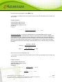
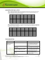
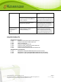
EPIGENTEK Complete Solutions for Epigenetics MethylFlash™ Methylated DNA Quantification Kit (Colorimetric) Base Catalog # P-1034 PLEASE READ THIS ENTIRE USER GUIDE BEFORE USE Uses: The MethylFlashTM Methylated DNA Quantification Kit (Colorimetric) is suitable for detecting global DNA methylation status using DNA isolated from any species such as mammalians, plants, fungi, bacteria, and viruses in a variety of forms including, but not limited to, cultured cells, fresh and frozen tissues, paraffin-embedded tissues, plasma/serum samples, and body fluid samples. Input DNA: The amount of DNA for each assay can be 50 to 200 ng. For optimal quantification, the input DNA amount should be 100 ng, as methylated DNA varies from tissue to tissue and can be less than 1% of total DNA in some species. Starting Materials: Starting materials can include various tissue or cell samples such as cells from flask or microplate cultured cells, fresh and frozen tissues, paraffin-embedded tissues, plasma/serum samples, body fluid samples, etc. Internal Control: Both negative and positive DNA controls are provided in this kit. A standard curve can be performed (range: 1 to 10 ng) or a single quantity of methylated DNA can be used as a positive control. Because global methylation can vary from tissue to tissue, and from normal and diseased states, it is advised to run replicate samples to ensure that the signal generated is validated. This kit will allow the user to quantify an absolute amount of methylated DNA and determine the relative methylation states of two different DNA samples. Precautions: To avoid cross-contamination, carefully pipette the sample or solution into the strip wells. Use aerosol-barrier pipette tips and always change pipette tips between liquid transfers. Wear gloves throughout the entire procedure. In case of contact between gloves and sample, change gloves immediately. 110 Bi County Blvd. Ste. 122, Farmingdale, NY 11735 Tel: 1-877-374-4368 ■ Fax: 1-718-484-3956 ■ E-mail: [email protected] ■ Web: www.epigentek.com © Epigentek Group Inc. All rights reserved. Products are for research use only. Page 1 Printed 2014-06-19 P-1034 EPIGENTEK Complete Solutions for Epigenetics Kit Contents Component 48 Assays Cat. #P-1034-48 96 Assays Cat. #P-1034-96 Storage Upon Receipt ME1 (10X Wash Buffer) 14 ml 28 ml 4°C ME2 (Binding Solution) 5 ml 10 ml RT ME3 (Negative Control, 20 µg/ml)* 10 µl 20 µl –20°C ME4 (Positive Control, 20 µg/ml)* 10 µl 20 µl –20°C ME5 (Capture Antibody, 1000 µg/ml* 4 µl 8 µl 4°C ME6 (Detection Antibody, 400 µg/ml)* 8 µl 16 µl –20°C ME7 (Enhancer Solution)* 8 µl 16 µl –20°C ME8 (Developer Solution) 5 ml 10 ml 4°C ME9 (Stop Solution) 5 ml 10 ml RT 8-Well Assay Strips (With Frame) 6 12 4°C User Guide 1 1 RT * Spin the solution down to the bottom prior to use. Note: The ME3 Negative Control is an unmethylated polynucleotide containing 50% of cytosine. The ME4 Positive Control is a methylated polynucleotide containing 50% of 5-methylcytosine. SHIPPING & STORAGE The kit is shipped in two parts: the first part at ambient room temperature and the second part on frozen ice packs at 4°C. Upon receipt: (1) Store ME3, ME4, ME6, and ME7 at –20°C away from light; (2) Store ME1, ME5, ME8, and 8-Well Assay Strips at 4°C away from light; (3) Store remaining components (ME2 and ME9) at room temperature away from light. All components of the kit are stable for 6 months from the date of shipment, when stored properly. Note: Check ME1 (10X Wash Buffer) contains salt precipitates before use. If so, briefly warm at room temperature or 37°C and shake the buffer until the salts are re-dissolved. MATERIALS REQUIRED BUT NOT SUPPLIED Adjustable pipette, multi-channel recommended Aerosol resistant pipette tips Microplate reader capable of reading absorbance at 450 nm 1.5 ml microcentrifuge tubes Incubator for 37°C incubation 110 Bi County Blvd. Ste. 122, Farmingdale, NY 11735 Tel: 1-877-374-4368 ■ Fax: 1-718-484-3956 ■ E-mail: [email protected] ■ Web: www.epigentek.com © Epigentek Group Inc. All rights reserved. Products are for research use only. Page 2 Printed 2014-06-19 P-1034 EPIGENTEK Complete Solutions for Epigenetics Plate seal or Parafilm M Distilled water 1X TE buffer pH 7.5 to 8.0 Isolated DNA of interest GENERAL PRODUCT INFORMATION Quality Control: Each lot of the MethylFlash™ Methylated DNA Quantification Kit (Colorimetric) is tested against predetermined specifications to ensure consistent product quality. Epigentek guarantees the performance of all products in the manner described in our product instructions. Product Warranty: If this product does not meet your expectations, simply contact our technical support unit or your regional distributor. We also encourage you to contact us if you have any suggestions about product performance or new applications and techniques. Safety: Suitable lab coat, disposable gloves, and proper eye protection are required when working with this product. Product Updates: Epigentek reserves the right to change or modify any product to enhance its performance and design. The information in this User Guide is subject to change at any time without notice. Thus, only use the User Guide that was supplied with the kit when using that kit. Usage Limitation: The MethylFlash™ Methylated DNA Quantification Kit (Colorimetric) is for research use only and is not intended for diagnostic or therapeutic applications. Intellectual Property: The MethylFlash™ Methylated DNA Quantification Kit (Colorimetric) and methods of use contain proprietary technologies by Epigentek. A BRIEF OVERVIEW DNA methylation occurs by the covalent addition of a methyl group at the 5-carbon of the cytosine ring by DNA methyltransferases, resulting in 5-methylcytosine (5-mC). In somatic cells, 5-mC is found almost exclusively in the context of paired symmetrical methylation of the dinucleotide CpG, whereas in embryonic stem (ES) cells, a substantial amount of 5-mC is also observed in non-CpG contexts. The biological importance of 5-mC as a major epigenetic modification in phenotype and gene expression has been recognized widely. For example, global decrease in 5-mC content (DNA hypomethylation) is likely caused by methyl-deficiency due to a variety of environmental influences, and has been proposed as a molecular marker in multiple biological processes such as cancer. It has been well demonstrated that the decrease in global DNA methylation is one of the most important characteristics of cancer. Thus, the quantification of 5-mC content or global methylation in cancer cells could provide very useful information for detection and analysis of this disease. Quite recently, a novel modified nucleotide, 5-hydroxymethylcytosine (5-hmC) has been detected to be abundant in mouse brain and embryonic stem cells. In mammals, it can be generated by oxidation of 5-methylcytosine, a reaction mediated by the Tet family of enzymes and Dnmt proteins. It is a hydroxylated and methylated form of cytosine. 110 Bi County Blvd. Ste. 122, Farmingdale, NY 11735 Tel: 1-877-374-4368 ■ Fax: 1-718-484-3956 ■ E-mail: [email protected] ■ Web: www.epigentek.com © Epigentek Group Inc. All rights reserved. Products are for research use only. Page 3 Printed 2014-06-19 P-1034 EPIGENTEK Complete Solutions for Epigenetics Unmethylated DNA Methylated DNA Hydroxymethylated DNA T-C-G-T-C-G-A-C-G T-mC-G-T-mC-G-A-mC-G T-hmC-G-T-hmC-G-A-hmC-G The broader functions of 5-hmC in epigenetics are still a mystery today. However, a line of evidence does show that 5-hmC plays a role in DNA methylation structures and patterns. Because of the presence of both 5-mC and 5-hmC in DNA with possibly different functions, it is important to determine the contents of these two modified nucleotides and their ratios in different cell types and in different compartments of the genome of mammalians. It is particularly important to identify that in healthy and diseased human cell/tissues, the epigenetic change at the DNA level is due to methylation or hydroxymethylation. Several chromatography-based techniques such as HPLC, TLC mass spectrometry are used for detecting 5-mC and 5-hmC. However, these methods are time consuming and have low throughput with high costs. To address this problem, Epigentek offers the MethylFlash™ Methylated DNA Quantification Kit (Colorimetric) to quantify 5-mC or methylated DNA. This kit is optimized for paired use with our MethylFlash™ Hydroxymethylated DNA Quantification Kit for simultaneously quantifying both methylated and hydroxymethylated DNA, or for quantifying methylated DNA by itself. The kit has the following advantages and features: Colorimetric assay with easy-to-follow steps for convenience and speed. The entire procedure can be finished within 4 hours. Innovative kit composition enables background signals to be extremely low, which eliminates the need for plate blocking and allows the assay to be simple, accurate, reliable, and consistent. High sensitivity, of which the detection limit can be as low as 0.2 ng of methylated DNA (methylation). Optimized antibody and enhancer solutions allow high specificity to 5-mC, with no cross-reactivity to unmethylated cytosine and no or negligible cross-reactivity to hydroxymethylcytosine within the indicated concentration range of the sample DNA. Universal positive and negative controls are included, which are suitable for quantifying methylated DNA from any species. Strip-well microplate format makes the assay flexible: manual or high throughput analysis. References 1. Robertson, K. D. Nat Rev Genet. 6: 597-610, 2005. 2. Kriaucionis, S. et al: Science. 324: 929-930, 2009. 3. Wyatt, G.R. et al: Biochem J. 55: 774-8, 1953. 4. Tahiliani, M. et al: Science. 324: 930-935, 2009. 5. Valinluck, V. et al: Nucleic Acids Res. 32: 4100-4108, 2004. 6. Valinluck, V. et al: Cancer Res. 67: 946-50, 2007. 7. Jin, S. G. et al: Nucleic Acids Res. 38: e125, 2010. 110 Bi County Blvd. Ste. 122, Farmingdale, NY 11735 Tel: 1-877-374-4368 ■ Fax: 1-718-484-3956 ■ E-mail: [email protected] ■ Web: www.epigentek.com © Epigentek Group Inc. All rights reserved. Products are for research use only. Page 4 Printed 2014-06-19 P-1034 EPIGENTEK Complete Solutions for Epigenetics PRINCIPLE & PROCEDURE The MethylFlashTM Methylated DNA Quantification Kit (Colorimetric) contains all reagents necessary for the quantification of global DNA methylation. In this assay, DNA is bound to strip wells that are specifically treated to have a high DNA affinity. The methylated fraction of DNA is detected using capture and detection antibodies and then quantified colorimetrically by reading the absorbance in a microplate spectrophotometer. The amount of methylated DNA is proportional to the OD intensity measured. 1.2 Methylated DNA OD450 nm 1 Unmethylated DNA 0.8 0.6 0.4 0.2 0 0 0.2 0.5 1 2 5 10 100 Input DNA (ng) Schematic procedure of the MethylFlash™ Methylated DNA Quantification Kit (Colorimetric) Demonstration of high sensitivity and specificity of methylated DNA detection achieved by the MethylFlash™ kit. Synthetic unmethylated DNA (contains 50% of cytosine) and methylated DNA (contains 50% of 5-methylcytosine) were added into the assay wells at different concentrations and then measured with the MethylFlash™ Methylated DNA Quantification Kit (Colorimetric). ASSAY PROTOCOL Starting Materials Input DNA Amount: DNA amount can range from 50 ng to 200 ng per reaction. An optimal amount is 100 ng per reaction. Starting DNA may be in water or in a buffer such as TE. DNA Isolation: You can use your method of choice for DNA isolation. Epigentek offers a series of genomic DNA isolation kits for your convenience. 110 Bi County Blvd. Ste. 122, Farmingdale, NY 11735 Tel: 1-877-374-4368 ■ Fax: 1-718-484-3956 ■ E-mail: [email protected] ■ Web: www.epigentek.com © Epigentek Group Inc. All rights reserved. Products are for research use only. Page 5 Printed 2014-06-19 P-1034 EPIGENTEK Complete Solutions for Epigenetics DNA Storage: Isolated genomic DNA can be stored at 4°C (short term) or –20°C (long term) until use. 1. Preparation of 1X Wash Buffer (ME1) 48 Assays Kit: Add 13 ml of ME1 10X Wash Buffer to 117 ml of distilled water (pH 7.2-7.5). 96 Assays Kit: Add 26 ml of ME1 10X Wash Buffer to 234 ml of distilled water (pH 7.2-7.5). Note: This Diluted ME1 1X Wash Buffer can now be stored at 4°C for up to six months. All other diluted solutions should be kept on ice at all times and should be discarded if not used within the same day. 2. Preparation of Diluted Positive Control (ME4) Single Point Control Preparation: Dilute ME4 Positive Control with 1X TE to 5 ng/µl (1 µl of ME4 + 3 µl of TE). Suggested Standard Curve Preparation: First, dilute ME4 to 10 ng/µl (5 µl of ME4 + 5 µl of 1X TE). Then, further prepare five different concentrations with the 10 ng/µl diluted ME4 and 1X TE into 0.5, 1, 2, 5, and 10 ng/µl according to the following dilution chart: Tube ME4 (10 ng/µl) 1X TE Resulting ME4 Concentration 1 1.0 µl 19.0 µl 0.5 ng/µl 2 1.0 µl 9.0 µl 1.0 ng/µl 3 1.0 µl 4.0 µl 2.0 ng/µl 4 2.5 µl 2.5 µl 5.0 ng/µl 5 4.0 µl 0.0 µl 10.0 ng/µl 3. DNA Binding a. Predetermine the number of strip wells required for your experiment. Carefully remove un-needed strip wells from the plate frame and place them back in the bag (seal the bag tightly and store at 4°C). b. Add 80 µl of ME2 Binding Solution to each well. c. Add 1 µl of ME3, 1 µl of Diluted ME4 (see note below), and 100 ng of your Sample DNA (1-8 µl) into the designated wells depicted in Table 1 or Table 2 . Mix solution by gently tilting from side to side or shaking the plate several times. Ensure the solution coats the bottom of the well evenly. Note: (1) For a single point control, add 1 µl of ME4 at a concentration of 5 ng/µl, as prepared in Step 2; for the standard curve, add 1 µl of Diluted ME4 at concentrations of 0.5 to 10 ng/µl (see the chart in Step 2). The final amounts should be 0.5, 1, 2, 5, and 10 ng per well. (2) For optimal binding, sample DNA volume added should not exceed 8 µl. (3) To ensure that ME3, Diluted ME4, and sample DNA are completely added into the wells, the pipette tip should be placed into the ME2 solution in the well and aspirated in/out 1-2 times. (4) Add replicate samples to the same column according to the suggested strip well set up in Table 1 or Table 2. d. Cover strip plate with plate seal or Parafilm M and incubate at 37°C for 90 min. 110 Bi County Blvd. Ste. 122, Farmingdale, NY 11735 Tel: 1-877-374-4368 ■ Fax: 1-718-484-3956 ■ E-mail: [email protected] ■ Web: www.epigentek.com © Epigentek Group Inc. All rights reserved. Products are for research use only. Page 6 Printed 2014-06-19 P-1034 EPIGENTEK Complete Solutions for Epigenetics e. Remove the binding reaction solution from each well. Wash each well three times with 150 µl of Diluted ME1 1X Wash Buffer. To wash the wells, pipette Diluted ME1 1X Wash Buffer into the wells then remove it from the wells and discard it. Repeat for a total of three washes. 4. Methylated DNA Capture a. Dilute ME5 (at 1:1000 dilution) with Diluted ME1. b. Add 50 µl of the Diluted ME5 to each well, then cover and incubate at room temperature for 60 min. c. Remove the Diluted ME5 solution from each well. d. Wash each well three times with 150 µl of Diluted ME1 1X Wash Buffer. e. Dilute ME6 (at 1:2000 dilution) with the Diluted ME1. f. Add 50 µl of the Diluted ME6 to each well, then cover and incubate at room temperature for 30 min. g. Remove the Diluted ME6 solution from each well. h. Wash each well four times with 150 µl of Diluted ME1 1X Wash Buffer. i. Dilute ME7 (at 1:5000 dilution) with the Diluted ME1. j. Add 50 µl of the Diluted ME7 to each well, then cover and incubate at room temperature for 30 min. k. Remove the Diluted ME7 solution from each well. l. Wash each well five times with 150 µl of Diluted ME1 1X Wash Buffer. 5. Signal Detection a. Add 100 µl of ME8 to each well and incubate at room temperature for 1 to 10 min away from light. Begin monitoring color change in the sample wells and control wells. The ME8 solution will turn blue in the presence of sufficient methylated DNA. b. Add 100 µl of ME9 to each well to stop enzyme reaction when color in the positive control wells turns medium blue. Mix the solution by gently shaking the frame and wait 1-2 min to allow the color reaction to be completely stopped. The color will change to yellow after adding ME9 and the absorbance should be read on a microplate reader at 450 nm within 2 to 15 min. Note: If the strip-well plate frame does not fit in the microplate reader, transfer the solution to a standard 96-well microplate. 6. 5-mC Calculation Relative Quantification: To determine the relative methylation status of two different DNA samples, simple calculation of percentage of 5-mC in total DNA can be carried out using the following formula: (Sample OD – ME3 OD) ÷ S 5-mC % = (ME4 OD – ME3 OD) x 2* ÷ P x 100% S is the amount of input sample DNA in ng. 110 Bi County Blvd. Ste. 122, Farmingdale, NY 11735 Tel: 1-877-374-4368 ■ Fax: 1-718-484-3956 ■ E-mail: [email protected] ■ Web: www.epigentek.com © Epigentek Group Inc. All rights reserved. Products are for research use only. Page 7 Printed 2014-06-19 P-1034 EPIGENTEK Complete Solutions for Epigenetics P is the amount of input positive control (ME4) in ng. * 2 is a factor to normalize 5-mC in the positive control to 100%, as the positive control contains only 50% of 5-mC. Example calculation: Average OD450 of ME3 is 0.075 Average OD450 of ME4 is 0.675 Average OD450 of Sample is 0.475 S is 100 ng P is 5 ng 5-mC % = (0.475 – 0.075) ÷ 100 x 100% = 1.67% (0.6750 – 0.075) x 2 ÷ 5 Absolute Quantification: To quantify the absolute amount of methylated DNA using an accurate calculation, first generate a standard curve and plot the OD values versus the amount of ME4 at each concentration point. Next, determine the slope (OD/ng) of the standard curve using linear regression (Microsoft Excel’s linear regression functions are suitable for such calculation) and the most linear part (at least 4 concentration points including 0 point ) of the standard curve for optimal slope calculation. Now calculate the amount and percentage of methylated DNA (5-mC) in total DNA using the following formulas: Sample OD – ME3 OD 5-mC (ng) = Slope x 2* 5-mC Amount (ng) 5-mC % = x 100% S S is the amount of input sample DNA in ng. * 2 is a factor to normalize 5-mC in the positive control to 100%, as the positive control contains only 50% of 5-mC. Example calculation: Average OD450 of ME3 is 0.075 Average OD450 of sample is 0.475 Slope is 0.12 OD/ng S is 100 ng 5-mC (ng) = 0.475 – 0.075 = 1.67 ng 0.12 x 2 1.67 5-mC % = 100 x 100% = 1.67% 110 Bi County Blvd. Ste. 122, Farmingdale, NY 11735 Tel: 1-877-374-4368 ■ Fax: 1-718-484-3956 ■ E-mail: [email protected] ■ Web: www.epigentek.com © Epigentek Group Inc. All rights reserved. Products are for research use only. Page 8 Printed 2014-06-19 P-1034 EPIGENTEK Complete Solutions for Epigenetics SUGGESTED STRIP WELL SETUP Table 1. Single Point Positive Control. The suggested strip-well plate setup using a single point positive control in a 48-assay format (for a 96-assay format, Strips 7 to 12 can be configured as Sample). The controls and samples can be measured in duplicate Well # A B C D E F G H Strip 1 ME3 ME4 Sample 1 Sample 1 Sample 2 Sample 2 Sample 3 Sample 3 Strip 2 ME3 ME4 Sample 4 Sample 4 Sample 5 Sample 5 Sample 6 Sample 6 Strip 3 Sample 7 Sample 7 Sample 8 Sample 8 Sample 9 Sample 9 Sample 10 Sample 10 Strip 4 Sample 11 Sample 11 Sample 12 Sample 12 Sample 13 Sample 13 Sample 14 Sample 14 Strip 5 Sample 15 Sample 15 Sample 16 Sample 16 Sample 17 Sample 17 Sample 18 Sample 18 Strip 6 Sample 19 Sample 19 Sample 20 Sample 20 Sample 21 Sample 21 Sample 22 Sample 22 Table 2. Standard Curve Preparation. The suggested strip-well plate setup for standard curve preparation in a 48-assay format (for a 96-assay format, Strips 7 to 12 can be configured as Sample). The controls and samples can be measured in duplicate. Well # A B C D E F G H Strip 1 ME3 ME4 0.5 ng ME4 1.0 ng ME4 2.0 ng ME4 5.0 ng ME4 10.0 ng Sample 1 Sample 1 Strip 2 ME3 ME4 0.5 ng ME4 1.0 ng ME4 2.0 ng ME4 5.0 ng ME4 10.0 ng Sample 2 Sample 2 Strip 3 Sample 3 Sample 3 Sample 4 Sample 4 Sample 5 Sample 5 Sample 6 Sample 6 Strip 4 Sample 7 Sample 7 Sample 8 Sample 8 Sample 9 Sample 9 Sample 10 Sample 10 Strip 5 Sample 11 Sample 11 Sample 12 Sample 12 Sample 13 Sample 13 Sample 14 Sample 14 Strip 6 Sample 15 Sample 15 Sample 16 Sample 16 Sample 17 Sample 17 Sample 18 Sample 18 TROUBLESHOOTING Problem Possible Cause Suggestion No signal in both the positive control and sample wells Reagents are added incorrectly. Check if reagents are added in the proper order and if any steps in the protocol may have been omitted by mistake. The well is incorrectly washed before DNA binding. Ensure the well is not washed prior to adding the positive control and sample. The bottom of the well is not completely covered by the ME2 Binding Solution. Ensure the solution coats the bottom of the well by gently tilting from side to side or shaking the plate several times. Incubation time and temperature are incorrect. Ensure the incubation time and temperature described in the protocol are followed correctly. 110 Bi County Blvd. Ste. 122, Farmingdale, NY 11735 Tel: 1-877-374-4368 ■ Fax: 1-718-484-3956 ■ E-mail: [email protected] ■ Web: www.epigentek.com © Epigentek Group Inc. All rights reserved. Products are for research use only. Page 9 Printed 2014-06-19 P-1034 EPIGENTEK Complete Solutions for Epigenetics Insufficient input materials. Ensure that a sufficient amount of positive control (> 1 ng) and samples (>100 ng) is added into the wells. Incorrect absorbance reading. Check if appropriate absorbance wavelength (450 nm) is used. Kit was not stored or handled properly. Ensure all components of the kit were stored at the appropriate temperature and the cap is tightly capped after each opening or use. No signal or weak signal in only the positive control wells The positive control DNA is insufficiently added to the well in Step 3c. Ensure a sufficient amount of positive control DNA is added. The ME4 Positive Control is degraded due to improper storage conditions. Follow the Shipping & Storage guidance in this User Guide for storage of ME4 Positive Control. High background present in the negative control wells Insufficient washing of wells. Check if washing recommendations at each step is performed according to the protocol. Contaminated by sample or positive control DNA. Ensure the well is not contaminated from adding sample or positive control DNA accidentally or from using contaminated tips. Incubation time is too long. The incubation time at Step 3d should not exceed 2 h. Over development of color. Decrease the development time in Step 5a before adding ME9 Stop Solution in Step 5b. Color reaction is not evenly stopped due to an inconsistency in pipetting time. Ensure ME8 Developer Solution and ME9 Stop Solution is added at the same time between replicates or otherwise maintain a consistent timing in between each addition of solutions. Follow the suggested strip well setup to reduce variability between replicates. Color reaction is not evenly stopped due to an inconsistent order of adding solutions. Ensure all solutions, particularly ME8 Developer Solution and ME9 Stop Solution, are added in the same order each time as all other solutions. The solutions are not evenly added due to inconsistency in pipetting volume. Ensure the solution in each pipette tip is equal in the multi-channel pipette. Equilibrate the pipette tip in any solutions before adding them. Ensure the solutions, especially those with small volumes (e.g., 1 ul) are completely added into the wells. Solutions or antibodies were not actually added into the wells. Do not allow pipette tip to touch the outer edges or inner sides of the wells to prevent solutions from sticking to the Large variation between replicate wells 110 Bi County Blvd. Ste. 122, Farmingdale, NY 11735 Tel: 1-877-374-4368 ■ Fax: 1-718-484-3956 ■ E-mail: [email protected] ■ Web: www.epigentek.com © Epigentek Group Inc. All rights reserved. Products are for research use only. Page 10 Printed 2014-06-19 P-1034 EPIGENTEK Complete Solutions for Epigenetics surface. Capture antibody vial appears to be empty or insufficient in volume Did not sufficiently shake the solutions in the wells evenly after adding ME9 Stop Solution in Step 5b. Gently and evenly shake the plate frame across a flat surface so that the solutions in the wells are better distributed. Do not stir. Did not use the same pipette device throughout the experiment. Use the same multi-channel pipette device throughout the entire experiment, as different pipette devices may have slight variations in performance. Buffer evaporated due to the very small volumes, resulting in a higher concentrated antibody. Add 1X PBS buffer into the Capture Antibody vial until you restore the correct, intended volume according to the Kit Contents described in this User Guide. Mix and centrifuge prior to use. RELATED PRODUCTS DNA Sample Preparation P-1003 FitAmp™ General Tissue Section DNA Isolation Kit P-1004 FitAmp™ Plasma/Serum DNA Isolation Kit P-1006 DNA Concentrator Kit P-1007 FitAmp™ Gel DNA Isolation Kit P-1009 FitAmp™ Paraffin Tissue Section DNA Isolation Kit P-1017 FitAmp™ Urine DNA Isolation Kit P-1018 FitAmp™ Blood and Cultured Cell DNA Extraction Kit DNA Methylation Quantification P-1035 MethylFlash™ Methylated DNA Quantification Kit (Fluorometric) P-1036 MethylFlash™ Hydroxymethylated DNA Quantification Kit (Colorimetric) P-1037 MethylFlash™ Hydroxymethylated DNA Quantification Kit (Fluorometric) 110 Bi County Blvd. Ste. 122, Farmingdale, NY 11735 Tel: 1-877-374-4368 ■ Fax: 1-718-484-3956 ■ E-mail: [email protected] ■ Web: www.epigentek.com © Epigentek Group Inc. All rights reserved. Products are for research use only. Page 11 Printed 2014-06-19 P-1034