1
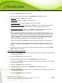
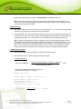
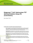
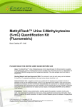
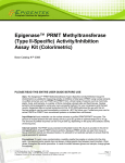
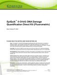
EPIGENTEK Complete Solutions for Epigenetics Epigenase™ Thymine DNA Glycosylase Activity/Inhibition Assay Kit (Colorimetric) Base Catalog # P-3094 PLEASE READ THIS ENTIRE USER GUIDE BEFORE USE Uses: The Epigenase™ Thymine DNA Glycosylase Activity/Inhibition Assay Kit (Colorimetric) is suitable for measuring the activity/inhibition of total TDG enzyme using nuclear extracts or purified TDG enzymes from a broad range of species such as mammals, plants, fungi, and bacteria, in a variety of forms including, but not limited to cultured cells and fresh and frozen tissues. Starting Materials: Input materials can be nuclear extracts or purified TDG enzymes. The amount of nuclear extracts for each assay can be between 2 µg to 10 µg with an optimal range of 5-6 µg. The amount of purified enzymes can be between 20 ng to 1 µg, depending on the purity and catalytic activity of the enzymes. Internal Control: The TDG assay standard is provided in this kit for the quantification of TDG enzyme activity. Because TDG activity can vary from tissue to tissue, and from normal and diseased states, it is advised to run replicate samples to ensure that the signal generated is validated. Precautions: To avoid cross-contamination, carefully pipette the sample or solution into the strip wells. Use aerosol-barrier pipette tips and always change pipette tips between liquid transfers. Wear gloves throughout the entire procedure. In case of contact between gloves and sample, change gloves immediately. 110 Bi County Blvd. Ste. 122, Farmingdale, NY 11735 Tel: 1-877-374-4368 ■ Fax: 1-718-484-3956 ■ E-mail: [email protected] ■ Web: www.epigentek.com © Epigentek Group Inc. All rights reserved. Products are for research use only. Page 1 Printed 2014-09-22 P-3094 EPIGENTEK Complete Solutions for Epigenetics KIT CONTENTS Component 48 Assays Cat. #P-3094-48 96 Assays Cat. #P-3094-96 Storage Upon Receipt WB (10X Wash Buffer) 14 ml 28 ml 4°C TAB (TDG Assay Buffer) 3 ml 6 ml RT TS (10X TDG Substrate)* 10 µl 20 µl –20°C BS (Binding Solution) 5 ml 10 ml RT TAS (TDG Assay Standard, 10 µg/ml)* 10 µl 20 µl –20°C CA (Capture Antibody, 1000X)* 4 µl 8 µl 4°C DA (Detection Antibody, 2000X)* 5 µl 10 µl –20°C ES (Enhancer Solution)* 5 µl 10 µl –20°C DS (Developer Solution) 5 ml 10 ml 4°C SS (Stop Solution) 5 ml 10 ml RT 8-Well Assay Strips (With Frame) 6 12 4°C User Guide 1 1 RT * For maximum recovery of the products, centrifuge the original vial prior to opening the cap. SHIPPING & STORAGE The kit is shipped in three parts: the first part at ambient room temperature, and the second and third parts on frozen ice packs at 4°C. Upon receipt: (1) Store TS, TAS, DA, and ES at –20°C away from light; (2) Store WB, CA, DS, and 8-Well Assay Strips at 4°C away from light; (3) Store remaining components (TAB, BS and SS) at room temperature away from light. Note: Check if WB 10X Wash Buffer contains salt precipitates before use. If so, warm at room temperature or 37°C and shake the buffer until the salts are re-dissolved. All components of the kit are stable for 6 months from the date of shipment, when stored properly. MATERIALS REQUIRED BUT NOT SUPPLIED Adjustable pipette or multiple-channel pipette Multiple-channel pipette reservoirs Aerosol resistant pipette tips Microplate reader capable of reading absorbance at 450 nm 1.5 ml microcentrifuge tubes Incubator for 37°C incubation Distilled water Nuclear extract or purified enzymes 110 Bi County Blvd. Ste. 122, Farmingdale, NY 11735 Tel: 1-877-374-4368 ■ Fax: 1-718-484-3956 ■ E-mail: [email protected] ■ Web: www.epigentek.com © Epigentek Group Inc. All rights reserved. Products are for research use only. Page 2 Printed 2014-09-22 P-3094 EPIGENTEK Complete Solutions for Epigenetics Parafilm M or aluminum foil GENERAL PRODUCT INFORMATION Quality Control: Each lot of Epigenase™ Thymine DNA Glycosylase (TDG) Activity/Inhibition Assay Kit (Colorimetric) is tested against predetermined specifications to ensure consistent product quality. Epigentek guarantees the performance of all products in the manner described in our product instructions. Product Warranty: If this product does not meet your expectations, simply contact our technical support unit or your regional distributor. We also encourage you to contact us if you have any suggestions about product performance or new applications and techniques. Safety: Suitable lab coat, disposable gloves, and proper eye protection are required when working with this product. Product Updates: Epigentek reserves the right to change or modify any product to enhance its performance and design. The information in this User Guide is subject to change at any time without notice. Therefore, only use the User Guide that was supplied with the kit when using that kit. Usage Limitation: The Epigenase™ Thymine DNA Glycosylase (TDG) Activity/Inhibition Assay Kit (Colorimetric) is for research use only and is not intended for diagnostic or therapeutic application. Intellectual Property: The Epigenase™ Thymine DNA Glycosylase (TDG) Activity/Inhibition Assay Kit (Colorimetric) and methods of use are intellectual property of Epigentek. A BRIEF OVERVIEW DNA methylation occurs by the covalent addition of a methyl group at the 5-carbon of the cytosine ring by DNA methyltransferases, resulting in 5-methylcytosine (5mC). In somatic cells, 5mC is found almost exclusively in the context of paired symmetrical methylation of the dinucleotide CpG, whereas in embryonic stem (ES) cells, a substantial amount of 5mC is also observed in non-CpG contexts. The biological importance of 5mC as a major epigenetic modifier of phenotype and gene expression has been widely recognized. DNA demethylation has been demonstrated to occur through at least two different pathways: (1) active deamination of 5-mC to T by an AID/APOBEC enzyme, giving a G/T mismatch that is converted to G/C by thymine DNA glycosylase (TDG) and then subsequently base excision repair (BER); and (2) iterative oxidation of 5-mC by TET enzyme to 5-hmC and further to 5-formycytosine (5-fC) and 5carboxylcytosine (5-caC). Both 5-fC and 5-caC can be rapidly excised by TDG to allow subsequent BER processing which results in 5-fC and 5-caC converting back to unmodified cytosine. 110 Bi County Blvd. Ste. 122, Farmingdale, NY 11735 Tel: 1-877-374-4368 ■ Fax: 1-718-484-3956 ■ E-mail: [email protected] ■ Web: www.epigentek.com © Epigentek Group Inc. All rights reserved. Products are for research use only. Page 3 Printed 2014-09-22 P-3094 EPIGENTEK Complete Solutions for Epigenetics TDG belongs to the TDG/mug DNA glycosylase family. Besides playing a critical role in active DNA methylation, TDG also removes thymine moieties from G/T mismatches by hydrolyzing the carbonnitrogen bond between the sugar-phosphate backbone of DNA and the mismatched thymine. It has been shown that TDG participates in the regulation of embryonic and germ cell development, mediates cellular defense against genetic and epigenetic mutations, and mediates DNA-directed cytotoxicity. In addition, TDG is a proposed tumor suppressor candidate. Thus, the measurement of TDG activity would be important as TDG may be involved in preventing tumorigenesis and determining cellular response to chemotherapy through DNA demethylation and BER, thereby benefiting cancer diagnostics and new target-based cancer therapeutics. The currently used methods for TDG activity detection involve DNA cleavage followed by electrophoresis, which can be time consuming and inconvenient, with low throughput and high cost. To address this issue, Epigentek developed and offers the Epigenase™ Thymine DNA Glycosylase (TDG) Activity/Inhibition Assay Kit (Colorimetric). Colorimetric assay with easy-to-follow steps for convenience and speed. The entire procedure can be finished within 5 hours. Directly measures TDG activity without the need for DNA cleavage, electrophoresis, or chromatography. Both cell/tissue extracts and purified TDG proteins can be used, which allows for detection of inhibitory effects of TDG inhibitors in vivo and in vitro Novel assay principle allows high sensitivity to be achieved. The activity/inhibition of TDG can be detected from as little as 20 ng of purified TDG proteins. TDG assay standard is conveniently included, which allows for specific quantification of TDG activity. Strip microplate format makes the assay flexible: manual or high throughput analysis (96 assays) PRINCIPLE & PROCEDURE In this assay, a 5-fC DNA substrate is stably coated onto microplate wells. Active TDG binds to the substrate and excises 5-fC from the substrate by a nicking reaction. The remaining un-excised 5-fC in the substrate will be recognized with a high affinity 5-fC DNA antibody. The ratio or amount of 5-fC in the substrate, which is inversely proportional to enzyme activity, can then be colorimetrically measured by reading the absorbance in a microplate spectrophotometer at a wavelength of 450 nm. The activity of the TDG enzyme is in turn inversely proportional to the optical density intensity measured. 110 Bi County Blvd. Ste. 122, Farmingdale, NY 11735 Tel: 1-877-374-4368 ■ Fax: 1-718-484-3956 ■ E-mail: [email protected] ■ Web: www.epigentek.com © Epigentek Group Inc. All rights reserved. Products are for research use only. Page 4 Printed 2014-09-22 P-3094 EPIGENTEK Complete Solutions for Epigenetics Fig. 1. Illustrated standard curve generated with the TDG assay standard from the Epigenase™ Thymine DNA Glycosylase (TDG) Activity/Inhibition Assay (Colorimetric). Demonstration of high sensitivity and specificity of the TDG activity assay achieved by using nuclear extracts with the the Epigenase ™ Thymine DNA Glycosylase (TDG) Activity/Inhibition Assay Kit (Colorimetric). Hela Nuclear extracts were used in the assay. Fig. 2. ASSAY PROTOCOL For the best results, please read the protocol in its entirety prior to starting your experiment. Starting Materials Input Amount: The amount of nuclear extracts for each assay can be between 2 µg and 10 µg with an optimal range of 5-6 µg. The amount of purified enzymes can be between 20 ng and 1 µg, depending on the purity and catalytic activity of the enzymes. Nuclear Extraction: You can use your method of choice for preparing nuclear extracts. Epigentek offers a nuclear extraction kit (Cat # OP-0002) optimized for use with this kit. Nuclear Extract or Purified TDG Protein Storage: Nuclear extract or purified TDG enzyme should be stored in aliquots at –80°C until use. 1. Buffer Solution & Preparation a. Prepare Diluted WB 1X Wash Buffer: 48-Assay Kit: Add 13 ml of WB 10X Wash Buffer to 117 ml of distilled water and adjust pH to 7.2-7.5. 96-Assay Kit: Add 26 ml of WB 10X Wash Buffer to 234 ml of distilled water and adjust pH to 7.2-7.5. This Diluted WB 1X Wash Buffer can now be stored at 4°C for up to six months. b. Prepare 0.5X TS TDG Substrate: Add 1 µl of TS 10X TDG Substrate to 19 µl of TAB Assay Buffer. About 2 µl of 0.5X TS will be required for each assay well. 110 Bi County Blvd. Ste. 122, Farmingdale, NY 11735 Tel: 1-877-374-4368 ■ Fax: 1-718-484-3956 ■ E-mail: [email protected] ■ Web: www.epigentek.com © Epigentek Group Inc. All rights reserved. Products are for research use only. Page 5 Printed 2014-09-22 P-3094 EPIGENTEK Complete Solutions for Epigenetics c. Prepare Diluted CA Capture Antibody Solution: Dilute CA Capture Antibody with Diluted WB 1X Wash Buffer at a ratio of 1:1000 (i.e., add 1 µl of CA to 1000 µl of Diluted WB 1X Wash Buffer). About 50 µl of Diluted CA will be required for each assay well. d. Prepare Diluted DA Detection Antibody Solution: Dilute DA Detection Antibody with Diluted WB 1X Wash Buffer at a ratio of 1:2000 (i.e., add 1 µl of DA to 2000 µl of Diluted WB 1X Wash Buffer). About 50 µl of Diluted DA will be required for each assay well. e. Prepare Diluted ES Enhancer Solution: Dilute ES Enhancer Solution with Diluted WB 1X Wash Buffer at a ratio of 1:5000 (i.e., add 1 µl of ES to 5000 µl of WB 1X Wash Buffer). About 50 µl of Diluted ES will be required for each assay well. f. Prepare Diluted TAS Standard Solution: Suggested Standard Curve Preparation: First, dilute TAS with TAB to 1 ng/µl by adding 1 µl of TAS to 9 µl of TAB. Then, further prepare five concentrations by combining the 1 ng/µl Diluted TAS with TAB into final concentrations of 0.05, 0.1, 0.2, 0.5, and 1.0 ng/µl according to the following dilution chart: TAB Resulting TAS Concentration Tube TAS (1 ng/µl) 1 1.0 µl 19.0 µl 0.05 ng/µl 2 1.0 µl 9.0 µl 0.1 ng/µl 3 1.0 µl 4.0 µl 0.2 ng/µl 4 2.0 µl 2.0 µl 0.5 ng/µl 5 4.0 µl 0.0 µl 1 ng/µl Note: Keep each of the diluted solutions except Diluted WB 1X Wash Buffer on ice until use. Any remaining diluted solutions other than Diluted WB should be discarded if not used within the same day. 2. Enzymatic Reaction a. Predetermine the number of strip wells required for your experiment. Carefully remove un-needed strip wells from the plate frame and place them back in the bag (seal the bag tightly and store at 4°C). b. Add 80 µl of BS Binding Solution to each well. c. Add 2 µl of TAB into the blank wells. Add 2 µl of 0.5X TS into internal control wells and each sample well. Add 1 µl of Diluted TAS into the standard curve wells (see the designated wells depicted in Table 1 under “Suggested Strip Well Setup” below). Mix solution by gently tilting from side to side or shaking the plate several times. Ensure the solution coats the bottom of the well evenly. Note: For the standard curve, add 1 µl of Diluted TAS at concentrations of 0.05 to 1 ng/µl (see the chart in Step 1g). The final concentrations should be 0.05, 0.1, 0.2, 0.5, and 1 ng per well. d. Cover strip plate with plate seal or Parafilm M and incubate at 37°C for 90 min. 110 Bi County Blvd. Ste. 122, Farmingdale, NY 11735 Tel: 1-877-374-4368 ■ Fax: 1-718-484-3956 ■ E-mail: [email protected] ■ Web: www.epigentek.com © Epigentek Group Inc. All rights reserved. Products are for research use only. Page 6 Printed 2014-09-22 P-3094 EPIGENTEK Complete Solutions for Epigenetics e. Remove the BS Binding Solution from each well. f. Wash each well three times with 150 µl of the Diluted WB 1X Wash Buffer each time. g. Blank Wells: Add 50 µl of TAB to each blank well. h. Standard Wells: Add 50 µl of TAB to each standard well. i. Internal Control Wells: Add 50 µl of TAB to each internal control well j. Sample Wells Without Inhibitor: Add 46 to 49 µl of TAB and 1 to 4 µl of nuclear extracts or purified TDG enzyme to each sample well without inhibitor. Total volume should be 50 µl per well. k. Sample Wells With Inhibitor: Add 41 to 44 µl of TAB, 1 to 4 µl of nuclear extracts or purified TDG enzyme, and 5 µl of inhibitor solution. Total volume should be 50 µl per well. Note: (1) Follow the suggested well setup diagrams under “Suggested Strip Well Setup”; (2) It is recommended to use 5 µg to 10 µg of nuclear extract per well or 50 ng to 500 ng of purified enzyme per well; (3) The concentration of inhibitor to be added into the sample wells can be varied (1 µM to 1000 µM). However, the final concentration of the inhibitors before adding to the wells should be prepared with TAB at a 1:10 ratio (i.e., add 0.5 µl of inhibitor to 4.5 µl of TAB) so that the original solvent of the inhibitor can be reduced to 1% of the reaction solution or less. l. Tightly cover strip plate with Parafilm M to avoid evaporation and incubate at 37°C for 45-60 min. Note: (1) The incubation time may depend on intrinsic TDG activity. However, in general, 45 min incubation is suitable for active purified TDG enzyme and 60 min incubation is required for nuclear extract. m. Remove the reaction solution from each well. Wash each well three times with 150 µl of the Diluted WB 1X Wash Buffer each time. 3. Antibody Binding & Signal Enhancing a. Add 50 µl of the Diluted CA to each well, then cover with Parafilm M or aluminium foil and incubate at room temperature for 60 min. b. Remove the Diluted CA solution from each well. c. Wash each well three times with 150 µl of the Diluted WB 1X Wash Buffer each time. d. Add 50 µl of the Diluted DA to each well, then cover with Parafilm M or aluminium foil and incubate at room temperature for 30 min. e. Remove the Diluted DA solution from each well. f. Wash each well four times with 150 µl of the Diluted WB 1X Wash Buffer each time. g. Add 50 µl of the Diluted ES to each well, then cover with Parafilm M or aluminum foil and incubate at room temperature for 30 min. h. Remove the Diluted ES solution from each well. 110 Bi County Blvd. Ste. 122, Farmingdale, NY 11735 Tel: 1-877-374-4368 ■ Fax: 1-718-484-3956 ■ E-mail: [email protected] ■ Web: www.epigentek.com © Epigentek Group Inc. All rights reserved. Products are for research use only. Page 7 Printed 2014-09-22 P-3094 EPIGENTEK Complete Solutions for Epigenetics i. Wash each well five times with 150 µl of the Diluted WB 1X Wash Buffer each time. Note: Ensure any residual wash buffer in the wells is thoroughly removed at each wash step. The wash can be carried out by simply pipetting the wash buffer into the wells and then pipetting the buffer out from the wells (discard the buffer). 4. Signal Detection a. Add 100 µl of DS to each well and incubate at room temperature for 1 to 10 min away from light. Begin monitoring color changes in the sample wells and control wells. b. Add 50 µl of SS to each well to stop enzyme reaction when the color in the positive control wells turns medium blue. The color will change to yellow after adding SS and the absorbance should be read on a microplate reader within 2 to 10 min at 450 nm with an optional reference wavelength of 655 nm. Note: (1) Most microplate readers have the capability to carry out dual wavelength analysis and will automatically subtract reference wavelength absorbance from the test wavelength absorbance. If your plate reader does not have this capability, the plate can be read twice – once at 450 nm and once at 655 nm. Then manually subtract the 655 nm ODs from 450 nm ODs; (2) If the stripwell microplate frame does not fit in the microplate reader, transfer the solution to a standard 96-well microplate. 5. TDG Activity Calculation a. Calculate the average duplicate readings for sample wells and blank wells. b. Calculate TDG activity or inhibition using the following formulas: For simple calculation: (Internal Control OD- blank OD) - (Sample OD – Blank OD) x 1000 TDG Activity (OD/min/mg) = (Protein Amount (µg)* x min**) * Protein amount added into the reaction at step 2J. ** Incubation time at step 2L (in minutes). Example calculation: Average OD450 of internal control is 0.75 Average OD450 of sample is 0.45 Average OD450 of blank is 0.1 Protein amount is 5 µg Incubation time is 1 hour (60 min) TDG activity = (0.65 – 0.35) x 1000 = 1 OD/min/mg (5 x 60) 110 Bi County Blvd. Ste. 122, Farmingdale, NY 11735 Tel: 1-877-374-4368 ■ Fax: 1-718-484-3956 ■ E-mail: [email protected] ■ Web: www.epigentek.com © Epigentek Group Inc. All rights reserved. Products are for research use only. Page 8 Printed 2014-09-22 P-3094 EPIGENTEK Complete Solutions for Epigenetics For accurate or specific activity calculation: First, generate a standard curve and plot the OD values versus the amount of TAS at each concentration point. Then determine the slope as OD/ng using linear regression (Microsoft Excel’s linear regression or slope functions are suitable for such calculations) and the most linear part (include at least 4 concentration points) of the standard curve for optimal slope calculation. Now calculate the amount of excised 5-fC DNA using the following formulas: (Internal Control OD- blank OD) - (Sample OD – Blank OD) Excised 5–fC DNA (ng) = Slope Excised Substrate (ng) TDG Activity (ng/min/mg) = x 1000 (Protein Amount (µg)* x min**) * Protein amount added into the reaction at step 2J. ** Incubation time at step 2L (in minutes). For inhibition calculation: Inhibition % = [ (Internal Control OD- blank OD) - (Inhibitor Sample OD – Blank OD) 1– (Internal Control OD- blank OD) - (No Inhibitor Sample OD – Blank OD) ] x 100% SUGGESTED STRIP WELL SETUP Table 1. The suggested strip-well plate setup for standard curve preparation in a 48-assay format (in a 96-assay format, Strips 7 to 12 can be configured as Sample). The controls and samples can be measured in duplicate. Well # A B C D E F G H Strip 1 Blank TAS 0.05 ng/µl TAS 0.1 ng/µl TAS 0.2 ng/µl TAS 0.5 ng/µl TAS 1 ng/µl Internal control Sample Strip 2 Blank TAS 0.05 ng/µl TAS 0.1 ng/µl TAS 0.2 ng/µl TAS 0.5 ng/µl TAS 1 ng/µl Internal control Sample Strip 3 Sample Sample Sample Sample Sample Sample Sample Sample Strip 4 Sample Sample Sample Sample Sample Sample Sample Sample 110 Bi County Blvd. Ste. 122, Farmingdale, NY 11735 Tel: 1-877-374-4368 ■ Fax: 1-718-484-3956 ■ E-mail: [email protected] ■ Web: www.epigentek.com © Epigentek Group Inc. All rights reserved. Products are for research use only. Strip 5 Sample Sample Sample Sample Sample Sample Sample Sample Strip 6 Sample Sample Sample Sample Sample Sample Sample Sample Page 9 Printed 2014-09-22 P-3094 EPIGENTEK Complete Solutions for Epigenetics TROUBLESHOOTING Problem Possible Cause Suggestion No signal or weak signal in both the standard and sample wells Reagents are added incorrectly. Check if reagents are added in the proper order with the right amount, and if any steps in the protocol may have been omitted by mistake. The substrate and standard are not properly bound to the wells. Ensure that (1) the TS and TAS are added into the wells; (2) the wells are completely covered with sufficient BS Binding Solution; and (3) binding time is sufficient (90 min). Incubation time and temperature are incorrect. Ensure the incubation time and temperature described in the protocol are followed correctly. Incorrect absorbance reading. Check if the appropriate absorbance wavelength (450 nm filter) is used. Kit was not stored or handled properly. Ensure all components of the kit are stored at the appropriate temperatures and tightly capped after each opening or use. The standard amount is insufficiently added to the well in Step 2c. Ensure a sufficient amount of standard is added. The standard is degraded due to improper storage conditions. Follow the Shipping & Storage guidance of this User Guide for storage of TAS (TDG Assay Standard). Insufficient washing of wells. Check if washing at each step is performed according to the protocol. Contaminated by sample or standard. Ensure the well is not contaminated from adding sample or standard accidentally or from using contaminated tips. Incubation time with detection antibody is too long. The incubation time at Step 3d should not exceed 45 minutes. Over development of color. Decrease the development time in Step 4a before adding SS Stop Solution in Step 4b. Protein sample is not properly extracted or purified. Ensure your protocol is suitable for TDG protein extraction. For the best results, it is advised to use Epigentek’s Nuclear Extraction Kit (Cat. No. OP-0002). Also, use fresh cells or tissues for protein extraction, as frozen cells or tissues could lose enzyme activity. Sample amount added into the wells is insufficient. Ensure a sufficient amount of purified enzymes or nuclear extracts is used as No signal or weak signal in only the standard curve wells High background present in the blank wells No signal or weak signal only in sample wells 110 Bi County Blvd. Ste. 122, Farmingdale, NY 11735 Tel: 1-877-374-4368 ■ Fax: 1-718-484-3956 ■ E-mail: [email protected] ■ Web: www.epigentek.com © Epigentek Group Inc. All rights reserved. Products are for research use only. Page 10 Printed 2014-09-22 P-3094 EPIGENTEK Complete Solutions for Epigenetics indicated in Steps 2J and 2K. The sample can be titrated to determine the optimal amount to use in the assay. Uneven color development Sample was not stored properly or has been stored for too long. Ensure sample is stored in aliquots at –80°C, for no more than 6 weeks for nuclear extracts and 6 months for purified enzymes. Avoid repeated freezing/thawing. Little or no activity of TDG contained in the sample. This problem may be a result of many factors. If the affecting factors cannot be determined, use new or re-prepared nuclear extracts or purified enzymes. Insufficient wash of the wells. Ensure the wells are washed according to the guidance of washing and residual wash buffer is removed as much as possible. Delayed color development or delayed stopping of color development in the wells. Ensure color development and stop solutions are added sequentially and consistent with the order you added the other reagents (e.g., from well A to G or from well 1 to 12). RELATED PRODUCTS Nuclear Extract Preparation OP-0002-1 EpiQuik™ Nuclear Extraction Kit DNA Methylation P-1015 P-2019 P-2020 P-1034 P-1035 P-1039 P-1040 P-3009 P-3010 Methylamp™ Methylated DNA Capture Kit EpiQuik™ Methylated DNA Immunoprecipitation Kit EpiQuik™ Tissue Methylated DNA Immunoprecipitation Kit MethylFlash™ Methylated DNA Quantification Kit (Colorimetric) MethylFlash™ Methylated DNA Quantification Kit (Fluorometric) MethylFlash™ Urine 5-Methylcytosine Quantification Kit (Colorimetric) MethylFlash™ Urine 5-Methylcytosine Quantification Kit (Fluorometric) EpiQuik™ DNMT Activity/Inhibition Assay Ultra Kit (Colorimetric) EpiQuik™ DNMT Activity/Inhibition Assay Ultra Kit (Fluorometric) DNA Demethylation P-1036 MethylFlash™ Hydroxymethylated DNA Quantification Kit (Colorimetric) P-1037 MethylFlash™ Hydroxymethylated DNA Quantification Kit (Fluorometric) P-1038 EpiQuik™ Hydroxymethylated DNA Immunoprecipitation (hMeDIP) Kit P-3087 Epigenase™ 5mC-Hydroxylase TET Activity/Inhibition Kit (Fluorometric) P-1041 MethylFlash™ 5-Formylcytosine (5-fC) DNA Quantification Kit 110 Bi County Blvd. Ste. 122, Farmingdale, NY 11735 Tel: 1-877-374-4368 ■ Fax: 1-718-484-3956 ■ E-mail: [email protected] ■ Web: www.epigentek.com © Epigentek Group Inc. All rights reserved. Products are for research use only. Page 11 Printed 2014-09-22 P-3094