1
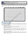
CODA-CERVA
Centrum voor Onderzoek in Diergeneeskunde en Agrochemie
Centre d’Etude et de Recherches Vétérinaires et Agrochimiques
SOP/VIR/PRE/41
Vnr: 01
Titel/Titre: Nucleic acid extraction from liquid or solid samples
Datum/Date: 10/01/11
Pg 1/18
Doel / Objet:
This standard operating procedure describes the extraction of nucleic acids from samples sent to the Virological
Platform for diagnosis by (RT)-qPCR analysis. The procedure delineates the following steps: (i) preparation of reagents,
(ii) extraction of nucleic acids, (iii) storage of nucleic acids and (iv) elimination of all reagents and samples.
Toepassingsgebied / Domaine d’application:
This procedure is applicable to all liquid (e.g. blood, serum) or solid (e.g. tissue) samples that are sent to the Virological
Platform for diagnosis by (RT-)qPCR analysis.
Bestemmelingen / Destinataires: zie distributielijst /
Voir liste de distribution
Aantal copies in omloop / Copies en circulation: 3
Historiek / Historique
Datum vrijgave
Date
d’application
Reden(en) wijziging / Motif(s) de changement
01
10/01/11
Eerste editie / Première édition
U
nc
o
nt
ro
lle
d
co
py
Vnr
Auteur
Naam / Nom
Vandenbussche F.
Kwaliteitsverantwoordelijke
Responsable Qualité
Knapen K.
Technisch verantwoordelijke
Responsable technique
Vandenbussche F.
CODA-CERVA
Centrum voor Onderzoek in Diergeneeskunde en Agrochemie
Centre d’Etude et de Recherches Vétérinaires et Agrochimiques
SOP/VIR/PRE/41
Vnr: 01
Titel/Titre: Nucleic acid extraction from liquid or solid samples
Datum/Date: 10/01/11
Pg 2/18
Contents
U
nc
o
nt
ro
lle
d
co
py
Contents ........................................................................................................ 2
1 Object ....................................................................................................... 3
2 Scope........................................................................................................ 3
3 Definitions, abbreviations, references and standards ........................................ 3
3.1
List with reference documents ................................................................ 3
3.2
Definitions and abbreviations ................................................................. 3
4 Principle .................................................................................................... 4
4.1
Liquid samples ..................................................................................... 4
4.2
Solid samples ...................................................................................... 4
4.3
Quality control ..................................................................................... 4
5 Validation parameters.................................................................................. 4
6 Supplies .................................................................................................... 5
6.1
Small laboratory material ...................................................................... 5
6.2
Reagents ............................................................................................ 5
6.3
Controls.............................................................................................. 5
7 Equipments................................................................................................ 6
8 Instruction ................................................................................................. 6
8.1
Guidelines to avoid contamination........................................................... 6
8.1.1
Sample handling ............................................................................. 6
8.1.2
Daily decontamination of the automated workstation ........................... 6
8.1.3
Weekly decontamination of the automated workstation ........................ 6
8.1.4
As-needed maintenance of the automated workstation ......................... 7
8.2
Preparation and quality control of the external extraction control ................ 8
8.3
Manual nucleic acid extraction ................................................................ 9
8.3.1
Labelling instructions ....................................................................... 9
8.3.2
Preparation of reagents.................................................................... 9
8.3.3
Manual extraction of liquid samples ................................................... 9
8.3.4
Manual extraction of solid samples ...................................................10
8.4
Automated nucleic acid extraction..........................................................11
8.4.1
Labelling instructions ......................................................................11
8.4.2
Preparation of reagents...................................................................12
8.4.3
Preparation of JANUS Automated Workstation ....................................13
8.4.4
Automated extraction of liquid samples .............................................14
8.4.5
Automated extraction of solid samples ..............................................15
8.5
Storage of sample remnants and nucleic acids .........................................16
8.6
Removal of samples/waste....................................................................16
9 Specific safety requirements........................................................................16
10
Internal control ......................................................................................17
11 Reporting..................................................................................................17
12
Annexes................................................................................................18
CODA-CERVA
Centrum voor Onderzoek in Diergeneeskunde en Agrochemie
Centre d’Etude et de Recherches Vétérinaires et Agrochimiques
SOP/VIR/PRE/41
Vnr: 01
Titel/Titre: Nucleic acid extraction from liquid or solid samples
1
Datum/Date: 10/01/11
Pg 3/18
Object
This standard operating procedure describes the extraction of nucleic acids from
samples sent to the Virological Platform for diagnosis by (RT)-qPCR analysis. The
procedure delineates the following steps: (i) preparation of reagents, (ii) extraction of
nucleic acids, (iii) storage of nucleic acids and (iv) elimination of all reagents and
samples.
2
Scope
This procedure is applicable to all liquid (e.g. blood, serum) or solid (e.g. tissue)
samples that are sent to the Virological Platform for diagnosis by (RT-)qPCR analysis.
Definitions, abbreviations, references and standards
3.1 List with reference documents
co
py
3
U
nc
o
nt
ro
lle
d
• SOP/VIR/ANA/17 : Detection of bluetongue virus RNA by RT-qPCR analysis
• Viral RNA Isolation, NucleoSpin RNA Virus user manual (version November
2009/Rev.08)
• Viral DNA/RNA Isolation, NucleoSpin 96 Virus Core Kit user manual (version
March 2010/Rev.03)
• TriPure Isolation Reagent, instruction manual (version 3, August 2004)
• JANUS Automated Liquid Handling System, user manual (publication No.
1694282 Rev.E)
• Vandemeulebroucke E., De Clercq K., Van der Stede Y., and Vandenbussche F.
(2010). A proposed validation method for automated nucleic acid extraction and
RT-qPCR analysis: an example using Bluetongue virus. J. Virol. Methods, 165:
76-82.
• PRO/5.1/04 : Afvalverwijdering / Elimination des déchets
• PRO/5.1/03 : Flow van de stalen in de eenheden / Flux des échantillons dans les
unités
3.2 Definitions and abbreviations
•
•
•
•
•
•
•
•
•
•
•
•
BTV: bluetongue virus
DMSO: dimethyl sulfoxide
EC: external control
EC-EXTR: external control of extraction
GAPDH : Glyceraldehyde 3-phosphate dehydrogenase
IC: internal control
mRNA : messenger ribonucleic acid
NAC: negative amplification control
NEC: negative extraction control
PAC: positive amplification control
RNA: ribonucleic acid
RT-qPCR: reverse transcription real-time polymerase chain reaction
CODA-CERVA
Centrum voor Onderzoek in Diergeneeskunde en Agrochemie
Centre d’Etude et de Recherches Vétérinaires et Agrochimiques
SOP/VIR/PRE/41
Vnr: 01
Titel/Titre: Nucleic acid extraction from liquid or solid samples
4
Datum/Date: 10/01/11
Pg 4/18
Principle
4.1 Liquid samples
co
py
Nucleic acids are extracted from liquid samples (e.g. blood, serum/plasma or cell
culture) by a silica membrane-based extraction procedure (e.g. NucleoSpin RNA virus
kit). Cells and virions are first lysed by adding both a highly concentrated guanidine
isothiocyanate solution (e.g. RAV1 buffer) and proteinase K solution. Carrier RNA is
added to the RAV1 buffer to improve the binding and recovery of low concentrated
viral nucleic acids. Following lysis, binding conditions are adjusted by adding 96-100%
(v/v) ethanol which improves binding of nucleic acids to the silica membrane.
Contaminants like salts, metabolites and soluble macromolecular cellular components
are removed in a series of wash steps using several wash buffers (e.g. RAW and RAV3
buffer). Finally, the purified nucleic acids are eluted in an elution solution (e.g. water
or water supplemented with 10% (v/v) DMSO).
4.2 Solid samples
U
nc
o
nt
ro
lle
d
Nucleic acids are extracted from solid samples (e.g. tissues) by combining a guanidine
thiocyanate:phenol:chloroform extraction procedure (e.g. TriPure Isolation Reagent)
with a silica membrane-based extraction procedure (e.g. NucleoSpin RNA virus kit).
Cells and virions are first simultaneously disrupted and homogenised by bead-milling
in the presence of a guanidine thiocyanate:phenol solution (e.g. TriPure Isolation
Reagent). Subsequently, chloroform is added to the homogenate and the mixture is
centrifuged to separate the inorganic/organic phases. After centrifugation, three
phases can be distinguished: a colourless aqueous (upper) phase containing the RNA,
a white interphase containing the DNA and a red organic (lower) phase containing the
proteins. The upper phase is transferred to a separate microtube and nucleic acids are
further purified using the above-described silica membrane-based extraction
procedure without the initial lysis step.
4.3 Quality control
All (RT-)qPCR assays of the Virological Platform contain an internal/external quality
control system to detect (RT-)qPCR failure due to poor sample quality, improper
nucleic acid extraction and/or PCR inhibition. As described in SOP/VIR/ANA/17, all
(RT-)qPCRs are based upon a standardized triplex format which allows the
simultaneous detection of viral nucleic acids, an internal control (IC) and an external
control (EC). As the IC consists of endogenous nucleic acids, a negative IC test result
is indicative of poor sample quality. Absence of IC amplification can, however, also be
due to an improper nucleic acid extraction and/or PCR inhibition. To exclude this
possibility, a fixed amount of EC-EXTR is added to each sample prior to extraction
which allows monitoring of the extraction and (RT-)qPCR efficiency in more detail.
5
Validation parameters
See validation file.
CODA-CERVA
Centrum voor Onderzoek in Diergeneeskunde en Agrochemie
Centre d’Etude et de Recherches Vétérinaires et Agrochimiques
SOP/VIR/PRE/41
Vnr: 01
Titel/Titre: Nucleic acid extraction from liquid or solid samples
6
Datum/Date: 10/01/11
Supplies
6.1 Small laboratory material
disposable gloves
homogenisation particles (metal beads or silicon carbide sharp particles)
microtubes (1.5 ml and 2.0 ml)
PCR-grade tips for manual pipettes
PCR-grade tips for automated pipetting system
round-well plates
sealing foil
scalpels
vortex
co
py
•
•
•
•
•
•
•
•
•
6.2 Reagents
U
nc
o
nt
ro
lle
d
• bleach 100%
stored at 20°C (± 5°C)
• bleach 10%
stored at 20°C (± 5°C)
• chloroform
stored at 20°C (± 5°C)
• dimethyl sulfoxide 100%
stored at 20°C (± 5°C)
• dimethyl sulfoxide (DMSO) 10%
stored at 20°C (± 5°C)
• EC-EXTR
stored at < -72°C
• ethanol 96-100%
stored at 20°C (± 5°C)
• NucleoSpin 96 Virus Core Kit (Macherey-Nagel)
stored at 20°C (± 5°C)
• proteinase K, lyophilized (Macherey-Nagel)
stored at 20°C (± 5°C)
• proteinase K, resuspended (Macherey-Nagel)
stored at 5°C (± 3°C)
• TriPure Isolation Reagent (Roche Diagnostics)
stored at 5°C (± 3°C)
• water, molecular biology grade
stored at 20°C (± 5°C)
6.3 Controls
• negative extraction control: water, molecular biology grade
stored at 20°C (± 5°C)
Pg 5/18
CODA-CERVA
Centrum voor Onderzoek in Diergeneeskunde en Agrochemie
Centre d’Etude et de Recherches Vétérinaires et Agrochimiques
SOP/VIR/PRE/41
Vnr: 01
Titel/Titre: Nucleic acid extraction from liquid or solid samples
7
Datum/Date: 10/01/11
Pg 6/18
Equipments
analytical balance
biosafety cabinet (class II)
centrifuge for microtubes and round-well blocks
chemical fumehood
freezer, < -16°C
freezer, < -72°C
JANUS Automated Workstation
pipettes with an accuracy range between 2 – 20 µl, 20 – 200 µl and 100 – 1000
µl (separate sets per cabinet/fumehood)
• refrigerator, 5°C (± 3°C)
• TissueLyser
• ThermoMixer
8
Instruction
nt
ro
lle
8.1.1 Sample handling
d
8.1 Guidelines to avoid contamination
co
py
•
•
•
•
•
•
•
•
nc
o
In order to avoid cross-contamination, all nucleic acid extractions are performed in
room D0.39/D0.40 which has its own designated lab coats, gloves, pipettes,
microtube holders, etc. Designated lab coats and gloves must be worn during all
manipulations and removed whenever leaving the room. Samples are only handled in
a biological safety cabinet (class II). After each manipulation, the work zone of the
biological safety cabinets, the shaft of the pipettes and the inside of the centrifuges
must be cleaned with Descosept AF, Rad-Con Surface Cleaner or an equivalent
product.
U
8.1.2 Daily decontamination of the automated workstation
The automated workstation should be cleaned and decontaminated at the end of each
working day. Perform the following steps:
1. Clean all tiles, racks and parts of the deck surface that have become dirty
during the consecutive extraction runs. Use Rad-Con Surface Cleaner or an
equivalent product. Do not use bleach as this can damage the automated
workstation.
2. Clean the shims and vacuum manifold base using Rad-Con Surface Cleaner or
an equivalent product.
3. Fill the system liquid container with deionised water.
4. Empty the waste containers.
5. Open/execute the daily decontamination protocol from the following folder:
C:\Packard\JANUS\bin\SOPs.
8.1.3 Weekly decontamination of the automated workstation
The automated workstation should be cleaned and decontaminated thoroughly at the
end of each week. Perform the following steps:
1. Remove all reagent troughs, tiles, racks and vacuum manifolds from the deck.
CODA-CERVA
Centrum voor Onderzoek in Diergeneeskunde en Agrochemie
Centre d’Etude et de Recherches Vétérinaires et Agrochimiques
SOP/VIR/PRE/41
Vnr: 01
Titel/Titre: Nucleic acid extraction from liquid or solid samples
Datum/Date: 10/01/11
Pg 7/18
co
py
2. Clean the deck surface using Rad-Con Surface Cleaner or an equivalent
product. Do not use bleach as this can damage the deck surface.
3. Clean all tiles and racks using Rad-Con Surface Cleaner or an equivalent
product. Do not use bleach as this can damage the tiles and racks.
4. Return all reagent troughs, tiles and racks to the deck.
5. Remove all sleeves from the tip adaptors.
6. Clean all sleeves and tip adaptors using Rad-Con Surface Cleaner or an
equivalent product. Do not use bleach as this can damage the sleeves and tip
adaptors.
7. Reassemble all sleeves/tip adaptors.
8. Clean the shims and vacuum manifold base using Rad-Con Surface Cleaner or
an equivalent product.
9. Clean and fill the system liquid container with deionised water.
10. Empty the waste containers.
11. Open/execute the weekly decontamination protocol from the following folder:
C:\Packard\JANUS\bin\SOPs.
8.1.4 As-needed maintenance of the automated workstation
nt
ro
lle
d
This section describes the maintenance procedures that do not need to be performed
on a regular basis.
U
nc
o
8.1.4.1 Sanitizing of drip pans
Drip pans are plastic troughs that occupy the space immediately below the deck.
These pans help contain spills by catching any liquids that is spilled on the deck
surface. Perform the following steps to sanitize the drip pans:
1. Remove all labware from the deck.
2. Remove each of the four screws connecting the deck plate to the frame.
3. Lift the deck plate up and away from the frame.
4. Lift the drip pans up and out of the frame base.
5. Wash and rinse the pans with mild laboratory detergent and water.
6. Drain, dry, and replace the drip pan in the frame base.
7. Place the deck plate on top of the frame. Make sure the deck plate is oriented
properly.
8. Reattach the deck plate to the frame using the four deck plate screws.
8.1.4.2 Flush/wash of the automated workstation
Air bubbles, deposits and/or residues can greatly influence the accuracy of pipetting.
Perform the following steps to flush/wash the automated workstation:
1. Fill the system liquid container with deionised water.
2. Empty the waste container.
3. Open/execute the FlushSysLiq protocol from the following folder:
C:\Packard\JANUS\bin.
4. Repeat the procedure until no air bubbles, deposits and/or residues are visible.
8.1.4.3 Replacement of a fixed tip
Damaged tips can greatly influence the accuracy of pipetting. Perform the following
steps to replace a damaged tip:
CODA-CERVA
Centrum voor Onderzoek in Diergeneeskunde en Agrochemie
Centre d’Etude et de Recherches Vétérinaires et Agrochimiques
SOP/VIR/PRE/41
Vnr: 01
Titel/Titre: Nucleic acid extraction from liquid or solid samples
Datum/Date: 10/01/11
Pg 8/18
1. Gently pull down the damaged tip adaptor and remove its sleeve.
2. Clean the sleeve and tip adaptor using Rad-Con Surface Cleaner or an
equivalent product. Do not use bleach as this can damage the sleeve and tip
adaptor.
3. Gently remove the fixed tip and disconnect the tubing.
4. Connect the tubing to a new fixed tip and reassemble the tip adaptor with its
sleeve.
5. Thoroughly flush/wash the automated workstation.
8.2 Preparation and quality control of the external extraction control
d
Overview of the primers used in the overlap extension PCR.
name
sequence (5’-3’)
pf EC-EXTR/overlap1
nt
ro
lle
Table 1:
co
py
The external extraction control is a single-stranded RNA molecule encoding an
amplicon that is unrelated to any of the tested pathogens. The amplicon was obtained
by overlap extension PCR using the primers pf EC-EXTR/overlap1 and pr ECEXTR/overlap1 (Table 1), cloned in a pCR-Blunt vector and sequenced to verify the
primer/probe binding sites. The resulting positive E. coli clones and their
corresponding plasmid DNA are stored at < -16 °C.
TTTGACGTTTGTAATGTCCGCTCTTTTTTGTACCACCTCCCACCGAC
CATC
nc
o
pr EC-EXTR/overlap1 AAACCAGTTGCTACCGATTTTACATAAAAGATGGTCGGTGGGAGGT
GGTACAAA
U
The transcription template DNA is obtained by a semi-specific PCR reaction on the
pCR-Blunt plasmid using the M13 forward primer of the vector and the reverse primer
of the specific RT-qPCR reaction. The single-stranded RNA molecules are subsequently
in vitro transcribed from the transcription template DNA using a T7 RNA polymerase
based transcription kit according to the manufacturer’s instructions. All residual DNA
is removed by an excessive DNase treatment and phenol:chloroform:isoamylalcohol
(25:24:1, v/v) extraction. The resulting RNA stock is stored at < -72°C until use. To
prepare the EC-EXTR, the RNA stock is diluted in RNA storage buffer (RNA storage
solution containing 0.01% (v/v) Tween-20 and 1 mg carrier RNA) to obtain a Cpvalue of approximately 30, aliquoted in volumes of 400 µl (liquid samples) or 250 µl
(solid samples) and stored at < -72°C.
Each new lot of external extraction control should be tested at least 10 times in
parallel with the previous one before being released for routine use. The results of
these comparative tests should be registered on document “SOP/VIR/PRE/41/DOC01:
Production and quality control of EC-EXTR” (see annex 1) .
CODA-CERVA
Centrum voor Onderzoek in Diergeneeskunde en Agrochemie
Centre d’Etude et de Recherches Vétérinaires et Agrochimiques
SOP/VIR/PRE/41
Vnr: 01
Titel/Titre: Nucleic acid extraction from liquid or solid samples
Datum/Date: 10/01/11
Pg 9/18
8.3 Manual nucleic acid extraction
8.3.1 Labelling instructions
All microtubes and NucleoSpin RNA virus columns should be clearly labelled to avoid
misidentification of samples. The microtubes/columns containing the negative
extraction control should be labelled as NEC. The microtubes/columns containing the
samples should be labelled by a letter (A-H) followed by a number (1-12) which refers
to the position of the sample on the 96-well (RT)-qPCR plate. As the first 3 positions
(i.e. A1, B1 and C1) of each 96-well (RT)-qPCR plate are reserved for the
negative/positive controls, samples should be labelled starting from D1 to H12. The
link between the sample label and the LIMS sample number should be registered
electronically in Excel using the sample identification macro.
co
py
8.3.2 Preparation of reagents
nt
ro
lle
d
All reagents should be prepared entirely in room D0.39/D0.40. With the exception of
the lysis solution, all reagents are used for all types of samples. Perform the following
steps to prepare the reagents:
1. lysis buffer / liquid samples:
Add 1 ml RAV1 buffer to a vial of lyophilized carrier RNA. Dissolve the carrier
RNA and transfer it back to the RAV1 bottle. Thaw one aliquot of ECEXTR/liquid and dilute 1:100 (v/v) into the RAV1 bottle. Label the
RAV1/cRNA/EC-EXTR bottle and store at 5°C (±3°C) for up to 2 days.
U
nc
o
lysis buffer / solid samples:
Thaw one aliquot of EC-EXTR/solid and dilute 1:100 (v/v) into TriPure Isolation
Reagent. Label the TriPure Isolation Reagent bottle/EC-EXTR and store at 5°C
(±3°C) for up to 2 days.
2. wash buffer:
Add 100 ml ethanol (96-100%) to 25 ml of the concentrated RAV3 buffer.
Label the RAV3 bottle and store at 20°C (± 5°C) for up to 4 weeks.
3. elution solution:
Dilute DMSO (100%) 1:10 (v/v) in water (molecular biology grade) and aliquot
in 50 ml tubes. Label the DMSO (10%) tubes and store at 20°C (± 5°C) for up
to 4 weeks.
8.3.3 Manual extraction of liquid samples
Nucleic acids are extracted from liquid samples by a silica membrane-based extraction
procedure (i.e. NucleoSpin RNA virus kit). All steps should be performed in a
biological safety cabinet (class II).
1. Switch on the ThermoMixer, set the temperature to 80°C and allow the
instrument to equilibrate.
2. Label a 1.5 ml microtube and add 150 µl of the corresponding liquid sample.
Repeat this step for each sample that needs to be extracted. Include at least 1
NEC per extraction run.
3. Pre-heat the necessary amount of RAV1 buffer containing carrier RNA and
EC-EXTR for 5 min at 70°C (± 5°C). Do not heat the RAV1 buffer longer than 5
CODA-CERVA
Centrum voor Onderzoek in Diergeneeskunde en Agrochemie
Centre d’Etude et de Recherches Vétérinaires et Agrochimiques
SOP/VIR/PRE/41
Vnr: 01
Titel/Titre: Nucleic acid extraction from liquid or solid samples
Datum/Date: 10/01/11
Pg 10/18
nc
o
nt
ro
lle
d
co
py
min or at higher temperatures as this will accelerate the degradation of the
carrier RNA and EC-EXTR.
4. Add 600 µl of the pre-heated RAV1 buffer to each microtube and mix by
vortexing for 10-15 sec. Incubate the microtubes for 5 min at 70°C (± 5°C) in
the ThermoMixer.
5. Add 600 µl of ethanol (96-100%) to each microtube and mix by vortexing for
10-15 sec.
6. Label a NucleoSpin RNA virus column and place it in a clean collection tube.
Repeat this step for each sample that needs to be extracted. Load 700 µl of
each lysed sample into the corresponding NucleoSpin RNA virus column and
centrifuge for 1 min at 8000 x g. Discard the flow-through and place the
Nucleospin RNA virus column into a clean collection tube.
7. Load the residual lysed sample onto the NucleoSpin RNA virus column and
centrifuge for 1 min at 8000 x g. Discard the flow-through and place the
NucleoSpin RNA virus column into a clean collection tube.
8. Add 500 µl RAW buffer to the NucleoSpin RNA virus column and centrifuge for
1 min at 8000 x g. Discard the flow-through and place the NucleoSpin RNA
virus column into the same collection tube.
9. Add 600 µl RAV3 buffer and centrifuge for 1 min at 8000 x g to remove all
remaining RAV3 buffer. Discard the flow-through and place the Nucleospin RNA
virus column into a clean collection tube.
10. Add 200 µl RAV3 buffer and centrifuge for 5 min at 11000 x g to remove all
remaining RAV3 buffer.
11. Pre-heat the necessary amount of water containing 10% (v/v) DMSO for at
least 10 min at 70°C (± 5°C). Label 1.5 ml microtubes and place the
NucleoSpin RNA virus columns into the corresponding microtubes. Add 100 µl
pre-heated water and incubate the microtubes for 2 min at 20°C (± 5°C).
Centrifuge for 1 min at 11000 x g.
U
8.3.4 Manual extraction of solid samples
Nucleic acids are extracted from solid samples by combining a guanidine
thiocyanate:phenol:chloroform extraction procedure (i.e. TriPure Isolation Reagent)
with a silica membrane-based extraction procedure (i.e. NucleoSpin RNA virus kit).
For (bio)safety reasons, steps 2 and 3 should be performed in a biological safety
cabinet (class II), whereas steps 4, 6, 7 and 9 should be performed in a chemical
fume hood.
1. Switch on the ThermoMixer, set the temperature to 75°C and allow the
instrument to equilibrate.
2. Label a 2 ml microtube and add 3-5 mm silicon carbide sharp particles. Repeat
this step for each sample that needs to be extracted. Include at least 1 NEC
per extraction run.
3. Cut/weigh a piece of 40 mg (± 5 mg) of sample and place it into the
corresponding 2 ml microtube. Use a clean scalpel and petri dish for each
sample!
4. Add 800 µl TriPure Isolation Reagent containing 1:100 (v/v) EC-EXTR to each
microtube.
CODA-CERVA
Centrum voor Onderzoek in Diergeneeskunde en Agrochemie
Centre d’Etude et de Recherches Vétérinaires et Agrochimiques
SOP/VIR/PRE/41
Vnr: 01
Titel/Titre: Nucleic acid extraction from liquid or solid samples
Datum/Date: 10/01/11
Pg 11/18
U
nc
o
nt
ro
lle
d
co
py
5. Disrupt the samples in the TissueLyser by shaking for 3 x 2 min at 25 Hz.
Disassemble the TissueLyser adapter set and rotate the rack of microtubes
after each shaking step to ensure uniform disruption. Samples that are nearer
to the TissueLyser move more slowly than samples further away from the
TissueLyser.
6. Incubate the disrupted samples for 5 min at 20°C (± 5°C).
7. Add 160 µl chloroform to each microtube, shake vigourously for 15 sec and
incubate for 5 min at 20°C (± 5°C).
8. Centrifuge the microtubes at 12000 x g for 15 min at < 10°C to separate the
solution into 3 phases.
9. Label a 2 ml microtube and transfer 400 µl of the corresponding upper
aqueous phase. Do not disturb the white interphase or red organic phase!
Repeat this step for each sample that needs to be extracted.
10. Add 400 µl of ethanol (96-100%) to each microtube and mix by vortexing for
10-15 sec.
11. Label a NucleoSpin RNA virus column and place it in a clean collection tube.
Repeat this step for each sample that needs to be extracted. Load 700 µl of
each aqueous phase/ethanol mixture into the corresponding NucleoSpin RNA
virus column and centrifuge for 1 min at 8000 x g. Discard the flow-through
and place the Nucleospin RNA virus column into a clean collection tube.
12. Load the residual lysed sample onto the NucleoSpin RNA virus column and
centrifuge for 1 min at 8000 x g. Discard the flow-through and place the
NucleoSpin RNA virus column into a clean collection tube.
13. Add 500 µl RAW buffer to the NucleoSpin RNA virus column and centrifuge for
1 min at 8000 x g. Discard the flow-through and place the NucleoSpin RNA
virus column into the same collection tube.
14. Add 600 µl RAV3 buffer and centrifuge for 1 min at 8000 x g to remove all
remaining RAV3 buffer.
15. Add 200 µl RAV3 buffer and centrifuge for 5 min at 11000 x g to remove all
remaining RAV3 buffer.
16. Pre-heat the necessary amount of water containing 10% (v/v) DMSO for at
least 10 min at 70°C (± 5°C). Label 1.5 ml microtubes and place the
NucleoSpin RNA virus columns into the corresponding microtubes. Add 300 µl
pre-heated water and incubate the microtubes for 2 min at 20°C (± 5°C).
Centrifuge for 1 min at 11000 x g.
8.4 Automated nucleic acid extraction
8.4.1 Labelling instructions
All round-well blocks and microtubes should be clearly labelled to avoid
misidentification of samples. The round-well blocks should be labelled with a preprinted label containing a unique ZZ-block number and barcode. The microtubes
containing the negative extraction control should be labelled as NEC. The microtubes
containing the samples should be labelled by a letter (A-H) followed by a number (112) which refers to the position of the sample on the 96-well (RT)-qPCR plate (Figure
1). As the first 3 positions (i.e. A1, B1 and C1) of each 96-well (RT)-qPCR plate are
reserved for the negative/positive controls, samples should be labelled starting from
CODA-CERVA
Centrum voor Onderzoek in Diergeneeskunde en Agrochemie
Centre d’Etude et de Recherches Vétérinaires et Agrochimiques
SOP/VIR/PRE/41
Vnr: 01
Titel/Titre: Nucleic acid extraction from liquid or solid samples
Datum/Date: 10/01/11
Pg 12/18
D1 to H12. The link between the sample label, LIMS sample number and ZZ-block
number should be registered electronically in Excel using the sample identification
macro.
2
3
4
5
6
7
8
9
10
11
12
A
NEC
6
14
22
30
38
46
54
62
70
78
86
B
NAC
7
15
23
31
39
47
55
63
71
79
87
C
PAC
8
16
24
32
40
48
56
64
72
80
88
D
1
9
17
25
33
41
49
57
65
73
81
89
E
2
10
18
26
34
42
50
58
66
74
82
90
F
3
11
19
27
35
43
51
59
67
75
83
91
G
4
12
20
H
5
13
21
nt
ro
lle
d
co
py
1
28
36
44
52
60
68
76
84
92
29
37
45
53
61
69
77
85
93
nc
o
Figure 1: Plate layout showing the position of the 3 controls and the 93 samples.
8.4.2 Preparation of reagents
U
All reagents should be prepared entirely in room D0.39/D0.40. With the exception of
the lysis solution, all reagents are used for all types of samples. Perform the following
steps to prepare the reagents:
1. decontamination solution:
Dilute 200 ml bleach (100 %) in 1800 ml deionised water. Label the bleach
bottle and store at 20°C (± 5°C) for up to 3 months.
2. lysis buffer / liquid samples:
Add 1 ml RAV1 buffer to a vial of lyophilized carrier RNA. Dissolve the carrier
RNA and transfer it back to the RAV1 bottle. Thaw one aliquot of ECEXTR/liquid and dilute 1:100 (v/v) into the RAV1 bottle. Label the
RAV1/cRNA/EC-EXTR bottle and store at 5°C (±3°C) for up to 4 weeks.
lysis buffer / solid samples:
Thaw one aliquot of EC-EXTR/solid and dilute 1:100 (v/v) into TriPure Isolation
Reagent. Label the TriPure Isolation Reagent/EC-EXTR bottle and store at 5°C
(±3°C) for up to 4 weeks.
CODA-CERVA
Centrum voor Onderzoek in Diergeneeskunde en Agrochemie
Centre d’Etude et de Recherches Vétérinaires et Agrochimiques
SOP/VIR/PRE/41
Vnr: 01
Titel/Titre: Nucleic acid extraction from liquid or solid samples
Datum/Date: 10/01/11
Pg 13/18
co
py
3. proteinase K solution:
Add 2.5 ml of proteinase buffer PB to a vial of lyophilized proteinase K. Label
the proteinase K vial and store at 5°C (±3°C) for up 6 months.
4. diluent buffer:
Add 400 ml ethanol (96-100%) and 100 ml water (molecular biology grade) to
400 ml RAV1. Label the diluent bottle and store at 20°C (± 5°C) for up to 4
weeks.
5. wash buffer:
Add 800 ml ethanol (96-100%) to 200 ml of the concentrated RAV3 buffer.
Label the RAV3 bottle and store at 20°C (± 5°C) for up to 4 weeks.
6. elution solution:
Dilute DMSO (100%) 1:10 (v/v) in water (molecular biology grade) and aliquot
in 50 ml tubes. Label the DMSO (10%) tubes and store at 20°C (± 5°C) for up
to 4 weeks.
8.4.3 Preparation of JANUS Automated Workstation
U
nc
o
nt
ro
lle
d
Perform the following steps to prepare the JANUS Automated Workstation:
1. If necessary, switch on the JANUS Automated Workstation and/or the
connected computer.
2. If necessary, switch on the vacuum pump, the 4 heaters and/or ThermoMixer.
Set the temperature of the ThermoMixer to 75°C and allow the instrument(s)
to equilibrate.
3. Launch the WinPrep software by clicking on the icon on the desktop.
4. Check the level of the system liquid in the system liquid container and fill with
deionised water if necessary. Check the level of liquid waste in both waste
containers and empty the waste containers if necessary. Check the
autoclavable bags containing all kinds of solid waste and replace with 2 empty
bags if necessary.
5. Visually inspect each sampling tip and replace all damaged tips (see section
8.1.4 for detailed instructions). Visually inspect the tubing for the presence of
air bubbles, deposits and/or residues. Flush/wash the automated workstation
thoroughly if any of these conditions are present (see section 8.1.4 for detailed
instructions).
6. Open/execute the corresponding nucleic acid extraction protocol (i.e.
BTV/blood or BTV/organ RNA extraction) from the following folder:
C:\Packard\JANUS\bin\SOPs.
7. Click on Reset Tip Boxes… button in the Initial User Query pop-up screen to go
to the Reset Tip Boxes pop-up screen. Select the various tip racks that will be
used and click on the Fill button to reset the tip box counter. Click on the Close
button to return to the Initial User Query pop-up screen. Click on the Next
button to go to the General Information pop-up screen.
8. Enter the number of prime cycles (i.e. initial flush/wash cycles). Enter 5 for the
first run of the day or 3 for all subsequent runs. Enter the number of samples
and the start position on the NucleoSpin Virus Binding Plate for the first and
second plate. Click on the Start button to go to the Plastics pop-up screen.
CODA-CERVA
Centrum voor Onderzoek in Diergeneeskunde en Agrochemie
Centre d’Etude et de Recherches Vétérinaires et Agrochimiques
SOP/VIR/PRE/41
Vnr: 01
Titel/Titre: Nucleic acid extraction from liquid or solid samples
Datum/Date: 10/01/11
Pg 14/18
9. Place the various plastic disposables on the deck positions that are indicated on
the Plastics pop-up screen. Click on the OK button to go to the Manifold 1 popup screen.
10. Build the vacuum manifold(s) as explained in the Manifold 1 and Manifold 2
pop-up screens. If less than 92 samples are to be processed, all unused wells
of the NucleoSpin Binding Plate should be sealed with a self-adhering foil to
ensure proper vacuum during all filtration steps. Click on the OK button to go
to the Reagents pop-up screen.
11. Place the various reagents on the deck positions that are indicated on the
Reagents pop-up screen. Click on the OK button to go to the Temperature
Check (liquid samples, section 8.4.4) or Sample/Elution Plate (solid samples,
section 8.4.5) pop-up screen.
co
py
8.4.4 Automated extraction of liquid samples
U
nc
o
nt
ro
lle
d
Nucleic acids are extracted from liquid samples by a silica membrane-based extraction
procedure (i.e. NucleoSpin 96 Virus Core Kit). Step 1 should be performed in a
biological safety cabinet (class II).
1. Label 2 round-well blocks with a pre-printed label. The first block will contain
the samples at the beginning of the run (i.e. sample plate) whereas the second
block will contain the extracted nucleic acids at the end of the run (i.e. elution
plate). Add 100 µl of the liquid samples to the sample plate using the plate
layout shown in Figure 1.
2. Wait until the temperature of the round-well block heater has reached 65°C.
Click on the OK button to go to the Sample/Elution Plate pop-up screen.
3. Pre-heat the necessary amount of RAV1 buffer containing carrier RNA and
EC-EXTR for 5 min at 70°C (± 5°C) and place on the indicated deck position.
Do not heat the RAV1 buffer longer than 5 min or at higher temperatures as
this will accelerate the degradation of the carrier RNA and EC-EXTR.
4. Place the sample and elution plate(s) on the deck positions that are indicated
in the Sample/Elution Plate pop-up screen. Click on the OK button.
5. Check whether the filter membranes are not clogged after the first and second
sample transfer. If necessary, pause the robot by clicking on the Pause button
and remove all remaining sample. Register the clogged filter membranes on
document “SOP/VIR/PRE/41/DOC02: Registration of clogged membranes” (see
annex 2) and in the sample identification macro at the end of the extraction
run.
6. Clean the vacuum manifold base(s) as explained in the Build Vacuum Manifold
pop-up screen(s) and click on the OK button.
7. Remove the elution plate(s) with NucleoSpin Binding Plate(s) on top from the
vacuum manifold base and centrifuge as explained in the Remove
Elution/Binding Plate pop-up screen. Click on the OK button.
8. Switch off the vacuum pump, all heaters and ThermoMixer as explained in the
Instruments pop-up screen and click on the OK button.
9. Clean the deck, empty the waste containers, remove the sample plates from
the deck and refill the system liquid container as explained in the Remove
Waste/Refill System Liquid pop-up screen. Click on the OK button.
CODA-CERVA
Centrum voor Onderzoek in Diergeneeskunde en Agrochemie
Centre d’Etude et de Recherches Vétérinaires et Agrochimiques
SOP/VIR/PRE/41
Vnr: 01
Titel/Titre: Nucleic acid extraction from liquid or solid samples
Datum/Date: 10/01/11
Pg 15/18
10. Close the microtubes containing the proteinase K solution and store the rack in
refrigerator LAB-21005. Empty the troughs containing the RAV1/cRNA/EXEXTR buffer and elution solution and rinse them with deionised water. Close all
other troughs containing the diluent, RAW and RAV3 buffers with the
corresponding lids.
8.4.5 Automated extraction of solid samples
U
nc
o
nt
ro
lle
d
co
py
Nucleic acids are extracted from solid samples by combining a guanidine
thiocyanate:phenol:chloroform extraction procedure (i.e. TriPure Isolation Reagent)
with a silica membrane-based extraction procedure (i.e. NucleoSpin 96 Virus Core
Kit). For (bio)safety reasons, steps 1, and 2 should be performed in a biological safety
cabinet (class II), whereas steps 3, 5, 6 and 8 should be performed in a chemical
fume hood.
1. Label a 2 ml microtube and add 3-5 mm silicon carbide sharp particles. Repeat
this step for each sample that needs to be extracted.
2. Cut/weigh a piece of 40 mg (± 5 mg) of sample and place it into the
corresponding 2 ml microtube. Use a clean scalpel and petri dish for each
sample!
3. Add 800 µl TriPure Isolation Reagent containing 1:100 (v/v) EC-EXTR to each
microtube.
4. Disrupt the samples in the TissueLyser by shaking for 3 x 2 min at 30 Hz.
Disassemble the TissueLyser adapter set and rotate the rack of microtubes
after each shaking step to ensure uniform disruption. Samples that are nearer
to the TissueLyser move more slowly than samples further away from the
TissueLyser.
5. Incubate the disrupted samples for 5 min at 20°C (± 5°C).
6. Add 160 µl chloroform to each microtube, shake vigourously for 15 sec and
incubate for 5 min at 20°C (± 5°C).
7. Centrifuge the microtubes at 12000 x g for 15 min at < 10°C to separate the
solution into 3 phases.
8. Label 2 round-well blocks with a pre-printed label. The first block will contain
the samples at the beginning of the run (i.e. sample plate) whereas the second
block will contain the extracted nucleic acids at the end of the run (i.e. elution
plate). Transfer 400 µl of the upper aqueous phase to the sample plate using
the plate layout shown in Figure 1. Do not disturb the white interphase or red
organic phase!
9. Place the sample and elution plate(s) on the deck positions that are indicated
in the Sample/Elution Plate pop-up screen. Click on the OK button.
10. Check whether the filter membranes are not clogged after the sample transfer.
If necessary, pause the robot by clicking on the Pause button and remove all
remaining sample. Register the clogged filter membranes on document
“SOP/VIR/PRE/41/DOC02: Registration of clogged membranes” (see annex 2)
and in the sample identification macro at the end of the extraction run.
11. Clean the vacuum manifold base(s) as explained in the Build Vacuum Manifold
pop-up screen(s) and click on the OK button.
CODA-CERVA
Centrum voor Onderzoek in Diergeneeskunde en Agrochemie
Centre d’Etude et de Recherches Vétérinaires et Agrochimiques
SOP/VIR/PRE/41
Vnr: 01
Titel/Titre: Nucleic acid extraction from liquid or solid samples
Datum/Date: 10/01/11
Pg 16/18
12. Remove the elution plate(s) with NucleoSpin Binding Plate(s) on top from the
vacuum manifold base and centrifuge as explained in the Remove
Elution/Binding Plate pop-up screen. Click on the OK button.
13. Switch off the vacuum pump, all heaters and ThermoMixer as explained in the
Instruments pop-up screen and click on the OK button.
14. Clean the deck, empty the waste containers, remove the sample plates from
the deck and refill the system liquid container as explained in the Remove
Waste/Refill System Liquid pop-up screen. Click on the OK button.
15. Empty the trough containing the elution solution and rinse it with deionised
water. Close all other troughs containing the RAW and RAV3 buffers with the
corresponding lids.
co
py
8.5 Storage of sample remnants and nucleic acids
d
All sample remnants should be stored in a refrigerator for at least 3 weeks. If
additional analyses are needed or samples are to be stored longer, the sample
remnants are transferred to the corresponding Reference Laboratory (see
PRO/5.1/03/DOC02 for detailed instructions).
nt
ro
lle
Nucleic acids can be stored in a refrigerator for no longer than 7 hrs to minimize nonspecific degradation. If the (RT)-qPCR analysis cannot be performed on the same day,
the nucleic acids should be stored at < -72°.
8.6 Removal of samples/waste
9
nc
o
All samples/waste should be removed from the laboratory according to the rules
described in PRO/5.1/04.
Specific safety requirements
Table 2:
U
The following components of the procedures described in sections 8.3 and 8.4 contain
hazardous substances (Table 2).
Overview of components containing hazardous substances.
component
RAV1
RAW
hazardous content
guanidine thiocyanate
guanidine hydrochloride
and ethanol < 50%
proteinase K
proteinase K, lyophilized
chloroform :
isoamyl alcohol
chloroform and
3-methyl butan-1-ol
TriPure Isolation
Reagent
phenol and guanidine
thiocyanate
risk phrases
R20/21/22
safety phrases
S13
R10-22-36/38
S7-13
R36/37/38-42
S22-24-2636/37
R22, R38, R40,
R48/20/22, R10,
R20, R37, R66
R24/25, R34,
R20/21/22, R32
S36/37, S60
S1/2, S28, S45,
S13
R10:
Flammable.
R20/21/22: Harmful by inhalation, in contact with the skin and if swallowed.
R22:
Harmful if swallowed.
CODA-CERVA
Centrum voor Onderzoek in Diergeneeskunde en Agrochemie
Centre d’Etude et de Recherches Vétérinaires et Agrochimiques
SOP/VIR/PRE/41
Vnr: 01
Titel/Titre: Nucleic acid extraction from liquid or solid samples
Datum/Date: 10/01/11
Pg 17/18
R24/25:
Toxic in contact with skin and if swallowed.
R32:
Contact with acids liberates very toxic gas.
R34:
Causes burns.
R36/37/38: Irritating to eyes, respiratory system and skin.
R38:
Irritating to skin.
R36/38:
Irritating to eyes and skin.
R40:
Limited evidence of carcinogenic effect.
R48/20/22: Danger of serious damage to health by prolonged exposure through
inhalation and if swallowed.
S60:
co
py
d
S28:
S36/37:
S45:
Keep locked-up and out of reach of children.
Keep container tightly closed.
Keep away from food, drink and animal feedstuffs.
Keep away from sources of ignition – no smoking.
Do not breathe dust.
Avoid contact with the skin.
In case of contact with eyes, rinse immediately with plenty of water and
seek medical advice.
After contact with skin, wash immediately with plenty of water.
Wear suitable protective clothing and gloves.
In case of accident or if you feel unwell, seek medical advice immediately
(show label where possible).
This material and its container must be disposed of as hazardous waste.
not applicable
11 Reporting
nc
o
10 Internal control
nt
ro
lle
S1/2:
S7:
S13:
S16:
S22:
S24:
S26:
U
All technical parameters of the nucleic acid extraction protocol should be registered
electronically in Excel using the extraction parameters macro. The technician should
always perform steps 1-5; step 6 is only optional. The scientist in charge should
always perform steps 6-7.
1. Launch Excel on one of the desktop computers in room D0.39/D0.40.
2. Launch the extraction parameters macro by clicking on the gear wheel icon on
the toolbar.
3. Select/fill in the technician, number of runs and block information of each run
in the first pop-up screen.
4. Select/fill in the equipment/reagents used in the different runs in the following
pop-up screens.
5. Select end in the last pop-up screen to save the file in the data_input folder of
the MolPlat network drive. All files are saved using the following standardized
format: yyyymmdd_first ZZ-block number to last ZZ-block number.
6. Open the saved file to add comments and/or select the scientist in charge in
the pop-up screen.
7. Print the report, sign all pages (both technician and scientist in charge) and
store in the file folder located in room D0.47.
CODA-CERVA
Centrum voor Onderzoek in Diergeneeskunde en Agrochemie
Centre d’Etude et de Recherches Vétérinaires et Agrochimiques
SOP/VIR/PRE/41
Vnr: 01
Titel/Titre: Nucleic acid extraction from liquid or solid samples
Datum/Date: 10/01/11
Pg 18/18
12 Annexes
U
nc
o
nt
ro
lle
d
co
py
Annex 1: SOP/VIR/PRE/41/DOC01/V01: Production and quality control of EC-EXTR
Annex 2: SOP/VIR/PRE/41/DOC02/V01: Registration of clogged membranes
Annex 3: SOP/VIR/PRE/41/DOC03/V01: Registration of extraction parameters (report
generated by Excel macro)
Annex 1:
PRODUCTION AND QUALITY
CONTROL OF EC-EXTR
SOP/VIR/PRE/41/DOC01/V01
nt
ro
lle
3. Aliquot preparation
• date:
• dilution factor:
• batch number:
• volume per aliquot:
• number of aliquots:
• technician:
d
2. RNA purification
• date:
• technician:
co
py
1. RNA synthesis
• date:
• plasmid:
• technician:
U
run
nc
o
4. Results of comparative testing
5. Release for routine use
• date:
• conclusion: YES/NO
• scientist in charge:
(name and signature)
EC-EXTR
[Cp-value]
EC-EXTR
[Cp-value]
Annex 2:
REGISTRATION OF CLOGGED MEMBRANES
SOP/VIR/PRE/41/DOC02/V01
general information
• date:
• ZZ-block:
• technician:
2
3
4
5
6
7
A
NEC
6
14
22
30
38
46
54
62
B
NAC
7
15
23
31
39
47
55
C
PAC
8
16
24
32
40
D
1
9
17
25
33
E
2
10
18
26
34
F
3
11
19
27
G
4
12
20
H
5
21
9
10
11
12
70
78
86
63
71
79
87
d
56
64
72
80
88
41
49
57
65
73
81
89
42
50
58
66
74
82
90
35
43
51
59
67
75
83
91
28
36
44
52
60
68
76
84
92
29
37
45
53
61
69
77
85
93
nt
ro
lle
48
nc
o
U
13
8
co
py
1