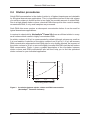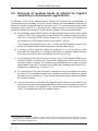
1
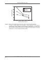
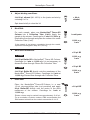
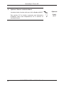
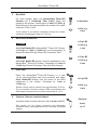
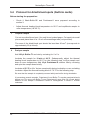
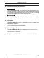
Genomic DNA
from tissue
User manual
A034521 R04en1/0/8/02.12 PD
Printed in Germany
NucleoSpin® Tissue XS
www.mn-net.com
MACHEREY-NAGEL
February 2012 / Rev. 04
MACHEREY-NAGEL
MACHEREY-NAGEL
MN
EN ISO 9001: 2008
CERTIFIED
Genomic DNA from tissue Protocol-at-a-glance (Rev. 04)
NucleoSpin® Tissue XS
1
Prepare sample
Up to 2.5 mg tissue
2
Pre-lyse sample
80 μL T1
8 μL Proteinase K
56 °C,
1–4 h
3
Lyse sample
80 μL B3
70 °C,
5 min
4
5
Adjust binding
conditions
80 μL ethanol
Bind DNA
Load lysate
11,000 x g,
1 min
6
7
Wash silica
membrane
50 μL B5
1st wash
11,000 x g,
1 min
2nd wash
11,000 x g,
2 min
50 μL B5
Elute DNA
20 μL BE
11,000 x g,
1 min
8
Optional:
Remove residual
ethanol
Optional:
90 °C,
8 min
MACHEREY-NAGEL GmbH & Co. KG · Neumann-Neander-Str. 6–8 · 52355 Düren · Germany
Tel.: +49 24 21 969-270 · Fax: +49 24 21 969-199 · [email protected] · www.mn-net.com
Genomic DNA from tissue
Table of contents
1 Components 4
1.1 Kit contents
4
1.2 Reagents, consumables, and equipment to be supplied by user
5
1.3 About this user manual
5
2 Product description
6
2.1 The basic principle
6
2.2 Kit specifications
6
2.3 Handling of sample material 7
2.4 Elution procedures
8
2.5 Removal of residual traces of ethanol for highest sensitivity in
downstream applications
9
3 Storage conditions and preparation of working solutions
11
4 Safety instructions
12
4.1 Risk and safety phrases
12
4.2 GHS classification
13
5 Protocols 15
5.1 Protocol for human or animal tissue
15
5.2 Protocol for cultured cells
18
5.3 Protocol for paraffin-embedded tissue
21
5.4 Protocol for dried blood spots (Guthrie cards)
24
5.5 Protocol for buccal swabs
26
5.6 Protocol for laser-microdissected tissue
28
5.7 Protocol for blood samples
29
6 Appendix
30
6.1 Troubleshooting
30
6.2 Ordering information
31
6.3 Product use restriction / warranty
31
MACHEREY-NAGEL – 02 / 2012, Rev. 04
3
Genomic DNA from tissue
1Components
1.1 Kit contents
NucleoSpin® Tissue XS
10 preps
50 preps
250 preps
740901.10
740901.50
740901.250
Lysis Buffer T1
5 mL
10 mL
50 mL
Lysis Buffer B3
5 mL
10 mL
50 mL
Wash Buffer B5
(Concentrate)*
1 mL
2 mL
6 mL
Elution Buffer BE**
5 mL
5 mL
15 mL
Proteinase K
(lyophilized)*
6 mg
20 mg
2 x 50 mg
0.8 mL
1.8 mL
8 mL
NucleoSpin® Tissue XS
Columns (green rings)
10
50
250
Collection Tubes (2 mL)
20
100
500
User manual
1
1
1
REF
Proteinase Buffer PB
* For preparation of working solutions and storage conditions see section 3.
**Composition of Elution Buffer BE: 5 mM Tris/HCl, pH 8.5
4
MACHEREY-NAGEL – 02 / 2012, Rev. 04
Genomic DNA from tissue
1.2 Reagents, consumables, and equipment to be
supplied by user
Reagents
•
96–100% ethanol
Consumables
•
•
1.5 mL microcentrifuge tubes for sample lysis and DNA elution
Disposable tips
Equipment
•
Manual pipettors
•
Vortex mixer
•
•
•
Centrifuge for microcentrifuge tubes
Thermal heating-block
Personal protection equipment (lab coat, gloves, goggles)
1.3 About this user manual
It is strongly recommended reading the detailed protocol sections of this user manual if
the NucleoSpin® Tissue XS kit is used for the first time. Experienced users, however,
may refer to the Protocol-at-a-glance instead. The Protocol-at-a-glance is designed
to be used only as a supplemental tool for quick referencing while performing the
purification procedure.
All technical literature is available on the internet at www.mn-net.com.
Please contact Technical Service ([email protected]) regarding information about
changes of the current user manual compared to previous revisions.
MACHEREY-NAGEL – 02 / 2012, Rev. 04
5
Genomic DNA from tissue
2
Product description
2.1 The basic principle
The NucleoSpin® Tissue XS kit is designed for the efficient isolation of genomic DNA
from small samples of different kinds of cells and tissues, such as laser-microdissected
samples, small amounts of blood, or dried blood spots and forensic samples. Due to a
special funnel design the NucleoSpin® Tissue XS Columns allow very small elution
volumes (5–30 μL) which results in highly concentrated DNA.
Lysis is achieved by incubation of the sample material in a Proteinase K supplemented
lysis buffer. Appropriate conditions for binding of the DNA to a silica membrane are
created by adding ethanol. The mixture is applied to the NucleoSpin® Tissue XS
Column and DNA binds to a silica membrane. Two subsequent washing steps
efficiently remove contaminations and highly pure DNA is finally eluted with 5–30 μL of
a slightly alkaline elution buffer of low ionic strength (5 mM Tris-HCl, pH 8.5).
2.2 Kit specifications
6
•
NucleoSpin® Tissue XS is recommended for the isolation of genomic DNA
from very small samples. Typical sample material comprises fresh or frozen
cells, tissues, blood spots on Guthrie / NucleoCard® / FTA cards (see ordering
information), buccal swabs, forensic samples, and others, see specifications at
a glance (Table 1, page 7).
•
NucleoSpin® Tissue XS is designed for high recovery of small amounts of
DNA due to a special column design.
•
The special column design is connected with a significantly reduced dead
volume which allows elution in as little as 5–30 μL elution buffer. DNA is ready
to use for downstream applications like real-time PCR and others.
•
The preparation time is approximately 40– 45 min for 6–12 samples (exclusive
incubation for lysis).
•
The DNA yield strongly depends on the sample type, quality, and amount, see
specifications at a glance (Table 1, page 7).
•
The length of the purified genomic DNA fragments depends on the quality of the
sample material and may vary between 500 bp from laser-microdissected, or
forensic samples, and up to 30 kb from fresh tissues or cultured cells.
mLkllllll
MACHEREY-NAGEL – 02 / 2012, Rev. 04
Genomic DNA from tissue
Table 1: Kit specifications at a glance
Parameter
NucleoSpin® Tissue XS
Technology
Silica-membrane technology
Format
Mini spin columns (XS design)
Typical sample size
Tissue samples: 0.025–10 mg tissue
(e.g., laser-microdissected, fresh, frozen),
Blood samples: 1–30 μL fresh or frozen blood,
Cultured cells: 10–10,000 cultured cells,
Paraffin-embedded tissue: 0.001–10 mg tissue
Guthrie card: spots of 15–30 mm2 (~ 4.4–6.2 mm),
Buccal swab: one
Fragment size
200 bp–approx. 50 kbp
Typical yield
Typical yields for selected samples are listed below:
100 HeLa cells: 0.1–0.5 ng DNA
1000 HeLa cells: 1–5 ng DNA
10000 HeLa cells: 10–50 ng DNA
0.025 mg mouse liver: 20–100 ng DNA
0.25 mg mouse liver: 200–1000 ng DNA
2.5 mg mouse liver: 600–3000 ng DNA
A260/A280
1.7–1.9
Elution volume
5–30 μL
Preparation time
~ 20 min/prep (excl. lysis)
Binding capacity
50 μg
2.3 Handling of sample material
The NucleoSpin® Tissue XS procedure is designed for very small samples and the
typical downstream applications are thus very sensitive. It is highly recommended
performing sampling and DNA purification with special care, in order to avoid a
contamination of the sample and the purified DNA with unwanted DNA-containing
material (e.g., fingerprints, hair particles, aerosol, dust).
Moreover, a cross-contamination between samples has to be excluded. The following
precautions are recommended:
•
Wear personal protection equipment (lab coat, gloves, goggles).
•
Always change pipette tips between liquid transfers.
•
•
Use aerosol resistant pipette tips.
Briefly centrifuge after mixing steps in order to remove droplets from tube lid.
MACHEREY-NAGEL – 02 / 2012, Rev. 04
7
Genomic DNA from tissue
2.4 Elution procedures
A high DNA concentration in the elution fraction is of highest importance and desirable
for all typical downstream applications. This is of particular interest if the total volume
of a reaction mixture is limited as this in turn limits the possible amount of added DNA.
Due to a high default elution volume, classical DNA clean-up kits often result in weakly
concentrated DNA, if only small samples are processed.
Such DNA often even requires a subsequent concentration before it can be used for
typical downstream applications.
In contrast to classical kits, NucleoSpin® Tissue XS allows an efficient elution in a very
small volume which results in highly concentrated DNA.
0.5
2.0
0.4
1.5
0.3
1.0
0.2
0.1
Yield [rel. units]
Concentration [rel. units]
An elution volume of 20 μL is recommended by default although volumes as small as
5 μL are feasible. A reduction of the elution volume from 20 μL to 5–15 μL will increase
DNA concentration whereas the total DNA yield is only slightly affected. An increase of
the elution volume to 30 μL or more will slightly increase total DNA yield but will reduce
DNA concentration. Figure 1 gives a graphic description of the correlation between
elution volume and DNA concentration and will thus help you to find the optimized
elution volume for your individual application.
0.5
20 µL
10 µL
5 µL
Decreasing elution volume
Figure 1: Correlation between elution volume and DNA concentration
(NucleoSpin® Tissue XS Columns)
8
MACHEREY-NAGEL – 02 / 2012, Rev. 04
Genomic DNA from tissue
2.5 Removal of residual traces of ethanol for highest
sensitivity in downstream applications
A reduction of the 20 μL default elution volume will increase the concentration of
residual ethanol in the eluate. For 20 μL elution volumes a heat incubation of the elution
fraction (incubate eluate with open lid for 8 min at 90 °C) is recommended if the eluate
comprises more than 20 % of the final PCR volume in order to avoid an inhibition of
sensitive downstream reactions. In this context, please mind the remarks below:
a) An incubation of the elution fraction at higher temperatures will increase signal
output in PCR. This is especially of importance if the template represents more
than 20 % of the total PCR reaction volume (e.g., more than 4 μL eluate used
as template in a PCR reaction with a total volume of 20 μL).
The template may represent up to 40 %* of the total PCR reaction volume, if the
eluate is incubated at elevated temperature as described above.
b) A volume of 20 μL used for elution will evaporate to 12–14 μL during a heat
incubation for 8 min at 90 °C. If a higher final volume is required, please increase
the volume of elution buffer, for example from 20 μL to 30 μL.
c) An incubation of the elution fraction for 8 min at 90 °C will denature DNA. If non
denatured DNA is required (for downstream applications other than PCR; for
example ligation / cloning), we recommend an incubation for a longer time at a
temperature below 80 °C as most of the DNA has a melting point above 80 °C.
Suggestion: Incubate for 17 min at 75 °C.
d) The incubation of the eluate at higher temperatures may be adjusted according
to Figure 2. The incubation times and conditions shown will reduce an elution
volume of 20 μL to about 12–14 μL and will effectively remove traces of ethanol
as described above.
e) If the initial volume of elution buffer applied to the column is less than 20 μL,
heat incubation times should be reduced in order to avoid complete dryness. If
the elution volume is, for example 5 μL, a heat incubation of the eluate for 2 min
at 80 °C will adequately remove residual ethanol.
* The maximum percentage of template volume in a PCR reaction may vary depending on the robustness of the
PCR system; 40 % template volume were tested using LightCyclerTM PCR (Roche) with the DyNAmoTM Capillary
SYBR® Green qPCR Kit (Finnzymes).
MACHEREY-NAGEL – 02 / 2012, Rev. 04
9
Genomic DNA from tissue
25
without shaking
700 rpm
1400 rpm
Incubation time [min]
20
15
10
Incubation time [min]
5
0
65
70
75
80
85
90
95
Incubation temperature [°C]
Figure 2: Removal of residual ethanol from the elution fraction by heat treatment.
In order to obtain highest PCR sensitivity, a heat incubation of the eluate is
recommended. Heat incubation may be performed at temperatures of 70–90 °C in a
heat block with or without shaking. Effective conditions (temperature, time, and shaking
rate) for ethanol removal can be read from the diagram; an initial volume of 20 μL will
evaporate to 12–14 μL during the incubation shown.
10
MACHEREY-NAGEL – 02 / 2012, Rev. 04
Genomic DNA from tissue
3
Storage conditions and preparation of working
solutions
Attention:
Buffer B3 contains chaotropic salt and detergents. Wear gloves and goggles!
CAUTION: Buffer B3 contains guanidine hydrochloride which can form highly reactive
compounds when combined with bleach (sodium hypochlorite). DO NOT add bleach or
acidic solutions directly to the sample-preparation waste.
•
All kit components can be stored at room temperature (18–25 °C) and are
stable up to one year.
•
Upon storage, especially at low temperatures, a white precipitate may form in
Buffers T1 or Buffer B3. Such precipitates can be easily dissolved by incubating
the bottle at 50–70 °C before use.
Before starting any NucleoSpin® Tissue XS protocol prepare the following:
•
Wash Buffer B5: Add the indicated volume (see bottle or table below) of
ethanol (96–100 %) to Buffer B5 Concentrate. Mark the label of the bottle to
indicate that ethanol was added. Store Wash Buffer B5 at room temperature
(18–25 °C) for up to one year.
•
Before first use of the kit, add the indicated volume (see bottle or table below)
of Proteinase Buffer PB to dissolve lyophilized Proteinase K. Proteinase K
solution is stable at -20 °C for 6 months.
NucleoSpin® Tissue XS
10 preps
50 preps
250 preps
740901.10
740901.50
740901.250
Wash Buffer B5
(Concentrate)
1 mL
Add 4 mL ethanol
2 mL
Add 8 mL ethanol
6 mL
Add 24 mL ethanol
Proteinase K
6 mg
Add 260 μL
Proteinase Buffer
20 mg
Add 1 mL
Proteinase Buffer
2 x 50 mg
Add 2.5 mL
Proteinase Buffer
to each vial
REF
MACHEREY-NAGEL – 02 / 2012, Rev. 04
11
Genomic DNA from tissue
4
Safety instructions
The following components of the NucleoSpin® Tissue XS kits contain hazardous
contents.
Wear gloves and goggles and follow the safety instructions given in this section.
4.1 Risk and safety phrases
Component
Hazard contents
Hazard
symbol
Risk
phrases
Safety
phrases
Inhalt
Gefahrstoff
Gefahrstoffsymbol
R-Sätze
S-Sätze
Guanidine hydrochloride
Xn*
R 22-36
26-39
Proteinase K, lyophilized
Xn
R 36/37/3842
S 22-2426-36/37
B3
Guanidinhydrochlorid
Proteinase K
Proteinase K, lyophilisiert
Risk phrases
R 22
Harmful if swallowed.
R 36
Irritating to eyes.
R 36/37/38
Irritating to eyes, respiratory system, and skin.
R 42
May cause sensitization by inhalation
Gesundheitsschädlich beim Verschlucken.
Reizt die Augen.
Reizt die Augen, Atmungsorgane und die Haut.
Sensibilisierung durch Einatmen möglich.
Safety phrases
S 22
Do not breathe dust.
S 24
Avoid contact with skin.
S 26
In case of contact with eyes, rinse immediately with plenty of water and seek
medical advice.
S 36/37
Wear suitable protective clothing and gloves.
S 39
Wear eye / face protection.
Staub nicht einatmen.
Berührung mit der Haut vermeiden.
Bei Berührung mit den Augen gründlich mit Wasser abspülen und Arzt konsultieren.
Bei der Arbeit geeignete Schutzhandschuhe und Schutzkleidung tragen.
Schutzhandschuhe / Gesichtsschutz tragen.
* Hazard labeling not neccessary if quantity per bottle below 125 g or mL (certificate of exemption
according to 67/548/EEC Art. 25, 1999/45/EC Art. 12 and German GefStoffV § 20 (3) and TRGS 200 7.1).
For further information see Material Safety Data Sheet.
12
MACHEREY-NAGEL – 02 / 2012, Rev. 04
Genomic DNA from tissue
4.2 GHS classification
Only harmful features need not be labeled with H and P phrases until 125 mL or 125 g.
Mindergefährliche Eigenschaften müssen bis 125 mL oder 125 g nicht mit H- und P-Sätzen gekennzeichnet
werden.
Component Hazard contents
GHS symbol
Hazard Precaution
phrases phrases
Inhalt
Gefahrstoff
GHS Symbol
H-Sätze
P-Sätze
B3
Guanidine hydrochloride 36–50 %
302, 319
280, 301+312,
305+351+338, 330,
337+313
315, 319,
334, 335
261, 280, 302+352,
304+340,
305+351+338, 312,
332+313, 337+313,
342+311, 403+233
Proteinase K
Warning
Guanidinhydrochlorid
36–50 %
Achtung
Proteinase K, lyophilized
Danger
Proteinase K, lyophilisiert
Gefahr
Hazard phrases
H 302
Harmful if swallowed.
H 315
Causes skin irritation.
H 319
Causes serious eye irritation.
H 334
May cause allergy or asthma symptoms or breathing difficulties if inhaled.
H 335
May cause respiratory irritation.
Gesundheitsschädich bei Verschlucken.
Verursacht Hautreizungen.
Verursacht schwere Augenreizung.
Kann bei Einatmen Allergie, asthmaartige Symptome oder Atembeschwerden verursachen.
Kann die Atemwege reizen.
Precaution phrases
P 261
Avoid breathing dust.
P 280
Wear protective gloves / eye protection.
P 301+312
IF SWALLOWED: Call a POISON CENTER or doctor / physician if you feel
unwell.
Einatmen von Staub vermeiden.
Schutzhandschuhe / Augenschutz tragen.
BEI VERSCHLUCKEN: Bei Unwohlsein GIFTINFORMATIONSZENTRUM oder Arzt
anrufen.
P 302+352
IF ON SKIN: Wash with soap and water.
P 304+340
IF INHALED: Remove victim to fresh air and keep at rest in a position comfortable for breathing.
BEI EINATMEN: An die frische Luft bringen und in einer Position ruhigstellen, die das
Atmen erleichtert.
MACHEREY-NAGEL – 02 / 2012, Rev. 04
13
Genomic DNA from tissue
Precaution phrases
P 304+341
IF INHALED: If breathing is difficult, remove to fresh air and keep at rest in a
position comfortable for breathing.
BEI EINATMEN: Bei Atembeschwerden an die frische Luft bringen und in einer Position
ruhigstellen, die das Atmen erleichtert.
P 305+351+338 IF IN EYES: Rinse cautiously with water for several minutes. Remove contact
lenses, if present and easy to do. Continue rinsing.
Bei Kontakt mit den Augen: Einige Minuten lang behutsam mit Wasser spülen.
Vorhandene Kontaktlinsen nach Möglichkeit entfernen. Weiter spülen.
P 312
Call a POISON CENTER or doctor / physician if you feel unwell.
P 330
Rinse mouth.
P 332+313
IF skin irritation occurs: Get medical advice / attention.
P 337+313
If eye irritation persists: Get medical advice / attention.
P 342+311
If experiencing respiratory symptoms: Call a POISON CENTER or doctor /
physician.
P 403+233
Store in a well ventilated place. Keep container tightly closed.
Bei Unwohlsein GIFTINFORMATIONSZENTRUM oder Arzt anrufen.
Mund ausspülen.
Bei Hautreizung: Ärztlichen Rat einholen / ärztliche Hilfe hinzuziehen.
Bei anhaltender Augenreizung: Ärztlichen Rat einholen / ärztliche Hilfe hinzuziehen.
Bei Symptomen der Atemwege: GIFTINFORMATIONSZENTRUM oder Arzt anrufen.
Behälter dicht verschlossen an einem gut belüfteten Ort aufbewahren.
For further information please see Material Safety Data Sheets (www.mn-net.com).
Weiterführende Informationen finden Sie in den Sicherheitsdatenblättern (www.mn-net.com).
14
MACHEREY-NAGEL – 02 / 2012, Rev. 04
NucleoSpin® Tissue XS
5Protocols
5.1 Protocol for human or animal tissue
Before starting the preparation:
1
•
Check if Wash Buffer B5 and Proteinase K were prepared according to
section 3.
•
Adjust thermal heating block temperature to 56 °C and equilibrate sample to
room temperature (18–25 °C).
Prepare sample
Place the sample of up to 2.5 mg into a 1.5 mL
microcentrifuge tube (not provided).
For samples from 2.5–10  mg double the volumes of Buffer T1,
Buffer B3, and ethanol in steps 2, 3, and 4 to 160 μL each.
2
Pre-lyse sample
Add 80 μL Buffer T1 and 8 μL Proteinase K solution
and mix by vortexing 2 x 5 s. Be sure that the sample is
completely covered with lysis solution.
If processing several samples, Proteinase K and Buffer T1
may be premixed directly before use. Do never mix Buffer T1
and Proteinase K more than 10–15 min before addition to
the sample: Proteinase K tends to self-digestion in Buffer T1
without substrate.
Incubate at 56 °C until complete lysis is obtained
(approximately 1–4 h or overnight). Vortex occasionally
during incubation or use a shaking incubator. At the
end of the incubation, adjust the thermal heating block
temperature to 70 °C for the following step.
If RNA-free DNA is crucial for downstream applications, an
RNase digest may be performed: Add 20 μL RNase A (20 mg/
mL) solution (not included; see ordering information) and
incubate for additional 5 min at room temperature.
MACHEREY-NAGEL – 02 / 2012, Rev. 04
+ 80 μL T1
+ 8 μL
Proteinase K
56 °C,
1–4 h
or
56 °C,
overnight
15
NucleoSpin® Tissue XS
3
Lyse sample
Add 80 μL Buffer B3, vortex 2 x 5 s and incubate at
70 °C for 5 min. Vortex briefly at the end of the incubation.
Optional: Adjust the thermal heating block temperature to
90 °C for the last step of the protocol.
Let the lysate cool down to room temperature.
A white precipitate may form in the lysate upon addition
of Buffer B3, especially if very small samples are used.
Precipitates will dissolve during the incubating step at 70 °C.
+ 80 μL B3
70 °C,
5 min
If insoluble particles are visible after the heat incubation
steps, centrifuge for 5 min at high speed (e.g., 11,000 x g)
and transfer the supernatant to a new microcentrifuge tube
(not provided).
4
Adjust DNA binding conditions
Add 80 μL ethanol (96–100 %) to the lysate and mix by
vortexing 2 x 5 s.
+ 80 μL
ethanol
Spin down briefly to clear the lid.
5
Bind DNA
For each sample, place one NucleoSpin® Tissue XS
Column into a Collection Tube (2 mL). Apply the
sample to the column. Centrifuge for 1 min at 11,000 x g.
Discard the flow-through and place the column into a new
Collection Tube (2 mL).
If the sample is not drawn completely through the matrix,
repeat the centrifugation step at 11,000 x g.
6
11,000 x g,
1 min
Wash silica membrane
+ 50 μL B5
1st wash
Add 50 μL Buffer B5 to NucleoSpin® Tissue XS Column.
Centrifuge for 1 min at 11,000 x g. It is not necessary to
discard the flow-through. Reuse the Collection Tube.
16
Load lysate
MACHEREY-NAGEL – 02 / 2012, Rev. 04
11,000 x g,
1 min
NucleoSpin® Tissue XS
2nd wash
Add 50 μL Buffer B5 to the NucleoSpin® Tissue XS
Column. Centrifuge for 2 min at 11,000 x g. Discard
Collection Tube with flow-through.
7
11,000 x g,
2 min
Elute DNA
Place the NucleoSpin® Tissue XS Column in a new
1.5 mL microcentrifuge tube (not provided) and apply
20 μL Buffer BE directly onto the center of the silica
membrane of the column. Centrifuge for 1 min at
11,000 x g.
Elution volume may be varied from approximately 5–30 μL.
For a correlation of elution volume, DNA concentration and
DNA amount eluted from the column see section 2.4–2.5.
8
+ 50 μL B5
Optional: Remove residual ethanol
Incubate elution fraction with open lid for 8 min at 90 °C.
See section 2.5 for further comments and alternative
incubation times and temperatures for a removal of residual
ethanol.
+ 20 μL BE
11,000 x g,
1 min
Optional:
8 min,
90 °C
MACHEREY-NAGEL – 02 / 2012, Rev. 04
17
NucleoSpin® Tissue XS
5.2 Protocol for cultured cells
Before starting the preparation:
1
•
Check if Wash Buffer B5 and Proteinase K were prepared according to
section 3.
•
Adjust thermal heating block temperature to 56 °C and equilibrate sample to
room temperature (18–25 °C).
Prepare sample
Resuspend up to 105 cells in a final volume of 80 μL
Buffer T1.
2
Pre-lyse sample
Add 8 μL Proteinase K solution and mix by vortexing
2 x 5 s.
Incubate at 56 °C for 10 min. Adjust the thermal heating
block temperature to 70 °C at the end of the incubation for
the following step.
If processing several samples, Proteinase K and Buffer T1
may be premixed directly before use. Do never mix Buffer T1
and Proteinase K more than 10–15 min before addition to
the sample: Proteinase K tends to self-digestion in Buffer T1
without substrate.
3
+ 80 μL T1
+ 8 μL
Proteinase K
56 °C,
10 min
Lyse sample
Add 80 μL Buffer B3, vortex 2 x 5 s and incubate at
70 °C for 5 min. Vortex briefly at the end of the incubation.
Optional: Adjust the thermal heating block temperature to
90 °C for the last step of the protocol.
Let the lysate cool down to room temperature.
A white precipitate may form in the lysate upon addition
of Buffer B3, especially if very small samples are used.
Precipitates will dissolve during the incubating step at 70 °C.
If insoluble particles are visible after the heat incubation,
centrifuge for 5 min at high speed (e.g., 11,000 x g) and
transfer the supernatant to a new microcentrifuge tube (not
provided).
18
MACHEREY-NAGEL – 02 / 2012, Rev. 04
+ 80 μL B3
70 °C,
5 min
NucleoSpin® Tissue XS
4
Adjust binding conditions
Add 80 μL ethanol (96–100 %) to the lysate and mix by
vortexing 2 x 5 s.
+ 80 μL
ethanol
Spin down briefly to clear the lid.
5
Bind DNA
For each sample, place one NucleoSpin® Tissue XS
Column into a Collection Tube (2 mL). Apply the
sample to the column. Centrifuge for 1 min at 11,000 x g.
Discard the flow-through and place the column into a new
Collection Tube (2 mL).
If the sample is not drawn completely through the matrix,
repeat the centrifugation step at 11,000 x g.
6
Wash silica membrane
1 wash
st
Add 50 μL Buffer B5 to NucleoSpin® Tissue XS Column.
Centrifuge for 1 min at 11,000 x g. It is not necessary to
discard the flow-through. Reuse the Collection Tube.
11,000 x g,
1 min
+ 50 μL B5
11,000 x g,
1 min
+ 50 μL B5
2nd wash
Add 50 μL Buffer B5 directly onto the membrane of the
NucleoSpin® Tissue XS Column. Centrifuge for 2 min at
11,000 x g. Discard flow-through with Collection Tube.
7
Load lysate
11,000 x g,
2 min
Elute DNA
Place the NucleoSpin® Tissue XS Column in a new
1.5 mL microcentrifuge tube (not provided) and apply
20 μL Buffer BE directly onto the center of the silica
membrane of the column. Centrifuge for 1 min at
11,000 x g.
Elution volume may be varied from approximately 5–30 μL.
For a correlation of elution volume, DNA concentration and
DNA amount eluted from the column see section 2.4–2-5.
MACHEREY-NAGEL – 02 / 2012, Rev. 04
+ 20 μL BE
11,000 x g,
1 min
19
NucleoSpin® Tissue XS
8
Optional: Remove residual ethanol
Incubate elution fraction with open lid for 8 min at 90 °C.
See section 2.5 for further comments and alternative
incubation times and temperatures for a removal of
residual ethanol.
20
MACHEREY-NAGEL – 02 / 2012, Rev. 04
Optional:
8 min,
90 °C
NucleoSpin® Tissue XS
5.3 Protocol for paraffin-embedded tissue
1
Prepare sample
As an alternative to this protocol, the NucleoSpin® FFPE
DNA kit (see ordering information) is recommended for
FFPE samples.
Prepare small sections (up to 3 mg; for larger samples
please see the indications below) from blocks of fixed,
embedded tissue. If possible, trim excess paraffin from
the block before slicing. Handle the sections with tweezers
or toothpicks and place the samples into microcentrifuge
tubes (not provided).
Add 300 μL n-octane or xylene to each tube. Vortex
vigorously and incubate at room temperature for about
30 min. Vortex occasionally.
Centrifuge at 11,000 x g for 3 min. Pipette off supernatant.
Add 1 mL ethanol (96–100 %) to each tube. Close and
mix by inverting several times. Centrifuge at 11,000 x g
for 3 min. Pipette off supernatant.
Repeat the ethanol washing step. Pipette off as much of
the ethanol as possible.
Incubate the open tube at 37 °C until the ethanol has
evaporated (~ 15  min).
Note: For samples from 3–10 mg the volumes of Buffer
T1, B3, and ethanol in steps 2, 3, and 4 should be doubled
(160 μL each). However, also for larger samples the indicated
volume (300 μL) of n-octane or xylene can be used.
MACHEREY-NAGEL – 02 / 2012, Rev. 04
21
NucleoSpin® Tissue XS
2
Pre-lyse sample
+ 80 μL T1
Add 80 μL Buffer T1 and 8 μL Proteinase K solution
and mix by vortexing 2 x 5 s. Be sure that the sample is
completely covered with lysis solution.
If processing several samples, Proteinase K and Buffer T1
may be premixed directly before use. Do never mix Buffer T1
and Proteinase K more than 10–15 min before addition to
the sample: Proteinase K tends to self-digestion in Buffer T1
without substrate.
Incubate at 56 °C until complete lysis is obtained
(approximately 1–4 h or overnight). Vortex occasionally
during incubation or use a shaking incubator. At the
end of the incubation, adjust the thermal heating block
temperature to 70 °C for the following step.
3
+ 8 μL
Proteinase K
56 °C,
1–4 h
or
56 °C,
overnight
Lyse sample
Add 80 μL Buffer B3, vortex 2 x 5 s and incubate at
70 °C for 5 min. Vortex briefly at the end of the incubation.
Optional: Adjust the thermal heating block temperature to
90 °C for the last step of the protocol.
Let the lysate cool down to room temperature.
A white precipitate may form in the lysate upon addition
of Buffer B3, especially if very small samples are used.
Precipitates will dissolve during the incubating step at 70 °C.
+ 80 μL B3
70 °C,
5 min
If insoluble particles are visible after the heat incubation,
centrifuge for 5 min at high speed (e.g. 11,000 x g) and
transfer the supernatant to a new microcentrifuge tube (not
provided).
4
Adjust binding conditions
Add 80 μL ethanol (96–100 %) to the lysate and mix by
vortexing 2 x 5 s.
Spin down briefly to clear the lid.
22
MACHEREY-NAGEL – 02 / 2012, Rev. 04
+ 80 μL
ethanol
NucleoSpin® Tissue XS
5
Bind DNA
For each sample, place one NucleoSpin® Tissue XS
Column into a Collection Tube (2 mL). Apply the
sample to the column. Centrifuge for 1 min at 11,000 x g.
Discard the flow-through and place the column into a new
Collection Tube (2 mL).
If the sample is not drawn completely through the matrix,
repeat the centrifugation step at 11,000 x g.
6
Wash silica membrane
1st wash
Add 50 μL Buffer B5 to NucleoSpin® Tissue XS Column.
Centrifuge for 1 min at 11,000 x g. It is not necessary to
discard the flow-through. Reuse the Collection Tube.
Add 50 μL Buffer B5 directly onto the membrane of the
NucleoSpin® Tissue XS Column. Centrifuge for 2 min at
11,000 x g. Discard Collection Tube with flow-through.
+ 50 μL B5
11,000 x g,
1 min
11,000 x g,
2 min
Elute DNA
Place the NucleoSpin® Tissue XS Column in a new
1.5 mL microcentrifuge tube (not provided) and apply
20 μL Buffer BE directly onto the center of the silica
membrane of the column. Centrifuge for 1 min at
11,000 x g.
Elution volume may be varied from approximately 5–30 μL.
For a correlation of elution volume, DNA concentration and
DNA amount eluted from the column see section 2.4–2.5.
8
11,000 x g,
1 min
+ 50 μL B5
2nd wash
7
Load lysate
Optional: Remove residual ethanol
Incubate elution fraction with open lid for 8 min at 90 °C.
See section 2.5 for further comments and alternative
incubation times and temperatures for a removal of residual
ethanol.
+ 20 μL BE
11,000 x g,
1 min
Optional:
8 min,
90 °C
MACHEREY-NAGEL – 02 / 2012, Rev. 04
23
NucleoSpin® Tissue XS
5.4 Protocol for dried blood spots (Guthrie cards)
Before starting the preparation:
1
•
Check if Wash Buffer B5 and Proteinase K were prepared according to
section 3.
•
Adjust thermal heating block temperature to 56 °C and equilibrate sample to
room temperature (18–25 °C).
Prepare sample
Cut out one dried blood spot. Use only blood soaked paper. Cut spots into small
pieces and place them in a 1.5 mL microcentrifuge tube (not provided).
The area of the dried blood spot should be less than 30 mm2 (corresponds to
approximately 20 μL blood).
2
Pre-lyse sample
Add 160 μL Buffer T1 and mix by vortexing for 2 x 5 s.
Incubate the sample for 10 min at 94 °C. Subsequently, adjust the thermal
heating block temperature to 56 °C for the following step. Let the sample cool
down to room temperature. Add 16 μL Proteinase K solution. Mix by vortexing
and spin the sample down briefly.
Incubate at 56 °C for 1 h. Vortex occasionally during incubation or use a shaking
incubator. Adjust the thermal heating block to 70 °C for the following step.
Be sure that the sample is completely covered with lyses buffer during incubation.
If processing several samples, Proteinase K and Buffer T1 may be premixed directly
before use. Do never mix Buffer T1 and Proteinase K more than 10–15 min before
addition to the sample: Proteinase K tends to self-digestion in Buffer T1 without
substrate.
24
MACHEREY-NAGEL – 02 / 2012, Rev. 04
NucleoSpin® Tissue XS
2 a
Separate lysis solution from paper pieces
Alternative A:
Place a NucleoSpin® Filter (not provided; see ordering information) into a
Collection Tube (2 mL). Transfer the complete lysate including paper pieces with
a 1 mL pipette tip onto the NucleoSpin® Filter. Centrifuge for 1 min at 11,000 x g.
Discard the NucleoSpin® Filter. Continue with flow-through.
Alternative B:
Transfer as much as possible of the lysate solution to a 1.5 mL microcentrifuge
tube (not provided). Discard paper pieces and continue with recovered solution.
3
Lyse sample
Add 160 μL Buffer B3, vortex 2 x 5 s and incubate at 70 °C for 5 min. Vortex
briefly after the incubation.
Let the lysate cool down to room temperature.
4
Adjust binding conditions
Add 160 μL ethanol (96–100 %) to the sample and mix by vortexing 2 x 5 s.
Spin down briefly to clear the lid.
Proceed with step 5 (Bind DNA) of the standard protocol (see section 5.1).
MACHEREY-NAGEL – 02 / 2012, Rev. 04
25
NucleoSpin® Tissue XS
5.5 Protocol for buccal swabs
Before starting the preparation:
•
Check if Wash Buffer B5 and Proteinase K were prepared according to
section 3.
•
Adjust thermal heating block temperature to 56 °C and equilibrate sample to
room temperature (18–25 °C).
1
Prepare sample
Collect the samples with cotton, dacron® (Daigger) or C.E.P. swabs (Gibco BRL).
Scrape firmly against the inside for each cheek several times and let the swabs
air dry.
The respective individual should not have consumed food or drink within 30 min
before collection of the samples.
2
Pre-lyse sample
Place the dry swab material in 1.5 mL microcentrifuge tubes (not provided).
Add a mixture of 200–400 μL Buffer T1 and 20–40 μL Proteinase K solution.
Additional amount of Buffer T1 and Proteinase K is required for this application (see
ordering information, section 6.2).
The suitable amount of Buffer T1 depends on the actual size of the buccal swab type.
Be sure that the buccal swab is completely covered with lysis buffer during incubation.
Mix by vortexing 2–5 s and incubate 10 min at 56 °C.
2 a
Separate lysis solution from buccal swabs
Alternative A:
Place a NucleoSpin® Filter (not provided; see ordering information) into a
Collection Tube (2 mL). Transfer the swab tip (cut off swab shaft) and the
remaining solution onto the NucleoSpin® Filter. Centrifuge for 1 min at 11,000 x g.
Discard the NucleoSpin® Filter. Continue with flow-through.
Alternative B:
Transfer as much as possible of the lysate solution to a 1.5 mL microcentrifuge
tube (not provided). Discard swab and continue with recovered solution.
26
MACHEREY-NAGEL – 02 / 2012, Rev. 04
NucleoSpin® Tissue XS
3
Lyse sample
Add one volume of Buffer B3 (200–400 μL), vortex 2 x 5 s and incubate at
70 °C for 5 min. Vortex briefly after the incubation.
Let the lysate cool down to ambient temperature.
4
Adjust binding conditions
Add one volume of ethanol (96–100 %; 200–400 μL) to each sample and mix
by vortexing 2 x 5 s.
Spin down briefly to clear the lid.
Proceed with step 5 (Bind DNA) of the standard protocol (see section 5.1).
MACHEREY-NAGEL – 02 / 2012, Rev. 04
27
NucleoSpin® Tissue XS
5.6 Protocol for laser-microdissected tissue
Before starting the preparation:
1
•
Check if Wash Buffer B5 and Proteinase K were prepared according to
section 3.
•
Adjust thermal heating block temperature to 56 °C and equilibrate sample to
room temperature (18–25 °C).
•
The extraction of genomic DNA from laser-microdissected samples is a
challenge: the sample amount is very small and the DNA quality is adversely
affected by fixation and staining procedures. The use of cryosections or different
fixation and staining procedures should always be considered as alternatives.
Prepare sample
Place laser-microdisseced sample into a 1.5 mL microcentrifuge tube (not
provided).
2
Pre-lyse sample
Add 80 μL Buffer T1 and 8 μL Proteinase K solution.
Mix by vortexing 2–5 s and incubate for approximately 1–4 h or overnight at
56 °C.
The optimal incubation time may vary depending on sample type and amount.
3
Lyse sample
Add 80 μL Buffer B3, vortex 2 x 5 s and incubate at 70 °C for 5 min. Vortex
briefly after the incubation.
Let the lysate cool down to room temperature.
4
Adjust binding conditions
Add 80 μL ethanol (96–100 %) to each sample and mix by vortexing 2 x 5 s.
Spin down briefly to clear the lid.
Proceed with step 5 (Bind DNA) of the standard protocol (see section 5.1).
28
MACHEREY-NAGEL – 02 / 2012, Rev. 04
NucleoSpin® Tissue XS
5.7 Protocol for blood samples
Before starting the preparation:
1
•
Check if Wash Buffer B5 and Proteinase K were prepared according to
section 3.
•
Adjust thermal heating block temperature to 56 °C and equilibrate sample to
room temperature (18–25 °C).
Prepare sample
Not necessary
2
Lyse blood samples
Pipette 8 μL Proteinase K solution and up to 20 μL blood into a 1.5 mL
microcentrifuge tube (not provided).
Add 60 μL Buffer T1.
Note: For larger blood samples (20–30 μL), add 16 μL Proteinase K and add 120 μL
Buffer T1. The volumes of Buffer B3 and ethanol have to be increased to 160 μL
during the following procedure.
3
Lyse sample
Add 80 μL Buffer B3 to the sample and vortex the mixture vigorously (10–20 s).
Incubate samples at 70 °C for 10–15 min.
4
Adjust binding conditions
Add 80 μL ethanol (96–100 %) to the lysate and mix by vortexing 2 x 5 s.
Spin down briefly to clear the lid.
5
Bind DNA
For each sample, place one NucleoSpin® Tissue XS Column into a Collection
Tube (2 mL). Apply the sample to the column. Centrifuge for 1 min at 11,000 x g.
Discard the flow-through and place the column into a new Collection Tube (2 mL).
Proceed with step 6 (Wash silica membrane) of the standard protocol (see
section 5.1).
MACHEREY-NAGEL – 02 / 2012, Rev. 04
29
Genomic DNA from tissue
6Appendix
6.1Troubleshooting
Problem
Low DNA yield
Possible cause and suggestions
Low DNA content of the sample
•
The content of DNA depends very much on sample type,
amount, and quality.
Sample contains residual cell debris or cells
The lysate may have contained residual particular matter.
Make sure to proceed after the lysis step only with clear
lysate before adding ethanol to create binding conditions.
Column clogging
•
No increase
of PCR signal
despite of an
increased volume
of eluate used as
template in PCR
Residual ethanol in eluate
•
Please see the detailed description of removal of residual
traces of ethanol in section 2.6.
Silica abrasion from the membrane
•
Discrepancy
between A260
quantification
values and PCR
quantification
values
Due to the typically low DNA content in very small samples
and the resulting low total amount of isolated DNA, a DNA
quantification via A260 absorption measurement is often
hampered due to the low sensitivity of the absorption
measurement. When performing absorption measurements close to the detection limit of the photometer, the
measurement may be influenced by minor amounts of
silica abrasion. In order to prevent incorrect A260 quantification of small DNA amounts centrifuge the eluate for
30 s at > 11,000 x g and take an aliquot for measurement
without disturbing any sediment. Alternatively, use a silica
abrasion insensitive DNA quantification method (e.g.,
PicoGreen® fluorescent dye).
Measurement not in the range of photometer detection limit
Unexpected
A260 / A280 ratio
30
•
In order to obtain a significant A260/A280 ratio it is necessary
that the initially measured A260 and A280 values are
significantly above the detection limit of the photometer
used. An A280 value close to the background noise of the
photometer will cause unexpected A260/A280 ratios.
MACHEREY-NAGEL – 02 / 2012, Rev. 04
Genomic DNA from tissue
6.2 Ordering information
Product
REF
Pack of
NucleoSpin® Tissue XS
740901.10 / .50 / .250
10 / 50 / 250
NucleoSpin® Tissue
740952.10 / .50 / .250
10 / 50 / 250
NucleoSpin® FFPE DNA
740980.10 / .50 / .250
10 / 50 / 250
Buffer T1
740940.25
25 mL
Buffer B3
740920
100 mL
740921
20 mL
740306.100
100 mL
740506
100 mg
740505.50
7410505
50 mg
100 mg
740403.10 / .100
10 / 100
NucleoSpin® Filters
740606
50
Collection Tubes (2 mL)
740600
1000
Buffer B5 Concentrate
(for 100 mL Buffer B5)
Buffer BE
Proteinase K
RNase A
NucleoCard®
Visit www.mn-net.com for more detailed product information.
6.3 Product use restriction / warranty
NucleoSpin® Tissue XS kit components are intended, developed, designed, and
sold FOR RESEARCH PURPOSES ONLY, except, however, any other function of the
product being expressly described in original MACHEREY-NAGEL product leaflets.
MACHEREY-NAGEL products are intended for GENERAL LABORATORY USE
ONLY! MACHEREY-NAGEL products are suited for QUALIFIED PERSONNEL ONLY!
MACHEREY-NAGEL products shall in any event only be used wearing adequate
PROTECTIVE CLOTHING. For detailed information please refer to the respective
Material Safety Data Sheet of the product! MACHEREY-NAGEL products shall
exclusively be used in an ADEQUATE TEST ENVIRONMENT. MACHEREY-NAGEL
does not assume any responsibility for damages due to improper application of our
products in other fields of application. Application on the human body is STRICTLY
FORBIDDEN. The respective user is liable for any and all damages resulting from such
application.
MACHEREY-NAGEL – 02 / 2012, Rev. 04
31
Genomic DNA from tissue
DNA/RNA/PROTEIN purification products of MACHEREY-NAGEL are suitable for
IN VITRO-USES ONLY!
ONLY MACHEREY-NAGEL products specially labeled as IVD are also suitable
for IN VITRO-diagnostic use. Please pay attention to the package of the product.
IN VITRO-diagnostic products are expressly marked as IVD on the packaging.
IF THERE IS NO IVD SIGN, THE PRODUCT SHALL NOT BE SUITABLE FOR
IN VITRO-DIAGNOSTIC USE!
ALL OTHER PRODUCTS NOT LABELED AS IVD ARE NOT SUITED FOR ANY
CLINICAL USE (INCLUDING, BUT NOT LIMITED TO DIAGNOSTIC, THERAPEUTIC
AND/OR PROGNOSTIC USE).
No claim or representations is intended for its use to identify any specific organism
or for clinical use (included, but not limited to diagnostic, prognostic, therapeutic, or
blood banking). It is rather in the responsibility of the user or - in any case of resale of
the products - in the responsibility of the reseller to inspect and assure the use of the
DNA/RNA/protein purification products of MACHEREY-NAGEL for a well-defined and
specific application.
MACHEREY-NAGEL shall only be responsible for the product specifications and the
performance range of MN products according to the specifications of in-house quality
control, product documentation and marketing material.
This MACHEREY-NAGEL product is shipped with documentation stating specifications
and other technical information. MACHEREY-NAGEL warrants to meet the stated
specifications. MACHEREY-NAGEL´s sole obligation and the customer´s sole remedy
is limited to replacement of products free of charge in the event products fail to perform
as warranted. Supplementary reference is made to the general business terms and
conditions of MACHEREY-NAGEL, which are printed on the price list. Please contact
us if you wish to get an extra copy.
There is no warranty for and MACHEREY-NAGEL is not liable for damages or defects
arising in shipping and handling (transport insurance for customers excluded), or
out of accident or improper or abnormal use of this product; defects in products or
components not manufactured by MACHEREY-NAGEL, or damages resulting from
such non-MACHEREY-NAGEL components or products.
MACHEREY-NAGEL makes no other warranty of any kind whatsoever, and
SPECIFICALLY DISCLAIMS AND EXCLUDES ALL OTHER WARRANTIES OF
ANY KIND OR NATURE WHATSOEVER, DIRECTLY OR INDIRECTLY, EXPRESS
OR IMPLIED, INCLUDING, WITHOUT LIMITATION, AS TO THE SUITABILITY,
REPRODUCTIVITY, DURABILITY, FITNESS FOR A PARTICULAR PURPOSE OR
USE, MERCHANTABILITY, CONDITION, OR ANY OTHER MATTER WITH RESPECT
TO MACHEREY-NAGEL PRODUCTS.
In no event shall MACHEREY-NAGEL be liable for claims for any other damages,
whether direct, indirect, incidental, compensatory, foreseeable, consequential, or
special (including but not limited to loss of use, revenue or profit), whether based upon
warranty, contract, tort (including negligence) or strict liability arising in connection with
the sale or the failure of MACHEREY-NAGEL products to perform in accordance with
32
MACHEREY-NAGEL – 02 / 2012, Rev. 04
Genomic DNA from tissue
the stated specifications. This warranty is exclusive and MACHEREY-NAGEL makes
no other warranty expressed or implied.
The warranty provided herein and the data, specifications and descriptions of this
MACHEREY-NAGEL product appearing in MACHEREY-NAGEL published catalogues
and product literature are MACHEREY-NAGEL´s sole representations concerning
the product and warranty. No other statements or representations, written or oral, by
MACHEREY-NAGEL´s employees, agent or representatives, except written statements
signed by a duly authorized officer of MACHEREY-NAGEL are authorized; they should
not be relied upon by the customer and are not a part of the contract of sale or of this
warranty.
Product claims are subject to change. Therefore please contact our Technical Service
Team for the most up-to-date information on MACHEREY-NAGEL products. You
may also contact your local distributor for general scientific information. Applications
mentioned in MACHEREY-NAGEL literature are provided for informational purposes
only. MACHEREY-NAGEL does not warrant that all applications have been tested in
MACHEREY-NAGEL laboratories using MACHEREY-NAGEL products. MACHEREYNAGEL does not warrant the correctness of any of those applications.
Last updated: 07 / 2010, Rev. 03
Please contact:
MACHEREY-NAGEL GmbH & Co. KG
Tel.: +49 24 21 969-270
[email protected]
Trademarks:
Dacron® is a registered trademark of Daigger
DyNAmo™ is a trademark of Finnzymes Oy
LightCycler™ is a trademark of a member of the Roche Group
NucleoSpin® is a registered trademark of MACHEREY-NAGEL GmbH & Co KG
PicoGreen® is a registered trademark of Molecular Probes, Inc.
SYBR® is a registered trademark of Molecular Probes, Inc.
All used names and denotations can be brands, trademarks, or registered labels of their respective
owner – also if they are not special denotation. To mention products and brands is only a kind of
information (i.e., it does not offend against trademarks and brands and can not be seen as a kind
of recommendation or assessment). Regarding these products or services we can not grant any
guarantees regarding selection, efficiency, or operation.
MACHEREY-NAGEL – 02 / 2012, Rev. 04
33