1
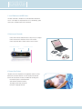
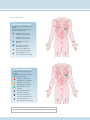
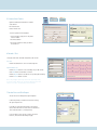
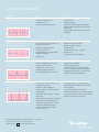
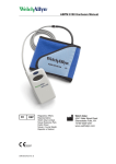
C A R D I O P E R F E C T
RESTING ECG
QUICK START GUIDE
i
CARDIOPERFECT ECG PATIENT INFORMATION
What is an electrocardiogram?
An electrocardiogram—often abbreviated, as EKG or ECG—is a test
that measures the electrical activity of the heartbeat. With each beat,
an electrical impulse (or “wave”) travels through the heart. This wave
causes the muscle to squeeze and pump blood from the heart.
Why is it done?
An ECG gives two major kinds of information. First, by measuring time
intervals on the ECG, a doctor can determine how long the electrical
wave takes to pass through the heart. Finding out how long the wave
takes to travel from one part of the heart to the next shows if the
electrical activity is normal or slow, fast or irregular. Second, by
measuring the amount of electrical activity passing through the heart
muscle, a cardiologist may be able to find out if parts of the heart are
too large or are overworked.
How is it done?
Several sensors called electrodes will pick up the electrical activity in the
heart. You will be asked to lie down, and technicians will put several
patches (electrodes) on the chest, arms and legs. Usually the electrodes
are soft and don’t cause any discomfort when they are put on or taken off
by the technician. The electrodes are connected to wires called leads,
which are connected to the ECG machine. The electrical activity of the
heart is then recorded on a moving strip of paper in the ECG machine.
During the ECG recording, you should lie quietly for 10-20 seconds,
because the electrocardiograph will detect any muscle or body movement.
Does it hurt?
No. There is no pain or risk associated with having an electrocardiogram.
Is it harmful?
No. The machine only records the ECG.
C A R D I O P E R F E C T™
PC-BASED ECG
QUICK START GUIDE
1 Install Software and USB Drivers
This Quick Start Guide is intended for use with CardioPerfect Workstation
version 1.6.0 or higher. For complete directions for use and warnings, please
consult your user manual located on the software CD.
2 Connect your Recorder
• Connect
the PC interface cable (ProLink) to a USB port on your computer.
• Connect
the other end to the black connector on the recorder.
• Connect
the patient cable to the blue connector on the recorder.
Patient Cable Connection
PC Interface Connection
On/Off Switch
3 Prepare Your Patient
The quality of an ECG is dependent on the preparation and the resistance
between the skin and the electrode. To ensure a good quality ECG and
minimize the skin/electrode resistance, remember the following points:
• Ensure
that the patient is warm and relaxed.
• Shave
electrode area before cleaning.
• Thoroughly
• Let
clean the area with alcohol.
dry prior to applying electrodes.
4 Connect Electrodes
AHA Electrode Placement
RA and LA electrodes should be placed on
the wrists.
RL and LL electrodes should be placed a
few inches above the ankle.
V1
Fourth intercostal space at
the right border of the sternum
V2
Fourth intercostal space at
the left border of the sternum
V3
Midway between locations
V2 and V4
V4
At the mid-clavicular line in
the fifth intercostal space
V5
At the anterior axillary line on
the same horizontal level as V4
V6
At the mid-axillary line on the
same horizontal level as V4 and V5
IEC Electrode Placement
R and L electrodes should be placed on
the wrists.
N and LF electrodes should be placed a
few inches above the ankle.
C1
Fourth intercostal space at
the right border of the sternum
C2
Fourth intercostal space at
the left border of the sternum
C3
Midway between locations
C2 and C4
C4
At the mid-clavicular line in
the fifth intercostal space
C5
At the anterior axillary line on
the same horizontal level as C4
C6
At the mid-axillary line on the
same horizontal level as C4 and C5
AAMI
V1 l
V2 l
V3 l
V4 l
V5 l
V6 l
RA l
LA l
RL l
LL l
IEC
C1 l
C2 l
C3 l
C4 l
C5 l
C6 l
Rl
Ll
Nl
Fl
5 Create a New Patient
• Open
the CardioPerfect Workstation Software.
• Select
• Fill
in Patient Card.
• Click
• To
Patient.
on the ECG Icon.
select a patient from the database:
1. In the Search box, type parts of the patient
name or number.
2. Click the Go button.
3. Click on the patient for whom you want to
record the test.
6 Record a Test
Select physician names and lead configuration, and click OK.
Auto ECG
• Click the Record button to start a 12-lead resting ECG.
Rhythm/Manual ECG
• Click
the Rhythm button to start a recording of up to 300 seconds.
• Click
Event button to mark areas of interest.
• Click
the Rhythm button a second time to stop the rhythm recording.
• Click
the Cancel button to cancel.
Note: The pretrigger option helps catch events by already saving
5 seconds of traces before you start the recording.The notch filter
removes noise that is caused by AC power interference.
7 Review Data and Print Report
• You
can choose from multiple ECG report templates.
• If
automatic printing is checked in the ECG Print settings,
the report will print now.
• If
you did not select Automatic printing in the Settings,
click on the down arrow next to the Print icon and choose
“Print Selected Formats” to initiate printing.
you just want to print one type of report, click on the
Print icon, and select the desired report page.
• If
LEAD QUALITY PROBLEMS
Condition
Causes
Actions
Lead-off information is displayed on the screen.
OR
One or more leads prints as a square wave:
• Electrode contact may be poor.
• Reattach the lead.
• A lead may be loose.
• Replace the electrode.
• A lead is disconnected from patient.
• Verify that the electrode area has been properly
prepared: shaved, cleaned with alcohol or acetone,
allowed to dry.
• Verify that electrodes have been properly stored
and handled.
Wandering baseline (an upward and
downward fluctuation of the waveforms):
• Electrodes that are dirty, corroded, loose, or
positioned on a bony area.
• Clean skin with alcohol or acetone.
• Insufficient or dried electrode gel.
• Help patient relax.
• Oily skin or body lotions.
• If wandering baseline persists, turn the
baseline filter on.
• Rising and falling of chest during rapid or
apprehensive breathing.
• Reposition or replace electrodes.
• Ask patient to remain still and relaxed.
• Patient moved.
Muscle tremor interference (random, irregular
voltage superimposed on the waveforms). May
resemble or coincide with AC interference:
• Patient is uncomfortable, tense, nervous.
• Patient is cold and shivering.
• Exam bed is too narrow or short to
comfortably support arms and legs.
• Arm or leg electrode straps are too tight.
AC interference (even-peaked, regular voltage
superimposed on the waveforms).
May resemble or coincide with muscle tremor
interference:
• Electrodes that are dirty, corroded, loose, or
positioned on a bony area.
• Insufficient or dried electrode gel.
• Patient or technician touching an electrode
during recording.
• Patient touching any metal parts of an exam
table or bed.
• Broken lead wire, patient cable, or power cord.
• Electrical devices in the immediate area,
lighting, concealed wiring in walls or floors.
• Improperly grounded electrical outlet.
• Incorrect AC filter frequency setting or
AC filter is turned off.
4341 State Street Road, PO Box 220, Skaneateles Falls, NY 13153-0220 USA
(p) 800.535.6663 (f) 315.685.2174 www.welchallyn.com
© 2007 Welch Allyn
REF 101910 Mat. Number: 708550, Ver: B
• Help patient get comfortable.
• Check all electrode contacts.
• If interference persists, turn the muscle-tremor
filter on. If interference still persists, the problem
is probably electrical in nature. See the following
suggestions for reducing AC interference.
• Verify that the patient is not touching any metal.
• Verify that the AC power cable is not touching the
patient lead cable.
• Verify that the proper AC filter is selected.
• If interference still persists, the noise may be caused
by other equipment in the room or by poorly grounded
power lines. Try moving to another room.