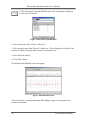
1
Heart Rhythm
Scanner
Heart Rate Variability
Analysis System
User’s Manual
Version 2.0
Copyright© 1998-2005 Biocom Technologies
All rights reserved.
Biocom Technologies makes no claim that its products treat or heal. Biocom’s products are designed to measure and monitor
physiological change. Professional should work within the scope of their licenses and/or certifications when interpreting or applying
data derived from Biocom’s products. This product is to be used for research and educational purposes only.
2
Biocom Heart Rhythm Scanner User’s Manual
TABLE OF CONTENTS
Credits..................................................................................................................................6
Software concept: .........................................................................................................6
Software design and user interface:..............................................................................6
Programming: ...............................................................................................................6
Quality Assurance:........................................................................................................6
Authoring online help and User’s manual: ...................................................................6
Special note...................................................................................................................6
1. Getting Started .................................................................................................................7
1.1. General Information..................................................................................................7
1.2. Check Package Contents ...........................................................................................8
1.3. Minimum System Requirements ...............................................................................9
1.4. Connect Sensor Device .............................................................................................9
1.5. Installation of the Heart Rhythm Scanner Software..................................................9
1.6. Uninstallation of the Heart Rhythm Scanner Software ...........................................10
2. Background Information ...............................................................................................11
2.1. Measurement of Internal Change ............................................................................11
2.2. Photoplethysmograph..............................................................................................11
2.3. Electrocardiograph..................................................................................................12
2.4. Heart Rate Monitoring and Health..........................................................................12
2.5. Heart Rate Variability .............................................................................................13
2.5.1. Time Domain Parameters: Heart Rate (HR).....................................................14
2.5.2. Time Domain Parameters: Mean NN ...............................................................14
2.5.3. Time Domain Parameters: SDNN ....................................................................14
2.5.4. Time Domain Parameters: RMS-SD ................................................................15
2.5.5. Frequency Domain Parameters: Total Power (TP)...........................................15
2.5.6. Frequency Domain Parameters: Very Low Frequency (VLF) .........................15
2.5.7. Frequency Domain Parameters: Low Frequency (LF) .....................................15
2.5.8. Frequency Domain Parameters: High Frequency (HF) ....................................16
2.5.9. Frequency Domain Parameters: LF/HF Ratio ..................................................16
2.5.10. Frequency Domain Parameters: Normalized Low Frequency........................16
(LF Norm)...................................................................................................................16
2.5.11. Frequency Domain Parameters: Normalized High Frequency (HF Norm) ....16
3. Start Heart Rhythm Scanner Session.............................................................................17
3.1. Proper Pulse Wave Sensor Placement And Use......................................................17
3.2. Proper ECG Sensor Placement And Use.................................................................17
3.2.1. Biocom 3000 Interface Unit .............................................................................17
3.2.2. ActiveECG Interface Unit ................................................................................19
3.3. Start the Heart Rhythm Scanner software ...............................................................21
4. Basic Menu Operations .................................................................................................22
4.1. Client Menu Mode ..................................................................................................22
4.1.1. Create new client record ...................................................................................22
4.1.2. Open existing client record ...............................................................................23
4.2. Protocol Menu Mode...............................................................................................23
Table of Contents
3
Biocom Heart Rhythm Scanner User’s Manual
4.2.1. Create new protocol..........................................................................................23
4.2.2. Open existing protocol......................................................................................24
5. Online Data Recording ..................................................................................................25
5.1. Start online data recording ......................................................................................26
5.2. Run next trial...........................................................................................................27
5.4. Finish trial or session recording ..............................................................................28
5.5. View trial summary.................................................................................................30
5.6. Zoom display window.............................................................................................30
5.7. Adjust graph scales..................................................................................................31
6. View Session Summary.................................................................................................33
6.1. Autonomic Balance Report .....................................................................................34
6.2. Parameter bar charts................................................................................................35
6.3. Autonomic balance diagram....................................................................................36
6.4. Heart rate trend ........................................................................................................37
6.5. Numerical HRV data sheet......................................................................................37
6.6. Zoom display windows ...........................................................................................38
6.7. Adjust graph scales..................................................................................................38
6.8. Display legend .........................................................................................................38
6.9. Review and edit trial note........................................................................................38
7. View Progress Summary...............................................................................................40
7.1. HRV Progress Report..............................................................................................40
7.2. Selected HRV parameter bar chart ..........................................................................41
7.3. Numerical HRV data sheet......................................................................................42
7.4. Adjust graph scales..................................................................................................42
7.5. Display legend .........................................................................................................42
8. Edit Heart Rate Data......................................................................................................43
9. Client Data Management ...............................................................................................46
9.1. Create new client record..........................................................................................46
9.2. Select client record ..................................................................................................46
9.3. Edit client record .....................................................................................................47
9.4. Delete Client Record ...............................................................................................47
9.5. Delete Session.........................................................................................................47
10. Protocol Management..................................................................................................48
10.1. Create new protocol ..............................................................................................48
10.2. Select protocol.......................................................................................................49
10.3. Edit protocol..........................................................................................................49
10.4. Delete protocol......................................................................................................49
11. Session Management ...................................................................................................51
11.1. Move session from one client record to another ...................................................51
11.2. Delete session........................................................................................................52
11.3. Put session data into external file ..........................................................................52
11.4. Get session data from external file ........................................................................53
11.5. Put session data into other database ......................................................................54
11.6. Get session data from other database ....................................................................55
11.7. Export session data into Excel worksheet.............................................................56
12. Database Management .................................................................................................57
4
Table of Contents
Biocom Heart Rhythm Scanner User’s Manual
12.1. Compact database..................................................................................................57
12.2. Backup database ....................................................................................................57
12.3. Restore database ....................................................................................................58
13. Printing ........................................................................................................................59
13.1. Standard Printing...................................................................................................59
13.2. Extra Printing Features..........................................................................................59
13.2.1. Print Setup ......................................................................................................59
13.2.2. Print Preview ..................................................................................................60
13.2.3. Printing ...........................................................................................................61
14. Program Settings .........................................................................................................62
14.1. Setting up an ECG device port ..............................................................................62
14.2. Program options ....................................................................................................63
14.2. Color selection.......................................................................................................64
15. Custom questionnaire ..................................................................................................65
15.1. Initial questioning..................................................................................................65
15.2. Final questioning ...................................................................................................68
15.3. View questionnaire................................................................................................68
16. Using Help ...................................................................................................................70
16.1. Help Contents ........................................................................................................70
16.2. Context Help .........................................................................................................70
17. Technical Support........................................................................................................72
Table of Contents
5
Biocom Heart Rhythm Scanner User’s Manual
Credits
Software concept:
§
§
§
Vadim Pougatchev, M.D., Biocom Technologies
Yevgeniy Zhirnov, M.D. Ph.D., Biocom Technologies ‘
Yevgeniy Gribkov, M.D., Biocom Technologies
Software design and user interface:
§
Vadim Pougatchev, M.D., Biocom Technologies
Programming:
§
§
§
Yevgeniy Gribkov, M.D., Biocom Technologies
Yevgeniy Zhirnov, M.D. Ph.D., Biocom Technologies
Vadim Pougatchev, M.D., Biocom Technologies
Quality Assurance:
§
§
Vadim Pougatchev, M.D., Biocom Technologies
Mark Ferris, BS, Biocom Technologies
Authoring online help and User’s manual:
§
§
§
Vadim Pougatchev, M.D., Biocom Technologies
Carmen V. Russoniello, Ph.D., TRS/CTRS, LPC, BCIA-EEG Associate Fellow
East Carolina University, School of Health and Human Performance
Mark Ferris, BS, Biocom Technologies
Special note
ActiveECG is a registered trademark of Active Corporation. All rights reserved.
This manual uses materials from ActiveECG User’s Manual with permission of Active
Corporation.
6
Credits
Biocom Heart Rhythm Scanner User’s Manual
1. Getting Started
1.1. General Information
Overall functioning of a living organism is controlled by the autonomic nervous
system. It has two antagonistic branches – the sympathetic and parasympathetic nervous
systems. Every organ is activated by one branch and inhibited by the other. Generally,
when the organism is in a calm state (relaxation, sleep, etc.), organs such as the heart,
lungs, and blood vessels are under parasympathetic control. When active (during physical
activity, psycho-emotional arousal or stress) these organs are dominated by the
sympathetic nervous system. A healthy organism is capable of adjusting to any external
influence by means of quick and adequate sympathetic response. Once that factor
disappears, parasympathetic activity increases, which balances overall autonomic
activity.
It is important to have a tool to evaluate the level of balance between the branches
of the autonomic nervous system, their reaction to specific provocative test factors, and
the overall condition of both branches. Such a tool can help people learn how to cope
with stress by achieving autonomic balance.
One of the most informative methods of evaluating the autonomic nervous
system, including both branches, is heart rate variability analysis. It measures the time
intervals between each two consecutive heartbeats, which vary under control of the
autonomic nervous system. When the parasympathetic system is dominant, the heart
interbeat intervals (IBI) are oscillating with higher frequencies (0.15 – 0.40 Hz). When
sympathetic arousal occurs, the lower frequency oscillations take place.
There are two other physiological modalities that reflect the state of autonomic
regulation: skin conductance and peripheral skin temperature. Skin conductance reflects
changes in sweat gland activity driven by involuntary arousal of the sympathetic nervous
system. These changes include both rapid changes, the organism reacting to external
events, and slow changes, reflecting variation in the overall tone of the sympathetic
system. The skin temperature shows the degree of vasoconstriction or dilation of the
peripheral blood vessels, an indication of the long-term process of interaction between
both branches of the autonomic nervous system. There is a great deal of ambiguity in
using these two modalities for evaluation of autonomic balance, so we use them for
reference purposes only.
There is a standard mathematical procedure for short-term HRV evaluation,
suggested by the Task Force of the European Society of Cardiology and the North
American Society of Pacing and Electrophysiology (1996). It provides both time and
frequency domain analysis of the IBI time series. There are three important parameters of
frequency domain analysis within HRV that reflect the levels of sympathetic and
parasympathetic activity and their balance. The high frequency range (0.15 Hz – 0.40 Hz)
of the IBI power spectrum (HF) reflects parasympathetic influence on heart rate. The low
frequency range (0.04 Hz – 0.15 Hz) of the IBI power spectrum (LF) displays
Getting Started
7
Biocom Heart Rhythm Scanner User’s Manual
sympathetic influence. The LF/HF ratio is used to show the balance between both
branches of the autonomic nervous system.
To perform the HRV analysis, an electrocardiograph (ECG) signal is usually
measured. The IBI are derived from the ECG as the intervals between consecutive Rpeaks. This method is very accurate and reliable but has a serious disadvantage – it
requires using complex ECG equipment and performing inconvenient multiple electrode
placement. The alternative way is to use a photoplethysmograph (PPG) measurement by
means of a portable and convenient finger sensor. It emits an IR light on the skin. The
emitted light is partially consumed by the blood flow. The degree of light consumption /
reflection is proportional to the changes in blood flow. The PPG signal has periodic peaks
that represent blood vessel pulsation. It can be also used to derive the IBI as the time
between two PPG peaks.
The Heart Rhythm Scanner is a versatile instrument for measuring and evaluating
heart rate variability (HRV). It does live recording of PPG signals, computing the
instantaneous changes in heart rate, and provides full analysis of heart rate variability
after the session is complete. Heart Rhythm Scanner allows the creation of any specific
protocol of HRV evaluation as a sequence of trials – periods of data recordings in simple
data units. For instance, let’s suppose we need to evaluate the effect of some drug on the
autonomic nervous system. Heart Rhythm Scanner allows building a protocol consisting
of two trials, each five minutes long. The first trial is to evaluate HRV condition before
drug intake. The second one is taken a certain amount of time after drug intake. Heart
Rhythm Scanner does full HRV analysis on both trials and brings up detailed reports,
which can be easily compared.
1.2. Check Package Contents
When you receive Heart Rhythm Scanner package, it must include the following
items:
Pulse Wave Sensor Version:
1. HRM-02 pulse wave sensor device with an ear-clip probe
2. “Heart Rhythm Scanner” software CD
3. User’s manual
Biocom 3000 ECG Version:
1.
2.
3.
4.
8
Biocom 3000 ECG interface unit
Y-type ECG electrode cable
“Heart Rhythm Scanner” software CD
User’s manual
Getting Started
Biocom Heart Rhythm Scanner User’s Manual
ActiveECG Version:
1.
2.
3.
4.
5.
6.
ActiveECG interface unit
Set of disposable pre-gelled ECG electrodes
Serial cable to connect ECG unit to PC
Test Cable
“Heart Rhythm Scanner” software CD
User’s manual
Contact Biocom Technologies or your local dealer if you have any concern regarding
content of the product received.
1.3. Minimum System Requirements
The Heart Rhythm Scanner has the following hardware requirements to ensure its reliable
functioning:
• CPU Pentium III 800 MHz
• 256 MB of RAM
• SVGA video card with at least 800x600, Hi-Color resolution
• At least 100 MB of free hard disk space
• CD ROM drive or Floppy Disk Drive
• One available USB port (COM port for Active ECG)
• Windows XP Operating system
If you are not sure about your computer, contact your local computer specialist to verify
whether your computer complies to the above requirements.
1.4. Connect Sensor Device
Simply attach the USB cable connector of the sensor to any USB port available in the
computer.
Sensor device can be connected with computer on or off. The device will be detected
automatically when software starts.
1.5. Installation of the Heart Rhythm Scanner Software
The Heart Rhythm Scanner software is Windows Me/2000/XP compatible (XP is
preferred). It is delivered on a CD-ROM.
To install it:
1) Insert the CD-ROM into the drive.
2) Click the “Start” button on the taskbar and select the “Run” menu option.
3) In the “Run” dialog box, type the following command line:
Getting Started
9
Biocom Heart Rhythm Scanner User’s Manual
D:\SETUP.EXE
4) Click the “OK” button to start installation.
5) Follow the program instructions to complete installation.
Fig.1.1. Installation dialog box
1.6. Uninstallation of the Heart Rhythm Scanner Software
To uninstall the Heart Rhythm Scanner software:
1)
2)
3)
4)
5)
6)
Click “Start” button on the taskbar.
Select “Settings” item from the system menu.
Select “Control Panel” option from the “Settings” menu.
Double-click “Add/Remove Programs” icon.
Select “Heart Rhythm Scanner” from the list and click “Add/Remove” button.
When InstallShield wizard window appears, select “Remove” option, click “Next”
button and follow on-screen instructions.
10
Getting Started
Biocom Heart Rhythm Scanner User’s Manual
2. Background Information
2.1. Measurement of Internal Change
Biocom research indicates that a highly accurate way to monitor internal physiological
changes related to the regulatory activity of the human autonomic nervous system (ANS)
is through the evaluation of variation in heart rate - Heart Rate Variability Analysis
(HRVA). “Heart Rhythm Scanner” includes sophisticated, technologically sound, but
user friendly software that measures and monitors HRV by means of using either
Photoplethysmograph PPG (simple less expensive product for the client use) or
Electrocardiograph ECG (professional product) monitoring devices. “Heart Rhythm
Scanner” also works dynamically with respiration and has a variety of phys ical,
psychological and sports medicine applications. Heart Rate Variability is a physiological
measurement that directly reflects a balance of the autonomic nervous system regulation,
which has control over all human body. HRV reflects the state of sympathetic (stress,
anxiety) or parasympathetic (relaxation, calmness) activation in the body.
2.2. Photoplethysmograph
Photoplethysmograph (PPG) was developed in the 1960's and 1970's by
psychophysiology researchers. The instrument is used for assessment of peripheral blood
flow to the extremities. When the client is stressed their blood vessels constrict (less
blood flow in fingers). Photoplethysmograph is based upon the premise that all living
tissue and blood have different light-absorbing properties. PPG works by placing an
individual finger between two parts of a transducer consisting of a light source and a
photocell (which converts light to electrical energy). A beam of infrared light is projected
toward the photocell. The blood in the finger scatters light in the infrared range, and the
amount of light reaching the cell is inversely related to the amount of blood in the finger.
Hence, when blood vessels in the finger dilate, the increased blood flow allows less light
to reach the photocell, when blood vessels constrict, blood flow is decreased and
increased light reaches the photocell.
Pulse volume measurements are related beat-to-beat variations in the force of blood flow.
Phasic changes made on a beat-to-beat basis, are called pulse volume measurements and
are related to beat variations in the force of blood flow. These beat-to-beat changes in
peripheral blood flow reflect ANS regulatory activity and can be used for the purpose of
evaluation of sympathetic activity or stress. (“Measuring Stress” by S. Cohen, R. Kessler
& L. U. Gordon, 1995).
Heart Rhythm Scanner employs a different method of analysis and is based upon beat-tobeat changes in inter-beat time intervals to reflect ANS regulatory activity.
Background Information
11
Biocom Heart Rhythm Scanner User’s Manual
2.3. Electrocardiograph
Electrocardiograph (ECG) is a tracing, which represents the heart electrical activity
derived by amplifying minutely small electrical impulses generated by the heart. The
electrical impulses generated in the heart muscle are recorded and transmitted to the
electrocardiograph by electrodes or leads attached to the body by small disposable metal
plates. The moistened plates are attached to the arms, legs, and cardiac areas.
ECG is a widely accepted and important diagnostic tool. ECG is used in some routine
medical examinations when a heart disorder is present or suspected. ECG helps diagnose
the damage that may have been inflicted on the heart muscle as a result of coronary
occlusion, rheumatic fever, the presence of abnormal rhythms, and the effect of drugs.
Moreover, ECG recordings provide information on the current fitness status of the heart
as well as reflect HRV as a result of ANS regulatory balance.
2.4. Heart Rate Monitoring and Health
It is well known from many scientific studies, that Heart Rate (HR) is one of the most
important parameters used for the evaluation of human psycho-physiological stability and
optimum performance. HR in fact, is an integral parameter obtained as a result of the
input of three factors:
•
•
•
Sympathetic Neural Regulation
Parasympathetic Neural Regulation
Hormonal Regulation
HR is highly dynamic, significantly informative, and easy to use for continuous
monitoring purposes. The rhythmograph is a sequence of heart inter-beat intervals
calculated in milliseconds. The analysis of rhythmograph provides information to
determine heart rate variability (HRV). Heart rate variability is determined by a statistical
algorithm and is a very robust measurement of the status of the heart as well as ANS
regulatory balance.
There are many internal and external factors that cause changes in heart rate. For
example, fear, be it real or perceived, is experienced by an increase in heart rate. Left
unchecked the individual will eventually adapt to this dysfunctional pattern and fatigue
(both mental and physical) and emotional distress will eventually occur. Consequently,
hormonal and neurological overwork occurs and causes a decrease in life satisfaction and
individual performance. Long-term exposure to these same processes will cause illness
and disease such as cardiovascular disease, adult onset diabetes, anxiety, depression panic
disorders, and so forth. Thus, the measurement and monitoring of HRV becomes a
critical window in the evaluation and amelioration of negative conditions. Given HRV is
one of the most informative methods of evaluating mental stress and fatigue it is easy to
see why.
12
Background Information
Biocom Heart Rhythm Scanner User’s Manual
Upon evaluation of the HRV parameters, a professional can interpret the individuals
actual physiological stress status as:
•
•
•
•
Normal
Functional Stress
Distress
Exhaustion
Thus, HRV analysis gives the evaluator the ability to assess the level of psychoemotional stress (be it their own or a clients), and provides information to forecast a level
of mental distress, fatigue, and possible exhaustion. Stress levels can be identified, even
when the user is not aware of his or her physiological reaction to stress or their
consequential decrease in performance. Information derived from HRV can be used in
conjunction with coping tools such as physical and mental breaks, physical exercise,
breathing (relaxation) exercises, and \ or games to assist the individual in becoming
“physiologically relaxed”. HRV is, therefore, an important tool in assisting individuals
increase their productivity and elevate their mood while helping them predict, prevent,
and treat their stress related medical conditions.
2.5. Heart Rate Variability
There are different methods of evaluation of the variations in heart rate. Two of them are
most common and are recommended by the Task Force of the European Society of
Cardiology and the North American Society of Pacing and Electrophysiology:
•
•
Time Domain Methods
Frequency Domain Methods
Time domain methods are commonly used for 24-hour data recordings. However,
according to the Task force short-term recordings (e.g. 5- min length) can also be
analyzed.
Frequency domain methods can be easily used for short-term recordings as well.
The following heart rate variability parameters are calculated by Heart Rhythm Scanner
based on short-term data record (at least 2 minutes long):
Time Domain Parameters:
•
•
•
•
Heart Rate
Mean NN
SDNN
RMS-SD
Background Information
13
Biocom Heart Rhythm Scanner User’s Manual
Frequency Domain Parameters:
•
•
•
•
•
•
•
Total Power
VLF
LF
HF
LF/HF Ratio
LF Norm
HF Norm
All HRV parameters are calculated on ‘normal-to- normal’ (NN) interbeat intervals (or
NN intervals) caused by normal heart contractions paced by sinus node depolarization.
All time domain parameters are derived directly from NN intervals recorded during the
trial.
The frequency domain parameters are derived from the power spectral density (PSD)
calculated by means of Fast Fourier Transform (FFT).
2.5.1. Time Domain Parameters: Heart Rate (HR)
This is a mean heart rate value averaged on entire recording (trial).
HR is measured in beats per minute (BPM).
2.5.2. Time Domain Parameters: Mean NN
This is a mean interbeat interval value averaged on entire recording (trial).
Mean NN is measured in milliseconds.
2.5.3. Time Domain Parameters: SDNN
SDNN is a standard deviation of the NN intervals, which is the square root of their
variance.
A variance is mathematically equivalent to the total power of spectral analysis, so it
reflects all cyclic components of the variability in recorded series of NN intervals.
The actual values of SDNN depend on the length of recording - the longer recording is,
the higher SDNN values are. Thus, in practice it is inappropriate to compare SDNN
values derived from the NN recording of different length.
SDNN is measured in milliseconds.
14
Background Information
Biocom Heart Rhythm Scanner User’s Manual
2.5.4. Time Domain Parameters: RMS-SD
RMS-SD is the square root of the mean squared differences of successive NN intervals.
This measure estimate high- frequency variations in heart rate in short-term NN
recordings that reflects an estimate of parasympathetic regulation of the heart.
RMS-SD is measured in milliseconds.
2.5.5. Frequency Domain Parameters: Total Power (TP)
In fact, Total Power is a short-term estimate of the total power of power spectral density
in the range of frequencies between 0 and 0.4 Hz.
This measure reflects overall autonomic activity where sympathetic activity is a primary
contributor.
Total Power is calculated in milliseconds squared (ms^2).
2.5.6. Frequency Domain Parameters: Very Low Frequency (VLF)
Very Low Frequency is a band of power spectrum range between 0.0033 and 0.04 Hz.
This measure is not well defined in terms of physiological mechanisms causing VLF
component of the power spectrum. Generally it is known this parameter indicates overall
activity of various slow mechanisms of sympathetic function.
Very Low Frequency band is calculated in milliseconds squared (ms^2).
2.5.7. Frequency Domain Parameters: Low Frequency (LF)
Low Frequency is a band of power spectrum range between 0.04 and 0.15 Hz.
This measure reflects both sympathetic and parasympathetic activity.
Generally it is a strong indicator of sympathetic activity in long-term recordings.
Parasympathetic influence is represented by LF when respiration rate is lower than 7
breaths per minute or during taking a deep breath. Thus, when subject is in the state of
relaxation with a slow and even breathing, the LF values can be very high indicating
increased parasympathetic activity rather than increase of sympathetic regulation.
Low Frequency band is calculated in milliseconds squared (ms^2).
Background Information
15
Biocom Heart Rhythm Scanner User’s Manual
2.5.8. Frequency Domain Parameters: High Frequency (HF)
High Frequency is a band of power spectrum range between 0.15 and 0.4 Hz.
This measure reflects parasympathetic (vagal) activity. HF is also known as a
‘respiratory’ band because it corresponds to the NN variations caused by respiration (this
phenomena is known as respiratory sinus arrhythmia (RSA)). Heart rate is increased
during inhalation and dropped during exhalation. Slower and even breathing causes an
increase in the amplitude of HF peak on power spectrum.
High Frequency band is calculated in milliseconds squared (ms^2).
2.5.9. Frequency Domain Parameters: LF/HF Ratio
This is the ratio between the power of Low Frequency and High Frequency bands.
This measure indicates overall balance between sympathetic and parasympathetic
systems. Higher values reflect domination of the sympathetic system, while lower ones domination of the parasympathetic system. However, when deep and even breathing
occurs, the elevation of this parameter reflects increase of parasympathetic regulation due
to effect of RSA.
LF/HF Ratio is calculated in normalized units.
2.5.10. Frequency Domain Parameters: Normalized Low Frequency
(LF Norm)
Normalized Low Frequency is the ratio between absolute value of the Low Frequency
and difference between Total Power and Very Low Frequency.
This measure minimizes an effect of changes in Very Low Frequency power and
emphasizes changes in sympathetic regulation.
Normalized LF is calculated in percentile units.
2.5.11. Frequency Domain Parameters: Normalized High Frequency
(HF Norm)
Normalized High Frequency is the ratio between absolute value of the High Frequency
and difference between Total Power and Very Low Frequency.
This measure minimizes an effect of changes in Very Low Frequency power and
emphasizes changes in parasympathetic regulation.
Normalized HF is calculated in percentile units.
16
Background Information
Biocom Heart Rhythm Scanner User’s Manual
3. Start Heart Rhythm Scanner Session
3.1. Proper Pulse Wave Sensor Placement And Use
Heart Rhythm Scanner measures HRV data by means of a pulse wave sensor with an earclip probe. It is a small plastic box. A separate ear-clip sensor probe is connected to the
device box. The device box has a long USB cable.
Connect the device USB cable to any USB port socket on the computer. The Heart
Rhythm Scanner software will detect it automatically.
Place an ear-clip on the left or right ear lobe. Make sure it is placed so that small glass
windows on the inside surfaces of the clip are in good contact with soft tissue of the ear
lobe. Be aware that proper placement will affect the quality of pulse wave signal. Small
ear lobes or earrings may make placement more difficult.
Fig. 3.1. Pulse wave sensor placement
3.2. Proper ECG Sensor Placement And Use
The Heart Rhythm Scanner supports two types of ECG interface units:
•
•
Biocom 3000 unit
ActiveECG unit
3.2.1. Biocom 3000 Interface Unit
Biocom 3000 is a portable unit connected to your PC with a USB cable and a standard set
of ECG sensors:
Option 1: Y-type cable terminated with two small finger pads (that embed small silver /
silver chloride dry ECG electrodes) with elastic Velcro straps (See. Fig. 3.2.).
Starting Heart Rhythm Scanner Session
17
Biocom Heart Rhythm Scanner User’s Manual
Option 2: Y-type cable terminated with two small flat plastic pads (that embed large
silver / silver chloride dry ECG electrodes) with two elastic wristbands (See. Fig. 3.3.).
Connect the device USB cable to any USB port socket on the computer. The Heart
Rhythm Scanner software will detect it automatically. Connect a flat RJ-45 connector of
the Y-cable to input “B” on the front panel of Biocom 3000 unit (opposite side to LED
indicator).
Option 1: Place two fingers of both hands on top of the rounded surface of the finger
pads. Use the Velcro straps to secure pads on the fingers. The fingers should be fixed on
pads to avoid movement artifacts. However they should not be too tight since that can
reduce blood flow. Loosen it slightly if pulsation can be felt in the fingers.
Fig. 3.2. ECG finger pad electrode placement
Option 2: Put elastic wristbands on both wrists. Place both sensor pads marked as “Left”
and “Right” on respective wrists with electrode disks face down so that wristbands secure
those pads firmly.
Fig. 3.3. ECG wrist electrode placement
18
Starting Heart Rhythm Scanner Session
Biocom Heart Rhythm Scanner User’s Manual
3.2.2. ActiveECG Interface Unit
The ActiveECG hardware has features, which make it exceptional as a portable ECG
device. The unit is designed for quick assessment. There are no buttons, switches, or
controls - everything is controlled from the PC. The enclosure itself is designed to hold
the ECG leadwires in a compact, easy-to-use platform. Additionally, no latex is used
anywhere within the ActiveECG hardware to help avoid allergic reactions from personnel
and clients.
The front of the ActiveECG hardware houses a “raceway” which the leadwires are
wrapped around. This provides a simple way to manage the leadwires and guarantee that
they are always with you. An integrated elastomeric cover surrounds the raceway holding
in the leadwire snap connectors. The cover can be replaced by slightly flexing the bottom
of the cover and popping out one of the two ends of the cover hinges.
After the leadwires have been unwound from the raceway and attached to electrodes, the
elastomeric cover can be closed if you would like to secure it. Leaving the cover open
allows you to see that the leadwires stay connected to the raceway connectors.
When you have completed session, disconnect the electrodes, clean the leadwires if
necessary, and wrap them around the opened raceway. When there is an inch or so of the
leadwires that has not been wound onto the raceway, tuck the remainder of the leadwires
into one of the raceway openings and close the cover to secure them. Alternatively, the
snap connectors at the end of the leadwires can drape outside the raceway, held in place
by the elastomeric cover.
Note that when you connect the leadwires, the colors of the leadwires should match the
colors of the connectors.
The components of ActiveECG should be inspected on a regular basis and prior to each
use. These inspections are important in order to guarantee the proper working condition
of your system.
Perform the following steps to inspect ActiveECG:
1. Inspect the physical condition. Check to ensure that cases and controls are clean
and in good condition.
2. Inspect cables, connectors, and electrodes. Make sure that the ECG leadwires are
attached inside the raceway. Inspect cables for cracks, breaks, or frayed wires. If
any damage is found, replace the cables immediately. Check for adequate supply
of ECG electrodes. Make sure the electrodes have not exceeded their expiration
date (if available) or that the packages have not been opened or damaged.
3. Make sure that a new spare battery is always available. Check that all batteries
being used and the spare battery are not beyond their expiration date.
Background Information
19
Biocom Heart Rhythm Scanner User’s Manual
Leadwire and electrode connection
The ActiveECG hardware has three ECG monitoring leadwires that provide the
monitoring of lead II only. The leadwires and connectors are all color-coded according to
the American Heart Association (AHA) standards. The following table provides a list of
AHA color codes for lead II connections:
Lead
II
Bipolar
RA - right arm
LA - left arm
LL - left leg
Lead and reference
Negative electrode
Reference
Positive electrode
Color
White
Black
Red
The connection from ActiveECG to each leadwire is an industry standard single .060 pin
connector. This allows you to easily replace the leadwires with other compatible leadwire
cables. Make sure that the leadwire cables are inserted fully into the ActiveECG
hardware. Loose or poor connections can cause intermittent results.
The leadwires provided with ActiveECG utilize standard snap connectors allowing you to
use a wide variety of commercially available electrodes. For best ECG monitoring
results, silver/silver chloride (Ag/AgCl) electrodes should be used with this equipment.
Electrodes of dissimilar metals should not be used.
Take care when attaching or removing the leadwires and electrodes to not damage the
wires or connectors.
Connection to PC
The ActiveECG hardware connects to a PC with a null- modem serial cable. After
connecting the cable to the ActiveECG hardware, make sure to screw in the thumbscrews
securely attaching the ActiveECG hardware to the cable.
To ensure accurate ECG recording, it is important that the electrodes be properly attached
to the client. Follow the instructions below to prepare the client.
Attaching electrodes to a client:
1. Prepare the client’s skin for effective electrode adhesion. Clean and dry the skin.
Briskly wipe skin dry with a towel or gauze to mildly abrade the skin and remove
oils, dirt, etc. Note that for proper electrode adhesion, never use alcohol, tincture
of bezoin or antiperspirant to prepare the skin.
2. Remove the disposable electrodes from their package. Attach the leadwires to the
electrodes and then peel off the protective backing on the electrodes. Inspect the
electrodes for any obvious signs of damage. Ensure that the gel on the electrodes
has not dried out. If you note damage or dry electrodes, use a new set of
electrodes.
20
Starting Heart Rhythm Scanner Session
Biocom Heart Rhythm Scanner User’s Manual
3. Place two electrodes on the client’s left wrist and one electrode on right wrist.
Apply the electrode flat to the skin and smooth the tape outward in all directions.
Do not press the center of the electrode.
4. Connect black and red leadwires (LA and LL) to the electrodes on left wrist and
white (RA) leadwire to the electrode on right wrist.
5. Attach the ActiveECG enclosure to the client’s clothing, belt, or something in
close proximity using the spring- loaded clip on the back. Position the device so as
to limit any potential strain put on the leadwires and electrodes.
After each client use:
1. Discard used electrodes including electrode sets that were opened and not used.
Electrodes dry out in less than 24 hours.
2. Replace the battery in the ActiveECG device if a low battery is indicated in the
test program. Make sure you have a spare battery.
3. Clean the device.
4. Inspect the device and cables for damage. Repair or replace damaged items
immediately.
5. Check that the leadwires are well connected to the connectors on the ActiveECG
device. Wrap the leadwires around the raceway and tuck the snap connector ends
inside before closing the elastomeric cover.
6. Recheck disposable supplies and replace whatever is needed.
3.3. Start the Heart Rhythm Scanner software
Double-click this shortcut icon on the desktop screen to start the Heart Rhythm
Scanner.
Once the program is loaded, the following screen comes up:
Fig.3.4. Main application window
Background Information
21
Biocom Heart Rhythm Scanner User’s Manual
4. Basic Menu Operations
Heart Rhythm Scanner provides two menu modes:
•
•
Client Mode
Protocol Mode
You can switch between these modes clicking respective buttons on the toolbar.
4.1. Client Menu Mode
Click this button to switch to client menu mode. It brings a list of client
records represented by separate “client folders”. Click on any of them to select
client record you need. Then you can proceed with any of the following
operations:
•
•
•
•
•
•
Create a new client record
Edit selected client record
Delete selected client record
Enter pre-test questionnaire
Enter post-test questionnaire
View questionnaire data
4.1.1. Create new client record
Click this button to create a new client record. It brings up the following
dialog box:
Fig.4.1. Client personal information dialog box
Enter data in the fields and click the “OK” button.
22
Basic Menu Operations
Biocom Heart Rhythm Scanner User’s Manual
A new “client folder” icon appears in the main application window once a
new client record ha been created.
4.1.2. Open existing client record
Click a client folder in the list to highlight it. Then you can
accomplish any of the remaining operations with client record
by clicking respective button on the toolbar that are available
while you are in client menu mode.
4.2. Protocol Menu Mode
Click this button to switch to protocol menu mode. It brings a list of HRV
evaluation protocols available in the system that are represented by separate
“protocol folders”. Click on any of them to select a protocol you need. Then
you can proceed with any of the following operations:
•
•
•
•
•
•
Create a new protocol
Edit selected protocol
Delete selected protocol
Run selected protocol session
View selected protocol sessions
View selected protocol progress
4.2.1. Create new protocol
What is a protocol? It is a specific procedure of using HRV data recording and analysis.
It has a predetermined timing structure – a set of single trials of specified duration.
A trial is a single unit of a protocol. It defines a contiguous data recording that can be
processed to get a standard HRV evaluation. The time length describes it.
Click this button on the client toolbar to create a new protocol. It brings up the
following dialog box:
Fig.4.2. Protocol properties dialog box
Basic Menu Operations
23
Biocom Heart Rhythm Scanner User’s Manual
Enter a new protocol name.
Use the “Add” and “Insert” commands to add trials to this protocol. The “Add” command
adds a trial to the end of a trial list. The “Insert” command adds a new trial to a specific
position highlighted in the list of trials.
Use the “Delete” command to eliminate a trial from the list.
Use the “Properties” command to modify the settings of an existing trial.
Check “Automatically run questionnaire before and after each session” check-box to
enable automatic run of questionnaire.
Use “Trial Duration” radio-buttons to select trial duration in this protocol. All trials will
have the same duration. It is required to be able to compare HRV data across trials.
Important Note! If you are using Biocom 3000 ECG interface unit, it is recommended to
set trial duration either 5- or 10- minutes long. The HRV population norms can only be
used with records 5- or 10-minutes long.
Click the “OK” command to complete construction of the protocol.
When adding a new trial or editing an existing one, the following dialog box is displayed:
Fig.4.3. Trial properties dialog box
Enter at least a new trial name, then click the “OK” button.
4.2.2. Open existing protocol
Click a protocol folder in the list to highlight it. Then you can accomplish any of the
operations with selected protocol by clicking respective button on the toolbar that are
available while you are in the protocol menu mode.
24
Basic Menu Operations
Biocom Heart Rhythm Scanner User’s Manual
5. Online Data Recording
To start an online data recording session, do as follows:
1) Open the client record you need.
2) Select an appropriate protocol.
3) Click this button to open the online monitoring screen.
The Heart Rhythm Scanner allows suspending current session and leaving it incomplete
with assumption that it will be resumed later (e.g. next day). If you previously ran this
protocol for selected client and left at least one session not complete, the following dialog
box appears requesting to specify whether one of suspended sessions should be resumed
or new one started:
Fig.5.1. Select session to be resumed
Select session to be resumed from the list and click “OK”.
If you wish to start new session, click “New Session” button.
If you resume session selected from the list, the following dialog box is displayed:
Fig.5.2. Select trial to run
Select next trial to run and click “Start Trial” button.
Online Data Recording
25
Biocom Heart Rhythm Scanner User’s Manual
When online monitoring starts, the following screen comes up:
Fig.5.3. HRV monitoring screen
This is a standard online screen. It includes the following windows:
1)
2)
3)
4)
Heart rate graph (upper left).
Heart rate value indicator (upper right).
Heart rate scatter chart (middle right).
Pulse wave or ECG graph (bottom).
The heart rate graph displays instantaneous changes in heart rate, updated every 0.5
seconds.
The heart rate value indicator displays the last HR value, updated every 0.5 seconds.
The heart rate scatter graph is also updated every 0.5 seconds. It reflects the variability of
the heart rate, graphically displaying current HR value versus the previous one. The more
variation in HR, the wider the “cloud” that is displayed. If the heart rate were constant,
the “cloud” would be convoluted into a single dot.
The pulse wave graph is updated 64 times per second. It displays live pulse wave signals
acquired by the finger sensor. It is helpful to observe this signal to be sure the sensor is
placed appropriately.
5.1. Start online data recording
Click this button to initiate data recording.
Observe pulse wave signals scrolling in the bottom window to make sure that the sensor
is placed correctly. When placement is correct, you will see a smooth signal with periodic
peaks:
26
Online Data Recording
Biocom Heart Rhythm Scanner User’s Manual
Fig.5.4. Normal pulse wave
Here is an example of artifacts caused by poor sensor placement:
Fig.5.5. Abnormal pulse wave
If this happens, reposition the finger on the sensor and secure it with the Velcro strip until
you see clear pulse waves. Then restart the current trial to get rid of the data containing
artifacts.
If you are using Biocom 3000 ECG interface unit, make sure that ECG signal is clean and
free of movement artifacts. When ECG electrode placement is correct, you should see an
ECG trace looking like that:
Fig.5.6. Normal ECG signal
Click this button to restart the trial.
The status bar displays information about progress for the current session:
Fig.5.7. Session progress indicator
It displays the current trial number, time elapsed, time remaining, and a progress
indicator.
5.2. Run next trial
Once the current trial is complete or manually terminated, the program prompts you to
advance to the next trial or chose other options. The following dialog box appears:
Online Data Recording
27
Biocom Heart Rhythm Scanner User’s Manual
Fig.5.8. Run trial dialog box
The next trial is automatically highlighted. Click “Start Trial” button to initiate it.
Use the “Add”, “Insert”, “Delete” and “Properties” buttons to modify the protocol of the
ongoing session. Refer to the protocol creation section for information on how to use
these functions. This feature allows you to create protocols on the fly.
Enter a text note for selected trial into trial note entry field.
If any modification occurs, the program will prompt you to save it when quitting online
mode.
5.4. Finish trial or session recording
Normally Heart Rhythm Scanner automatically finishes current trial once its time expires,
and terminates the session once all trials are completed.
However, you have an option to terminate current trial prematurely or even terminate an
entire session at any time.
Click this button to stop the current trial.
If the trial was prematurely stopped, its data set is considered incomplete and cannot be
used for HRV evaluation.
When trial stops (automatically or by command) the program displays a dialog box
illustrated on Fig.5.8.
Click “Stop sessio n” button to finish this session. This will bring a session preview
screen. It allows for reviewing the results of session just completed and decide whether
data should be saved in client record.
28
Online Data Recording
Biocom Heart Rhythm Scanner User’s Manual
Fig.5.9. Session preview screen
Click this button to save session.
Click this button to switch to main mode menu.
If there is data recorded, the program prompts you to save this session to the current
client record:
Fig.5.10. Save session data
If you forgot to select right client before session started, select one from the combo-box
in this dialog box.
If this is a new client, click “New Client” button to create new client record.
Click “Yes” to save current session data to selected client record.
Click “No” to leave online mode without saving session data.
Click “Cancel” to go back to online mode.
When leaving run session mode, you can specify whether you wish to do final close of
this session or want to run it again. This feature is particularly helpful if the protocol you
are running requires considerable time between trials (e.g. next day) and it is not possible
to proceed with next trial without quitting program. The following dialog box appears
allowing for this selection:
Online Data Recording
29
Biocom Heart Rhythm Scanner User’s Manual
Fig.5.11. Select session termination option
5.5. View trial summary
Sometimes it is important to see HRV data immediately following the end of current trial.
If the protocol assumes stops between trials, you can view current trial summary. When
protocol dialog box (See Fig.5.8.) appears between trials, click “Summary button” and
view last trial summary:
Fig.5.12. Trial summary screen
Click this button to get back to online screen.
5.6. Zoom display window
The online screen contains several display windows. You can enlarge any of them
(except the heart rate value indicator window) to better see details.
To enlarge a window:
1) Select the window you need to enlarge. You can make your selection either by
clicking the area of the window you need or by repeatedly pressing the “Tab” key
30
Online Data Recording
Biocom Heart Rhythm Scanner User’s Manual
until the window is selected. The selected window has a title name in italics, shaded
in white.
Fig.5.13. Selected window title
2) Click this button to zoom the selected window.
Click it again to return the enlarged window to its normal size.
5.7. Adjust graph scales
Each graph or chart has two scales: a vertical Y-Scale (representing amplitude) and a
horizontal X-Scale (usually a time scale except on the scatter chart).
The Y-Scale is determined by the scale range – the difference between maximum and
minimum signals. It is adjusted to different values to make the range wider or narrower,
depending on the variation within the signal. If the range were large and the signals had a
small amount of variation, it would be hard to see the signal details. On the other hand, if
the variation in signals were too big, the signal might be bounced out of this range.
The Y-Scale is also determined by the scale offset (origin) which positions the current
range within the full range of possible signal values.
For instance, the full range of heart rate is between 30 and 150 beats per minute. The
current range might be 40 with an offset at 50. It could give a scale between 50 and 90
beats per minute.
To adjust the scales:
1) Select the window you need to adjust. You can make your selection either by clicking
the area of the window you need or by repeatedly pressing the “Tab” key until the
window is selected.
2) Click this button to open the scale adjustment dialog box:
If you wish the Y-Scale to be self-adjusting during the online signal display, set the
“Autoscale” check-box to an “ON” state. Clear it if you wish to have the scale range
fixed.
Online Data Recording
31
Biocom Heart Rhythm Scanner User’s Manual
Fig.5.14. Scale adjustment dialog box
When the “Autoscale” setting is OFF, you can use the scale range slider bar to set an
appropriate scale range. The selected range is displayed above the slider bar.
If you want to have amplitude scale with the scale origin fixed at certain level, check
“Anchor” check-box. If it is cleared, the scale origin will be automatically updating if
“Autoscale” function is active.
If “Anchor” is checked, you can manually adjust scale origin using an amplitude scale
offset slider bar. The selected offset is displayed above the slider bar.
To adjust the time scale, use the “X-Scale settings” control in the same way as to adjust
the Y-Scale. The setting is displayed to the left of the slider bar. Check “Trial” check-box
to set time scale equal to the trial length.
Check “Save settings as default to current client” check-box if you want to have current
settings automatically invoked when the same client is selected for the session.
Click “Set default settings” button to alter any customized settings to the factory preset
settings”.
You can also see the result of a scale adjustment in a preview window within this dialog
box
Click “OK” button to apply the new scale settings to the selected window.
Click “Cancel” button to discard all setting changes made.
32
Online Data Recording
Biocom Heart Rhythm Scanner User’s Manual
6. View Session Summary
The session summary allows you to view the details of the HRV data collected during a
specific session of a selected protocol for the current client.
To view the summary of a specific HRV evaluation session:
1) Open the client record you need.
2) Select an appropriate protocol.
3) Select an appropriate session:
Fig.6.1. Session selection dialog box
4) Click this button to open the session summary screen.
Once this is done, the following screen is displayed:
Fig.6.2. Session summary screen
The session summary consists of six pages of data reports. Click the appropriate page
tabs at the bottom of the screen to switch between pages.
View Session Summary
33
Biocom Heart Rhythm Scanner User’s Manual
6.1. Autonomic Balance Report
The First (default) page displays a printable “Autonomic Balance Report” form that
consists of a number of letter-size pages, one page per trial accomplished. Each page
includes:
Fig.6.3. Client personal information section
Fig.6.4. Numerical HRV data section
Fig.6.5. Heart rate tachogram
Fig.6.6. Frequency domain summary section
34
View Session Summary
Biocom Heart Rhythm Scanner User’s Manual
Fig.6.7. Autonomic balance diagram
Fig.6.8. Power spectrum section
Fig.6.9. Trial note
6.2. Parameter bar charts
Pages 2 to 4 display a set of HRV parameter bar charts:
Fig.6.10. Example of HRV parameter bar chart
View Session Summary
35
Biocom Heart Rhythm Scanner User’s Manual
A parameter bar chart displays a set of enumerated bars representing the parameter values
for each enumerated trial.
6.3. Autonomic balance diagram
Page 4 displays the autonomic balance diagram, combined for all trials:
This diagram is the most important for HRV interpretation. It indicates the conditions of
both parasympathetic (vertical scale) and sympathetic (horizontal scale) activity of the
autonomic nervous system.
The diagram has a gradient fill background color scheme: green area in the middle
gradually transforming to yellow and finally to red color in all directions. This gradient
color scheme intuitively reflects zones of autonomic balance: the more green color is the
stronger autonomic balance and both branches are closer to normal levels.
If you are using Biocom 3000 ECG interface unit and recorded 5- or 10-minute long
trials, this diagram will be displayed with both scales divided into three parts, indicating
three conditions: Lower (or left) – below normal range; Middle – within normal range;
Upper (or right) – above normal range. It also indicates whether one branch of the
autonomic nervous system is in dominance over the other or they are balanced.
Fig.6.11. Autonomic balance diagram
36
View Session Summary
Biocom Heart Rhythm Scanner User’s Manual
There are 9 possible combinations:
LF
HF
Sympathetic
Parasympathetic
Autonomic Balance
Low
Low
Inhibited
Inhibited
Low
Norm
Inhibited
Optimal
Low
High
Inhibited
Activated
Norm
Low
Optimal
Inhibited
Norm
Norm
Optimal
Optimal
Norm
High
Optimal
Activated
High
Low
Activated
Inhibited
High
Norm
Activated
Optimal
High
High
Activated
Activated
Maintaining balance between sympathetic and
parasympathetic branches at their low levels of
regulatory activity
Moderate dominance of the parasympathetic nervous
system with low levels of sympathetic and normal levels
of parasympathetic regulation
Significant dominance of the parasympathetic nervous
system with low levels of sympathetic and high levels of
parasympathetic regulation
Moderate dominance of the sympathetic nervous system
with normal levels of sympathetic and low levels of
parasympathetic regulation
Maintaining balance between sympathetic and
parasympathetic branches at their normal levels of
regulatory activity
Moderate dominance of the parasympathetic nervous
system with normal levels of sympathetic and high
levels of parasympathetic regulation
Significant dominance of the sympathetic nervous
system with high levels of sympathetic and low levels of
parasympathetic regulation
Moderate dominance of the sympathetic nervous system
with high levels of sympathetic and normal levels of
parasympathetic regulation
Maintaining balance between sympathetic and
parasympathetic branches at their high levels of
regulatory activity
6.4. Heart rate trend
Page 5 displays a line graph of the heart rate recorded for all trials. Each trial segment of
the entire session trend is separated by a vertical dotted line.
Fig.6.12. Heart rate trend line graph
6.5. Numerical HRV data sheet
Page 6 displays numerical data for all HRV parameters across all trials:
View Session Summary
37
Biocom Heart Rhythm Scanner User’s Manual
Fig.6.13. HRV data sheet
6.6. Zoom display windows
You can enlarge any window of a report page if there is more than one window on that
page.
Refer to the online screen functions section to get information on how to use this
function.
6.7. Adjust graph scales
You can adjust the scales of any graph or chart on the report.
Refer to the online screen functions section to get information on how to use this
function.
6.8. Display legend
When you see a parameter bar chart or heart rate graph it is nice to know which trials are
displayed. The names of the trials can be displayed in a legend dialog box along with
color codes corresponding to the colors of the respective bars or lines.
Click this button to display a legend box:
Fig.6.14. Session trial legend window
Another click of this button will close the legend box.
6.9. Review and edit trial note
Any trial may have text notes entered during the session. In session review mode you can
view and edit any trial note of selected session. If there were no notes created during the
session, you can enter them here.
38
View Session Summary
Biocom Heart Rhythm Scanner User’s Manual
Click this button to display note edit dialog box:
Fig.6.15. Session trial legend window
Select trial from the combo-box, review an edit note, then click “OK” button.
View Session Summary
39
Biocom Heart Rhythm Scanner User’s Manual
7. View Progress Summary
The progress summary allows you to view the details of the HRV data collected during
all sessions of a selected protocol for the current client.
To view the progress summary of a specific HRV evaluation protocol:
1) Open the client record you need.
2) Select an appropriate protocol.
3) Click this button to open the session summary screen.
The progress report screen is displayed as the default.
Fig.7.1. Progress summary report screen
The progress summary consists of three pages of data reports. Click the appropriate page
tab at the bottom of the screen to switch between pages.
7.1. HRV Progress Report
The first (default) page displays a printable “HRV Progress Report” form that has one
letter-size page. It includes: client personal information and session dates, set of line
graphs displaying the major HRV parameters across all sessions.
Fig.7.2. Client personal information
40
View Progress Summary
Biocom Heart Rhythm Scanner User’s Manual
Fig.7.3. Progress report line graphs
Each session is represented by one node on the graph of a specific HRV parameter.
The progress report page displays data from the selected trial.
Use this trial selection combo-box to display
the progress report for different trials.
7.2. Selected HRV parameter bar chart
Page 2 displays a bar chart of a selected HRV parameter and selected trials across all
sessions of the protocol.
It includes a series of enumerated bars representing all sessions available. Use the legend
box to view which particular date/time corresponds to a specific bar on this chart.
Fig.7.4. HRV parameter bar chart
Use this parameter selection combo-box to display a bar
chart of other HRV parameters.
View Progress Summary
41
Biocom Heart Rhythm Scanner User’s Manual
Use this trial selection combo-box to display
a bar chart of the current HRV parameter for
other trials.
7.3. Numerical HRV data sheet
Page 3 displays numerical data for a selected HRV parameter for all trials across all
sessions available.
Fig.7.5. HRV progress data sheet
Use the parameter selection combo-box to display the data
in numerical sheet format.
7.4. Adjust graph scales
You can adjust the scales for any graph or chart on the report.
Refer to the online screen functions section to get information on how to use this
function.
7.5. Display legend
When you see a parameter bar chart, it is helpful to know the dates and times of the
sessions that are displayed there. This information can be displayed in a legend dialog
box along with color codes corresponding to the colors of the respective bars.
Click this button to displa y a legend box.
Fig.7.6. Progress session legend window
Another click of this button will close the legend box.
42
View Progress Summary
Biocom Heart Rhythm Scanner User’s Manual
8. Edit Heart Rate Data
It is very important to have heart rate recordings free of any abnormal readings to
perform correct heart rate variability analysis! Unfortunately there might be various
reasons for having unstable heart rate calculation like:
1) Improper sensor placement that produces weak or unstable signal;
2) Body movements producing movement artifacts on raw signal;
3) Ectopic heartbeats that cannot be involved into HRV evaluation.
In ECG version of the Heart Rhythm Scanner, the program stores both raw ECG signal
and heart rate data during the session. The PPG version stores heart rate data only.
After session you may visually review HR data and tag abnormal HR readings. If you
have ECG version, the program displays raw ECG trace as a line graph and HR readings
as big blue dots along the ECG trace. Thus you can see whether HR reading was
calculated properly.
The normal heartbeat should be detected in the near proximity to the R-peak (very sharp
and narrow peak) on ECG trace and its value should not deviate from previous normal
HR reading more than 30%. The software is capable of an automatic search of various
types of abnormal heartbeats irrelevant to their cause. It is based on standard statistical
procedure of exclusion of rough artifacts from data series. This procedure involves the
following steps:
1. Linear trend is removed from HR data record.
2. Mean HR and standard deviation of HR (SD) are calculated).
3. Each HR reading is tested as belonging to the normal HR data pool based on
Student t-test.
However this method only works if the number of artifacts found is a small part of entire
data series, otherwise the results are not reliable and such HR data should not be
evaluated for HRV at all.
Once abnormal HR reading are detected automatically and toggled into "bad" state
marked with big red dot, you can toggle any HR reading back into “normal” state and do
it back and forth many times.
In PPG version only HR readings are displayed. So the only way to make sure HR
reading is normal is to watch if rapid changes (over 30% difference in any two
consecutive HR readings) in HR are observed. Toggle those blue dots to mark them as
“abnormal” ones.
Edit Heart Rate Data
43
Biocom Heart Rhythm Scanner User’s Manual
1. Click this button to start edit HR data mode. This will display a dialog box
to select trial to be edited.
Fig.8.1. Select trial to edit HR data
2. Select client name from “Client” combo-box.
3. Select protocol name from “Protocol” combo-box. This will bring a list of trials of all
sessions available in selected client record for selected protocol.
4. Select trial to be edit ed.
5. Click “Edit” button.
The following edit HR data screen will appear:
Fig.8.2. Edit HR data screen
Click on any blue or red dot representing HR reading to toggle it into opposite state
(normal or abnormal).
44
View Progress Summary
Biocom Heart Rhythm Scanner User’s Manual
Use scroll bar at the bottom of screen to scroll data back and forth to review full HR
record.
Click this button to save results once editing is accomplished.
Click this button to quit HR edit mode.
Click this button to compress time scale.
Click this button to expand time scale.
Click this button to engage one-time automatic “abnormal” heartbeat search
function.
Click this button to remove all “abnormal” heartbeat tags.
The following information is specific for ECG version only!
Raw ECG signals are initially stored along with HR data to help you in editing HR
readings, removing movement artifacts and abnormal heartbeats. However saving ECG
signals takes a lot of disk space comparatively to HR data itself. Because of that it is wise
to delete ECG signal record from the session once you finished HR editing and are quite
sure it was done correctly and no more editing will be necessary.
Remember! Once you delete raw ECG signal, no more HR editing of this session
will be possible!
To delete raw ECG record, open “Select trial to edit HR data” dialog box, then select trial
and click “Remove raw data” button.
Edit Heart Rate Data
45
Biocom Heart Rhythm Scanner User’s Manual
9. Client Data Management
The Heart Rhythm Scanner manages multiple client records in a built- in database. This
allows for doing many HRV assessment sessions with many clients. The system keeps all
sessions in respective client records for selective access.
Click this button on the toolbar to switch to client menu mode before executing
any operation with client records.
9.1. Create new client record
Click this button to create a new client record. It brings up the following
dialog box:
Fig.9.1. Client personal information dialog box
Enter data in the fields and click the “OK” button.
A new “client folder” icon appears in the main application window once new
client record is created.
9.2. Select client record
Certain operations with client records require selection of the client record. Such
selection makes this record to be opened automatically.
To select client record click on any of the existing “client folder” icon to select it.
Selected icon is highlighted.
46
Client Data Management
Biocom Heart Rhythm Scanner User’s Manual
9.3. Edit client record
Click this button to edit personal client information in the selected client
record. The client personal information dialog box comes up on the screen
(See. Fig.8.1.). Edit any field, then click OK.
9.4. Delete Client Record
Click this button to delete selected client record. You will be prompted to
confirm deletion of the client record.
Fig.9.2. Prompt to confirm client record deletion
Click “Yes” button to confirm record deletion.
Note! Since a client record holds all session data associated with this client, all such
session data will be permanently lost along with client record.
9.5. Delete Session
If you need to delete any particular session of the selected client, do as follows:
- Click this button to switch to the protocol mode, unless it is already done.
This mode displays a list of protocols represented by “protocol folder” icons
in the main application window.
- Click this button to open a session selection dialog box (See Fig.6.1.).
- Select session to be deleted from the list of existing sessions.
- Click “Remove” button in the session selection dialog box.
Fig.9.3. Prompt to confirm session deletion
Click “Yes” button to confirm record deletion.
Note! Session data will be permanently lost!
Client Data Management
47
Biocom Heart Rhythm Scanner User’s Manual
10. Protocol Management
The Heart Rhythm Scanner manages multiple HRV test protocols in a built- in database.
This allows for doing many different types of HRV assessment sessions with any client.
Click this button on the toolbar to switch to protocol menu mode before
executing any operation with protocols.
10.1. Create new protocol
What is a protocol? It is a specific procedure of using HRV data recording and analysis.
It has a predetermined timing structure – a set of single trials of specified duration.
A trial is a single unit of a protocol. It defines a contiguous data recording that can be
processed to get a standard HRV evaluation. The time length describes it.
Note! Even though the program allows the creation of protocols with trials of the
different duration, keep in mind that it is not correct to compare HRV parameters
obtained from trials with different lengths!
Click this button on the toolbar to create a new protocol. It brings up the
following dialog box:
Fig.10.1. Protocol properties dialog box
Enter a new protocol name.
Set trial duration using the following radio-buttons: “5 minutes”, “10 minutes”, “Other”.
If “Other” option is selected, specify custom length of trials in minutes, which cannot be
shorter than 2 minutes.
Use the “Add” and “Insert” commands to add trials to this protocol. The “Add” command
adds a trial to the end of a trial list. The “Insert” command adds a new trial to a specific
position highlighted in the list of trials.
48
Protocol Management
Biocom Heart Rhythm Scanner User’s Manual
Use the “Delete” command to eliminate a trial from the list.
Use the “Properties” command to modify the settings of an existing trial.
Check “Automatically run questionnaire before and after each session” check-box to
enable automatic run of questionnaire.
Click the “OK” command to complete construction of the protocol.
When adding a new trial or editing an existing one, the following dialog box is displayed:
Fig.10.2. Trial properties dialog box
Enter at least a new trial name and the trial length in minutes, then click the “OK” button.
The program warns you if you enter a trial length other than five minutes. According to
the standards of HRV analysis, a five- minute recording is required to do short-term HRV
evaluation. Without at least five minutes of data, the recording cannot be used for
reference purposes.
10.2. Select protocol
Certain operations with protocols require selection one of the existing protocols. Such
selection makes it to be opened automatically.
Click any of the existing “protocol folder” icons to highlight selected protocol.
10.3. Edit protocol
Click this button to edit selected protocol. The protocol properties dialog box
comes up to edit protocol. See section 10.1. for details on editing protocol
properties.
10.4. Delete protocol
Click this button to delete selected protocol. You will be prompted to confirm
deletion of the protocol.
Protocol Management
49
Biocom Heart Rhythm Scanner User’s Manual
Fig.10.3. Prompt to confirm protocol deletion
Click “Yes” button to confirm protocol deletion.
50
Protocol Management
Biocom Heart Rhythm Scanner User’s Manual
11. Session Management
The Heart Rhythm Scanner manages numbers of sessions recorded for any HRV test
protocol in a built- in database. Upon collecting many sessions several useful operations
with sessions will give you a great deal of help.
11.1. Move session from one client record to another
Sometimes session data might be mistakenly saved in association with wrong client
record. The Heart Rhythm Scanner allows you to move any particular session data
between client records back and forth.
To move session:
1. Select “Move Session” option from the “Client” menu.
2. When “Move Session” dialog box appears on the screen, select client name on the left
panel where you are going to move session from.
Fig.11.1. Move sessions dialog box
3. Select a protocol name on the left panel associated with the session you are going to
move. This will bring a list of sessions of specified protocol saved in the selected client
record.
4. Select a session in the list on left panel you are going to move.
5. Select client name on the right panel you are going to move session to.
6. Click “Move Session” button to commit moving session.
7. Repeat steps 2 - 6 to move any other sessions.
8. Click “Close” button to close this dialog box.
Session Management
51
Biocom Heart Rhythm Scanner User’s Manual
11.2. Delete session
To delete any particular session:
1. Click this button to select protocol menu mode if you are not there yet. This
will display a list of icons representing HRV protocols available in the
database.
2. Click this button to display “View Session” dialog box.
Fig.11.2. Select session dialog box
3. Select session to be deleted.
4. Click “Remove” button to delete selected session.
5. Repeat steps 3-4 to delete more sessions if needed.
6. Click “Cancel” to close selection dialog box.
11.3. Put session data into external file
Sometimes certain sessions should be transferred from current database to another. The
Heart Rhythm Scanner allows for copying session data from one database to another if
they are on the same local disk or same local network. But what you need to transfer data
to your colleague for review but he or she is in the other city. Perfect way to
communicate is an electronic mail. The Heart Rhythm Scanner allows for putting session
data into separate file in compressed format. Such file can be easily attached to an email
message and sent anywhere. On the other side this file will be downloaded and brought
into other database for further review.
To put session into file:
1. Select “Put to File” option from the “Session” menu.
52
Session Management
Biocom Heart Rhythm Scanner User’s Manual
2. When “Put to File” dialog box appears on the screen, select client name from the
“Client” combo-box.
Fig.11.3. Put session to file dialog box
3. Select a protocol name from the “Protocol” combo-box. This will bring a list of
sessions of specified protocol saved in the selected client record.
4. Select a session from the list that you are going to put to file.
5. Using “Target File” enter filename you want to put session to. It is recommended to
use “Change File” button to specify exact location of the file along with its name.
6. Click “Put” button to commit putting session into file.
7. Repeat steps 2 - 6 to save other sessions in separate files.
8. Click “Close” button to close this dialog box.
11.4. Get session data from external file
Sometimes certain sessions should be transferred from current database to another. The
Heart Rhythm Scanner allows for copying session data from one database to another if
they are on the same local disk or same local network. But what you need to transfer data
to your colleague for review but he or she is in the other city. Perfect way to
communicate is an electronic mail. The Heart Rhythm Scanner allows for putting session
data into separate file in compressed format. Suc h file can be easily attached to an email
message and sent anywhere. On the other side this file will be downloaded and brought
into other database for further review.
To get session from file and put it into database:
1. Select “Get from File” option from the “Session” menu.
Session Management
53
Biocom Heart Rhythm Scanner User’s Manual
2. When “Get from File” dialog box appears on the screen, enter filename you want to get
session from using “Source File” entry field. It is recommended to use “Change File”
button to specify exact location of the file along with its name.
Fig.11.4. Get session from file dialog box
3. Click “Retrieve client records” button. It will bring a client name, protocol name and
session stored in file into respective combo-boxes and session list.
4. Select a session from the list that you are going to put to database.
5. Click “Add session to database” button to commit putting session into database.
6. Click “Close” button to close this dialog box.
If there is no client or protocol in your database that are identical to what was in the file,
the program will create an appropriate client record and adds protocol to this database.
11.5. Put session data into other database
Sometimes certain sessions should be transferred from current database to another. The
Heart Rhythm Scanner allows for copying session data from one database to another if
they are on the same local disk or same local network.
To put session into other database:
1. Select “Put to Other Database” option from the “Session” menu.
2. When “Put to File” dialog box (See Fig.11.3.) appears on the screen, select client name
from the “Client” combo-box.
54
Session Management
Biocom Heart Rhythm Scanner User’s Manual
3. Select a protocol name from the “Protocol” combo-box. This will bring a list of
sessions of specified protocol saved in the selected client record.
4. Select one or multiple sessions from the list that you are going to put to the database.
5. Using “Target File” enter name of the target database file you want to put session(s)
to. It is recommended to use “Change File” button to specify exact location of the
database file along with its name.
6. Click “Put” button to commit putting session into file.
6. Click “Close” button to close this dialog box.
11.6. Get session data from other database
Sometimes certain sessions should be transferred from current database to another. The
Heart Rhythm Scanner allows for copying session data from one database to another if
they are on the same local disk or same local network.
To get session(s) from other database and put it (them) into database:
1. Select “Get from Other Database” option from the “Session” menu.
2. When “Get from File” dialog box (See Fig.11.4.) appears on the screen, enter name of
the source database file you want to get session from using “Source File” entry field. It is
recommended to use “Change File” button to specify exact location of the file along with
its name.
3. Click “Retrieve client records” button.
4. Select an appropriate client name from “Client” combo-box.
5. Select protocol name from “Protocol” combo-box. This will bring a list of sessions
available for selected client and protocol.
6. Select one or multiple sessions from the list that you are going to put to your current
database.
7. Click “Add session to database” button to commit putting session into database.
8. Click “Close” button to close this dialog box.
If there is no client or protocol in your database that are identical to what was in the file,
the program will create an appropriate client record and adds protocol to this database.
Session Management
55
Biocom Heart Rhythm Scanner User’s Manual
11.7. Export session data into Excel worksheet
This feature is very useful if you want to put selected session data into Excel for further
manipulations with text and numerical data: formatting as custom reports, charting, basic
statistics, etc.
To export session into Excel format worksheet:
1. Select “Export data to Excel Sheet” option from “Session” menu.
2. When “Export to Excel” dialog box appears on the screen, select client name from
“Client” combo-box.
Fig.11.5. Export session to Excel dialog box
3. Select protocol name from “Protocol” combo-box. This will brings a list of sessions for
current selection of protocol and client name.
4. Select one session from the session list to be exported.
5. Set any of the following check-boxes ( “Client Information”, “Interbeat Intervals”,
“HRV Data” ) to specify what session data will be exported into Excel format. Each
selected component will be exported into separate sheet combining all together a new
workbook.
6. Click “OK” button.
This will close current dialog box and opens up Microsoft Excel program with a new and
empty workbook. The Heart Rhythm Scanner will automatically fill out all created sheets
with data being exported. Upon completion of the export process, Excel may suggest to
save current workbook. Specify any filename you want to keep this data in.
56
Session Management
Biocom Heart Rhythm Scanner User’s Manual
12. Database Management
The Heart Rhythm Scanner manages various types of data: client records, protocols,
HRV sessions in fairly complex database format. Time to time you should do several
procedures dealing with entire database.
12.1. Compact database
While intensive use of the system a lot of database operations happen. Multiple records
are created, accessed, modified and deleted. Because of that database files gradually
grows and its internal structure loses its optimal organization. However there is a way to
optimize a database file, which also causes its becoming more compact.
To compact database, select “Compact” menu option from “Database” menu. The
program automatically does this job.
12.2. Backup database
It is very important to keep data safe. Time to time any trouble with computer happen,
e.g. computer or hard disk crash. As many other software products Heart Rhythm
Scanner offers Backup / Restore feature that allows to make a copy of entire database in a
very convenient compressed format. It can be used to keep data copies just in case or to
transfer entire data storage to another computer with ease.
To backup entire database:
1. Select “Backup” menu item from “Database” menu.
2. The program prompts you with the question to perform backing up the database. Click
“Yes” button.
3. Standard "Save As" dialog box comes up to allow you to specify backup file name and
its location.
Fig.12.1. Backup database dialog box
4. Once the filename and location is specified, click “OK” button.
Database Manage ment
57
Biocom Heart Rhythm Scanner User’s Manual
12.3. Restore database
It is very important to keep data safe. Time to time any trouble with computer happen,
e.g. computer or hard disk crash. As many other software products Heart Rhythm
Scanner offers Backup / Restore feature that allows to make a copy of entire database in a
very convenient compressed format. It can be used to keep data copies just in case or to
transfer entire data storage to another computer with ease.
To restore entire database:
1. Select “Restore” menu item from “Database” menu.
2. The program prompts you with the question to perform restoring up the database. Click
“Yes” button.
3. Standard "Open" dialog box comes up to allow you to specify backup file name and its
location.
Fig.12.2. Restore database dialog box
4. Once the filename and location is specified, click “OK” button.
58
Session Management
Biocom Heart Rhythm Scanner User’s Manual
13. Printing
13.1. Standard Printing
Heart Rhythm Scanner allows for printing using standard Windows printing capabilities.
In fact, any type of session or progress report screen can be printed out as well in the
special printable format.
Session report can be printed from selected session review mode. See Section 6. for
details.
Progress report can be printed from the progress review mode. See Section 7. for details.
Click this button for printing in any of these modes.
13.2. Extra Printing Features
13.2.1. Print Setup
Direct printing described in previous section uses default printing setup parameters.
Print setup can be accessed through the main program menu as follows:
-
Click MODE menu option.
Select PRINT SETUP option.
Fig.13.1. Mode menu selections
The following dialog box comes up:
Printing
59
Biocom Heart Rhythm Scanner User’s Manual
Fig.13.2. Print setup dialog box
You can change a printing device, printer properties.
You can select a printing orientation: Portrait or Landscape.
You can select a size of paper and its source.
13.2.2. Print Preview
Before printing it might be helpful to see how a printout would look on paper. A print
preview feature allows seeing it WYSIWYG (“what you see is what you get”).
Print preview can be accessed through the main program menu as follows:
-
Get any printable report on screen (See details in above topics).
Click MODE menu option.
Select PRINT PREVIEW option.
It brings the following view on screen:
Fig.13.3. Print preview
60
Printing
Biocom Heart Rhythm Scanner User’s Manual
13.2.3. Printing
This function initiates printing on selected printer according to print setup. It is identical
to the direct printing described in Section 10.1.
Before printing the program displays the following dialog box:
Fig.13.4. Print dialog box
Here you can select printing device, set its properties, set a number of copies to print.
Click OK to start printing.
Printing
61
Biocom Heart Rhythm Scanner User’s Manual
14. Program Settings
Heart Rhythm Scanner has a number of settings that are used by the program. All of them
have certain preset values, which are determined when the software is installed.
Sometimes, you need to modify any of them. To access those settings:
Click VIEW menu option.
Select SETTINGS option from pull-down menu.
It brings a pop-up menu with various settings options.
Fig.14.1. Settings menu
14.1. Setting up an ECG device port
NOTE: This option is for ECG unit only!
Select DEVICE PORT item from settings menu to configure an ECG device.
Fig.14.2. Configure ECG unit dialog box
If you use this system on PC with Windows Me, you can choose either option under ECG
UNIT CONNECTED TO.
If COM PORT is selected, select proper COM port and set IRQ number. Consult your
computer technician to obtain those numbers for your computer.
62
Program Settings
Biocom Heart Rhythm Scanner User’s Manual
NOTE: USB PORT option requires connection of Biocom 1500 ECG unit to PC by
means of special COM-to-USB adapter optionally provided by Biocom Technologies.
NOTE: If you use computer with Windows 2000 or Windows XP (both Home and
Professional), USB option is required. Contact Biocom Technologies to obtain COM-toUSB adapter.
14.2. Program options
Select OPTIONS item from settings menu to view and modify several options.
Fig.14.2. Program options dialog box
Use “Raw data source” selector to choose type of the device as a source of heart rate
information. Three options are available: Simulator (for software test purpose), Pulse
wave sensor, Biocom 1500 ECG Unit and Active ECG Unit.
Use “List clients by” selector to choose the type of listing of the client records. The
following options are available: Real name and Alias name. Second one should be used
for confidentiality reason.
The “HR simulation file” and “PPG simulation file” entry fields are used strictly for
software testing purpose and should not be used.
“Sensor check-out before trial” check-box specifies whether sensor should be detected
before each trial start.
“Permit filtering of protocol / client list” check-box specifies accessibility to the special
filtering features. There are two filters available in the program - “Client filter” and
“Protocol filter”. Both of them can be enabled or disabled through VIEW menu.
Set “Display frequency domain parameters in Ln” check-box to enable display bars and
graphs of these parameters in logarithmic scales.
Program Settings
63
Biocom Heart Rhythm Scanner User’s Manual
If “Client filter” is enabled, those client records are listed in the client mode menu, that
have sessions recorded for currently selected protocol.
If “Protocol filter” is enabled, those protocols are listed in the protocol mode menu, that
were used with currently selected client.
14.2. Color selection
Many screen elements can have customized colors, e.g. window background color, bar
fill colors, etc.
Select COLORS from the settings menu to specify which color attribute to customize.
Fig.14.3. Color options menu
Select any menu option, then select an appropriate color in the color selection dialog box
and click OK.
Fig.14.4. Color selection dialog box
64
Program Settings
Biocom Heart Rhythm Scanner User’s Manual
15. Custom questionnaire
This section describes one of preexisting questionnaires provided with the Heart Rhythm
Scanner. It is dedicated to collect, review and compare various information that cannot be
measured automatically, e.g. symptoms, feelings, etc.
Generally questionnaire allows for the following procedures:
•
•
•
Collecting initial data (pre-session, pre-course, etc.)
Collecting final data (post-session, post-course, etc.)
Review questionnaire data and comparisons
Each session is associated with a single set of pre- and post- questionnaire data. But it
doesn’t mean each session must have this data collected. You have full freedom of using
questionnaire, e.g. initial data can be collected before the first session of the treatment
course and final data – after the last session of the course. Then both sets can be
compared to see the changes happened in the course of full set of sessions.
If questionnaire should be used before and after each session, it is a good idea to set a
special parameter of the protocol called “Automatically run questionnaire before and
after each session”. See Section 10.1. for details. In this case any set of pre- and postdata can be compared to see the effect of treatment on any stage.
15.1. Initial questioning
This procedure can be initiated automatically if “Automatically run questionnaire before
and after each session” feature is enabled. If not, you should do it manually as follows:
Click this button to switch to the client menu mode, unless it is already done.
Click this button on the client toolbar to start initial (or pre-session)
questionnaire.
Select an appropriate session associated with this questioning from the session selection
dialog box. Highlight a session in the list and click OK.
Custom Questionnaire
65
Biocom Heart Rhythm Scanner User’s Manual
Fig.15.1. Session selection dialog box
Then a sequence of dialog boxes will come up on the screen.
On the first page of the questionnaire use data entry fields to enter specific information
requested.
Fig.15.2. First page of the questionnaire
Click NEXT button to proceed to the next page.
Click CANCEL to stop questioning.
Click HELP to get help on using this dialog box.
After first page there will be a set of pages presenting various questions with 10 possible
options of the answers. Click respective button to choose an answer.
Click BACK to go to previous page.
Click NEXT button to proceed to the next page.
66
Custom Questionnaire
Biocom Heart Rhythm Scanner User’s Manual
Fig.15.3. Typical questionnaire page
There are pages allowing for multiple selections. Click on any selection button to choose.
Fig.15.4. Multiple selection page
On the last page of questionnaire you can enter up to five steps of some of your specific
procedure (the algorithm of actions) if any.
Fig.15.5. Last page of the questionnaire
Click FINISH button to complete questionnaire.
Custom Questionnaire
67
Biocom Heart Rhythm Scanner User’s Manual
15.2. Final questioning
This procedure can be initiated automatically if “Automatically run questionnaire before
and after each session” feature is enabled. If not, you should do it manually as follows:
Click this button to switch to the client menu mode, unless it is already done.
Click this button on the client toolbar to start final (or post-session)
questionnaire.
Select an appropriate session associated with this questioning from the session selection
dialog box (See Fig.15.1.). Highlight a session in the list and click OK.
Complete questionnaire in the same way as initial one, described in Section 12.1.
15.3. View questionnaire
Click this button to switch to the client menu mode, unless it is already done.
Click this button on the client toolbar to start final (or post-session)
questionnaire.
Use session selection dialog box (See Fig.12.1.) to select an appropriate session
associated with the data you want to view. Highlight a session in the list and click OK.
Fig.15.6. View questionnaire screen
This screen displays a long printable report that includes all the information collected
with initial and final questionnaires.
68
Custom Questionnaire
Biocom Heart Rhythm Scanner User’s Manual
It also includes numbers representing changes occurred between initial and final
questioning.
Since questioning can be done on pre- and post-session basis, you can select any
particular session associated with pre- and post- data you are interested in to do a
comparison. Use PRE and POST combo-boxes to select such sessions.
Fig.15.7. Pre - and post- session selection
Click this button to print out questionnaire report form.
Custom Questionnaire
69
Biocom Heart Rhythm Scanner User’s Manual
16. Using Help
Heart Rhythm Scanner has a built- in context sensitive help.
16.1. Help Contents
Access help content through the main menu as follows:
•
•
Click on main menu HELP option.
Select HELP TOPICS option from the pull-down menu.
The following window comes up:
16.1. Heart Rhythm Scanner help contents
Browse help contents to find an appropriate topic.
16.2. Context Help
To get context help use on of the following ways:
1. Click this button to get a context sensitive help. When it is clicked, a
mouse cursor changes its shape to a “help cursor” - an arrow with the
question mark. Point it to any object on the screen and click left button to get
a context help.
2. Press SHIFT-F1 key combination to get a “help cursor”.
3. Press F1 key to get help on the object currently pointed by the mouse cursor.
70
Using Help
Biocom Heart Rhythm Scanner User’s Manual
Here is an example of such help:
16.2. Help topic example
Using Help
71
Biocom Heart Rhythm Scanner User’s Manual
17. Technical Support
Customer Support Phone:
Web-site:
E- mail:
Contact your local dealer
www.biocomtech.com
[email protected]
Call your local dealer for:
•
•
•
•
•
System installation
Software installation
System troubleshooting
Hardware use
Software use
According to our customer support policy we charge customers $60.00 an hour with
$15.00 increments for all incoming calls and $75.00 an hour when we return calls.
Each new customer has one hour of technical support free. If a computer and peripherals
comply with our requirements and configured properly, there is enough time to get the
system up and running within this hour.
Any support related to hardware repair, accessories and supplies replacement is free of
charge.
Any reports on possible software bugs found, software comments and suggestions are
more than welcome. Please send them to us by fax or e- mail.
RETURN POLICY
While most software vendors do not allow returns once the software has been opened, we
allow a 15-day trial period on the Heart Rhythm Scanner and HeartTracker. If you are not
satisfied with your purchase within 15 days of receipt (AND AFTER YOU HAVE
SPOKEN WITH A CUSTOMER SUPPORT REPRESENTATIVE TO GET A RETURN
NUMBER), you may return your software and device for a full refund, less shipping
costs and credit card charge (approximately 2.5% if paid with credit card). Between 15 30 days after your purchase, we will refund your purchase price less a 15% restocking
fee. After 30 days there will be no returns allowed.
Technical Support
72