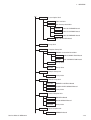
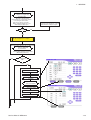
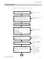
1
TENTATIVE
MEK- 8222J
MEK- 8222K
AUTOMATED
HEMATOLOGY ANALYZER
MEK-8222
0634-001851A
Model:
MEK-8222J/K
Manual code no.: 0634-001851A
Reader Comment Card
We welcome your comments about this manual. Your comments and suggestions help us improve our
manuals. Please circle the number for each of the following statements corresponding to your evaluation
and add comments in the space provided.
Fax or send your completed comment card to:
Fax: +81 (3) 5996-8100
International Div., Sales Promotion Section, Nihon Kohden Corp., 1-31-4, Nishiochiai Shinjuku-ku, Tokyo
161-8560, Japan
This manual is organized.
I can find the information I want.
The information is accurate.
I can understand the instructions.
The illustrations are appropriate and helpful.
The manual length is appropriate.
Strongly
Agree
1
Agree
Neutral
Disagree
2
3
4
Strongly
Disagree
5
1
1
1
1
1
2
2
2
2
2
3
3
3
3
3
4
4
4
4
4
5
5
5
5
5
cutting line
Comments:
Thank you for your cooperation. We appreciate it very much.
Name:
Occupation/Position:
Hospital/Company:
Address:
Phone:
CONTENTS
Contents
EMC Related Caution ..............................................................................................................i
Conventions Used in this Manual and Instrument ................................................................ iii
Warnings, Cautions and Notes .................................................................................... iii
Explanations of the Symbols in this Manual and Instrument ..................................... iv
On panel ............................................................................................................ iv
On screen and recorded data ........................................................................... v
Section 1
General....................................................................................1C.1
Introduction ......................................................................................................................... 1.1
Service Policy .................................................................................................................... 1.2
Specifications ..................................................................................................................... 1.3
Measured Parameters, Ranges and Reproducibility to
Specimen from Venous Blood ........................................................................ 1.3
Detection Method ........................................................................................... 1.3
Standardization Analysis Method .................................................................. 1.3
Interference Substances ................................................................................ 1.4
Dilution Ratio .................................................................................................. 1.6
Counting Time ................................................................................................ 1.6
Display ........................................................................................................... 1.6
Data Storage .................................................................................................. 1.6
Environmental Conditions .............................................................................. 1.6
Power Requirements ...................................................................................... 1.6
Dimensions and Weight .................................................................................. 1.7
Electromagnetic Compatibility ....................................................................... 1.7
Safety ............................................................................................................. 1.7
Panel Descriptions ............................................................................................................. 1.8
Front Panel ............................................................................................................... 1.8
Right Side Panel ....................................................................................................... 1.9
Rear Panel ...............................................................................................................1.10
Inside Panels ...........................................................................................................1.11
Composition .......................................................................................................................1.12
Installation Flow Chart ......................................................................................................1.15
How to Upgrade the Software ........................................................................................... 1.21
Replacing the ROM .......................................................................................1.21
Upgrading the Software with the Memory Card ........................................... 1.24
Connection and Wiring Diagram ........................................................................................ 1.27
Hydraulic System Diagram ...............................................................................................1.28
Tube List ............................................................................................................................ 1.29
Service Manual MEK-8222
C.1
CONTENTS
Section 2
Troubleshooting .....................................................................2C.1
Check Procedure ................................................................................................................ 2.1
Background Noise Check ................................................................................................... 2.2
Measuring Background Noise .................................................................................. 2.2
Measurement ................................................................................................. 2.2
Results ........................................................................................................... 2.3
Scattergram Check ............................................................................................................ 2.4
Counting Blood Samples of a Healthy Person ......................................................... 2.4
Reproducibility Check ........................................................................................................ 2.8
Check Procedure ...................................................................................................... 2.8
CV Data Check ......................................................................................................... 2.9
Check Items Before Instrument Check ............................................................................. 2.10
Check Items before Instrument Check ................................................................... 2.10
Sampled Whole Blood Handling Check ................................................................... 2.11
Blood Sample Handling for Counting ............................................................ 2.11
Blood Sample Handling for High Reliability ................................................... 2.11
Blood Sample from a Patient with Specific Conditions ................................. 2.11
Incorrect Data .................................................................................................................... 2.12
Background Noise ................................................................................................... 2.12
How to Decrease the Background Noise on the Instrument after
Long Term Storage ........................................................................................ 2.13
Laser Optical Unit Problems ................................................................................... 2.14
Problem 1. ..................................................................................................... 2.14
Problem 2. ..................................................................................................... 2.14
Scattergram Problems ............................................................................................ 2.15
Possible Causes and Countermeasures for Poor WBC Reproducibility ................. 2.27
Troubleshooting an Alarm .................................................................................................. 2.28
Reagent Alarm ......................................................................................................... 2.28
Section 3
Board/Unit Description ..........................................................3C.1
Sub Bath Unit .....................................................................................................................
CBC Measuring Unit ...........................................................................................................
Laser Optical Unit ...............................................................................................................
Complex Pump Unit ............................................................................................................
Sample Pump Unit ..............................................................................................................
Sheath Pump Unit ...............................................................................................................
Closed Sampler Unit ..........................................................................................................
Open Sampler Unit .............................................................................................................
Table Unit ............................................................................................................................
Display Unit ........................................................................................................................
MASTER Board ..................................................................................................................
SLAVE CBC Board .............................................................................................................
SLAVE MS Board ...............................................................................................................
C.2
3.1
3.1
3.1
3.1
3.2
3.2
3.2
3.2
3.2
3.2
3.3
3.3
3.3
Service Manual MEK-8222
CONTENTS
Section 4
Disassembly and Assembly ..................................................4C.1
Removing the PV-820V Display Unit .................................................................................. 4.1
Removing the MC-820V CBC Measuring Unit ................................................................... 4.2
Removing the MB-820V Sub Bath Unit .............................................................................. 4.4
Removing the MP-820V Complex Pump Unit .................................................................... 4.5
Removing the MS-820V Closed Sampler Unit ................................................................... 4.6
Removing the MS-821V Open Sampler Unit ...................................................................... 4.7
Removing the MS-822V Table Unit .................................................................................... 4.8
Removing the MO-820V Laser Optical Unit ....................................................................... 4.9
Removing the MP-821V Sample Pump Unit .....................................................................4.10
Removing the MP-822V Sheath Pump Unit ...................................................................... 4.11
Removing the MC-821V Flow Cytometry Piping Unit ....................................................... 4.12
Removing the MP-520V Pump Unit ...................................................................................4.14
Section 5
Socket Pin Assignment ..........................................................5C.1
Socket Pin Assignment ......................................................................................................
RS-232C Socket ............................................................................................
ZK-820V Socket .............................................................................................
USB Socket ....................................................................................................
Section 6
5.1
5.1
5.1
5.1
Adjustment .............................................................................6C.1
Adjusting the Flow Cell Position ......................................................................................... 6.1
Adjusting the FS/FL/SD Gain and FS Threshold ................................................................ 6.4
Adjusting Sensors .............................................................................................................. 6.7
SENSOR MONITOR screen .................................................................................... 6.7
How to call up the SENSOR MONITOR screen ............................................ 6.7
CBC Manometer ....................................................................................................... 6.8
WBC manometer upper sensor adjustment ................................................... 6.9
WBC manometer lower sensor adjustment ................................................... 6.9
RBC manometer upper sensor adjustment .................................................... 6.9
RBC manometer lower sensor adjustment .................................................... 6.9
CBC Electrode .........................................................................................................6.10
HGB Sensor ............................................................................................................6.11
HGB sensor adjustment ................................................................................6.11
Calibration Switch ...................................................................................................6.12
Liquid Sensor ...........................................................................................................6.13
4-channel liquid sensor unit adjustment ....................................................... 6.15
Pressure Sensor .....................................................................................................6.19
Temperature Sensor ................................................................................................6.20
Setting on the UT-7159 WARMER CONTROL board ....................................6.21
Calibrating the Touch Screen ............................................................................................6.22
Service Manual MEK-8222
C.3
EMC RELATED CAUTION
This equipment and/or system complies with the International Standard EN61326-1 for electromagnetic
compatibility for medical electrical equipment and/or system. However, an electromagnetic environment that
exceeds the limits or levels stipulated in the EN61326-1, can cause harmful interference to the equipment
and/or system or cause the equipment and/or system to fail to perform its intended function or degrade its
intended performance. Therefore, during the operation of the equipment and/or system, if there is any
undesired deviation from its intended operational performance, you must avoid, identify and resolve the
adverse electromagnetic effect before continuing to use the equipment and/or system.
The following describes some common interference sources and remedial actions:
1. Strong electromagnetic interference from a nearby emitter source such as an authorized radio station or
cellular phone:
Install the equipment and/or system at another location if it is interfered with by an emitter source such
as an authorized radio station. Keep the emitter source such as cellular phone away from the equipment
and/or system.
2. Radio-frequency interference from other equipment through the AC power supply of the equipment and/
or system:
Identify the cause of this interference and if possible remove this interference source. If this is not
possible, use a different power supply.
3. Effect of direct or indirect electrostatic discharge:
Make sure all users and patients in contact with the equipment and/or system are free from direct or
indirect electrostatic energy before using it. A humid room can help lessen this problem.
4. Electromagnetic interference with any radio wave receiver such as radio or television:
If the equipment and/or system interferes with any radio wave receiver, locate the equipment and/or
system as far as possible from the radio wave receiver.
If the above suggested remedial actions do not solve the problem, consult your Nihon Kohden Corporation
subsidiary or distributor for additional suggestions.
The CE mark is a protected conformity mark of the European Community. The products herewith comply
with the requirements of the IVD Directive 98/79/EEC.
This equipment complies with International Standard EN55011 (1999) Group 1, Class B. Class B
EQUIPMENT is equipment suitable for use in domestic establishments and in establishments directly
connected to a low voltage power supply network which supplies buildings used for domestic purposes.
Service Manual MEK-8222
i
This page is intentionally left blank.
ii
Service Manual MEK-8222
Conventions Used in this Manual and Instrument
Warnings, Cautions and Notes
Warnings, cautions and notes are used in this manual to alert or signal the reader to specific information.
WARNING
A warning alerts the user to the possible injury or death associated with the use or misuse of the
instrument.
CAUTION
A caution alerts the user to possible injury or problems with the instrument associated with its use or
misuse such as instrument malfunction, instrument failure, damage to the instrument, or damage to other
property.
NOTE
A note provides specific information, in the form of recommendations, prerequirements, alternative
methods or supplemental information.
Service Manual MEK-8222
iii
Explanations of the Symbols in this Manual and Instrument
The following symbols found in this manual/instrument bear the respective descriptions as given.
On panel
Symbol
Description
Description
Main power lamp
HEMOLYNAC•3
(hemolysing reagent)
“On” only for part of the equipment
HEMOLYNAC•5
(hemolysing reagent)
“Off” only for part of the equipment
WASTE
Laser ON
Hand positions for carrying the
instrument
Reset
RS-232C socket
Manual
USB socket
Clean
IVD
IVD conformity
Eject
Fuse
Start
AC power off
(Disconnection from the mains)
Emergency
AC power on
(Connection to the mains)
Do not touch the sample rack
during measurement
Alternating current
Inlet
Equipotential terminal
Outlet
Year of manufacture
Attention, consult operator's manual
Serial number
ISOTONAC•3
(diluent)
CLEANAC
(detergent)
CLEANAC•3
(detergent)
iv
Symbol
The CE mark is a protected
conformity mark of the European
Community. The products
herewith comply with the
requirements of the IVD Directive
98/79/EEC.
Service Manual MEK-8222
On screen and recorded data
Symbol
Description
Beside WBC or RBC measured
value: Sample error
Beside HGB measured value: Dirty
measurement baths
Beside WBC measured value: Poor
hemolyzation
Beside HGB measured value: HGB
voltage adjustment error
Service Manual MEK-8222
Symbol
Description
Beside HGB measured value: HGB
circuit error
C
Beside WBC or PLT measured
value: Platelet coagulation
v
Section 1 General
Introduction ........................................................................................................................ 1.1
Service Policy .................................................................................................................... 1.2
Specifications ..................................................................................................................... 1.3
Measured Parameters, Ranges and Reproducibility to
Specimen from Venous Blood ....................................................................... 1.3
Detection Method .......................................................................................... 1.3
Standardization Analysis Method .................................................................. 1.3
Interference Substances ................................................................................ 1.4
Dilution Ratio ................................................................................................. 1.6
Counting Time ............................................................................................... 1.6
Display ........................................................................................................... 1.6
Data Storage ................................................................................................. 1.6
Environmental Conditions .............................................................................. 1.6
Power Requirements ..................................................................................... 1.6
Dimensions and Weight ................................................................................. 1.7
Electromagnetic Compatibility ....................................................................... 1.7
Safety ............................................................................................................ 1.7
Panel Descriptions ............................................................................................................. 1.8
Front Panel .............................................................................................................. 1.8
Right Side Panel ...................................................................................................... 1.9
Rear Panel ............................................................................................................. 1.10
Inside Panels .......................................................................................................... 1.11
Composition ..................................................................................................................... 1.12
Installation Flow Chart ..................................................................................................... 1.15
How to Upgrade the Software .......................................................................................... 1.21
Replacing the ROM ..................................................................................... 1.21
Upgrading the Software with the Memory Card ........................................... 1.24
Connection and Wiring Diagram ...................................................................................... 1.27
Hydraulic System Diagram .............................................................................................. 1.28
Tube List .......................................................................................................................... 1.29
Service Manual MEK-8222
1C.1
1. GENERAL
Introduction
CAUTION
To maintain the instrument in normal condition, the user must perform
the periodic maintenance. Refer to “Maintenance” of the operator’s
manual.
This tentative service manual consists of the “General” and “Socket Pin
Assignment” sections only.
NOTE
If the instrument has a problem and there has been no periodic
maintenance, the instrument will usually be normal again by cleaning
the fluid paths or replacing a consumable with a new one.
Service Manual MEK-8222
1.1
1. GENERAL
Service Policy
CAUTION
• Be careful not to directly touch any place where blood is or may
spread to.
• Wear rubber gloves to protect yourself from infection before doing
maintenance.
Nihon Kohden Corporation’s basic policy for technical service is to replace faulty
units, printed circuit boards or parts. We do not support component-level repair of
boards and units outside the factory.
NOTE
• When ordering parts or accessories from your nearest Nihon Kohden
Corporation’s distributor, please quote the NK code number and part
name which is listed in this service manual, and the name or model
of the unit in which the required part is located. This will help us to
promptly attend to your needs.
• Always use parts and accessories recommended or supplied by
Nihon Kohden Corporation to assure maximum performance from
your instrument.
1.2
Service Manual MEK-8222
1. GENERAL
Specifications
Measured Parameters, Ranges and Reproducibility to Specimen from Venous Blood
Specifications were determined using hematology control blood (MEK-3DN), counted 10 times consecutively.
Measured Parameters
WBC: White blood cell count
Measuring Range
0 to 300 × 10 /µL
3
NE%: Neutrophil percent
0 to 99.9%
LY%: Lymphocyte percent
0 to 99.9%
MO%: Monocyte percent
0 to 99.9%
EO%: Eosinophil percent
0 to 99.9%
BA%: Basophil percent
0 to 99.9%
NE: Neutrophil count
0 to 99.9 × 10 /µL
LY: Lymphocyte count
0 to 99.9 × 10 /µL
MO: Monocyte count
0 to 99.9 × 10 /µL
EO: Eosinophil count
0 to 99.9 × 10 /µL
BA: Basophil count
0 to 99.9 × 10 /µL
RBC: Red blood cell count
0 to 14.9 × 10 /µL
3
3
3
3
3
6
HGB: Hemoglobin concentration
HCT: Hematocrit
0 to 29.9 g/dL
within 1.5%CV (5.0 × 10 /µL)
6
within 1.5%CV (16 g/dL)
0 to 99.9%
MCV: Mean cell volume
20 to 199 fL
MCH: Mean cell hemoglobin
10 to 50 pg
MCHC: Mean cell hemoglobin concentration
RDW: Red blood cell distribution width
0 to 50%
3
PCT: Platelet crit
within 1.0%CV (70 to 120 fL)
10 to 50 g/dL
0 to 1490 × 10 /µL
PLT: Platelet count
within 4.0%CV (3.0 × 10 /µL)
3
0 to 2.9%
MPV: Mean platelet volume
PDW: Platelet distribution width
Detection Method
Blood cell count:
Hemoglobin:
Hematocrit:
WBC population:
Platelet crit:
RBC distribution width:
Platelet distribution width:
Reproducibility to Specimen from Venous
Blood
(CV: Coefficient of Variation)
3
within 2.0%CV (4.0 to 9.0 × 10 /µL)
3
within 5.0%CV (WBC: 4.0 to 9.0 × 10 /µL,
NE%: 40 to 70%)
3
within 5.0%CV (WBC: 4.0 to 9.0 × 10 /µL,
LY%: 20 to 45%)
3
within 12.0%CV (WBC: 4.0 to 9.0 × 10 /µL,
MO%: 2 to 10%)
3
within 20.0%CV (WBC: 4.0 to 9.0 × 10 /µL,
EO%: 2 to 10%)
within CV30.0% (>2%) or average value ±1% (0 to
3
2%) (WBC: 4.0 to 9.0 × 10 /µL, BA%: 0 to 3%)
0 to 20.0 fL
0 to 50%
Electrical resistance detection
Cyanmethemoglobin optical detection
Histogram calculation
Light scatter by laser
Histogram calculation
Histogram calculation
Histogram calculation
Standardization Analysis Method
WBC: ICSH1988 ICSH: The assignment of values to fresh blood used for calibrating automated blood cell counters. Clin
Lab Haematol, 10:203-212, 1988
RBC: ICSH1988 ICSH: The assignment of values to fresh blood used for calibrating automated blood cell counters. Clin
Lab Haematol, 10:203-212, 1988
Service Manual MEK-8222
1.3
1. GENERAL
HGB: NCCLS H15-A2 H15-A2: Reference and Selected Procedures for the Quantitative Determination of Hemoglobin in
Blood Second Edition; Approved Standard (1994)
HCT: NCCLS H7-A2 H7-A2: Procedure for Determining Packed Cell Volume by the Microhematocrit Method Second
Edition; Approved Standard (1993)
PLT: Brecher & Cronkite
Interference Substances
WBC:
Unlysed red cells
In some rare occasions, the RBC in the blood sample may not completely lyse and these non-lysed RBC
may be detected as WBC and cause increase in WBC count.
Multiple myeloma
The precipitation of proteins in multiple myeloma patients may increase the WBC count.
Leukemia
WBC is fragile in leukemia patient and WBC may be destroyed during measurement. These WBC
fragments may also interfere with WBC differential measurement.
Chemotherapy
Cytotoxic and immunosuppressive drugs cause low WBC count.
Cryoglobulins
Cryoglobulin may be increased in patients who are or have myeloma, cancer, leukemia, macroglobulinemia,
lymphoproliferative disorders, metastatic tumors, autoimmune disorders, infections, anerurism, pregnant,
thromboembolic phenomena, diabetes, etc, which cause increase in WBC, RBC or PLT counts and HGB
concentration. In such cases, warm the blood sample to 37°C in a water bath for 30 minutes and measure
the sample immediately.
RBC:
Leukemia
An increase in WBC in leukemia patient causes increase in RBC.
Agglutinated RBC
Agglutinated RBC may decrease RBC count. This can be checked by abnormal MCH and MCHC values
and examination of the stained blood film.
Cold agglutinins
IgM immunoglobulins which are elevated in clod agglutinin disease may decrease RBC and PLT counts and
increase MCV.
HGB:
Turbidity of the blood sample
Any physiologic and/or therapeutic factors may increase HGB. In such a case, determine the cause of
turbidity and follow the appropriate method below.
1. Increased WBC
An extreme increase in WBC will cause excessive light scatter. In these cases, measure manually.
Centrifuge the diluted sample and measure the supernatant fluid with a spectrophotometer.
2. Increased lipids
The blood sample may be milky when there is excessive lipid. This may occur with hyperlipidemia,
hyperproteinemia and hyperbilirubinemia. Accurate HGB measurement can be achieved by manual
methods and a plasma blank.
3. Increased turbidity
When RBC are resistant to lysing, turbidity may increase causing increase in HGB. Observe if MCH
and MCHC values are abnormal. HGB result affects MCH and MCHC result.
4. Fetal bloods
The mixing of fetal and maternal blood may increase HGB value.
HCT:
Agglutinated RBC
RBC agglutination may cause erroneous HCT and MCV values. Observe if MCH and MCHC values are
abnormal. In such a case, measure manually.
1.4
Service Manual MEK-8222
1. GENERAL
MCV:
MCH:
MCHC:
RDW:
PLT:
MPV:
Agglutinated RBC
RBC agglutination may cause erroneous HCT and MCV values. Observe if MCH and MCHC values are
abnormal. In such a case, measure manually.
Excessive number of large PLT
Excessive number of large PLT and/or excessively high WBC may affect the MCV value. Check by careful
examination of the stained blood film.
MCH is determined from HGB and RBC values. Therefore, the limitations for HGB and RBC also affect
MCH value.
MCHC is determined from HGB and HCT values. Therefore, the limitations for HGB and HCT also affect
MCHC value.
RDW is determined from RBC value. Therefore, the limitations for RBC also affect RDW value.
Agglutinated RBC
Agglutinated RBC may decrease RBC count and erroneous RDW. This can be checked by abnormal MCH
and MCHC values and examination of the stained blood film.
Nutritional deficiency or blood transfusion
Iron and/or cobalamin and/or folate deficiency may increase RDW.
Very small fragments
Very small RBC, RBC fragments and WBC fragments may be the cause in increased PLT count.
Agglutinated RBC
PLT may be trapped in the agglutinated RBC resulting in decrease in PLT. This can be checked by abnormal
MCH and MCHC values and examination of the stained blood film.
Very large PLT
Large PLT may exceed the PLT threshold and may not be counted which results in low PLT count.
Chemotherapy
Cytotoxic and immunosuppressive drugs may increase the fragility of cells which may cause low PLT count.
In such a case, measure manually.
Hemolysis
Hemolyzed specimens contain red cell stroma which may increase PLT count.
Anticoagulated blood
Blood anticoagulated with acid-citrate-dextrose may have clumped PLT which may cause decrease in PLT
count.
Agglutinated PLT
Clumped PLT may decrease PLT count and/or increase WBC count. For such sample, collect the sample in
sodium citrate anticoagulant and measure only PLT. The PLT result must be corrected for the sodium citrate
dilution effect.
Very large PLT
Large PLT may exceed the PLT threshold and not be counted which results in low MPV.
Very small fragments
Very small RBC, RBC fragments and WBC fragments may interfere with MPV measurement.
Agglutinated RBC
PLT may be trapped in the agglutinated RBC resulting erroneous MPV. This can be checked by abnormal
MCH and MCHC values and examination of the stained blood film.
Chemotherapy
Cytotoxic and immunosuppressive drugs may affect MPV. In such a case, measure manually.
NOTE
Blood samples collected in EDTA does not maintain stable MPV because platelets swell
depending on the interval after collection and storage temperature.
Service Manual MEK-8222
1.5
1. GENERAL
WBC 5 part differential parameters are derived from the WBC count, therefore, the limitations for WBC also affect these
parameters.
LY and LY%: Erythroblasts, certain parasites and RBC that are resistant to lysis may interfere with an accurate LY count.
MO and MO%:
Large lymphocytes, atypical lymphocytes, blasts and excessive number of basophils may interfere with
an accurate MO count.
NE and NE%: Excessive eosinophils, metamyelocytes, myelocytes, promyelocytes, blasts and plasma cells may interfere
with an accurate NE count and NE%.
EO and EO%: Abnormal granules may interfere with an accurate EO count.
BA and BA%: Immature cell, metamyelocytes, myelocytes, promyelocytes, blasts and plasma cells may interfere with an
accurate BA count and BA%.
Dilution Ratio
• Venous blood
Sample volume:
WBC/HGB:
RBC/PLT:
• Capillary blood
Sample volume:
WBC/HGB:
RBC/PLT:
55 µL
200:1
40,000:1
10 µL
1200:1
240,000:1
20 µL
600:1
120,000:1
Counting Time
Approximately 45 s/sample, 80 samples/hr
Display
Display:
Resolution:
Screen size:
Display contents:
8.4 inch, TFT type color LCD with touch screen keys
800 × 600 dots
approx. 170.4 × 127.8 mm
Numerical data, scattergrams, histograms, measuring conditions, alarm message and other messages,
touch screen keys
Data Storage
Numerical data for all counted parameters for up to 400 samples and histograms and scattergrams for up to 60 samples
Environmental Conditions
Storage temperature:
Operating temperature:
Storage humidity:
Operating humidity:
Storage atmospheric pressure:
Operating atmospheric pressure:
Power Requirements
Power requirements:
Power consumption:
1.6
−20 to 60°C
15 to 30°C
10 to 95% (Non-condensing)
30 to 85% (Non-condensing)
70 to 106 kPa
70 to 106 kPa
MEK-8222J: 110, 117 or 127 V ±10% AC, 50/60 Hz
MEK-8222K:220, 230 or 240 V ±10% AC, 50/60 Hz
360 VA
Service Manual MEK-8222
1. GENERAL
Dimensions and Weight
Dimensions: 613 W × 550 D × 583 H (mm)
Net weight: approx. 55 kg
Electromagnetic Compatibility
IEC 61326-1 (1997) Amendment 1 (1998) Amendment 2 (2000)
EN 61326-1 (1997) Amendment 1 (1998)
CISPR11 (1997), Group 1, Class B
EN 55011 (1998) Amendment 1 (1999), Group 1, Class B
Safety
Safety standards:
IEC 61010-1 2nd Edition (2001)
EN 61010-1 (1993) Amendment 2 (1995)
Laser: IEC 60825-1 (1993) Amendment 1 (1997)
EN 60825-1 (1994) Amendment 11 (1996)
According to the type of protection against electrical shock: CLASS I EQUIPMENT
According to the degree of protection against harmful ingress of water: IPX0 (Ordinary EQUIPMENT)
According to the degree of safety of application in the presence of a FLAMMABLE ANAESTHETIC MIXTURE WITH AIR,
OR WITH OXYGEN OR NITROUS OXIDE: EQUIPMENT not suitable for use in the presence of FLAMMABLE
ANAESTHETIC MIXTURE WITH AIR, OR WITH OXYGEN OR
NITROUS OXIDE
According to the mode of operation: CONTINUOUS OPERATION
EQUIPMENT types (classification): Indoor stationary EQUIPMENT
Installation Category: II EQUIPMENT
Pollution Degree: 2 EQUIPMENT
Requirements for marking of in vitro diagnostic instruments: EN1658 (1996)
Service Manual MEK-8222
1.7
1. GENERAL
Panel Descriptions
Front Panel
6
7
8
9
10
11
1
2
12
3
13
14
4
5
15
16
CAUTION
Do not put your hand in the
rack table during
measurement.
No.
Name
1
Main power lamp
2
Power key
3
Reset key
Description
6
Power lamp
7
Laser lamp
8
Operation indicator
lamps
9
Manual mode key
10
LCD screen
11
Eject key
12
Start key
13
Emergency key
14
Clean key
15
Sample rack
Lights when the main power switch on the rear panel is turned on.
Turns the hematology analyzer power on or off when the main power switch on the rear panel
is turned on. When the power is turned on, priming and self-check are automatically performed,
and the READY screen is displayed.
Stops operation when pressed during operation. Returns to the READY screen when pressed
while changing settings.
Use this key only when an error occurs.
For manual mode only.
Press to aspirate the sample and start counting.
For manual mode only.
Aspirates the sample.
Lights when the main power switch on the rear panel and power key on the front panel are
turned on.
Lights when the laser switch on the right side panel is turned on.
For manual mode only.
When blinking: Aspirating the sample
When off: Counting the sample
When lit: Ready for next counting
Sets the measurement mode in manual mode when pressed. The sampling nozzle for manual
mode is lowered.
Displays measurement data, various messages and touch screen keys.
For rack mode only.
Press this key to slide the sample rack in or out.
For rack mode only.
Press this key to start measurement.
Press this key to count an emergency sample during routine counting.
Cleans the fluid path, aperture and manometer with detergent. Priming is performed
automatically after cleaning the fluid path. Press this key when clogging occurs, the manometer
becomes dirty or bubbles occur in the manometer.
Holds the sample tubes containing specimens. Up to 50 sample tubes can be accommodated.
16
Rack table
Holds the sample rack.
4
5
1.8
Manual count
switch
Manual sampling
nozzle
Service Manual MEK-8222
1. GENERAL
Right Side Panel
8
CAUTION
1
Do not insert an object, such a
pencil into these holes.
Otherwise the liquid sensor unit
may be damaged.
2
3
4
5
6
7
CAUTION
Always carry the hematology analyzer by more than two people and by
the left and right sides. Never hold it by the front and rear. Otherwise
the front panel may come off and the hematology analyzer may be
dropped, causing injury to the operator and damage to the hematology
analyzer.
No.
1
2
3
4
5
6
7
8
Service Manual MEK-8222
Name
Laser switch
ISO3
Diluent inlets
CLN
Detergent inlet
CLN3
Detergent inlet
HEMO3
Hemolysing reagent inlet
HEMO5
Hemolysing reagent inlet
WASTE
Waste outlets
Flow cell unit window
cover
Description
Turns the laser on or off with the laser key for WBC 5 part
differential measurement.
Inlets for the ISOTONAC•3 diluent.
Inlet for the CLEANAC detergent.
Inlet for the CLEANAC•3 detergent.
Inlet for the HEMOLYNAC•3 hemolysing reagent.
Inlet for the HEMOLYNAC•5 hemolysing reagent.
Outlets for waste such as used lyse, detergent and aspirated
samples.
When two screws and the cover are removed, you can see the
flow cell unit. Open the window only when adjusting the gain.
1.9
1. GENERAL
Rear Panel
Refer to warnings and cautions in “Connecting an External
Instrument to the Hematology Analyzer” in Section 2.
1
2
3
4
5
6
Refer to warnings and cautions in “Connecting the Power
Cord and Grounding the Hematology Analyzer” in Section 2.
No.
1
RS232C socket
2
Bar code socket
3
USB socket
4
Main power switch
5
Fuse holder
6
7
1.10
Name
AC SOURCE
AC source socket
Equipotential ground
terminal
7
Description
Connects to the optional WA-460V card printer, WA-711V
printer, WA-820V printer or PC. When a PC is connected to this
socket, histogram and scattergram data cannot be output.
Connects to the optional ZK-820V handy bar code reader.
Connects to a PC. Histogram and scattergram data can be output.
Supplies the power to the hematology analyzer when it is turned
on. Under normal conditions keep this switch turned on.
Contains the time lag fuse. To replace the fuse, contact your
Nihon Kohden distributor.
Connects the AC power cord to supply AC power to the
hematology analyzer.
Connects the ground lead to the equipotential ground terminal on
the wall for earth grounding.
Service Manual MEK-8222
1. GENERAL
Inside Panels
CAUTION
• For WBC 5 part differential measurement, the laser beam is used. Do
not open any part labeled “CAUTION”. The laser can cause burns
and blindness.
• Do not remove the caution labels.
NOTE
• When attaching the filter joint assembly, be careful not to bend or
damage the filter packing at the bottom of the measurement bath.
• When there is a leakage, check that there is no scratch or damage to
the circumference of the filter.
Service Manual MEK-8222
1.11
1. GENERAL
Composition
MEK-8222J
MEK-8222K
JQ-820V
Inlet/Outlet Unit
XP-513V
JQ-821V
Left Side Piping Unit
XP-513V
JQ-822V
JQ-823V
3-way Valve
Center Piping Unit
UT-7171
LIQUID LEVEL SENSOR Board
XP-513V
3-way Valve
4-channel Liquid Sensor Unit
UT-7179
4WAY LIQUID SENSOR Board
MB-820V
Sub Bath Unit
MC-820V
CBC Measuring Unit
UT-7143
PRE AMP Board
UT-7180
CBC Measuring Board Set
* Not available
MC-821V
1.12
3-way Valve
UT-7181*
HGB AMP Board
UT-7182*
HGB LED Board
UT-7183*
MANOMETER PTR Board
UT-7184*
MANOMETER LED Board
XP-512V
2-way Valve
XP-513V
3-way Valve
Flow Cytometry Piping Unit
UT-7152
DRIVER1 Board
XP-512V
2-way Valve
XP-513V
3-way Valve
Service Manual MEK-8222
1. GENERAL
MO-820V
Laser Optical Unit
MF-820V
Flow Cell Unit
UT-7172
MO Preamp Board Set
UT-7173
MO FS PREAMP Board
UT-7174
MO FL PREAMP Board
UT-7175
MO SD PREAMP Board
UT-7176
MP-520V
Pump Unit
MP-820V
Complex Pump Unit
UT-7177
MP-821V
MP-822V
MS-820V
MS-821V
MD/MP Connection Board Set
UT-7186
MD CONNECTION Board
UT-7187
MP CONNECTION Board
XP-503V
3-way Valve
XP-513V
3-way Valve
Sample Pump Unit
XP-512V
2-way Valve
Sheath Pump Unit
UT-7158
SHEATH CONTROL Board
UT-7171
LIQUID LEVEL SENSOR Board
XP-512V
2-way Valve
Closed Sampler Unit
UT-7154
DRIVER3 Board
UT-7185
3POS SENSOR Board
XP-513V
3-way Valve
Open Sampler Unit
UT-7065
Service Manual MEK-8222
MO LD DRIVER Board
SENSOR Board
1.13
1. GENERAL
MS-822V
PV-820V
Table Unit
UT-7086
SENSOR3 Board
UT-7156
DRIVER5 Board
UT-7157
TABLE SENSOR Board
Display Unit
UT-7178
* Not available
1.14
Front Panel Board Set
UT-7149*
LCD CONTROL Board
UT-7150*
MANUAL KEY Board
UT-7151*
RACK KEY Board
RK-820V
Chassis
SC-820VJ
SC-820VK
Power Supply Unit
UT-7146
MASTER Board
UT-7147
SLAVE CBC Board
UT-7148
SLAVE MS Board
UT-7153
DRIVER2 Board
UT-7155
DRIVER4 Board
UT-7160
POWER Board
UT-7161
INTERFACE Board
ZY-820V
Warmer Unit
UT-7159
WARMER CONTROL Board
XP-513V
3-way Valve
Service Manual MEK-8222
1. GENERAL
Installation Flow Chart
Place the instrument in a suitable place.
Connect the power cord, reagents and
external equipment to the instrument.
Turn on the instrument.
Prime the instrument.
MENU
OTHER OPERATIONS
PRIME ON INSTALLATION
[PRIME ON INSTALLATION] is
special procedure after storing
and transporting the instrument.
Change the OPERATOR to [FACTORY]
to service the instrument.
Display the
[OPERATORS & PASSWORDS] screen.
MENU
SETTINGS
OPERATORS & PASSWORDS
Select [FACTORY].
FACTORY
SELECT OPERATOR
Enter the passwords.
[4321]
[Enter]
The status bar on the screen is displayed
in orange.
Service Manual MEK-8222
MEK-8222 has 3 default operator types.
1. MANUFACTURER : password [ 4321 ] for servicing
Allows you to enter any screen and change any settings.
2. LAB TECHNICIAN: password [ 8222 ]
Allows you to enter any screen and change settings
except for the [SERVICE MAINTENANCE] screen.
3. OTHER: password [ 0000 ]
Allows you to only perform the measurement and view the data and settings.
Operator name [MEK-F]( Type of Operator: LAB TECHNICIAN ) is selected
as the default setting.
1.15
1. GENERAL
Check the laser optical unit and roughly adjust
the gain with 7 µm polymer microsphere
suspensions.
Display the
[DIFF GAIN (ROUGH)] screen.
MENU
SETTINGS
SENSITIVITY & THRESHOLD
DIFF GAIN
Measure the 7 µm polymer
microsphere suspensions.
Adjust the flow cell position.
Remove the flow cell unit
window cover and loosen the
screw fastening the flow cell
adjustment screw.
Perform [CLEAN FLOW CELL] procedure.
MENU
OTHER OPERATIONS
CLEAN FLOW CELLS
Turn the flow cell
adjustment screw
about 2 or 3˚
FS CV must be 7% or less. FL CV must be 7% or less.
Is CV OK ?
NO
YES
This result is optimum, the CV values of FS and FL are
below 7%. The histogram is narrow and scattergram is
small oval shape as shown below.
1.16
If the result is not optimum, the CV values of FS
and FL are over 7%. The histogram is wide and
scattergram is large as shown below.
Service Manual MEK-8222
1. GENERAL
Check the histogram PEAK.
FS PEAK = approx. 61 (FS IDEAL* PEAK)
FL PEAK = approx. 90 (FL IDEAL* PEAK)
*Optimum
Is PEAK OK ?
NO
Press AUTO GAIN key to change the
current gain to the target gain.
Then, measured PEAK changes to the
IDEAL (optimum) PEAK.
YES
AUTO GAIN
Press the OK key and the instrument
starts [CLEAN FLOWCELL] procedure
automatically.
OK
Rough adjustment of FS (size) and
FL (complexity) is complete.
Check the background noise.
Measure a few empty sample
tubes in the closed mode.
Perform [CLEAN]
procedure.
WBC: 0.2 or less (×103/uL)
RBC: 0.05 or less (×106/uL)
HGB: 0.1 or less (g/dL)
PLT: 10 or less (×103/uL)
TOC: 100 or less (count)
Is Data OK ?
NO
YES
Service Manual MEK-8222
1.17
1. GENERAL
YES
Perform fine adjustment of the gain
with human blood samples.
Prepare 10 blood samples of
different healty people within
12 hours after the blood collection.
Rough adjustment of FS (size) and FL (complexity)
with 7um particle is already completed .
Adjustment of SD (granularity) is not yet completed.
Measure the 10 human blood
samples in the closed mode.
Call up the
[DIFF GAIN (FINE)] screen.
MENU
SETTINGS
SENSITIVITY & THRESHOLD
DIFF GAIN
ADJUST GAIN (FINE)
Load the scattergram and
calculate the target gain.
Enter the measured sample's
sequence number.
Since the PEAK of Monocyte of human
blood is the most stable parameter in WBC
5 part-diff, the optical fine adjustment is
done with the MO PEAK.
LOAD NEW SCATTERGRAM
ADD TO LIST
MO PEAK (FS,FL,SD) is displayed.
But, the measured PEAK may be
different from the IDEAL PEAK and
the scattergram can touch the division
line.
Press the AUTO GAIN key to change
the current gain to the target gain.
Then, the measured PEAK changes to
the IDEAL PEAK.
AUTO GAIN
Draw the scattergram with the
changed GAIN.
REFRESH
1.18
Service Manual MEK-8222
1. GENERAL
Measure the human blood
samples once again and
check the data.
Check the following points.
Scattergrams are oval in shape and are
not touching the division line.
Flags do not appear, especially
Blasts, Immature Granulocyte, Left
Shift, Atypical Lymphocytes.
Is Data OK ?
Perform the fine adujstment of the gain
with human blood samples again.
NO
YES
All optical adjustment is complete.
Check the CBC calibration with the MEK-3DN.
Measure MEK-3DN
in closed mode.
Data should be within the assay values.
(Only CBC parameters)
YES
Are all parameters
OK ?
NO
NO
Auto calibration procedure.
MENU
CALIBRATION
CLOSED
Measure the MEK-3DN
in the closed mode.
AUTO CAL MODE
Enter the assay value.
AUTO CAL SET
Service Manual MEK-8222
1.19
1. GENERAL
NO
YES
Manual calibration procedure
Measure the MEK-3DN
in the closed mode at
least 3 times.
The service maintenance menu is
convenient for the manual calibration
and reproducibility check.
[CLOSED 10 SAMPLES] key
measures the sample 10 times
in closed mode.
[10 data X-CV] key displays the last
10 samples' average and CV.
MENU
MENU
OTHER OPERATIONS
OTHER OPERATIONS
SERVICE MAINTENANCE
SERVICE MAINTENANCE
CONTINUOUS MEASUREMENT
10 data X-CV
CLOSED 10 SAMPLES
Calculate the new
calibration coefficient.
New calibration coeffitient
=
Previous calibration coefficient
×
Assay value / Mean measured value
For example,
RBC previous calibration coefficient = 998
RBC assay value = 462
RBC Mean measured value = 440
RBC new calibration coeffitient = 998 × 462 / 440
= 1048
CBC calibration is complete.
The calibration of 5 part diff uses the same
method as the manual calibration procedure
of the CBC, but it must be done with human
blood samples.
New calibration coeffitient
=
Previous calibration coefficient
×
Mean other method's value
/
Mean measured value
For example,
LY% previous calibration coefficient = 1000
LY% mean other method's value = 35.8
LY% mean measured value = 32.6
LY% new calibration coefficient = 1000 × 35.8 / 32.6
= 1098
All adjustments and calibrations are
complete.
1.20
Service Manual MEK-8222
1. GENERAL
How to Upgrade the Software
Instruments with a serial number between 00001 and 000xx require the following
ROM replacement before the software upgrade. Therefore, check the serial number
on the rear panel of the instrument. If the serial number is not in the abovementioned serial numbers, go to “Upgrading the Software with Memory Card”.
Replacing the ROM
1. Turn off the main power switch on the rear panel of the instrument and unplug
the AC power cord.
CAUTION
Before removing any parts from the instrument, wait 10 minutes after
turning off the instrument and disconnecting the power cord from the
AC outlet.
2. Remove the screws which secure the side covers and top cover to the
instrument chassis. Remove the covers from the instrument.
Top cover
Left side cover
Right side cover
Unplug the power cord
Rear cover
Service Manual MEK-8222
1.21
1. GENERAL
3. Remove the screws which secure the rear cover to the instrument chassis.
Slowly pull the rear cover toward you until the cable connected to the rear
cover backside appears as shown below. Disconnect the cable at the
INTERFACE board which is attached to the rear cover. Completely remove the
rear cover from the instrument.
4. Insert a small flat-blade screwdriver between the ROM and its socket on the
MASTER board as shown below. Carefully lift the ROM with the screwdriver
until the ROM slightly tilts. Take out the screwdriver and insert it into the
opposite side of the IC socket. Continue lifting both sides of the ROM with the
screwdriver until the ROM is removed.
MASTER board
Flat-blade screwdriver
ROM
1.22
Service Manual MEK-8222
1. GENERAL
CAUTION
When you remove the ROM from the IC socket with a sharp edge tool
such as a flat-blade screwdriver, be sure that it does not damage the
printed pattern under the IC socket.
IC socket
Printed pattern
5. Put the new ROM on the IC socket and carefully insert the pins of the new
ROM into the IC socket.
CAUTION
• Before putting the new ROM on the IC socket, check that the ROM
position is the same as the old ROM.
• Do not break or bend a pin of the ROM. The pins must be correctly
inserted into the socket. Otherwise the instrument may not work.
Service Manual MEK-8222
1.23
1. GENERAL
Upgrading the Software with the Memory Card
1. Turn off the main power switch on the rear of the instrument.
2. Remove the right side cover as shown below.
Right side cover
Main power switch
3. Insert the memory card into the slot as shown below.
Memory card
Card slot
4. Turn on the main power switch on the rear of the instrument.
1.24
Service Manual MEK-8222
1. GENERAL
5. Turn on the Power key on the front panel of the instrument.
The “DOWNLOAD MEK-8222 PROGRAM” screen appears.
The MEK-8222 software has five parts.
MASTER BD (board)
SLAVE CBC BD (board)
SLAVE MD BD (board)
LANGUAGE: ENGLISH (local language program)
ANALYSIS PROGRAM
6. Install the software as follows:
To install all the software, press the DOWNLOAD ALL PROGRAM
touch screen key.
To install the MASTER BD software only, press the MASTER BD Vxxxx (xxxx) key on the screen.
To install the SLAVE CBC BD software only, press the SLAVE CBC BD
Vxx-xx (xxxx) key on the screen.
To install the SLAVE MS BD software only, press the SLAVE MS BD
Vxx-xx (xxxx) key on the screen.
To install the LANGUAGE software, press the LANGUAGE (local
language ) Vxx-xx (xxxx) key on the screen.
To install the ANALYSYS PROGRAM software only, press the
ANALYSYS PROGRAM Vxx-xx (xxxx) key on the screen.
Service Manual MEK-8222
1.25
1. GENERAL
After the software is installed, the following screen appears.
7. Press the OK key on the screen.
The instrument starts automatic priming.
8. Press the card eject key and remove the memory card from the instrument.
Memory card
Card eject key
9. Reassemble the instrument completely.
1.26
Service Manual MEK-8222
1. GENERAL
Connection and Wiring Diagram
CNA210
CN205
CN202
CN301
CN204
CNA209
Manual mode
CNA242
CN203
PHK7P PHD10P DF-30P SM04P SFW15R
CN103 PHD12P
PHD16P
LCD CONTROL board
HIF-60P CN101
DC line
LED
Switch
Eject
Start
Emergency
Clean
Eject
Start
Emergency
Clean
AC line
Serial communication Iine
CN102
CNA241
CN001 VH5P
RS-232C
DSUB 9P
CNA104
CNA009
CNA216
USB
CN101
CN004
CN001
HIF-60P
VH5P
CNA113
INTERFACE board UT-7161
CN104
CN002
PHD20P
CNA105
SHEATH CONTROL board
UT-7158
PHD28P
CNP207
2MV
2MV
2MV
2MV
2
3
4
5
DC
CN101
VH2P
CN004
CNA228
Position Sensor POS
Position Sensor CODE0
Position Sensor CODE1
Up and Down
(Nozzle)
Turn
MT2
PX
3MV
1
Sampling nozzle
UD0 Upper position
MT3
PK5
Depressure needle
UD1 Upper position
CN003
VH6P
Laser switch
Emergency stop switch
UD2
UT-7154
CNA227 MFC-26P
3MV
3
CNA227
CNA
109
CNA304
CN001
AMP2P PHD12P
40P CNA102
BL-185
PHD24P
CNA108
WARMER CONTROL board
UT-7159
CN002
CN205
HKP8P
DRIVER1 board
1
6
MT
PX
3MV 3MV 3MV
1
2
3
3MV
4
MD CONNECTION board
AMP4P
SL1
SL0
AMP10P
MD SETMD SET
Position sensor ENCODE
SW1 SW2
Position sensor ORIGIN
RL0,1,2,3
CN204
CN203
MTR
SPG
PHD10P
CNA221
CN206
AMP4P
AMP10P
CNA223
CN205
CNA243
CNA246
WBC
LED board
MANOMETER
PTR board
WBC
CN208
LED board
CNA202
UD0
1
Upper Position
2MV
2
Lower Position
UD1
Lower position
AMP8P
3MV
1
Spare
CNP201 CN201
MFC10P
MT1
PX
Turn
MT2
PX
CNA245
RBC
MANOMETER
LED board
PTR board
LED board
CNA247
HIF40P
40P
CNA107
RBC
HGB HGB
AMP LED
board board
CNP225
50P
CN101
HIF50P
VH2P
CN002
VH-2P
MFC16P CN201
CNP238 MFC20PCN202
Left side piping unit
3MV 3MV 3MV
JQ-821V
2
3
1
3MV 3MV 3MV 3MV
6
7
4
5
Pump unit MP-520V (RBC)
SEN
CN203
DRIVER2 board
UT-7153
Manual
count CNA239 CN102
switch
AMP3P
AMP9P
MT
PH
Position sensor
SEN
AMP3P
Pump unit MP-520V (WBC)
MT
PH
Position sensor
SEN
CNA237
CNA236
Service Manual MEK-8222
CNA106
CN004
CN103
Position Sensor
CNA240
HIF50P
VH6P
CN207
Up and Down
CN102
VH6P
CNP240
Pump unit MP-520V (5-part WBC)
MT
PH
CN003
CN001
3MV 2MV 3MV
5
6
7
MFC16P
CN202
CN101
HIF40P
CNA242
2MV 2MV 2MV 2MV
1
2
3
4
UD1 Middle position
UD2
VH8P
PHD18P
Manual sampling nozzle
UD0 Upper position
POS
CODE0
CODE1
CN005
VH8P
CN001
PRE AMP board UT-7143
SENSOR board UT-7065
Position sensor
Position sensor
Position sensor
MTW
SPG
WBC
CNA012
CNA224
CNA226
Open sampler unit MS-821V
Sub bath unit MB-820V
CNA220
PHD12P
CBC measuring unit MC-820V
for MD (Diluter)
for MP
MP CONNECTION board
CCD barcode
reader ZK-822V
(Option)
CNA011
MFC16P
CNP238
MFC10P
MFC20P
MT
PX
VH6P
RBC
CNP222
Up Position sensor UD0
Down Position sensor UD1
CN204
3MV
2
TABLE SENSOR board UT-7157
CNA010
UT-7152
Complex pump unit
MP-820V
PHR5
POS
CODE0
CODE1
CODE2
HIF-16P
CNA241
2MV
PHD12P
CN201
Position sensor
Position sensor
Position sensor
Position sensor
CNA233
MT
SPG
CN001
CN202
CNA219
CN204
PHD12
CNA013
UT-7155
3MV
1
SENSOR3 board
UT-7086
CN203
CNA234CN205
VH6P
64P
SLAVE CBC board
UT-7147
Back and Forth
CNA232
DRIVER4 board
Container
with
CNA218 heater
MT2
PX
AMP6P
CN203
Lower position
PHD10
CN102
CNA242
PK
243
HIF64P
Warmer unit ZY-820V
CNA004
3MV
8
2MV
CN101
HIF-64P
VH2P
2MV
Sample pump unit MP-821V
H8S
CPU
CN003
CN204
Sample tube
UD1
CNA
014
AMP7P
POS0
POS1
POS2
POS3
POS4
POS5
POS6
POS7
POS8
POS9
CN206
Flow cytometry piping unit
MC-821V
2MV
2MV
2
3
5
For mixing
Upper position
MFC-10P
CN101
AMP12P
CNA205
4
CNP227
CN101
CNA206
LIQUID LEVEL SENSOR board
UT-7171
PMV
UD0
Turn
Up and Down
CNA112
2MV
MT2
DC
MT1
PX
Center piping unit
3MV
JQ-822V
1
PMV
7
CN106
CN202
AMP8P
CNP235
VH6P
VH6P HIF40P
CN203
Inlet/outlet unit
JQ-820V
CN001
VH6P
CNP231
CN201
POS10
POS11
3MV
2
CN107
HIF50P
CN301
H8S
CPU
DRIVER5 board
Right and Left
UT-7156
CN102
50P
PHD10P
VH-6P
MT1
PK5
CNA229
AMP12P
AMP2P AMP4P
CN201
CN101
HKP6P
4WAY LIQUID SENSOR board
CNP306
UT-7179
3MV
1
HIF40P
CNA008
4-Channel liquid sensor unit JQ-823V
CN202
HIF50P
PHD10P CN302
CN001
CNA230
SLAVE MS board
UT-7148
CN002
HIF-60P
CNA015
VH6P
VH 7P
Table unit
MS-822V
CN101
60P
CN001
Pi
Pi
Pi
Pi
Pi
Pi
Pi
Pi
Pi
Pi
Pi
Pi
CNA305
CN103
VH 7P
CNA110
HIF60P
DRIVER3 board
CN201
UD4
Sampling nozzle Lower position
UD5
Depressure needle Lower position
MFC16P
LD DRIVER board UT-7176
CN007
CNA303
CN103
VH10P
CNA006
CNA303
CN302
PHD20P
CN101
CN105
350428-1
CN004
3MV
2
Sampling nozzle
Middle position
CN001
VH10P
CN202
UD3
Depressure needle Middle position
CNA103
PHD10P
PHD10P
Laser optical unit MO-820V
MO preamp board set UT-7172
VH10P
CN002
CNA302
Closed sampler unit MS-820V
MT1
PK5
CNA005
CN006
CN001
VH10P
172034-1
CNA007
CN005
CN003
CN301
3POS SENSOR board UT-7185
Up and Down
(Needle)
POWER board UT-7160
CN009
CNA301
PHD10P
MASTER board
UT-7146
MFC16P
MT1
CN301
PHD28P
CN201
CN202
PHK6P
VH10P
VH3P
CNA003
CNA302
VH3P
PHD10P
H8S
CPU
CN102
CN101
PHKS4P
Power supply unit SC-820VJ/VK
CNA101
CNA002
PHK6P
VH10P
CN302
CN303
Sheath pump unit MP-822V
LIQUID LEVEL
SENSOR board
UT-7171
CNA208
2MV
1
CNA211
RS-232C
DSUB 25P
Aperture
tube
CNA217
Aperture
tube
CNA243
Thermal Printer
WA-820V (Option)
CNA212
Operation indicator
CNA213
Card Printer
WA-460V (Option)
RACK KEY board
LCD unit
CNA214
Handy Barcode
Reader Unit
ZY-820V (Option)
Touch screen
Inverter board
CNA215
Data Management
Program for PC
Qi-820V (Option)
Display unit PV-820V
MANUAL KEY board
LED
Switch
Power key
Main power
Manual mode key
Power
Reset key
Laser
Impact Printer WA-711V
(Option) or locally
avialable printer LQ-300,
LQ-300+, LX-300+
AMP9P CN204
AMP9P CN203
1.27
1. GENERAL
Hydraulic System Diagram
MS-821V
3
1
MS-820V
4
6
6
7
1
10
8
2
2
JQ-820V-1
9
5
5
7
RINSE
UNIT
1
2
13
M
OPEN
4
1
1
3
3
1
2 2
1
11
1
ISOTONAC 3
19
E
5
18
2
CLEANAC
6
JQ-823V
5
6
2
JQ-822V
12
3
3
12
4
6
O
L
3
1
10
11
ZY-820V
Warmer
Unit
D
8
9
SAMPLE CUP
RINSE
UNIT
1
3
7
5
2
7
4
3
8
4
4
CLEANAC 3
HEMOLYNAC 3
HEMOLYNAC 5
ISOTONAC 3
MP-820V
2
1
MC-821V
2
9
1
1
3
19
15
2
3
9
10
1
4
6
5
7
14
1
4
15
4
19
6
3
MP-821V
2
17
13
4
8
1
1
20
14
5
10
5
13
7
11
9
2
4
3
7
9
8
2
6
8
4
7
1
5
6
MP-822V
8
OPEN
UT-7158
PRESSURE
SENSOR
16
4
3
5
2
1
5
11
12
4
3
2
8
2
3
1
4
16
MB-820V
7
2
15
JQ-820V-2
16
A
3
18
6
WASTE
MF-820V
18
7
9
11
2
K
17
16
17
10
12
4
2
17
14
5
1
N
10
12
15
13
JQ-821V
6
MP-520V
2
3
3
1
MP-520V
7
I
JQ-820V-3
20
5
1
6
H
11
OPEN
4
MP-520V
10
F
J
1.28
12
13
WASTE
14
G
Service Manual MEK-8222
Index
1
2
<MP-820V>
Model Number
Remarks
YKYKYK-
CO
CO
00002
180
180
4
5
6
7
8
9
10
11
12
13
14
15
16
17
18
YKYKYKYKYKYKYKYKYKYKYKYKYKYKYK-
OO
DO
DO
CC
CC
CD
CD
CO
CO
CD
CD
CO
CO
CO
00001
090
160
160
140
150
640
280
150
150
180
200
090
090
050
19
20
YKYK-
A
AO
270
140
3
Tube with ground terminal (180
mm long)
Index
Tube with capacitor and ground
terminal (240 mm long)
CO
CO
CO
CO
BO
DO
AD
AC
AD
AC
AD
AC
AC
090
090
090
085
250
160
210
350
170
300
300
280
200
1
2
3
4
Model Number
YKYKYKYK-
CO
CO
BD
AD
Index
1
2
3
4
5
6
7
8
9
10
11
12
13
14
15
16
17
Model Number
YKYKYKYKYKYKYKYKYKYKYK-
CO
BD
BO
CO
DO
BC
BC
CO
DO
DO
DO
559147
559147
559147
559147
534102
534102
Remarks
130
120
065
160
150
250
325
320
110
160
210
30
55
100
230
210
230
7.5 m tube
7.5 m tube
7.5 m tube
7.5 m tube
7.5 m tube
7.5 m tube
Remarks
085
120
190
320
<MS-820V>
Model Number
YKYKYKYKYKYKYKYKYKYKYKYKYKYKYKYKYK-
YKYKYKYKYKYKYKYKYKYKYKYKYK-
Remarks
OO
OO
CO
DO
AO
CO
CO
OO
DO
CO
OO
DO
DD
AC
AO
DO
CO
547953
547953
Remarks
085
084
095
205
400
140
085
125
080
235
090
090
100
150
300
080
080
55 7.5 m tube
60 7.5 m tube
Index
1
2
3
4
5
6
7
Model Number
YKYKYKYKYKYKYK-
DO
CO
DO
CO
AC
BC
CD
Remarks
305
305
270
330
400
450
170
Note: To use 7.5 m tube, cut it in the optimal length.
<MS-821V>
Index
Model Number
YK-
00003
Remarks
Tube with grommet (615 mm long)
1.29
1. GENERAL
1
2
3
4
5
6
7
8
9
10
11
12
13
14
15
16
17
18
19
1
2
3
4
5
6
7
8
9
10
11
12
13
<MP-822V>
Model Number
<MP-821V>
<MC-821V>
Index
Index
Tube List
Service Manual MEK-8222
<MC-820V>
Index
1
2
3
4
5
6
7
8
9
10
11
12
13
14
15
16
17
18
19
20
<MEK-8222>
<JQ-822V>
Model Number
YKYKYKYKYKYKYKYKYKYKYKYKYKYKYKYKYKYKYKYK-
FO
AO
AO
FO
DF
CF
FO
FO
BF
BO
BO
OO
FO
BO
AO
AO
BO
CD
AC
AL
Remarks
130
150
120
075
110
120
160
170
140
180
140
100
140
150
160
130
230
130
120
500
Index
1
2
3
4
5
6
7
8
9
10
11
12
Model Number
YKYKYKYKYKYKYKYKYKYKYKYK-
AC
AO
BO
BO
BO
CO
DO
OO
OO
OO
OO
OO
Remarks
Service Manual MEK-8222
1
2
3
4
5
6
7
8
9
10
Model Number
YKYKYKYKYKYKYKYKYKYK-
CD
CC
AC
DK
BC
DO
CC
DK
CC
BC
Index
1
2
3
4
Model Number
YKYKYKYK-
CO
AO
AO
AO
AB
AA
AO
AO
AO
CO
BO
BC
AC
BD
CK
BD
AO
AD
532639
Remarks
480
350
400
480
480
950
750
520
500
900
600
750
100
750
470
7.5 m tube
Remarks
190
170
170
170
<MF-820V>
Index
1
2
3
4
Remarks
150
100
330
330
310
030
100
320
100
290
Model Number
YKYKYKYKYKYKYKYKYKYKYKYKYKYK-
<JQ-823V>
<JQ-821V>
Index
Index
A
B
C
D
E
F
G
H
I
J
K
L
M
N
O
150
680
530
660
700
200
130
110
120
120
400
450
Model Number
YK-
AC
532639
532639
532612A
Remarks
470
200
200
30
7.5 m tube
7.5 m tube
Silicon tube 0.5 × 2.3 AE
<ZY-820V>
Index
1
2
3
4
5
6
Model Number
YKYKYKYKYKYK-
OO
CO
BC
CD
AC
AD
Remarks
210
110
300
130
160
350
Standard Accessory
YKYK-
00004
00005
Tube for HEMOLYNAC3 (400 mm long)
Tube for HEMOLYNAC5 (400 mm long)
Note: To use 7.5 m tube or 100 m tube,
cut it in the optimal length.
1. GENERAL
1.30
<JQ-820V>
Section 2 Troubleshooting
Check Procedure ............................................................................................................... 2.1
Background Noise Check .................................................................................................. 2.2
Measuring Background Noise .................................................................................. 2.2
Measurement ................................................................................................. 2.2
Results .......................................................................................................... 2.3
Scattergram Check ............................................................................................................ 2.4
Counting Blood Samples of a Healthy Person ......................................................... 2.4
Reproducibility Check ........................................................................................................ 2.8
Check Procedure ..................................................................................................... 2.8
CV Data Check ........................................................................................................ 2.9
Check Items Before Instrument Check ............................................................................ 2.10
Check Items before Instrument Check .................................................................. 2.10
Sampled Whole Blood Handling Check .................................................................. 2.11
Blood Sample Handling for Counting ............................................................ 2.11
Blood Sample Handling for High Reliability .................................................. 2.11
Blood Sample from a Patient with Specific Conditions ................................. 2.11
Incorrect Data .................................................................................................................. 2.12
Background Noise ................................................................................................. 2.12
How to Decrease the Background Noise on the Instrument after
Long Term Storage ...................................................................................... 2.13
Laser Optical Unit Problems .................................................................................. 2.14
Problem 1. ................................................................................................... 2.14
Problem 2. ................................................................................................... 2.14
Scattergram Problems ........................................................................................... 2.15
Possible Causes and Countermeasures for Poor WBC Reproducibility ................ 2.27
Troubleshooting an Alarm ................................................................................................ 2.28
Reagent Alarm ....................................................................................................... 2.28
Service Manual MEK-8222
2C.1
2. TROUBLESHOOTING
Check Procedure
Check the instrument according to the check procedure below.
Laser switch check
• Is the laser switch turned on?
No
Turn the laser switch on.
Yes
Environmental condition check
• Is there enough reagent for measurement?
• Are the correct or recommended reagents used?
• Is the operating temperature acceptable?
• Is the supplied main power voltage correct?
No
Refer to “Check Items before
Instrument Check”.
Yes
Turn on the instrument
Check with alarm messages
• Does the instrument generate any of the following
alarms?
NO DILUENT
NO DETERGENT
NO LYSING REAGENT
Yes
Refer to “Troubleshooting
an Alarm”.
No
Check with 7 µm polymer microsphere suspensions
No
• Do the CV values of FS and FL meet the
following conditions?
FS: ≤ 7%
FL: ≤ 7%
Adjust the flow cell unit
position. Refer to Section 6
“Adjustment”.
Yes
Background noise check
• Is the result out of the normal range?
Yes
No
Check with blood samples of a healthy person
• Is the obtained scattergram unacceptable?
Yes
• Is the data reproducibility poor for all measurement
parameters?
• Is there no flag?
No
Refer to “Background Noise
Check” and “Background
Noise”.
Refer to “Scattergram
Check”.
Refer to “Reproducibility
Check” and “Poor
Reproducibility”.
Refer to “Troubleshooting
an Alarm” for alarm
generation.
END
Service Manual MEK-8222
2.1
2. TROUBLESHOOTING
Background Noise Check
Measuring Background
Noise
Background noise increases in the following cases.
Problem
Old diluent. Germs begin to breed in the
diluent 6 months after opening.
Dust in the diluent container.
Extremely high or low diluent
temperature (normal range is 15 to 30°C).
The sampling nozzle is clogged and
bubbles occur in the sub bath.
Countermeasure
Replace diluent.
Replace diluent.
Adjust diluent temperature to 15 to
30°C.
Replace the sampling nozzle with a
new one.
Measurement
Count the diluent in closed mode and manual mode.
In Closed Mode
1. Press the Eject key on the front panel. The rack table slides out.
2. Set a few empty sample tubes in the rack.
3. Press the Start key on the front panel to count the empty samples.
In Manual Mode
1. Press the Manual mode key on the front panel. The sampling nozzle is
lowered.
2. Press the Manual count switch to count the diluent. There is no need to
aspirate the diluent from the sampling nozzle.
2.2
Service Manual MEK-8222
2. TROUBLESHOOTING
Results
The result is displayed on the screen after measurement. Make sure that the
values are less than or equal to the following values.
WBC: 0.2 (×103/µL)
RBC:
0.05 (×106/µL)
HGB:
0.1 (g/dL)
PLT:
10 (×103/µL)
TOC:
100 (count)
(TOC: total optical count. This parameter only appears in a background
noise measurement.)
Disregard the other parameter values because noise does not affect the other
parameters.
If the values are greater than the above values, check the following items,
press the Clean key to clean the fluid path and recount the diluent.
•
•
•
•
•
The diluent is clean.
No bubbles in the diluent.
The apertures and aperture tubes are clean.
The aperture is firmly attached.
The measurement baths and sample cup are clean.
If the scattergram count is still over 100, refer to the “Background Noise”
section.
Service Manual MEK-8222
2.3
2. TROUBLESHOOTING
Scattergram Check
Counting Blood Samples
of a Healthy Person
1. Reduce the background noise so that the scattergram count (TOC) is less
than or equal to 100.
2. Prepare a blood sample of a healthy person. The blood sample must be
within 8 hours after the blood sample is collected.
3. Turn the sample tube containing the blood sample upside down ten times
or more so that the plasma and red blood cells are evenly mixed.
4. Set the sample tube containing the blood sample in the rack position no. 1.
5. Press the Start key to count the blood sample.
6. Check that the 5 parts of WBC, i.e. neutrophil, lymphocyte, monocyte,
eosinophil, and basophil are scattered on each allotted area as shown
below. If a part of the 5-part WBC is scattered on an area different from
the allotted area, refer to Section 6 “Adjustment”.
02/FEB/99 13:15
02/FEB/99
IDNO. 000000000004
[%]
8.0 103/uL
[103/uL] WBC
4.62 106/uL
RBC
59.7
4.7
NE
g/dL
HGB 14.5
2.5
LY 31.5
43.2
%
HCT
0.5
MO 5.9
fL
MCV 93.5
0.2
EO 2.0
31.4
pg
MCH
0.9
0.1
BA
g/dL
MCHC 33.6
238 103/uL
PLT
RDW
PCT
MPV
PDW
2.4
11.4 L
0.16
7.0
18.3 H
%
%
fL
%
Service Manual MEK-8222
2. TROUBLESHOOTING
The following two cases require gain adjustment. Refer to Section 6
“Adjusting FS/FL/SD Gain and FS Threshold”.
1) Excessive high gain
02/FEB/99 13:15
02/FEB/99
IDNO. 000000000004
[%]
4.5 103/uL
[103/uL] WBC
4.96 106/uL
RBC
2.3
NE 49.8
HGB 15.1
g/dL
1.6
LY 35.9
46.1
HCT
%
0.2
MO 5.3
MCV 92.9
fL
0.4
EO 8.4 H
30.4
MCH
pg
0.0
BA 0.6
MCHC 32.8
g/dL
Blasts
214 103/uL
PLT
Immature GR
Left shift
RDW 11.5
%
0.33
PCT
%
MPV 15.8 H fL
PDW 21.3 H %
2) Excessive low gain
02/FEB/99 13:15
02/FEB/99
IDNO. 000000000004
[%]
4.7 103/uL
[103/uL] WBC
5.04 106/uL
RBC
2.4
NE 50.8
15.2
HGB
g/dL
34.7
1.6
LY
47.0
HCT
%
MO 5.9 F3 0.3
93.3
MCV
fL
H
8.6
0.4
EO
MCH 30.2
pg
0.0
BA 0.0
MCHC 32.3
g/dL
214 103/uL
PLT
RDW
PCT
MPV
PDW
Service Manual MEK-8222
11.7
0.34
15.9 H
21.3 H
%
%
fL
%
2.5
2. TROUBLESHOOTING
The following two cases require threshold gain adjustment. Refer to Section 6
“Adjusting FS/FL/SD Gain and FS Threshold”.
1) Excessive high threshold gain
02/FEB/99 13:15
02/FEB/99
IDNO. 000000000004
[%]
4.7 103/uL
[103/uL] WBC
5.00 106/uL
RBC
2.5
NE 53.6
14.9
HGB
g/dL
31.2
1.5
LY
46.5
HCT
%
0.4
MO 9.1 H
93.0
MCV
fL
2.1
0.1
EO
MCH 29.8
pg
0.2
BA 4.0 H
MCHC 32.0
g/dL
Left shift
204 103/uL
PLT
RDW
PCT
MPV
PDW
11.5
0.33
16.2 H
19.7 H
%
%
fL
%
2) Excessive low threshold gain
02/FEB/99 13:15
02/FEB/99
IDNO. 000000000004
[%]
4.7 103/uL
[103/uL] WBC
4.99 106/uL
RBC
2.2
NE 46.8
HGB 14.8
g/dL
1.5
LY 32.2
46.7
HCT
%
0.2
MO 4.8
MCV 93.6
fL
0.6
EO 12.9 H
29.7
H
MCH
pg
0.2
BA 3.3
MCHC 31.7
g/dL
196 103/uL
PLT
RDW
PCT
MPV
PDW
2.6
11.5
0.30
15.6 H
19.0 H
%
%
fL
%
Service Manual MEK-8222
2. TROUBLESHOOTING
The scattergram shown below results from one of the following reasons.
• The flow cell unit position is wrong or the flow cell is clogged.
• The blood sample has a problem.
• Problem such as leakage is somewhere in the fluid path.
02/FEB/99 13:15
02/FEB/99
IDNO. 000000000004
[%]
5.2 103/uL
[103/uL] WBC
5.17 106/uL
RBC
2.5
NE 49.7
HGB 15.0
g/dL
2.0
LY 37.5
46.1
HCT
%
0.3
MO 5.0
MCV 89.2
fL
0.4
EO 7.8 H
MCH 29.0
pg
0.0
BA 0.0
MCHC 32.5
g/dL
196 103/uL
PLT
RDW
PCT
MPV
PDW
11.9
0.28
14.6 H
17.8
%
%
fL
%
The above scattergram requires adjustment or component replacement. Refer
to “Scattergram Problems” section.
Service Manual MEK-8222
2.7
2. TROUBLESHOOTING
Reproducibility Check
Use blood sample of a healthy person for 10 measurements. The blood sample
must be within 8 hours after the blood sample is collected so that you can
check the data reproducibility with CV values. If the CV values are out of the
specified range, the instrument does not work normally.
Refer to “Installation Flowchart” CLOSED 10 SAMPLES and 10 data X-CV on
page 1.20.
Check Procedure
1. Reduce the background noise so that the scattergram count is less than or
equal to 100. Refer to “Background Noise Check” on page 2.2.
2. Prepare blood sample of a healthy person. The blood sample must be
within 8 hours after the blood samples are collected.
3. Turn the sample tube containing the blood sample upside down ten times
or more so that the plasma and red blood cells are evenly mixed.
4. Set the sample tube containing the blood sample in tube stand No. 1.
5. Press the following keys on each screen to display the CLOSED 10
SAMPLES screen.
MENU key → OTHER OPERATIONS key → SERVICE MAINTENANCE
key → CONTINUOUS MEASUREMENT key → CLOSED 10 SAMPLES
key
6. When the READY screen is displayed, press the following keys on each
screen to display the 10 data X-CV screen.
MENU key → OTHER OPERATIONS key → SERVICE MAINTENANCE
key → 10 data X-CV key
7. Press the PRINT key on the screen to print the 10 data X-CV table.
8. Press the QUIT key on each screen until the MENU screen appears.
9. Press the DATA EDITING key on the MENU screen to display the DATA
EDITING screen.
10. Delete the 10 sample data on tomorrow’s date. Refer to the MEK-8222J/K
operator’s manual, Section 6 “DeletingData”.
2.8
Service Manual MEK-8222
2. TROUBLESHOOTING
CV Data Check
Since the following parameters show you the data reproducibility of the
instrument, check the printout data.
•
•
•
•
•
CV of NE%: 5% or less (at NE% = 50%)
CV of LY%: 5% or less (at LY% = 40%)
CV of MO%: 12% or less (at MO% = 5%)
CV of EO%: 20% or less (at EO% = 4%)
CV of BA%: 40% or less (at BA% = 1%)
If the CV data of a parameter is higher than the above values, it means poor
reproducibility of the parameter.
The data reproducibility statistically depends on the number of cells. If there are
more of one type of cells, the CV is lower for the more numerous type of cells.
Since MO%, EO%, and BA% are much less than LY% and NE%, the CV for
MO%, EO% and BA% is much higher than for LY% and NE%. Especially, BA%
is less than 1% in a healthy person’s blood. Therefore, the CV of BA% can
reach up to 40%.
Service Manual MEK-8222
2.9
2. TROUBLESHOOTING
Check Items Before Instrument Check
Check Items before
Instrument Check
Use the instrument and diluent under the following operating conditions:
Around instrument
Diluent
- temperature: 15 to 30°C
- temperature: 15 to 30°C
- humidity: 30 to 90%
- atmospheric pressure: 850 to 1060 hPa
If the temperature is below 15°C, the WBC count may increase because the RBCs
are not sufficiently hemolyzed due to the lower temperature. Insufficiently
hemolyzed RBCs will be included in the WBC count as RBC ghosts.
When the sample, diluent, or hemolysing reagent temperature is lower than 15ºC,
the scattergram has ghosts in the lymphocyte area.
02/FEB/99 13:15
02/FEB/99
IDNO. 000000000004
[%]
7.7 103/uL
[103/uL] WBC
4.59 106/uL
RBC
55.9
4.4
NE
HGB 14.0
g/dL
2.5
LY 32.8
35.5 L %
HCT
0.4
MO 5.2
L
77.3
MCV
fL
0.2
EO 3.2 H
MCH 30.5
pg
0.2
BA 2.9 H
MCHC 39.4 H g/dL
320 103/uL
PLT
RDW
PCT
MPV
PDW
10.9 L
0.17
5.5
19.3 H
%
%
fL
%
When the sample, diluent, or hemolysing reagent temperature is higher than
30ºC, it will cause too many ghosts on the scattergrams. Too many ghosts will
decrease the count of white blood cells. The 5 parts of the white blood cells
are not clearly separated on the scattergrams. The percent of eosinophil may
be higher than the normal temperature range.
02/FEB/99 13:15
02/FEB/99
IDNO. 000000000004
[%]
4.8 103/uL
[103/uL] WBC
5.16 106/uL
RBC
1.7
NE 37.4 L
HGB 15.0
g/dL
1.6
LY 33.2
46.0
HCT
%
0.3
MO 5.3
89.1
MCV
fL
H
24.0
1.2
EO
MCH 29.1
pg
0.0
BA 0.1
MCHC 32.6
g/dL
193 103/uL
PLT
Eosinophilia
RDW
PCT
MPV
PDW
11.4 L
0.27
14.1H
18.3H
%
%
fL
%
Therefore, the room and fluid temperatures must be within the range of 15 to 30ºC.
2.10
Service Manual MEK-8222
2. TROUBLESHOOTING
Sampled Whole Blood
Handling Check
Blood Sample Handling for Counting
To measure the white blood cells under stable conditions, leave blood samples
under the temperature range of 15 to 30°C and count the blood samples within
8 hours after collecting them.
Blood Sample Handling for High Reliability
To get highly reliable data, do not count the blood samples which are left for more
than 8 hours after the collection.
Counting the blood samples which are left for more than 8 hours may cause the
5 part ratios of the WBC to be out of the normal range even if the blood
samples are collected from healthy persons.
Blood Sample from a Patient with Specific Conditions
There is a possibility that WBC counts are inaccurately obtained from patients
with specific conditions. Especially, the red blood cells from a patient who has
hepatopathy (liver disease), certain special treatment, or is neonatal have high
resistance of their membranes against hemolysing reagent which causes
insufficient hemolysing. This will cause a lot of ghosts within the WBC count
areas so that the ghosts can be counted as WBC. It requires a measurement method
other than the hematology analyzer, such as the smear method.
Service Manual MEK-8222
2.11
2. TROUBLESHOOTING
Incorrect Data
Background Noise
When the obtained diluent data just exceeds the tolerable value of the
parameter shown in the table in “Background Noise Check” of this section,
there are five possible causes of error:
(1) Dirty diluent
(2) Dirty fluid paths in the instrument
NOTE
Before storing the instrument for a long time, clean all fluid
paths in the instrument with distilled water. Otherwise, the
diluent salt adheres to the baths and cannot be removed easily.
(3) Dust which adheres to the flow cell wall causes the laser beam to widely
diverge and scatter.
(4) Electrical noise affecting the counting/calculation circuit:
AC line, peripheral equipment such as microwave treatment machine,
motors in the instrument.
(5) Mechanical noise affecting the counting/calculation circuit:
Vibration from motors in or around the instrument, such as a centrifuge.
As a countermeasure to the above 5 causes, take the following actions.
Cause (1): Replace the 18 L diluent container with a new one.
Cause (2): Press the Clean key 2 or 3 times.
Cause (3): Gently clean the flow cell wall with a cotton swab moistened
with alcohol and leave it to dry.
CAUTION
Never scratch the flow cell wall.
Cause (4): Securely ground the instrument, including any optional units such
as a card printer.
Cause (5): Keep the instrument away from the vibration source.
2.12
Service Manual MEK-8222
2. TROUBLESHOOTING
How to Decrease the Background Noise on the Instrument after Long
Term Storage
If the instrument is left for a long time without diluent draining, the diluent in the
measurement bath evaporates. The salt crystals remain and adhere to the mixing
chamber, sampling cup and other fluid paths. The salt crystals cannot be removed
with normal cleaning.
High background noise results from storing the instrument without cleaning it.
Even an instrument cleaned with distilled water has dust which is very difficult to
remove after long term storage.
To decrease the background noise on the instrument, perform the following
procedure.
1. Clean the sampling cup with the CLEANAC•3 detergent. Refer to Section
10 “Maintenance” of the operator’s manual.
2. Check the filters. Refer to Section 10 “Maintenance” of the operator’s
manual.
3. Check the pump tube and pinch valve tubes. Refer to Section 10
“Maintenance” of the operator’s manual.
4. Press the Clean key on the front panel to clean the inside of the
instruments.
5. Set a few empty sample tubes in the rack and perform the background noise
check to check if the background noise on specified parameters are within
the respective normal ranges.
NOTE
If the background noise is still out of the range, repeat the above
procedure.
Service Manual MEK-8222
2.13
2. TROUBLESHOOTING
Laser Optical Unit
Problems
Problem 1.
Scattergram and Histograms do not appear. The TOC (total optical count) is 0.
Possible Cause
The LASER switch is set to off.
Failure of MP-822V. No flow of the sheath
fluid.
Failure of MP-821V. No flow of the sample.
Wrong position of the MF-820V.
Countermeasure
Remove the cause.
Replace the MP-822V with a new one.
Replace the MP-821V with a new one.
Adjust the flow cell position.
Problem 2.
CV of FS and FL is above 7.0%.
Possible Cause
Wrong position of the MF-820V.
Countermeasure
Adjust the flow cell position.
Inside of the flow cell is dirty.
There are bubbles in the flow cell.
7 µm polymer microsphere suspensions are
wrong. (For example, it is too old.)
Sheath pressure gives excessively high or low
pressure. 95 to 105 kPa is acceptable.
The flow cell is clogged.
Perform the CLEAN FLOW CELL procedure.
Dust is adhered to the flow cell outside wall.
2.14
Change
7
µm polymer
microsphere
suspensions.
Replace the MP-822V with a new one.
Remove the cause.
Clean the outside wall of the flow cell with a
cotton swab moistened with alcohol.
Service Manual MEK-8222
2. TROUBLESHOOTING
Scattergram Problems
When the instrument works normally, it displays a normal scattergram such as
the following typical one after aspirating the healthy person’s blood.
02/FEB/99 13:15
02/FEB/99
IDNO. 000000000004
[%]
8.0 103/uL
[103/uL] WBC
4.62 106/uL
RBC
4.7
NE 59.7
HGB 14.5
g/dL
2.5
LY 31.5
43.2
HCT
%
0.5
MO 5.9
MCV 93.5
fL
0.2
EO 2.0
31.4
MCH
pg
0.1
BA 0.9
MCHC 33.6
g/dL
238 103/uL
PLT
RDW
PCT
MPV
PDW
11.4 L
0.16
7.0
18.3 H
%
%
fL
%
If a scattergram different from the above example is displayed, there are a lot of
possible causes as shown below.
• Scattergram 1
The following example shows that the 5 parts of WBC are grouped but they
are not in the area allotted by the division lines.
1
2
3
4
5
6
Possible Causes
Countermeasure
Gain of the amplifier which amplifies the scattered Adjust the gain correctly. Refer to
light pulses is set incorrectly. See Fig. 1.
Section 6 “Adjusting FS/FL/SD Gain
and FS Threshold”.
The inside wall of the flow cell is dirty. Albumin Press the Clean key to clean the wall
adhered to the inside wall of the flow cell can decrease inside the flow cell.
the scattered light intensity. See Fig. 1.
The blood sample collected from a person is left for Use the blood sample within 8 hours
more than 8 hours. It can cause hemolyzed WBC.
after collecting the blood sample.
See Fig. 2.
Even if the blood sample is collected from a healthy Not required because there is no
person, the scattergram may show a pattern different problem.
from normal because of specific blood characteristics.
See Fig. 3.
Laser beam intensity decreases without the above Replace the MO-820V laser optical unit
causes 1 and 2.
with a new one.
The wrong setting of the laser beam focus or
deterioration of the semiconductor laser can cause each
part of the WBC to not be in the allotted area and each
cell size to be in a lower position than the actual WBC.
See Fig. 1.
If the flow cell is set to a wrong position so that the Adjust the flow cell position. Refer to
center of the flow cell is slightly out of alignment with Section 6 “Adjusting the Flow Cell
the laser beam axis, the scattergram may show that Position”.
each cell size is in a lower position than the actual
WBC because the wrong position of the flow cell
decreases the scattered light intensity. See Fig. 1.
Service Manual MEK-8222
2.15
2. TROUBLESHOOTING
<Fig. 1>
<Fig. 2>
When the blood sample is left for more than 12 hours after the collection,
the 5 parts of white blood cells have not been clearly separated.
2.16
Service Manual MEK-8222
2. TROUBLESHOOTING
<Fig. 3>
The edge of neutrophil distribution is in the MO & BA area on the SizeComplexity scattergram because of specific blood characteristics.
It is not caused by a wrong setting or malfunction of the instrument.
Service Manual MEK-8222
2.17
2. TROUBLESHOOTING
• Scattergram 2
The following example shows that the 5 parts of WBC are not clearly
separated because of too many ghosts. Too many ghosts will decrease the
WBC count and stay at the NE & EO area so that the EO percentage may be
excessively higher.
02/FEB/99 13:15
02/FEB/99
IDNO. 000000000004
[%]
4.8 103/uL
[103/uL] WBC
5.16 106/uL
RBC
1.7
NE 37.4 L
HGB 15.0
g/dL
1.6
LY 33.2
46.0
HCT
%
0.3
MO 5.3
MCV 89.1
fL
1.2
EO 24.0 H
MCH 29.1
pg
0.0
BA 0.1
MCHC 32.6
g/dL
193
PLT
103/uL
Eosinophilia
RDW
PCT
MPV
PDW
11.4 L
0.27
14.1H
18.3H
%
%
fL
%
<Fig. 4>
1
2
3
4
2.18
Possible Causes
Countermeasure
Even if the blood sample is collected from a healthy Not required if the ghosts are scattered out of the
person, the scattergram may show that there are a lot of scattergrams.
ghosts because of specific blood characteristics.
There is a possibility that WBC counts are
inaccurately obtained from a patient with specific
conditions. Especially, the red blood cells from a
patient who has hepatopathy, certain special
treatment, or is neonatal with high resistance of
their membranes against hemolysing reagent, will
have insufficient hemolysing. This will cause too
many ghosts on the scattergrams. It requires a
measurement method other than the hematology
analyzer, such as the smear method.
The operating temperature is out of 15 to 30°C.
Use the instrument within the range of 15 to 30°C
according to the specifications.
If aspirating the blood sample takes a longer time than Remove the cause.
the usual aspiration time or stirring the blood sample in
the mixing chamber is insufficient, hemolysing the RBC
insufficiently can result in a lot of ghosts. The possible
causes are shown below.
- Clog in filter.
- Deterioration of pump tube at rotary pump unit MP520V.
- Loose tube joint.
- Clog at sampling cup.
- Leak at mixing chamber, especially at joint.
- Leak in fluid path.
The lyse pump unit has one of the following problems.
Remove the cause.
- The hemolysing reagent is not dispensed correctly
because of abnormal position sensor.
- The hemolysing reagent is not aspirated nor
dispensed correctly because of an abnormal valve.
- The hemolysing reagent leaks because of an
abnormal O-ring, piston, or cylinder.
Service Manual MEK-8222
2. TROUBLESHOOTING
• Scattergram 3
The following example shows that the 5 parts of WBC are too widely scattered to
group them.
<Fig. 5>
1
2
Possible Causes
The blood sample is from a person who has a specific blood characteristic.
The blood sample is from a person who has too many WBC to group
normally. See Fig. 6.
Wrong position of the flow cell unit.
If the blood cell which flows in the flow cell does not meet the center of
the laser beam, the scattered light intensity varies according to the blood
cell position in the flow so that each part of the WBC cannot be clearly
grouped. See Fig. 5.
Turbulent flow of the sheath (diluent) and diluted blood sample in the flow
cell.
Adjust the flow cell position by counting 7 µm polymer microsphere
suspensions according to Section 6 “Adjustment”. If one of the FS and FL
coefficients of variation is still more than 7.0% after the flow cell position
adjustment, one of the following causes will make turbulent flow in the
flow cell.
- The instrument is used where there is vibration.
- There is a leak in the fluid path or a tube is folded.
- There is foreign matter such as dust in the flow cell.
Countermeasure
These causes do not result in incorrect
data.
-
There are bubbles in the flow cell.
-
-
The straightening part of the flow cells is clogged by contamination.
Dust or contamination clogs somewhere in the fluid path.
Foreign matter such as dust is adhered to the flow cell outside wall.
Foreign matter such as dust is adhered to the lens.
Foreign matter such as dust is adhered to the detector.
MO-820V has one of the following problems.
Laser beam is thinner than the normal.
Laser beam axis is not aligned with the detector.
MP-822V has the following problem.
Since the sheath pump gives excessive high pressure, the sheath flow
is turbulent. If the sheath pump gives excessive low pressure, the
WBC cannot be kept in single file at the flow cell. See Fig. 5.
Adjust the flow cell position optimally.
Refer to Section 6 “Adjusting the Flow
Cell Position”.
Remove the causes.
-
Refer to operator’s manual Section
10 “Cleaning the Flow Cell”.
Refer to operator’s manual Section
10 “Cleaning the Flow Cell”.
3
-
Service Manual MEK-8222
Replace the MO-820V with a new
one.
Adjust the secondary pressure to 95
to 105 kPa using the regulator.
2.19
2. TROUBLESHOOTING
<Fig. 6>
Since the blood has too many WBC and NE, the NE is widely scattered
on the Size-Complexity scattergram.
2.20
Service Manual MEK-8222
2. TROUBLESHOOTING
• Scattergram 4
The following example shows that the 5 parts of WBC are quite differently
scattered from the normal one or not displayed at all.
Possible Causes
1
2
3
4
5
6
Specific blood characteristics
- When there are too many ghosts for the instrument to count the WBC, the
scattergram shows only the ghosts. High membrane resistance of the red blood
cells collected from the patient who has a disease such as liver disease or has red
blood cells resistant to hemolyzation allows such a scattergram. See Fig. 13.
- The blood sample of a leukemia patient has no normal scattergram. See Fig. 7A,
7B.
Wrong gain setting for the scattered light pulse.
If the amplifier gain for scattered light is set excessively high or low, most of the
white blood cells are shown at the end of the scattergram.
See Fig. 8.
Wrong reagent use
If a diluent other than ISOTONAC•3 or a hemolysing reagent other than
HEMOLYNAC•5 is used, the scattergram has no data reliability.
Use of MEK-3DN
Using the MEK-3DN does not allow the data reliability on the scattergram.
See Fig. 9.
Failure of diluent or blood sample aspiration. See Fig. 10.
The following problems can cause such a scattergram.
- The filter is clogged.
- The pump tube at MP-520V is broken.
- A tube joint is loose.
- The sampling cup outlet is clogged.
- There is a leak at the mixing chamber.
- A tube in the fluid path has a leak.
Failure of sample transfer to the flow cell unit from the mixing chamber. See Fig.
11.
The following problems can cause such a scattergram.
- The waste fluid tube is collapsed or clogged by dust or contamination.
- The flow cell is clogged by contamination.
- The sample needle is clogged at the straightening part in the flow cell.
- The MP-821V does not work.
- The silicon tube is not completely inserted into the bottom of the sample needle
under the flow cell or the silicon tube is folded.
Failure of flow cell unit MF-820V. See Fig. 12.
The following problems can cause such a scattergram.
- The flow cell is broken.
7
8
- The flow cell is clogged by contamination or dust.
- The straightening part of the flow cell has a leak or clog.
- Foreign matter such as dust is adhered to the flow cell outside wall.
Failure of MP-821V sample pump unit. See Fig. 11.
The following problems can cause the failure that the pump does not work.
- The piston position sensor (photo-interrupter) is broken.
- The piston rod coupler screw is loose.
- There is a poor contact between the connectors or the cable is disconnected.
The following problems can cause the failure that the pump works but the blood
sample is not transferred.
- The electromagnetic valve is broken (leak, wire disconnection, poor contact).
- The piston O-ring has deterioration.
- Tube joints at the electromagnetic valve are loose.
- Leak at the end of the tube.
Service Manual MEK-8222
Countermeasure
Perform
a
measurement
method, such as the smear
method, instead of using the
hematology analyzer.
Adjust the amplifier gain
optimally. Refer to Section 6
“Adjusting FS/FL/SD Gain
and FS Threshold”.
Use Nihon Kohden specified
reagents.
Not required.
Remove the cause.
Remove the cause.
Remove the cause.
- Replace the flow cell unit
with a new one.
Remove the cause.
2.21
2. TROUBLESHOOTING
9
10
11
Possible Causes
Countermeasure
Failure of MP-820V. See Fig. 13.
Remove the cause.
The following problems can cause the display of too many ghosts on the scattergram.
- Faulty piston position sensor does not allow the hemolysing pump to dispense the
correct volume.
- Faulty electromagnetic valve does not allow the hemolysing pump to aspirate or
dispense the correct volume.
- Faulty O-ring, faulty piston or faulty cylinder allows the hemolysing reagent to
leak.
MP-822V failure. No flow of the sheath fluid (diluent). See Fig. 14.
Remove the cause.
The following problems can cause such a scattergram.
- Poor contact at the connector or disconnection of the cable.
- A leak in the fluid path or tube disconnection.
- A leak in the air tank.
- Faulty regulator.
- Faulty electromagnetic valve.
- Tube joint is loose at the electromagnetic valve.
No fluid is in the air filter.
Remove the cause.
No display on the scattergram. See Fig. 12.
Remove the cause.
- The LASER switch on the left side of the instrument is set to OFF.
- The head cover if open.
- A faulty PREAMP board (especially FS PREAMP board).
- Faulty 5 DIFF board.
- Wrong position of the MF-820V.
- Adjust the flow cell
position. Refer to Section
6 “Adjusting the Flow
Cell Position”.
<Fig. 7A>
2.22
Service Manual MEK-8222
2. TROUBLESHOOTING
<Fig. 7B>
<Fig. 8>
<Fig. 9>
Service Manual MEK-8222
2.23
2. TROUBLESHOOTING
<Fig. 10>
Failure of diluent or blood sample aspiration causes the overflow of the
diluent or blood from the sampling cup to go in the RBC measurement bath.
It makes the RBC, HCT, and PLT higher and displays abnormal erythrocyte
index message flag such as Hypochromia.
<Fig. 11>
2.24
Service Manual MEK-8222
2. TROUBLESHOOTING
<Fig. 12>
<Fig. 13A>
High membrane resistance of the red blood cells collected from the patient
who has a disease, such as liver disease or has red blood cells resistance to
hemolyzation, allows such a scattergram. As another case, since there is
no hemolysing reagent in the mix chamber due to the failure of the
hemolysing pump, the data is scattered around the upper right corner on
the scattergrams. Then some message flags such as Immature GR will be
displayed.
Service Manual MEK-8222
2.25
2. TROUBLESHOOTING
<Fig. 13B>
High membrane resistance of the red blood cells collected from the patient
who has a disease, such as liver disease or has red blood cells resistance to
hemolyzation, allows such a scattergram. As another case, since there is
insufficient hemolysing reagent in the mix chamber due to the failure of
the hemolysing pump, the scattergram shows few data and many ghosts
which have been counted as EO.
<Fig. 14>
2.26
Service Manual MEK-8222
2. TROUBLESHOOTING
Possible Causes and
Countermeasures for Poor
WBC Reproducibility
If you get a poor reproducibility result even though each scattergram has no
problem after performing the “Reproducibility Check” section, consider the
following reasons and take the countermeasure to solve the reproducibility
problem.
1) Deterioration of blood sample
Since the blood sample is left for more than 12 hours after being collected,
the 5 part WBC counts can cause a poor reproducibility.
Countermeasure: Use the blood sample within 12 hours after collecting it.
2) Use of blood sample of which the WBC is less than normal
If the blood sample collected from a healthy person has a lower than
normal quantity of WBC, it can cause poor reproducibility. The data
reproducibility statistically depends on the quantities of each cell. If there
are more of one type cells than other type cells, the CV is lower than for
the other type cells. If the CV data of a parameter is higher than the CV
value on the specifications, it means poor reproducibility of the parameter.
Countermeasure:Use a blood sample of healthy person.
Use CV compensated according to the WBC quantity.
3) Use of unspecified reagents
Using a reagent different from the specified one can cause poor
reproducibility.
Countermeasure: Use only the specified reagents.
4) Faulty MP-821V
If there is a leak or a bubble in the MP-821V, it can cause poor
reproducibility because the diluted blood sample does not flow normally
and the WBC quantity decreases.
Countermeasure: Remove the leak or bubble from the MP-821V.
5) Wrongly diluted blood sample
If the diluent is left in the sampling cup or mixing chamber, the blood sample
is more diluted. It can cause poor reproducibility. If the waste fluid flows
backward and is left in the mixing chamber during the aeration for mixing the
blood sample with hemolysing reagent, it can cause poor reproducibility.
Countermeasure: Remove the leak or clog which causes the remaining diluent
or backward waste flow.
6) Noise
External noise comes in, the noise can be counted as white blood cells and
will cause poor reproducibility.
Countermeasure: Remove the cause of the noise.
Service Manual MEK-8222
2.27
2. TROUBLESHOOTING
Troubleshooting an Alarm
Reagent Alarm
The instrument generates the following alarms as reagent alarms when the 4
channel liquid detection sensors in the Inlet/Outlet unit cannot detect the
presence of diluent, detergent, and hemolysing reagents.
NO DILUENT
NO DETERGENT
NO LYSING REAGENT
1) NO DILUENT
Possible Causes
When the diluent cannot Damage in the diluent aspiration fluid
be aspirated through the path or loose tube joint.
ISOTONAC•3
diluent Faulty mixing chamber.
inlet.
The tube is disconnected or the tube has
a leak.
When the diluent can be
aspirated through the
ISOTONAC•3
diluent
inlet.
The pump tube for MP-520V has a
deterioration or leak.
Air bubbles stay on the liquid detector
sensor.
Wrong setting of the sensor voltage.
Faulty liquid detection sensor.
Countermeasure
Check the fluid path.
Replace the mixing chamber.
Completely connect the tube
between the instrument and
ISOTONAC•3
container
or
replace the tube with a new one.
Replace the pump tube with a
new one.
Press the Clean key on the front
panel.
Adjust the VR on the Liquid
Sensor board.
Replace the Liquid Sensor board.
2) NO DETERGENT
Possible Causes
When
the
detergent Damage in the detergent aspiration fluid
cannot
be
aspirated path or loose tube joint.
through the CLEANAC Faulty mixing chamber.
or
CLEANAC•3 One of the tubes is disconnected or the
detergent inlet.
tube has a leak.
The pump tube for MP-520V has a
deterioration or leak.
When the detergent can Air bubbles stay on the liquid detector
be aspirated through the sensor.
CLEANAC
or Wrong setting of the sensor voltage.
CLEANAC•3 detergent
inlet.
Faulty liquid detection sensor.
2.28
Countermeasure
Check the fluid path.
Replace the mixing chamber.
Completely connect the tube
between the instrument and
CLEANAC•3
container
or
replace the tube with a new one.
Completely connect the tube
between the instrument and
CLEANAC container or replace
the tube with a new one.
Replace the pump tube with a
new one.
Press the Clean key on the front
panel.
Adjust the VR on the Liquid
Sensor board.
Replace the Liquid Sensor board.
Service Manual MEK-8222
2. TROUBLESHOOTING
3) NO LYSING REAGENT
Possible Causes
When the hemolysing Faulty 3-way valve of the hemolysing
reagent
cannot
be pump
aspirated through the Faulty cylinder block of the hemolysing
HEMOLYNAC•5 inlet.
pump
Faulty O-ring of the hemolysing pump
Damage in the hemolysing reagent
aspiration fluid path or loose tube joint.
The tube is disconnected or the tube has
a leak.
When the hemolysing Wrong setting of the sensor voltage
reagent can be aspirated
through
the Faulty liquid detection sensor
HEMOLYNAC•5 inlet.
Service Manual MEK-8222
Countermeasure
Replace the 2-way valve.
Replace the cylinder block.
Replace the O-ring.
Check the fluid path.
Completely connect the tube
between the instrument and
HEMOLYNAC•5 container or
replace the tube with a new one.
Adjust the VR on the Liquid
Sensor board.
Replace the Liquid Sensor board.
2.29
Section 3 Board/Unit Description
Sub Bath Unit .....................................................................................................................
CBC Measuring Unit ..........................................................................................................
Laser Optical Unit ..............................................................................................................
Complex Pump Unit ...........................................................................................................
Sample Pump Unit .............................................................................................................
Sheath Pump Unit ..............................................................................................................
Closed Sampler Unit ..........................................................................................................
Open Sampler Unit ............................................................................................................
Table Unit ...........................................................................................................................
Display Unit ........................................................................................................................
MASTER Board .................................................................................................................
SLAVE CBC Board ............................................................................................................
SLAVE MS Board ..............................................................................................................
Service Manual MEK-8222
3.1
3.1
3.1
3.1
3.2
3.2
3.2
3.2
3.2
3.2
3.3
3.3
3.3
3C.1
3. BOARD/UNIT DESCRIPTION
The instrument consists of the following units and boards.
Sub Bath Unit
The sub bath unit has two sub baths and motors which rotate the sub baths. The
two sub baths are used for 200 times for WBC/HGB and 40,000 times for RBC/
PLT diluted samples, respectively. The diluted sample in each sub bath is
poured into the measurement bath of the CBC measuring unit when the sub
bath is rotated.
CBC Measuring Unit
The measuring unit has two measurement baths and manometers. Each
measurement bath has an aperture. The aperture size is different for the two
baths (WBC and RBC/PLT). Two electrodes are positioned so that the aperture
is in between them. When a constant current flows between the two electrodes
and blood cells are passing through the aperture, the voltage between the two
electrodes varies in proportion to the cell size (electrical resistance). The
voltage variation signals, such as pulses for passing cells, are obtained until
the constant volume of diluted sample is aspirated. A voltage which is
proportional to the hemoglobin concentration is also obtained. These data are
sent to the SLAVE CBC board.
Laser Optical Unit
The laser optical unit emits a laser beam that is applied to each blood cell in
the diluted and hemolysed sample which passes through the flow cell in a
single file. The cells reflect and scatter the laser according to their size and
shape. The scattered light is collected in three directions, i.e. forward small
angle scattered light component (FS), forward large angle scattered light
component (FL) and orthogonal scattered light component (SD). These
collected light intensities are converted to electrical signals and amplified on
the PRE AMP board.
Complex Pump Unit
The complex pump unit aspirates a whole blood sample and dilutes it 200
times and 40,000 times. This unit dispenses the constant volumes of the diluent
and hemolysing reagent.
Service Manual MEK-8222
3.1
3. BOARD/UNIT DESCRIPTION
Sample Pump Unit
The sample pump unit aspirates the diluent and dispenses it. This unit sends
the diluted and hemolysed sample to the flow cell at 3 to 6 µl/second.
Sheath Pump Unit
The sheath pump unit applies the constant pressure to the diluent and sends it
to the flow cell.
Closed Sampler Unit
The closed sampler unit releases a pressure inside the sample tube with the cap
pierce nozzle. The unit aspirates the sample in the tube and moves the
sampling nozzle to the sub bath or sampling cup so that the aspirated sample is
ready for the measurement. After the sample is aspirated, the rinse unit cleans
blood from the outside of the sampling nozzle.
Open Sampler Unit
The open sampler unit moves the manual sampling nozzle to the sub bath or
measurement bath so that the aspirated sample is ready for the measurement.
After the sample is aspirated, the manual rinse unit cleans blood from the
outside of the sampling nozzle.
Table Unit
The table unit moves a sample tube set in the rack to the stirring position or
blood aspiration position using x-axis and y-axis drive motors.
Display Unit
The display unit displays the numeric and graphic data and operation keys on
the color LCD screen. This unit controls the LED indicators for the status of
the switches on the front panel.
3.2
Service Manual MEK-8222
3. BOARD/UNIT DESCRIPTION
MASTER Board
The MASTER board controls the operations for the measurement with flow
cytometry method and performs the data processing for the user interface as
shown below.
- Control of the electromagnetic valves and motors
- Amplification and count of the pulses obtained from the scattered light
- LCD control
- USB control
- Memory card control
SLAVE CBC Board
The SLAVE CBC board controls the following operations for dilution and
blood cell count with the electrical resistance detection method.
- Control of the electromagnetic valves and motors
- Amplification and count of the pulses obtained from the electrical
resistance detection
- Dilution control
SLAVE MS Board
The SLAVE MS board controls sample transportation, stirring and dispensing such
as control of the electromagnetic valves and motors.
Service Manual MEK-8222
3.3
Section 4 Disassembly and
Assembly
Removing the PV-820V Display Unit ................................................................................. 4.1
Removing the MC-820V CBC Measuring Unit ................................................................... 4.2
Removing the MB-820V Sub Bath Unit .............................................................................. 4.4
Removing the MP-820V Complex Pump Unit .................................................................... 4.5
Removing the MS-820V Closed Sampler Unit ................................................................... 4.6
Removing the MS-821V Open Sampler Unit ..................................................................... 4.7
Removing the MS-822V Table Unit .................................................................................... 4.8
Removing the MO-820V Laser Optical Unit ....................................................................... 4.9
Removing the MP-821V Sample Pump Unit .................................................................... 4.10
Removing the MP-822V Sheath Pump Unit ...................................................................... 4.11
Removing the MC-821V Flow Cytometry Piping Unit ...................................................... 4.12
Removing the MP-520V Pump Unit ................................................................................. 4.14
Service Manual MEK-8222
4C.1
4. DISASSEMBLY AND ASSEMBLY
Before disassembling the instrument according to the following procedures,
remove the front, top, side and rear covers from the instrument.
Removing the PV-820V Display Unit
The display unit consists of the following.
- Color TFT LCD
- Touch screen
- RACK KEY board
- MANUAL KEY board
- LCD CONTROL board
1. Remove the 7 screws which secure the display unit to the chassis.
2. Slightly push the display unit toward the rear and unhook its inside hook.
Slowly remove the display unit from the instrument until you can
disconnect the 2 cables connected to the MASTER board at the LCD
CONTROL board.
3. Remove the 3 ferrite cores from the display unit by pressing both hooks of
each ferrite core inward. Disconnect the 2 cables from the display unit and
completely remove it from the instrument.
Front view
Service Manual MEK-8222
4.1
4. DISASSEMBLY AND ASSEMBLY
Removing the MC-820V CBC Measuring Unit
The CBC measuring unit consists of the following.
- Two apertures (80 µm in diameter for RBC, 100 µm in diameter for
WBC)
- PRE AMP board
- Two manometers and MANOMETER boards for RBC/PLT and WBC
- HGB measuring block
- HGB AMP board
- HGB LED board
- Two measurement baths
- Electromagnetic (solenoid) valves
- Air chamber
- Manual measurement switch
1. Remove the 7 tube joints (Index No. 15, 16, 17, 18, 20, 21, 22) from the
CBC measuring unit.
Sample cup
2. Remove the sample cup from the CBC measuring unit by pressing the hook
of the sample cup and turning the sample cup down.
4.2
Service Manual MEK-8222
4. DISASSEMBLY AND ASSEMBLY
3. Remove the 2 black screws which secure the CBC measuring unit to the
chassis as shown below.
4. Slowly remove the CBC measuring unit from the instrument while
removing the 2 sub baths from the shafts.
Front view
5. Disconnect the 2 cables connected to the SLAVE CBC board at the CBC
measuring unit.
6. Disconnect the 2 cables connected to the DRIVER2 board at this board.
7. Completely remove the CBC measuring unit from the instrument.
Service Manual MEK-8222
4.3
4. DISASSEMBLY AND ASSEMBLY
Removing the MB-820V Sub Bath Unit
1. Remove the 2 black screws which secure the sub bath unit to the chassis.
2. Disconnect the 2 cables connected to the DRIVER4 board at this board.
3. Remove the sub bath unit from the instrument.
Front view
4.4
Service Manual MEK-8222
4. DISASSEMBLY AND ASSEMBLY
Removing the MP-820V Complex Pump Unit
The complex pump unit consists of the following.
- Cylinder and piston
- 3-way valves (XP-513V and XP-503V)
- MP CONNECTION board
- MD CONNECTION board
- Stepping motors and ball screws
1. Disconnect the 2 cables connected to the DRIVER4 board at this board.
2. Remove the 8 tube joints from the JQ-822V center piping unit, ZY-820V
warmer unit and MS-820V closed sampler unit.
3. Remove the 2 black screws which secure the complex pump unit to the
chassis.
4. Remove the complex pump unit from the instrument.
Front view
Service Manual MEK-8222
4.5
4. DISASSEMBLY AND ASSEMBLY
Removing the MS-820V Closed Sampler Unit
The closed sampler unit consists of the following.
- Photo sensors
- Stepping motors and ball screws
- 3POS SENSOR board
- Rinse unit
- Mixing unit
1. Disconnect the 2 cables connected to the SLAVE MS board at the closed
sampler unit.
Front view
2. Remove the 5 tube joints from the closed sampler unit.
3. Remove the 4 black screws which secure the closed sampler unit to the
chassis.
4. Remove the closed sampler unit from the instrument.
CAUTION in Assembling the Instrument
Put the closed sampler unit back to the original position and move
the unit to the left as much as possible. Fasten the unit to the
chassis with the black screws.
4.6
Service Manual MEK-8222
4. DISASSEMBLY AND ASSEMBLY
Removing the MS-821V Open Sampler Unit
The open sampler unit consists of the following.
- Photo sensors
- Stepping motor and lead screw
- SENSOR board
- Rinse unit
1. Disconnect the 2 cables connected to the DRIVER4 board at this board.
Front view
2. Remove the black tube joint (No. 9) from the closed sampler unit.
3. Remove the 3 black screws which secure the open sampler unit to the
chassis.
4. Remove the open sampler unit from the instrument.
CAUTION in Assembling the Instrument
Put the open sampler unit back to the original position and move
the unit to the left as much as possible. Fasten the unit to the
chassis with the black screws.
Service Manual MEK-8222
4.7
4. DISASSEMBLY AND ASSEMBLY
Removing the MS-822V Table Unit
The table unit consists of the following.
- SENSOR3 board
- TABLE SENSOR board
- DRIVER5 board
- Stepping motors
- Linear slide
1. Remove the 3 screws which secure the table unit to the chassis as shown
below.
Front view
2. Slowly remove the table unit from the instrument until you can disconnect
the 2 cables connected to the SLAVE MS board at the table unit.
3. Disconnect the 2 cables from the table unit and remove the table unit from
the instrument.
CAUTION in Assembling the Instrument
Put the table unit back to the original position and move the unit to
the left as much as possible. Fasten the unit to the chassis with the
black screws.
4.8
Service Manual MEK-8222
4. DISASSEMBLY AND ASSEMBLY
Removing the MO-820V Laser Optical Unit
The laser optical unit consists of the following.
MO LD DRIVER board
MO preamp boards
Flow cell unit
Laser diode
1. Remove the 2 nuts and 2 spacers from the laser optical unit as shown
below.
Rear view
2. Disconnect the cable connected to the MASTER board at this board.
3. Disconnect the 3 tubes from the flow cell unit. Remove the tube joint
(No.4) from the MC-821V flow cytometry piping unit as shown below.
4. Remove the laser optical unit from the instrument.
Service Manual MEK-8222
4.9
4. DISASSEMBLY AND ASSEMBLY
Removing the MP-821V Sample Pump Unit
The sample pump unit consists of the following.
- Cylinder and piston
- 2-way valve XP-512V
- Stepping motor and lead screw
- Photo sensors
1. Remove the tube joint (No. 6) from the MC-821V flow cytometry piping
unit.
2. Remove the tube joint (No. 3) from the JQ-820V inlet/outlet unit
3. Remove the 2 black screws which secure the sample pump unit to the
chassis. Slowly pull down the top of this unit and pull it upward until you
can disconnect the 2 cables connected to the DRIVER1 board at this board.
Rear view
4. Disconnect the 2 cables from the sample pump unit and completely remove
it from the instrument.
4.10
Service Manual MEK-8222
4. DISASSEMBLY AND ASSEMBLY
Removing the MP-822V Sheath Pump Unit
The sheath pump unit consists of the following.
- Diaphragm pump
- 2-way valve XP-512V
- Air tank
- Regulator
- Pressure sensor and SHEATH CONTROL board
- Sheath tank and LIQUID LEVEL SENSOR board
1. Remove the 3 tube joints from the sheath pump unit.
2. Disconnect the cable connected to the MASTER board at the sheath pump
unit.
3. Remove the 2 black screws which secure the sheath pump unit to the
chassis. Remove the unit from the instrument.
Rear view
Service Manual MEK-8222
4.11
4. DISASSEMBLY AND ASSEMBLY
Removing the MC-821V Flow Cytometry Piping Unit
1. Remove the 6 tube joints (No. 4 to No. 9) from the flow cytometry piping
unit as shown below. Disconnect the 3 tubes from the laser optical unit if
connected.
2. Remove the tube joint (No. 1) from the JQ-820V inlet/outlet unit.
3. Disconnect the following cable from the DRIVER1 board.
- Cable connected to the pump unit
- Cable connected to the 4WAY LIQUID SENSOR board
- Cable connected to the JQ-822V center piping unit
- 2 Cables connected to the MP-821V sample pump unit
- 2 Cables connected to the MASTER board
Front view
4.12
Service Manual MEK-8222
4. DISASSEMBLY AND ASSEMBLY
4. Remove the 2 black screws which secure the flow cytometry piping unit to
the chassis. Remove the unit from the instrument.
Rear view
Service Manual MEK-8222
4.13
4. DISASSEMBLY AND ASSEMBLY
Removing the MP-520V Pump Unit
Front view
RBC pump unit
1. Remove the 2 tube joints from the pump unit.
2. Disconnect the cable connected to the DRIVER2 board at this board.
3. Remove the 3 black screws which secure the pump unit to the chassis
through the shield cover.
4. Remove the pump unit from the instrument.
WBC pump unit
1. Remove the 2 tube joints from the pump unit.
2. Disconnect the cable connected to the DRIVER2 board at this board.
3. Remove the 3 black screws which secure the pump unit to the chassis
through the shield cover.
4. Remove the pump unit from the instrument.
5-part WBC pump unit
1. Remove the 2 tube joints from the pump unit.
2. Disconnect the cable connected to the DRIVER1 board at this board.
3. Remove the 3 black screws which secure the pump unit to the chassis
through the shield cover.
4. Remove the pump unit from the instrument.
4.14
Service Manual MEK-8222
Section 5 Socket Pin Assignment
Socket Pin Assignment ......................................................................................................
RS-232C Socket ............................................................................................
ZK-820V Socket ............................................................................................
USB Socket ...................................................................................................
Service Manual MEK-8222
5.1
5.1
5.1
5.1
5C.1
5. SOCKET PIN ASSIGNMENT
Socket Pin Assignment
CAUTION
Connect only the specified instruments to the connectors or sockets
marked with
by following the specified procedure. Otherwise
electrical leakage current may harm the operator.
RS-232C Socket
D sub 25 pin (female)
SDBB-25S-SL-LNK (02)
Pin No.
Signal
Pin No.
Signal
Pin No.
Signal
1
NC
10
NC
19
NC
2
TXD1
11
NC
20
(To pin 6)
3
RXD1
12
NC
21
NC
4
RTS1
13
NC
22
NC
5
CTS1
14
NC
23
NC
6
(To pin 20)
15
NC
24
NC
7
SG
16
NC
25
NC
8
NC
17
NC
9
NC
18
NC
Pin No.
Signal
Pin No.
Signal
Pin No.
Signal
1
FG
4
(To pin 6)
7
CTS0
2
TXD0
5
SG
8
RTS0
3
RXD0
6
(To pin 4)
9
VCC
Pin No.
Signal
1
NC
2
USB-D+
3
USB-D−
4
ED
ZK-820V Socket
USB Socket
Service Manual MEK-8222
5.1
Section 6 Adjustment
Adjusting the Flow Cell Position ......................................................................................... 6.1
Adjusting the FS/FL/SD Gain and FS Threshold ............................................................... 6.4
Adjusting Sensors .............................................................................................................. 6.7
SENSOR MONITOR screen .................................................................................... 6.7
How to call up the SENSOR MONITOR screen ............................................ 6.7
CBC Manometer ...................................................................................................... 6.8
WBC manometer upper sensor adjustment ................................................... 6.9
WBC manometer lower sensor adjustment ................................................... 6.9
RBC manometer upper sensor adjustment ................................................... 6.9
RBC manometer lower sensor adjustment .................................................... 6.9
CBC Electrode ....................................................................................................... 6.10
HGB Sensor ........................................................................................................... 6.11
HGB sensor adjustment ............................................................................... 6.11
Calibration Switch .................................................................................................. 6.12
Liquid Sensor ......................................................................................................... 6.13
4-channel liquid sensor unit adjustment ...................................................... 6.15
Pressure Sensor .................................................................................................... 6.19
Temperature Sensor .............................................................................................. 6.20
Setting on the UT-7159 WARMER CONTROL board .................................. 6.21
Calibrating the Touch Screen ........................................................................................... 6.22
Service Manual MEK-8222
6C.1
6. ADJUSTMENT
Adjusting the Flow Cell Position
The following problems require adjustment of the flow cell position.
• When a blood sample collected from a healthy person is counted, the scattergram
shows that the 5 parts of WBC are not clearly separated.
• When the 7 µm polymer microsphere suspensions are counted in the Particle
Measurement mode, one of the CV values of FS and FL stays in the
following percentage.
CV of FS:
Higher than 7%
CV of FL:
Higher than 7%
Adjust the flow cell position (rough adjustment of the gain for WBC 5 part
differential measurement) by measuring 7 µm polymer microsphere
suspensions. The DIFF GAIN screen can only be entered when the type of
operator is lab technician or manufacturer.
Observe the instructions on the 7 µm polymer microsphere suspensions manual
to achieve optimum performance and safety.
CAUTION
• Do not swallow the polymer microsphere suspensions. If
swallowed, contact your physician immediately.
• Avoid contact with the mouth and eyes. If the polymer microsphere
suspensions contacts the mouth or eyes, wash thoroughly and
immediately with water, and contact your physician immediately.
1. Remove the flow cell unit window cover on the right side panel by
removing two screws.
Flow cell unit window cover
2. Loosen the screw fastening the flow cell unit adjustment screw with the
thick hex wrench. Turn the screw counterclockwise.
Hex wrench (thick type)
Service Manual MEK-8222
6.1
6. ADJUSTMENT
3. Insert the thin hex wrench into the flow cell adjustment screw.
Flow cell unit adjustment screw
Hex wrench (thin type)
4. Prepare the polymer microsphere suspensions in a sample tube and 10
blood samples of different healthy people within 12 hours after collection.
5. Press the SETTINGS key on the MENU screen to display the SETTINGS
screen.
6. Press the SENSITIVITY THRESHOLD key on the SETTINGS screen to
display the SENS & THRESH screen.
7. Press the DIFF GAIN key to display the DIFF GAIN (ROUGH) screen.
The ideal peak and current gain values are displayed on the screen.
6.2
Service Manual MEK-8222
6. ADJUSTMENT
8. Press the Manual mode key on the front panel to enter manual mode, put
the sampling nozzle into the bottom of the sample tube containing the
polymer microsphere suspensions so that the tip of the sampling nozzle
touches the bottom of the sample tube, and press the Manual count switch.
The polymer microsphere suspensions is aspirated and measurement starts.
The result appears on the screen. When the CV of FS and FL are below 7%,
go to step 10.
When the result is not optimum, press the CLEAN FLOW CELLS key to
clean the flow cell unit. After cleaning, measure the polymer microsphere
suspensions again.
When the result is still not optimum, do the next step.
9. Turn the flow cell adjustment screw about 2 or 3° and press the CLEAR
key to redraw the histograms.
about 2 to 3
The polymer microsphere suspensions measurement lasts for about 25
seconds. If adjusting the flow cell unit is not complete within this period,
measure polymer microsphere suspensions again.
If the histograms are narrower than before, turn the flow cell adjustment
screw in the same direction.
If the histograms are wider than before, turn the flow cell adjustment screw
in the opposite direction.
Repeat drawing histograms, adjusting flow cell unit and redrawing
histograms until you obtain the optimum result.
10. Fasten the flow cell unit adjustment screw.
11. Press the AUTO GAIN key to change the current gain to the target gain.
Service Manual MEK-8222
6.3
6. ADJUSTMENT
Adjusting the FS/FL/SD Gain and FS Threshold
Before performing this adjustment, check that the CV values of FS and FL meet
the following conditions by measuring the 7 µm polymer microsphere
suspensions.
FS ≤ 7%
FL ≤ 7%
Refer to “Adjusting the Flow Cell Position”.
1. Press the ADJUST GAIN (FINE) key to display the DIFF GAIN (FINE)
screen.
The “For fine adjustment, human blood must be measured. Go to READY
screen, measure blood and return to this screen. Go to READY screen?”
message is displayed.
2. Press the YES key and go to the READY screen.
3. Set the 10 human blood samples which were prepared in step 4 in the rack
and measure them. Make sure that “CBC + Diff” is selected in the
“PARAMETERS” box on the READY screen.
The samples can also be measured manually.
4. Display the DIFF GAIN (FINE) screen.
6.4
Service Manual MEK-8222
6. ADJUSTMENT
5. Press the LOAD NEW SCATTERGRAM key to draw scattergrams of the
sequence number displayed for “NEW SAMPLE”.
6. When the scattergrams are oval in shape and are not touching the division
line, press the ADD TO LIST key to register the data in the ACTUAL PEAK
LIST. It does not matter if the scattergrams are not within their allotted
area.
Check Points
• Check that the WBC distribution
is in the square ABED
and does not cover the lines AB, BE, ED and DA.
• Check that the WBC distribution
is in the square
BCHG and does not cover the lines BC, CH, HG and GB.
• Check that the WBC distribution
is in the square DEGF
and does not cover the lines DE, EG, GF and FD.
• Check that the WBC distribution
is in the square IJML
and does not cover the lines IJ, JM, ML and LI.
• Check that the WBC distribution
is in the triangle OPR
and does not cover the lines OP, PR and RO.
When the scattergrams are not optimum, do not register the data
in the gain list.
Draw scattergrams of all samples you measured in step 14. You
must have at least one data which has been added to the
ACTUAL PEAK LIST.
7. Auto gain adjustment
Press the AUTO GAIN key to change the current gain to the target gain.
Manual gain adjustment
You can also change the gain by using the numeric keys on the screen.
Press the MANUAL GAIN key, use the arrow keys to move the cursor to
the desired item and enter the number in the EDIT GAIN column.
Service Manual MEK-8222
6.5
6. ADJUSTMENT
To adjust size (vertical axis of the scattergram), adjust FS GAIN.
To adjust complexity (horizontal axis), adjust FL GAIN.
To adjust granularity (horizontal axis), adjust SD GAIN.
8. Press the REFRESH key to draw scattergrams with the gain entered in the
EDIT GAIN boxes.
9. Press the ADD TO LIST key to register the data in the ACTUAL PEAK
LIST.
10. When the gain is adjusted, go to the READY screen and measure human
blood sample to check that the scattergrams are optimum and no flags are
displayed.
6.6
Service Manual MEK-8222
6. ADJUSTMENT
Adjusting Sensors
SENSOR MONITOR screen
The SENSOR MONITOR screen displays the real-time outputs of the sensors in
the instrument. The displayed output values tell you whether the sensor is
functioning correctly, out of adjustment, or faulty.
How to call up the SENSOR MONITOR screen
1. READY screen.
2. Press the MENU touch screen key.
(MENU screen appears.)
3. Press the OTHER OPERATIONS touch screen key.
(OTHER OPERATIONS screen appears.)
4. Press the SERVICE MAINTENANCE touch screen key.
(SERVICE MAINTENANCE screen appears)
5. Press the MONITOR touch screen key.
6. Press the SENSOR MONITOR touch screen key.
The SENSOR MONITOR screen appears.
Service Manual MEK-8222
6.7
6. ADJUSTMENT
CBC Manometer
The output voltages from the sensors attached to the two manometers in the
CBC measuring unit are displayed.
1. WBC UP: Output voltage of the sensor located at the upper position of the
WBC manometer
2. WBC LOW: Output voltage of the sensor located at the lower position of
the WBC manometer
3. RBC UP: Output voltage of the sensor located at the upper position of the
RBC manometer
4. RBC LOW: Output voltage of the sensor located at the lower position of
the RBC manometer
Diluent or Sample
Normal Output Voltage
Present
1.5 V or less
Absent
3.5 V or more
RBC manometer
WBC manometer
6.8
Service Manual MEK-8222
6. ADJUSTMENT
WBC manometer upper sensor adjustment
• Adjust the sensor output control VR1 until the WBC UP value on the screen
shows 1.5 V or less when there is liquid at the sensor position level in the
manometer.
• Adjust the sensor output control VR1 until the WBC UP value on the screen
shows 3.5 V or more when there is no liquid at the sensor position level in
the manometer.
WBC manometer lower sensor adjustment
• Adjust the sensor output control VR2 until the WBC LOW value on the
screen shows 1.5 V or less when there is liquid at the sensor position level in
the manometer.
• Adjust the sensor output control VR2 until the WBC LOW value on the
screen shows 3.5 V or more when there is no liquid at the sensor position
level in the manometer.
VR1
VR2
RBC manometer upper sensor adjustment
• Adjust the sensor output control VR3 until the RBC UP value on the screen
shows 1.5 V or less when there is liquid at the sensor position level in the
manometer.
• Adjust the sensor output control VR3 until the RBC UP value on the screen
shows 3.5 V or more when there is no liquid at the sensor position level in
the manometer.
RBC manometer lower sensor adjustment
• Adjust the sensor output control VR4 until the RBC LOW value on the screen
shows 1.5 V or less when there is liquid at the sensor position level in the
manometer.
• Adjust the sensor output control VR4 until the RBC LOW value on the screen
shows 3.5 V or more when there is no liquid at the sensor position level in
the manometer.
VR3
VR4
Service Manual MEK-8222
6.9
6. ADJUSTMENT
CBC Electrode
The differential voltages between the two electrodes are displayed.
The normal range of the differential voltage of these electrodes is 18.3 V±0.3
V.
RBC electrode
6.10
WBC electrode
Service Manual MEK-8222
6. ADJUSTMENT
HGB Sensor
HGB sensor output voltage is displayed.
ON: Output voltage from the sensor in the HGB measuring block of the CBC
measuring unit when the LED is lit.
The HGB SENSOR ON voltage must be in the range of 3.0 V±0.3 V.
OFF: Output voltage from the sensor in the HGB measuring block of the CBC
measuring unit when the LED is not lit.
HGB measuring block
HGB sensor adjustment
1. Remove the front cover.
2. Remove the HGB measuring block cover.
3. Call up the SENSOR MONITOR screen.
4. Adjust the HGB sensor output control VR until the HGB SENSOR ON
value shows 3.0±0.1 V when the WBC measurement bath is filled with
diluent.
HGB SENSOR ON value
Service Manual MEK-8222
6.11
6. ADJUSTMENT
HGB measuring block cover
Calibration Switch
HGB sensor output control VR
The correction factors of the CBC measuring unit and complex pump unit are
displayed.
MP-820V: Correction factor which is set by the rotary switches of the complex
pump unit.
MC-820V WBC: Correction factor which is set by the rotary switches of the
WBC measuring block of the CBC measuring unit.
MC-820V RBC: Correction factor which is set by the rotary switches of the
RBC measuring block of the CBC measuring unit.
MP-820V
Rotary switches
MC-820V
Rotary switches
Rotary switches
6.12
Service Manual MEK-8222
6. ADJUSTMENT
Liquid Sensor
The output voltage from each liquid sensor is displayed.
DETERGENT: Voltage detected by the detergent (CLEANAC and
CLEANAC3) detection sensor in the JQ-823V 4-channel liquid sensor
unit.
HEMOLYNAC 3: Voltage detected by the hemolysing reagent
(HEMOLYNAC 3) detection sensor in the 4-channel liquid sensor unit.
HEMOLYNAC 5: Voltage detected by the hemolysing reagent
(HEMOLYNAC 5) detection sensor in the 4-channel liquid sensor unit.
SHEATH DILUENT: Voltage detected by the diluent (ISOTONAC 3)
detection sensor in the 4-channel liquid sensor unit.
JQ-823V
Liquid
Service Manual MEK-8222
Normal Output Voltage
Present
1.5 V or less
Absent
3.5 V or more
6.13
6. ADJUSTMENT
LIQUID TANK: Voltage detected by the diluent (ISOTONAC 3)
detection sensor on the UT-7171 LIQUID LEVEL SENSOR board in the
JQ-822V center piping unit.
SHEATH TANK: Voltage detected by the diluent (ISOTONAC 3)
detection sensor on the UT-7171 LIQUID LEVEL SENSOR board in the
P-822V sheath pump unit.
JQ-822V
MP-822V
Liquid
6.14
Normal Output Voltage
Present
3.5 V or more
Absent
1.5 V or less
Service Manual MEK-8222
6. ADJUSTMENT
4-channel liquid sensor unit adjustment
1. Call up the SENSOR MONITOR screen.
2. Adjust the DETERGENT sensor as follows:
a. When there is no detergent in the detergent sensor block, adjust the
DETERGENT sensor output control 1 until the value indicates 3.5 V or
more.
b. When the detergent sensor block is filled with detergent, adjust the
DETERGENT sensor output control 1 until the value indicates 1.5 V or
less
DETERGENT sensor
DETERGENT sensor block
Output control 1
Service Manual MEK-8222
6.15
6. ADJUSTMENT
3. Adjust the HEMOLYNAC 3 sensor as follows:
a. When there is no HEMOLYNAC 3 in the HEMOLYNAC 3 sensor block,
adjust the HEMOLYNAC 3 sensor output control 2 until the value
indicates 3.5 V or more.
b. When the detergent sensor block is filled with HEMOLYNAC 3, adjust
the HEMOLYNAC 3 sensor output control 2 until the value indicates
1.5 V or less.
HEMOLYNAC 3 sensor
HEMOLYNAC 3 sensor block
Output control 2
6.16
Service Manual MEK-8222
6. ADJUSTMENT
4. Adjust the HEMOLYNAC 5 sensor as follows:
a. When there is no HEMOLYNAC 5 in the HEMOLYNAC 5 sensor block,
adjust the HEMOLYNAC 5 sensor output control 3 until the value
indicates 3.5 V or more
b. When the HEMOLYNAC 5 sensor block is filled with HEMOLYNAC 5,
adjust the HEMOLYNAC 5 sensor output control 3 until the value
indicates 1.5 V or less
HEMOLYNAC 5 sensor
HEMOLYNAC 5 sensor block
Output control 3
Service Manual MEK-8222
6.17
6. ADJUSTMENT
5. Adjust the SHEATH DILUENT sensor as follows:
a. When there is no diluent in the SHEATH DILUENT sensor block,
adjust the SHEATH DILUENT sensor output control 4 until the value
indicates 3.5 V or more.
b. When the SHEATH DILUENT sensor block is filled with diluent,
adjust the SHEATH DILUENT sensor output control 4 until the value
indicates 1.5 V or less.
SHEATH DILUENT sensor
SHEATH DILUENT sensor block
Output control 4
6.18
Service Manual MEK-8222
6. ADJUSTMENT
Pressure Sensor
The pressure values detected by the pressure sensors in the sheath pump unit are
displayed.
1st PRESSURE: Pressure detected by the first pressure sensor on the UT7158 SHEATH CONTROL board in the MP-822V sheath pump unit.
2nd PRESSURE: Pressure detected by the second pressure sensor on the
SHEATH CONTROL board in the sheath pump unit.
MP-822V
The normal range of the first pressure sensor: 130 to 160 kPa
The normal range of the second pressure sensor: 80 to 120 kPa
Service Manual MEK-8222
6.19
6. ADJUSTMENT
Temperature Sensor
The temperature values from the temperature sensors in the ZY-820V warmer
unit are displayed.
WARMER TEMP: Temperature detected by the temperature sensor on
the heater of the warmer unit.
INTERNAL TEMP: Temperature detected by the temperature sensor on
the WARMER CONTROL board in the warmer unit.
6.20
Service Manual MEK-8222
6. ADJUSTMENT
Setting on the UT-7159 WARMER CONTROL board
The WARMER CONTROL board controls the heater by turning it on or off
according to the temperature set by resistance of the thermistor.
The temperature can be set to 30°C, 35°C, 40°C or OFF by the DIP switch
setting as shown below.
DIP switch
DIP switch setting
Set Temperature
30°C
35°C
40°C
Heater OFF
BIT 1
ON
OFF
OFF
OFF
BIT 2
OFF
ON
OFF
OFF
BIT 3
OFF
OFF
ON
OFF
BIT 4
OFF
OFF
OFF
ON
The DIP switch is set to 40°C at the factory.
Service Manual MEK-8222
6.21
6. ADJUSTMENT
Calibrating the Touch Screen
1. Press the Manual mode key while pressing the reset key.
The TOUCH SCREEN CALIBRATION screen appears.
reset key
Manual mode key
2. Touch the cross-point of the + mark at the upper left corner of the LCD.
Immediately after touching the touch point, the instrument generates a pip
tone and the + mark moves to the upper center point of the LCD.
6.22
Service Manual MEK-8222
6. ADJUSTMENT
3. Touch the cross-point of the + mark of the LCD whenever the mark moves
to a different position.
4. In the last touch the cross-point of the + mark at the lower right corner of
the LCD. The screen changes to the READY screen which shows you that
the touch screen calibration is completed.
Last point
Service Manual MEK-8222
6.23