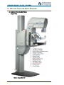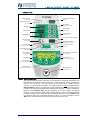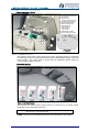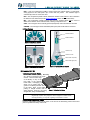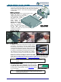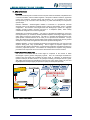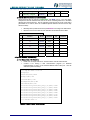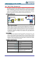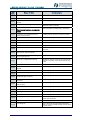
1
Diamond
A Medical Physicist’s Guide
to the
Diamond Breast Imaging System
MM03341b
A Medical Physicist’s Guide to the Diamond
Breast Imaging System
Prepared by:
Instrumentarium Imaging Inc
Instrumentarium gratefully acknowledges contributions to, and review of this Guide by:
Arthur G. Haus
Consulting Medical Physicist to Instrumentarium
and (AEC Performance Evaluation Procedure) by:
Melissa C. Martin, M.S., FACR
Consulting Medical Physicist
Member of the FDA NMQAAC
Copyright
Code:
MM03341b
Date:
October 2003
© 2003 Instrumentarium Imaging Inc
Authored By:
Tim Jaenchen
Senior Product Specialist
Instrumentarium Imaging Inc
Documentation, trademark and the software are copyrighted with all rights reserved. Under the
copyright laws the documentation may not be copied, photocopied, reproduced, translated, or
reduced to any electronic medium or machine-readable form in whole or part, without the prior
written permission of Instrumentarium Imaging.
Instrumentarium Imaging reserves the right to revise this publication from time to time and to make
changes in the content of it without obligation to notify any person of such revision or changes.
Manufactured By:
Instrumentarium Imaging
P.O. Box 20
FIN-04301 Tuusula
FINLAND
Tel. +358 10 394 6500
Fax. +358 10 394 6501
E-mail: [email protected]
Internet: http://www.InstrumentariumImaging.com
Diamond complies with UL and C-UL (File E218408).
North American Distribution:
Instrumentarium Imaging Inc
300 W Edgerton Avenue
Milwaukee, WI 53207
Tel: 800 558 6120
Fax: 414 481 8665
E-mail: [email protected]
Internet: http://usa.instrumentarium.com
Windows® is trademark of Microsoft Corporation in the United States of America and other
countries. HyperTerminal is a trademark of Hilgraeve Inc, Monroe, MI, USA.
iii
iv
A MEDICAL PHYSICIST’S GUIDE
TO THE
DIAMOND BREAST IMAGING SYSTEM
TABLE OF CONTENTS
4. Grid Specifications .....................................13
I. AN INTRODUCTION TO DIAMOND
G. MultiChoice™ Magnification............................13
A. Scope............................................................... 1
H. Physicist Test Mode .......................................13
B. Precautions/Warnings...................................... 1
1. Using the Service Mode Interface ..............13
2. Service Program Menu...............................13
3. Using the Physicist Mode Program ............14
1. Intended Use ............................................... 1
2. Radiation Safety .......................................... 1
3. Safety Features & Error Codes.................... 2
I. Collecting Data Using an External PC .............15
C. Other Sources Of Information.......................... 2
1. Data Formats..............................................15
2. Equipment Required...................................15
3. HyperTerminal Interface For Diamond .......15
4. Data Capture ..............................................16
1. On-Site OEM Documentation ...................... 2
2. Contact Information ..................................... 2
3. Calling For Help ........................................... 2
4. Reference Materials .................................... 3
J. Integrated Quality Control (IQC) .....................16
II. GETTING FAMILIAR WITH DIAMOND
IV. AUTOMATIC EXPOSURE CONTROL SYSTEM
A. Diamond Operating Controls ........................... 4
A. Features .........................................................18
1. System View ................................................ 4
2. Control Panel ............................................... 5
3. Positioning Control Centre........................... 6
4. Foot Pedal Control....................................... 6
5. Other Controls ............................................. 7
1. Absolute AEC™ ..........................................18
2. PaddleLogic™ .............................................18
3. Density Control...........................................18
4. AutoPoint™ Detector...................................18
B. Modes Of Operation .......................................18
B. Diamond Set-Up .............................................. 7
1. Manual........................................................19
2. Auto Time ...................................................19
3. Auto kV.......................................................19
4. Other Modes...............................................19
1. Attaching a Cassette Holder ........................ 7
2. Attaching a Compression Paddle ................ 7
3. Switching On the Unit .................................. 8
4. Inserting a Cassette..................................... 8
5. Selecting an Exposure Mode....................... 8
6. Selecting an X-Ray Beam Filter................... 9
7. Selecting a Detector .................................... 9
8. Adjusting Density Control ............................ 9
C. Technique Chart.............................................19
D. AEC Operation ...............................................20
1. Overview ....................................................20
2. Auto-kV Functional Description ..................20
3. AutoPoint™ Detector...................................21
C. Diamond Exposure .......................................... 9
E. AEC Performance Evaluation.........................23
1. Make an Exposure....................................... 9
2. Automatic Compression Release .............. 10
3. Display Accuracy & Brief Specifications .... 10
4. Post-Exposure Display Notes .................... 10
5. Duty Cycle ................................................. 10
1. Background ................................................23
2. Definitions ..................................................25
3. Testing Procedure—MQSA Method # 2 ......26
4. Optional—Detector Reproducibility .............28
III. DESCRIPTION OF MAIN FEATURES
V. PHANTOM IMAGING & ARTIFACT EVALUATION
A. ParkBack™ ..................................................... 11
VI. DIAMOND X-RAY TUBE
B. PaddleLogic™................................................. 11
C. Collimation..................................................... 11
A. Target and Filter Information ..........................31
1. Aperture Plates (Diaphragms) ................... 11
2. X-Ray Field Localizer Light........................ 11
B. Tube Geometry and Dimensions....................31
VII. THF X-RAY GENERATOR
D. Rotation ......................................................... 11
1. Anatomic C-Arm Rotation (ACR) ............... 12
2. AutoRotate................................................. 12
3. Mirror Angle ............................................... 12
A. Operating Principles .......................................32
B. Duty-Cycle ......................................................32
C. Generator Specifications ................................32
E. Alpha ID II Film-ID Label................................ 12
F. ProForm Synchro-Bucky™ ............................. 12
VIII. COMPRESSION SYSTEM
1. Patient Comfort and Safety........................ 12
2. Cassette Tray Operation............................ 12
3. Generator Synchronization ........................ 13
A. Foot Controls ..................................................34
v
B. Compression Force........................................ 34
D. Table — Maximum mA/mAS........................... 40
C. ECS Compression ......................................... 34
E. Varian X-Ray Tube Specifications ................. 40
F. Alpha ID II Flash Label Explanation ............... 41
IX. APPENDIX
G. Table — Diamond Error Code Guide.............. 41
H. Back Panel Connections ............................... 44
A. Article One — Physics Testing Procedures for
Diamond ............................................................. 35
I. Data Output Samples ...................................... 45
B. Article Two — AEC Density Control
Performance Clarification ................................... 37
C. Template — Detector Outline......................... 39
vi
A MEDICAL PHYSICIST'S GUIDE TO DIAMOND
AN INTRODUCTION TO DIAMOND
I. A N I NTRODUCTION T O D IAMOND
D
iamond Breast Imaging System is a high quality x-ray imaging device intended for
screening and diagnostic mammography examinations, and interventional procedures.
Among its many remarkable features are several new and innovative ones, which
include: AutoPoint™ AEC Detector, ParkBack™ tube head movement, PaddleLogic™, ACR
motorized C-Arm rotation and the Position Control Center.
Additionally Diamond
incorporates the highly acclaimed ECS™ bi-directional compression system, Delta32-TACT™
3-D digital and Stereotactic option, THF (Tuned High-Frequency) generator, a digital-ready
design, bi-angle rotating anode X-ray tube, ROC grid option, and easy patient positioning.
Despite the breadth and sophistication of fresh technology implemented in Diamond, it
continues the tradition of core qualities found in Instrumentarium’s Performa and Alpha IQ of
the past — comfort for the patient and ease of use for the technologist.
A. SCOPE
Instrumentarium recognizes the importance and supports the efforts of medical physics in the
delivery of quality mammography service. This Guide has been prepared to assist Medical
Physicists as they work with Diamond Breast Imaging System during Annual Mammography
Surveys and consultations.
This document is written for the practicing Medical Physicist who already possesses a basic
familiarity with the operation of mammographic x-ray systems. Features and functions specific
to Diamond are described so that the Medical Physicist may efficiently and effectively work with
the unit.
The reader is advised that although this Guide is generally suitable for worldwide use, portions
of it are directed toward and address the regulatory environment in the USA, namely the
Mammography Quality Standards Act (“MQSA’).
This effort is intended to be a ‘living document.’ The reader is encouraged to remit comments
and suggestions (see Contact Information) for items to be added to this Guide that will benefit
Medical Physicists and help facilities to obtain optimal results from their Diamond. We invite
you to discover the sophistication of design and simplicity of operation of the Diamond Breast
Imaging System.
B. PRECAUTIONS/WARNINGS
1. Intended Use
Diamond is intended only for mammography examinations, no other uses are permitted. Two
film/screen formats (18x24 and 24x30) and several cassette holder and digital options are
available. The unit has been designed for stationary use, however special design elements
have been engineered for clients choosing a mobile van application (contact Instrumentarium
for details).
When energized, this device emits filtered X-Ray radiation for medical purposes. The Diamond
system is CE-marked in accordance with MDD (93/42/EEC) and its design complies with IEC
60601-1. It fulfills the EMC requirements of IEC 60601-1-2, as a Class A device. The unit is
tested and certified according to UL 2601-1 and CAN/CSA C22.2 NO. 601.1.
WARNING!
Never use the equipment if it is suspected that any electrical, or radiation-emitting
component is defective or inoperative.
WARNING:
Only trained, licensed persons are allowed to operate the Diamond unit.
2. Radiation Safety
Since radiation is harmful to the human body, the radiation protection regulations of individual
countries must be complied with. The user must have permission to use an x-ray device for
mammography. Radiation protection measures should be used. Prior to examination, it is
recommended to make sure that the doctor knows if the patient is pregnant. If someone has to
be close to the patient, protective clothing must be used.
The maximum radiation that the system is capable of producing corresponds to 5% of the
typical CT-radiographic exposure. However, the user should wear a dose-recording device and
follow local regulations for radiation protection.
MM03341b
1
A MEDICAL PHYSICIST'S GUIDE TO DIAMOND
AN INTRODUCTION TO DIAMOND
The optional radiation shield must be used, or the unit must be operated from a shielded
control room (significant zone of occupancy is 2 meters). During the exposure, visual and
audible contact should be maintained with the patient. The freestanding radiation shield can be
positioned as required in the X-ray room. An Instrumentarium Imaging mammography
accessory storage unit is available as an option and may be placed behind the freestanding
radiation shield. Diamond with radiation protection is in accordance with IEC 601-1-3:1994.
3. Safety Features & Error Codes
Diamond incorporates many safety features designed to offer protection from physical injury
and unnecessary radiation dose to the operator and patient. A few examples:
Exposures without a cassette present or double exposure are prevented
Mechanical movement (vertical, rotation, etc.) is
inhibited if compression force is detected
If the proper optical density can not be achieved with
the technique selected, the Exposure Monitor will
terminate the exposure in 50 msec
Operational or safety-related errors that prevent exposure or
some other function are explained to the user in the form of an error code and description on
the control panel. If you encounter any errors during a survey, consult the Diamond Error Code
Guide in the appendix.
C. OTHER SOURCES OF INFORMATION
1. On-Site OEM Documentation
Each Diamond system is delivered with a complete set of original factory documentation,
including:
86500 Diamond Operation Instructions
86501 Diamond Installation Instructions
86502 Diamond Service Manual
86513 Diamond Technique Chart
86528 Diamond Quick User Guide
Check with your client or the equipment installer for their location.
2. Contact Information
Instrumentarium Imaging Inc
Product Support Division — Mammography
300 W Edgerton Avenue
Milwaukee WI 53207 USA
ph: 800.558.6120 • fax: 414.481-8665 • URL: http://usa.instrumentarium.com/
Ext.
Email
Brian Broncatti
Contact
National Service Mgr
Title
6314
[email protected]
Darin Grebe
Operations Coordinator
6318
[email protected]
Jason Kraft
Service Engineer
6326
[email protected]
Julia Hollweck
Service Admin
6336
[email protected]
Diane Sazama
Admin Assistant
6314
[email protected]
Tim Jaenchen
Senior Product Specialist
6312
[email protected]
3. Calling For Help
To better assist you in answering questions or resolving technical or operational issues, the
support staff at Instrumentarium may require additional information:
1. General
Serial number of the unit
Name of the operator who experienced the problem
Software revision level (on the LCD-display when power is switched on)
2. AEC issues
Film/screen combination in use
2
MM03341b
A MEDICAL PHYSICIST'S GUIDE TO DIAMOND
AN INTRODUCTION TO DIAMOND
3.
Processing time and developer temperature
What is the gross fog, speed index, and contrast index of the film developed in
the processor?
Was any work done on the film processor just prior to the problem?
Did problem appear suddenly or gradually?
Breast compression, kV selected, type of view, mAS of exposure, detector
positioning, filter selected, which imaging mode?
Other concerns
a. What is the history of the problem?
b. Error codes on the display?
c. What have you done to attempt a fix?
d. Is it permanent failure or intermittent?
e. If intermittent, how often per day?
f. Did problem appear as a result of some other work performed on the unit?
g. Is any other equipment exhibiting intermittent problems?
4. Reference Materials
1. Haus AG, Jaskulski SM. The Basics of Film Processing in Medical Imaging. Madison
WI: Medical Physics Publishing, 1997.
2.
MM03341b
Haus Arthur G. (ed). Advances in Film Processing Systems Technology and Quality
Control in Medical Imaging. Madison WI: Medical Physics Publishing, 2001.
3
A MEDICAL PHYSICIST'S GUIDE TO DIAMOND
GETTING FAMILIAR WITH DIAMOND
II. G ETTING F AMILIAR W ITH D IAMOND
A. DIAMOND OPERATING CONTROLS
1. System View
Figure 1. Diamond Main View
4
MM03341b
A MEDICAL PHYSICIST'S GUIDE TO DIAMOND
GETTING FAMILIAR WITH DIAMOND
2. Control Panel
Ready Indicator
Exposure Time Display
(green LED)
(seconds)
ready
x-ray
Radiation Indicator
timer
s
(amber LED)
KV Selector
(range: 20-39, step: 1kV)
KV Display
kV
MAS Selector
(range: 2-500)
MAS Display
mAs
Density Selector
(range: -7 to +7)
Density Display
Den
(3 green LEDs)
Filter Selector
Auto kV
Auto Time
Manual
(moly, rhodium, aluminum)
AEC Mode Indicator
mGy
MODE
Auto
Point
AEC Mode Selector
(AutoKV, AutoTime, Manual)
AutoPoint Indicator
(green LED)
Warning
This x-ray unit may
! be dangerous to
operator unless tisafet exposure
factors and operating instructions
d
b
d
(3 green LEDs)
Cancel Button
(for menu options)
Avg. Glandular Dose Display
Mo
Rho
Al
Filter Indicator
Cancel
LCD Display
Preprog1 Bucky 18x24
03.07.21 11:46
Auto
Rel
Menu
Set
Menu Selector & Set Button
(for menu options)
Function Key 3
(button, LED and LCD label)
Function Key 1
(button, LED and LCD label)
Function Key 2
(button, LED and LCD label)
Exposure Button
(single stage switch)
Figure 2. Operator Control Panel
The Diamond Operator Control Panel uses an LCD-display and 7-segment alphanumeric and
LED indicators to communicate status information, as well as various buttons to regulate the
functions of the X-ray unit. The control panel can be attached to the radiation shield or
remotely, in a control booth for example. An X-Ray Exposure can only be initiated from the
Exposure Button. When it is pressed, the computer responds and starts an exposure routine,
but when it is released the hardware itself terminates the exposure — a safety consideration.
Behavior of the Function Keys will vary depending on the menu display and required
functions. They are used to choose various imaging modes, to select the Integrated Quality
Control (IQC) function and to input date, time and hospital information. Use the Cancel button
to go back one step in the menu without selecting an option, or to cancel a selection. Use the
SET button to accept selections and to proceed to the next step in the menu.
MM03341b
5
A MEDICAL PHYSICIST'S GUIDE TO DIAMOND
GETTING FAMILIAR WITH DIAMOND
3. Positioning Control Centre
A.
B.
C.
D.
C
A
B
E.
F.
G.
H.
I.
J.
D1
F
E
G
D2
K.
L.
Collimator Lamp Button
Compression Force Selector
Filter Selector
Rotate Control
1-CCW, 2-CW (patient view)
AutoPoint Detector On/Off
Manual Detector Selector
Thickness Display (cm)
Force Display (N)
Angle Display (°)
Vertical Control
1-Up, 2-Down
Paddle Release Button
Manual Compression Knob
B
H
I
L
J1
J2
A
C
F
K
E
Figure 3. Positioning Control Centre
The Position Control Centre cleverly locates every function and display used by a technologist
for positioning a patient in close proximity with the patient, permitting better patient contact and
faster operation. The Centre moves in concert with the compression system making the
controls readily available at all times.
4. Foot Pedal Control
Figure 4. Foot Pedal Control
The Diamond Foot Pedal controls several systems of motion that run at various speeds,
depending on the process being called upon.
CAUTION!
The PAD and ECS controls will act to apply force between the compression
plate and the top of the cassette holder.
C-ARM — Only the C-Arm is shifted in the direction of the tube head at a ‘slow’ speed.
6
MM03341b
A MEDICAL PHYSICIST'S GUIDE TO DIAMOND
GETTING FAMILIAR WITH DIAMOND
PAD — Only the Compression Paddle is driven toward the cassette holder at ‘high-speed’
during approach and ‘slow-speed’ upon contact with the patient. The collimator light is
automatically turned on at the same time.
ECS — The compression paddle is driven toward the cassette holder and the C-Arm is shifted in
the direction of the tube head (see the ECS Compression section for more information).
REL — The compression paddle is driven away from the cassette holder and the C-Arm is
shifted in the direction of the cassette holder, relieving compression force.
Z-UP — The Carriage is driven vertically upward (dual-speed) until a limit switch is activated.
Z-DOWN — The Carriage is driven downward (dual-speed) until a limit switch is activated.
5. Other Controls
P
Parkback Retract
Parkback Extend
Figure 5. AutoRotate Controls
Center 0°
Vertical ‘Z’ Up
Vertical ‘Z’ Down
Rotate Tube Left (CCW)
Rotate Tube Right (CW)
Left (-15°)
Right (15°)
Figure 7. Tubehead Cover Controls
Figure 6. Stereotactic Tilt Controls
B. DIAMOND SET-UP
1. Attaching a Cassette Holder
All cassette holders (Bucky, Multi-Mag,
etc.) attach to Diamond in the same
manner. Rest the back edge
of the Bucky on top of the
front edge of the cassette
holder base, overlapping by
about 2-3 cm (helps align the
guides in the Bucky with the slots
in the base). Keep it level while
pushing it straight forward until the
detent engages. Remove it by pulling Figure 8. Attaching the Bucky
straight back, firmly and deliberately.
NOTE:
The drive motor (in the C-Arm) and the cassette tray (in the Bucky) are
magnetically coupled. If they happen to remain engaged for a moment when
removing the Bucky, you may hear a grinding sound, which is normal.
2. Attaching a Compression Paddle
Insert the compression paddle into the holes on the front of the Positioning Control Centre and
push the paddle horizontally until it locks.
MM03341b
7
A MEDICAL PHYSICIST'S GUIDE TO DIAMOND
GETTING FAMILIAR WITH DIAMOND
To remove a compression paddle, first push and release either of the two
release buttons (not both) located on each side of the Positioning
Control Centre to unlock it. Then remove the paddle by pulling it
out horizontally.
3. Switching On the Unit
Check that the power cord is connected and the circuit
breaker (see Fig1-’O’) is switched on.
Toggle the Diamond user power switch
to ON (behind the carriage left
side, patient view). During
initialization the control
panel
displays
the
software version, (i.e.
Diamond SW C1.02.7)
afterwhich
it
displays:
Timer = 0, kV = 25, MAS = - - -,
Density = +0, mGy = 0, Auto-kV =
on, filter = Mo.
(Note, this start-up
condition is the default and may have been
modified by the installer.)
Figure 9. Attaching a
Compression Paddle
4. Inserting a Cassette
1
2
3
Figure 10. Inserting a Cassette
(1) Push the cassette latch aside with the leading corner of
the cassette. (2) Continue to insert the cassette, holding it
level and square with the Bucky. (3) Gently slide the cassette
in until the latch springs back into place. Take care not to trap
your fingers. The cassette tray will automatically drive forward
for exposure, and afterwards automatically drive back again.
The cassette tray can be driven manually to the ‘loaded’ or
‘ejected’ position by sliding the cassette latch release handle.
LOAD EJECT
Remove the cassette by pushing the cassette latch handle Figure 11. Latch Release Handle
towards the front end of the Bucky. Hold it in the forward
position with your middle finger and pull the cassette out with your thumb and index finger.
See sections ProForm Synchro Bucky™ and MultiChoice™ Magnification for more information.
NOTE:
If a cassette is in the ‘load’ position when power fails, simply pull the Bucky off of
the base – the tray will shift to the ‘eject’ position and the cassette may be
retreived.
5. Selecting an Exposure Mode
Diamond’s Exposure Control system has three modes of operation: Auto kV, Auto Time, and Manual. Select the mode with
the Mode select button. The light at the left of the button indicates
the mode that is currently selected. See the AEC System section
for more information.
NOTE:
Auto kV
Auto Time
Manual
MODE
Figure 12. Mode Selector
An exposure monitor will terminate the exposure in 50 msec (with an error
message) if the AEC determines that a suitably exposed image cannot be
achieved within the back-up time.
8
MM03341b
A MEDICAL PHYSICIST'S GUIDE TO DIAMOND
GETTING FAMILIAR WITH DIAMOND
AEC Operating Range — kV Limits
Bucky:
Manual: 20-39 kV
Auto Time and Auto kV: 23-35 kV
Specimen: 15-39 kV
Magnification:
Manual: 20-39 kV
Auto Time and Auto kV: 23-35 kV
Specimen: 15 - 39 kV
Delta 32:
Manual: 20-39 kV
Specimen: 15 - 39 kV
Table 1. KV Limits in AEC
6. Selecting an X-Ray Beam Filter
Diamond has three X-ray beam filters: Molybdenum (‘Mo’), Rhodium (‘Rh’) and
Aluminum (‘Al’). A filter is selected with the button at the Position Control Centre
or on the Control Panel. The movement is motorized and the chosen filter is
indicated with an LED. Diamond can be configured to automatically select the most
Mo
Rho
appropriate X-Ray beam filter (Auto-kV and Auto-Time modes) by enabling the
Al
AutoFilter feature; manual control is the default setting. Mo and Rh filters are
available between 15 - 35 kV (all modes), Al between 36 - 39 kV (in manual Fig. 13. Filter
Selector
mode only). More Diamond X-ray Tube information may be found later on in
this document.
7. Selecting a Detector
Diamond’s AEC system has 8 detectors that can be
selected automatically or manually. In automatic mode
(AutoPoint LED on), it chooses the most appropriate
detector at each exposure, depending upon the
individual breast being examined. Toggle automatic
mode on/off by pressing the AutoPoint button on the
Detector Location Display. Select a detector manually
by first toggling AutoPoint ‘off’, and then pressing the
relevant detector button on the Detector Location
Display. The detector outline on the compression
paddle correlates with the detector buttons and their Figure 14. Detector Selector
location under the image receiver. Find more AutoPoint
information in the AEC Features and AEC Operation sections later on in this document.
8. Adjusting Density Control
Each AEC mode has 15 steps for density adjustment, where one
step corresponds to a 10% change in mAS (factory default).
Since the step size may be programmed from 5% to 20% by the
installer, consult with the client to verify if a special adjustment
was made. More information on the Density Control may be
found later on in this document.
Den
Figure 15. Density Selector
C. DIAMOND EXPOSURE
1. Make an Exposure
The Ready light illuminates when Diamond is prepared for an
ready
exposure. If the exposure button is pressed before the system is
s
timer
x-ray
‘ready,’ a message is displayed to the operator indicating the
reason why not.
Figure 16. Ready / X-Ray
Make an exposure by pressing the X-Ray Exposure Control, a Indicators
single-stage switch (no ‘prep’ position), and continue holding it until the X-ray exposure
terminates. Radiation production is made known to the operator by the illuminated ‘X-Ray’
light, the audible enunciator, and the accumulating value on the timer display. If the exposure
button is released prematurely, the exposure is immediately terminated and an error message
is displayed on the LCD screen.
MM03341b
9
A MEDICAL PHYSICIST'S GUIDE TO DIAMOND
GETTING FAMILIAR WITH DIAMOND
2. Automatic Compression Release
Diamond’s Automated Compression Release (‘AutoRel’)
Preprog1 Bucky 18x24
decompresses the breast without operator input
03.07.21 11:46
immediately after completing an exposure, or in the event of
Auto
a power failure. AutoRel can be toggled on/off by pressing
Rel
Function Key 3 on the control panel (labeled with ‘Auto Rel’
on the LCD display). If a power failure occurs while AutoRel
is off, the C-Arm and/or paddle will not move. Also, if the
Delta32 Stereotactic device or any of the biopsy paddles
are in place, AutoRel is inhibited, however compression can Figure 17. AutoRel Function Key
be released with the foot pedals or manual compression
wheels. Refer to the Compression System section for more information.
3. Display Accuracy & Brief Specifications
Display and several system specifications are included here for the convenience of the Medical
Physicist performing an annual survey. A complete list may be found in Section 10 of the
Diamond Operating Instructions.
kV Range:
15 - 39 kV, in 1 kV increments
kV Accuracy:
± 1.5 kV, ripple < 5%
mAS Range:
large focus: 2-500
small focus: 4-300
mAS Accuracy:
Timer Range:
Timer Accuracy:
± 4mAS (for mAS ≤ 40), ± 10% (for mAS >40)
large focus: 0.02 – 5.0 Sec
small focus: 0.04 – 10 Sec
± 0.10 seconds
Compression Force Display:
± 20 N
Compression Thickness Display:
± 1 cm
Angle Display:
1
mGy Display :
± 2°
± 30%
Table 2. Brief List of Specifications
4. Post-Exposure Display Notes
kV Display — If Auto kV mode was used, the value in the kV display reflects the final kV
selected, not necessarily the initial or intermediate kV at the beginning of an exposure
sequence. See AEC Function Description for more information about kV shifting.
Timer Display — If a Bucky device was used, the value in the Timer display reflects the net
exposure time, which accounts for the ‘off' time introduced by the Synchro-Bucky feature. See
ProForm Synchro-Bucky for more information.
NOTE:
Triggering and timer accuracy of non-invasive kVp Meters may be affected by
Diamond’s Synchro-Bucky feature.
5. Duty Cycle
Duty cycle limitation is calculated based on the actual target heat load, and therefore lasts only
as long as necessary to protect the tube. During this time an exposure cannot be made. If the
exposure button is pressed before the cooling time has elapsed, the timer display will indicate
the cooling time remaining, typically 10 to 50 seconds. See the Diamond X-Ray Tube section
for more information.
1
For further information about estimating absorbed dose, refer to:
Spectral Dependence of Glandular Tissue dose in Screen-Film Mammography, Wu Xizeng, Wu X,
Barnes Gary T, Tucker Douglas M, Radiology 1991; 179:143-148
Normalized Average Glandular dose in Molybdenum Target- Rhodium Filter and Rhodium Target Rhodium filter Mammography, Wu Xizeng, Wu X, Gingold Eric L, Barnes Gary T, Tucker Douglas M,
Radiology 1994; 193:83-89
10
MM03341b
A MEDICAL PHYSICIST'S GUIDE TO DIAMOND
DESCRIPTION OF MAIN FEATURES
III. D ESCRIPTION
OF
M AIN F EATURES
A. PARKBACK™
P
A unique feature of Diamond is the capability to move the tube head back
and away from the patient 19 cm (7 1/2”) for totally free access to the
b
breast from above. This is particularly useful when positioning oblique or
lateral projections, or for interventional procedures such as a biopsy.
Movement is controlled by buttons located (a) on either side, or (b) on top a
of the tube head. Pressing a button drives the tube head until it comes to
its end position.
ParkBack operation may set for unattended
(press/release) operation, or ‘dead-man’ (press/hold) operation,
depending on the local safety regulations and user preferences.
AutoPark — operation can be programmed to automatically park
when the C-arm rotates to projections over 15°.
Safety clutch/sensor — if tube head movement is obstructed, ParkBack operation is
aborted and an error message is displayed.
AutoDrive — operation can be programmed to automatically drive forward when the
exposure control is pressed (before exposure ‘prep’ begins). If the exposure control is
released prematurely, the forward movement stops. AutoDrive is inhibited if the paddle
or cassette holder is missing, or if biopsy paddles or the Delta 32 receiver are in use.
NOTE:
Pressing any button will stop all motor operation affecting patient safety.
B. PADDLELOGIC™
Diamond’s PaddleLogic feature facilitates (1) automatic adjustment of the collimator, and (2)
regulation of AutoPoint AEC detectors - separately for each compression paddle. This feature
can be customized to customer preferences and is especially useful for diagnostic imaging
such as spot compression views, or interventional procedures when using ‘biopsy’
compression paddles. When Diamond detects one of these special paddles, it automatically
collimates the X-ray beam to the region of interest and constrains the available detectors to
that same ROI. The factory default is set for full coverage of the receiver for all paddles.
NOTE:
If the paddle and the cassette holder suggest different X-ray field sizes, Diamond
will always select the smaller field size.
C. COLLIMATION
Diamond has a fully computerized Beam Limiting Device (collimator). The radiation field-size
is automatically collimated when Diamond determines which cassette holder (Bucky or MultiMagnification) and compression paddle (see PaddleLogic™) are being used.
1. Aperture Plates (Diaphragms)
The automatic motorized collimator allows any rectangular collimation. For other collimated
shapes, i.e. semi-circular spot sizes, corresponding special diaphragms must be inserted
manually.
2. X-Ray Field Localizer Light
The X-ray field illumination light is switched on automatically when the compression
foot pedal is pressed, the tube head is being driven forward or the collimator light
switch is pressed. It is inhibited when the tube head is in the ParkBack position. The
light is switched off automatically after 40 seconds (adjustable by service technician
from 20 - 60 sec), when the exposure button is pressed, or when the collimator light switch is
pressed.
D. ROTATION
The C-Arm rotates ±185° from the vertical position (0°). A number of
operator control are provided to easily adjust the rotation angle. Direct
control can be taken by pressing and holding the Rotate-CW or RotateCCW buttons on the Position Control Centre, (a) or the tube head cover,
(b) until the desired angle is reached. Automatic operation is achieved
with the ‘AutoRotate’ controls, (c).
MM03341b
11
b
c
a
A MEDICAL PHYSICIST'S GUIDE TO DIAMOND
DESCRIPTION OF MAIN FEATURES
1. Anatomic C-Arm Rotation (ACR)
The axis of c-arm rotation for Diamond is close to the film plane, permitting the image receiver
to closely follow the contour of the breast. This minimizes the need for the operator to adjust
the height (Z-Up/Down) of the c-arm when rotating from CC to OBL projections.
2. AutoRotate
Located on either side of the c-arm is a group of controls that conveniently rotate the c-arm to
one of five pre-programmed projection angles. The ‘automatic’ nature of these controls can be
set by a service technician to behave in one of four ways, depending on local safety regulations
and user preference.
OFF — ‘dead-man’ operation: press/hold button until rotation stops
ON — unattended operation: press/release button
PADL-UP — unattended operation: same as ON but only when paddle is fully released
C.PEDAL — hands-free ‘dead-man’ operation: press/release button, then press/hold
the CARM foot pedal until rotation stops
Auto-rotation is inhibited if (1) no paddle is attached, (2) an invasive procedure paddle is in
use, or (3) the system detects compression force (>30N).
3. Mirror Angle
The OBL button normally drives the C-Arm to a predefined default angle (for example 45°),
which may afterwards be fine-tuned to the patient’s anatomy. With the MirrorAngle feature
enabled, pressing the opposite OBL button (after an exposure) causes the C-Arm to
automatically rotate to the same, but opposing angle. The same principle holds true for the
LAT buttons, which normally drive to the default 90° projection. If the angle is adjusted with the
direct-control buttons and then an exposure is taken, pressing the opposite LAT causes the CArm to automatically rotate to the opposing lateral projection.
E. ALPHA ID II FILM-ID LABEL
The film identification system,
Alpha ID II, can be a helpful data
recording or troubleshooting tool
during an annual survey. The post
exposure technique information is
transferred to the label camera for
subsequent ‘flashing’ onto the film (see the Flash Label Explanation section in the appendix for
more info).
F. PROFORM SYNCHRO-BUCKY™
1. Patient Comfort and Safety
The Bucky system for Diamond has been carefully
engineered with interventional procedures and
patient comfort in mind. All patient contact surfaces
are smooth and rounded and absent of any seams
or screws, resulting in increased safety and comfort.
In order to achieve this, the cassette (DIN-standard)
is inserted into a lateral slot that is 13cm (7cm for
large Bucky) anterior from the chest wall end of the
Bucky. This design results in a completely sealed
enclosure under the patient, ideal for preventing fluid
entry during interventional procedures.
Figure 18. ProForm Bucky
2. Cassette Tray Operation
When the cassette is inserted, it is automatically
moved forward (‘loaded’) into position against the front edge of the Bucky, ready for exposure.
After an exposure (or under certain error conditions) the cassette is automatically moved back
(‘ejected’) to the opening ready for removal from either side. The system permits manual
cassette removal in the event of power failure or system malfunction. The ProForm SynchroBucky consists of a one-part carbon fiber cover with rounded corners. Buckies utilize a
common grid motor integrated into the Diamond’s C-Arm for both Bucky sizes.
12
MM03341b
A MEDICAL PHYSICIST'S GUIDE TO DIAMOND
DESCRIPTION OF MAIN FEATURES
3. Generator Synchronization
Grid reciprocation is monitored by Diamond’s exposure control system. If it calls for an
exposure time that extends into a grid reversal, the generator suspends radiation for
approximately 60 msec at the apex of grid ‘turnaround’. This brief cessation is transparent to
the operator and patient, but under certain conditions may cause some non-invasive kVp
meters to sense the abrupt fall in the radiation waveform and prematurely abort collecting data.
Conversely, some non-invasive devices may not detect the brief cessation of radiation and
display the time duration from initial exposure start to final termination. The Diamond postexposure mAS and Time values account for the sum of the ‘off-time’ at each grid turnaround
and display only the net exposure time. This synchronization is engineered to prevent gridline
artifact and may be observed on a kV waveform.
4. Grid Specifications
Bucky Atten. Equiv:
Standard Grid
R.O.C. Grid
≤ 25 µm Al (total of all layers)
≤ 25 µm Al (total of all layers)
36 lp/mm
36 lp/mm
5:1
6:1 equivalent
Grid Frequency:
Grid Ratio:
Table 3. Grid Specifications
G. MULTICHOICE™ MAGNIFICATION
For magnification studies, Diamond users may conveniently choose from one of three
magnification ratios using the same cassette holder (film format is 18 x 24 cm). The factors are
1.6x, 1.8x and 2.0x and are easily changed by the operator.
The magnification factor is indicated in a window and can be changed by pushing in the two
small knobs in the back of the tunnel frame and then lifting up or lowering the tunnel. The
small focus (0.1mm) is automatically selected for magnification exposures. In magnification
manual mode kV values starting from 20 kV can be used.
H. PHYSICIST TEST MODE
When it is operated in ‘Normal Mode’, Diamond automates many functions and processes for
easier and safer operation. For example, functions such as focal spot selection or beam
collimation are automatic and not adjustable by the routine user. Or, processes like cassette
insertion or kV selection are monitored and controlled to prevent wrong exposures. However,
during an annual inspection by a Medical Physicist or calibration by a service technician, these
automated actions tend to make certain tasks more time-consuming or even impossible.
Diamond software includes an operational mode, ‘Service Mode’, that limits these automated
actions, bypassing the normal function of the system. This mode enables the service
technician to perform various calibrations, adjustments and evaluations without the usual
safety constraints.
NOTE:
AEC and generator performance and duty-cycle limiting behave the same in
Service Mode as they do in Normal Mode.
1. Using the Service Mode Interface
To access Service Mode, simultaneously press kV- and
Den- buttons for 3 seconds until the display indicates
SERVICE. To return to Normal Mode, simultaneously press
kV- and Den- buttons for 3 seconds until SERVICE clears
from the display.
CAUTION!
Diamond may update the kV or density display before recognizing the second
button press. Verify the kV and Density settings before taking exposures.
2. Service Program Menu
The Service Mode Interface permits access to a Menu that contains special programs for
system setups, calibration and diagnostics. The menu has been organized into several
categories called ‘Groups.’ They are: A: System Setup, B: Defaults, C: Diagnostics, D: AEC E:
C-Arm Rotation, F: Collimator, G: Compression, H: Filter, I: Generator, J: Tube Park, K: Stereo
Movement, and L: Physicist Mode.
MM03341b
13
A MEDICAL PHYSICIST'S GUIDE TO DIAMOND
DESCRIPTION OF MAIN FEATURES
One of these service programs, L: Physicist Mode is designed to simplify inspection of
various non-standard settings and paddle/receiver combinations by Medical Physicists and
service technicians. The table below lists Diamond operation that can be altered.
WARNING!
The Service Program Menu contains programs with software parameters and
adjustments that affect the safe and proper performance of Diamond. Only
trained service technicians are authorized to access these other programs.
Parameter
Range
(Default)
Grid
Off /
System
Disable/Enable Grid Movement. (Ideal for non-invasive kVp Meters with
triggering problems relating to Synchro-Bucky.)
Large /
Small /
Focus Size Selection. Over-ride normal focal spot size for Bucky (large) or
Mag (small). Also sets collimator front plate to anterior (large) or posterior
(small) setting.
Focus
System
Notes
Paddle
Any
Paddle /
Holder
Any
Holder /
System
Cassette Holder Selection. Over-ride normal cassette holder detection and
beam collimation*. If no cassette holder is attached the collimator is driven
fully open.
Off /
System
Disable/Enable kV Limit. If disabled and manual mode is selected, any kV in
the range 15 to 39 can be used.
kV Limit
System
Paddle Selection. Over-ride normal paddle detection and PaddleLogic
operation (beam collimation and AutoPoint detector selection). *
* Over-ride of collimation is constrained to the actual size of a mounted receiver. Ex: The large
paddle and large Bucky are selected, but only an 18x24 Bucky is attached, so the X-ray field size
will be limited to 18 cm by 24 cm. The smaller field size for any paddle or receiver combination
prevails over the larger.
Table 4. Physicist Mode Adjustments
3. Using the Physicist Mode Program
a) Start Service Mode as described in step 1 above.
b) Access the Service Program Menu by pressing
the Menu/Set button. The LCD display shows a
portion of the list of Groups. Function Key 1
(‘Fkey1’) scrolls the list Down while FKey3 scrolls
Up.
Menu
Set
c) Press Up or Down until the ‘L: Physicist’ Group
is displayed between the pointers (‘> <’). Press
Accept (FKey2) to gain access to the program.
d) Within the L: Physicist group is a single
‘Procedure’ item called 1:Setup. Press Accept to
open it.
e) Step down through the list of ‘Parameters’ by
pressing Next until the desired item is between the
pointers (note that navigation has shifted to FKey2).
Change the parameter value with the Down or Up
keys according to the table above. Press Menu/Set
to approve the change.
f) Press Cancel at any time to exit without making a change; press it several times to return to
Service Mode.
NOTE:
Any changes to settings made in the Physicist Mode Program are restored to
normal ’System’ defaults when exiting Service Mode.
14
MM03341b
A MEDICAL PHYSICIST'S GUIDE TO DIAMOND
DESCRIPTION OF MAIN FEATURES
I. COLLECTING DATA USING AN EXTERNAL PC
Diamond is equipped with 2 serial data communication ports. The first, Com 1, is configured
for system calibration and programming, and for downloading AEC and parameter information.
The second port, Com 2, is configured for sending label data to the Alpha ID II film camera.
1. Data Formats
Test data may be gathered from one of three output formats (see Data Output Samples in the
appendix).
Output Type
Port
Notes
Exposure
Flash Label
COM2
Requires DB9 null-modem
adaptor
aecdump 1
COM1
Basic AEC data output — each detector’s signal strength and
limited post-exposure data: time, detectors used (pri and sec),
kV, exp. time, thickness, exp #.
COM1
Detailed AEC data output — same as above plus: mAS, density,
filter, cassette holder, kV-shift pattern, and service data (linearity
cal, pre-comp. exp. time, detector offset, target time, AkVLimit,
IQC coef.)
aecdump 2
Comments
Printout of essential post-exposure data: cassette holder, filter,
exp. time, kV, mAS, density, thickness, force, angle, laterality,
dose, detector, exp #.
Table 5. Data Output Types
2. Equipment Required
All three formats are output as standard ASCII text data and may be viewed or saved with a PC
communications program. Equipment required:
Laptop Computer
PC Communication program (such Windows HyperTerminal® by Hilgraeve Inc.)
Standard RS-232 Serial Cable, DB-9FM to DB-9M (available from Instrumentarium, part
code 85102)
DB-9 Null Modem Adaptor (if collecting the COM-2 label output)
3. HyperTerminal Interface For Diamond
The HyperTerminal interface and setup may vary somewhat depending on the version of the
program and operating system, however its setup should be similar to the information offered
here.
Figure 20. Connect To Dialog
Figure 19. Port Settings Dialog
MM03341b
15
A MEDICAL PHYSICIST'S GUIDE TO DIAMOND
DESCRIPTION OF MAIN FEATURES
a)
In HyperTerminal, select the desired COM-port with the File|Properties menu and the
‘Connect To’ tab sheet.
b) Next, click Configure to open the Port Settings dialog box. Adjust the parameters to
those shown in the table below.
c) Accept the changes with OK.
The serial connection is an ordinary asynchronous receiver/transmitter (RS- 232). The serial
ports in Diamond are initialized as follows:
Port
Baud
Rate
Data
Bits
Parity
Stop
Bits
Flow
Control
COM 1
(aecdump)
19200
8
None
1
None
COM 2
(Alpha ID II)
1200
8
None
2
None
Table 6. Serial Port Configuration
4. Data Capture
a) With Diamond switched off, connect
the serial cable between COM-1 and
the PC serial port (see back panel
connections in the appendix). —OR—
For COM-2 output, remove the serial
cable from the Alpha ID II, and
connect it with the null-modem
adaptor to the PC serial port.
b) On the PC using Notepad or a similar
text-editing program, create a blank
text file for recording data (example,
C:\param aecdump.txt).
c) Open a HyperTerminal session
d)
e)
f)
g)
h)
i)
NOTE:
Center ‘B’
connector
configured with the port settings Figure 21. Alpha ID Serial Ports
described above.
Switch on Diamond. After it starts and connects with the PC, HyperTerminal will
display a prompt, D1.05.7>, where ‘1.05.7’ is the software version. (Only for COM-1
output. No prompt with COM-2).
Select the HyperTerminal
menu, Transfer|Capture Text.
In the dialog box choose the
blank text file and click Start.
—
In
COM-1
Session
HyperTerminal type param
aecdump 1 (or aecdump 2)
and press Enter to send a
Figure 22. Capture Text Dialog
command to Diamond to
output AEC data. After each
exposure Diamond will send data to the PC.
COM-2 Session — Begin taking exposures. Diamond will send data to the PC.
When the session is completed, end the text capture with menu Transfer\Capture and
Stop. Switch off Diamond, close HyperTerminal, and disconnect the serial cable.
Switch on Diamond.
If the cable to the PC is still attached when Diamond is restarted, Diamond will reestablish a connection.
J. INTEGRATED QUALITY CONTROL (IQC)
Integrated Quality Control (‘IQC’) is a Diamond feature designed to keep image density the
same even if the film/processor environment has changed. Diamond can correct the AEC
exposure times by up to ±40% after which the unit gives an error code and prompts for a check
16
MM03341b
A MEDICAL PHYSICIST'S GUIDE TO DIAMOND
DESCRIPTION OF MAIN FEATURES
of the film processing system. The IQC test is done with a phantom exposure. After the
exposure the film is developed and its density checked with a densitometer. If the measured
optical density is different than the current setting in Diamond, the new value is entered in QA
(Quality Assurance), establishing a coefficient that modifies the AEC exposure time calculation.
MM03341b
17
A MEDICAL PHYSICIST'S GUIDE TO DIAMOND
AUTOMATIC EXPOSURE CONTROL SYSTEM
IV. A UTOMATIC E XPOSURE C ONTROL S YSTEM
Diamond Automatic Exposure Control (AEC) is a sophisticated, all solid-state, microprocessorbased system. Designed around the revolutionary AutoPoint™ Detector, the AEC System
automatically; (1) selects the most appropriate among eight detectors arrays, (2) selects the
beam filter (molybdenum or rhodium), (3) selects the initial kV and then adjusts the kV ‘on-thefly’ (a pre-exposure analysis isn’t necessary), and (4) calculates the mAS/time required for
proper optical density. Additional features such as PaddleLogic™, Exposure Monitor and
Synchro-Bucky™ combine with the operation of the AEC System to provide extremely hightech, yet user-friendly performance.
A. FEATURES
1. Absolute AEC™
Absolute AEC combines the technologies of Total Exposure Control (TEC), which originated
with Instrumentarium’s Performa Mammography System, together with the brand-new
AutoPoint technology. Auto Technique Parameters are selected during the exposure, which
means no test exposure is necessary. Exposure Monitor terminates the exposure in 50 msec
if proper image density cannot be achieved within the backup time; unnecessary patient dose
is minimized.
2. PaddleLogic™
Diamond PaddleLogic feature recognizes the compression paddle being used and automatically adjusts the collimation and regulates the detectors available for AutoPoint.
Interventional or special procedure paddles, like spot compression devices or small biopsy
paddles, can be individually configured to collimate radiation exposure only to the region of
interest, and constrain the AutoPoint detector to that same ROI.
3. Density Control
The density control function is a microprocessor-based feature that manipulates the AEC
exposure time calculation. On the operator control panel is a 15-step density selector ranging
from -7 steps to +7 steps. The amount of change in exposure time per step is programmable
by a service technician between 5% and 20%, in 1% increments. An article discussing Density
Control Performance may be found in the appendix.
4. AutoPoint™ Detector
The AutoPoint™ Detector is a highly complex component of the Diamond AEC system located
in the image receptor base. This unique circuit contains 8 discrete detector arrays that are
optimized in size, shape and location – 4 of them are positioned on the cassette centerline; four
of them are off centerline. A detector location is selected not by mechanical movement but
rather by software activation, either automatically by AutoPoint or manually by the operator.
AutoPoint selects the most appropriate detector to use for calculating the exposure time and
yields benefits such as:
Fewer retakes from choosing the wrong
detector
– Consistent selection of a detector based on
object radio-opacity, not on visual appearance
– More homogeneous optical densities over the
entire breast image
– More reliable performance when small breasts
are examined
The detectors are elliptical in shape; 3 are along the centerline,
2 are angled next to the centerline, and 3 half-ellipses are at
the chestwall edge. The active area of each detector array is
sampled by a minimum of 12 sub-detectors.
–
4
3
2
1
L
C
R
B. MODES OF OPERATION
NOTE:
For all three Modes of Operation, both the focal spot selection and tube current
(mA) are managed by Diamond and not adjustable by the operator.
18
MM03341b
A MEDICAL PHYSICIST'S GUIDE TO DIAMOND
AUTOMATIC EXPOSURE CONTROL SYSTEM
1. Manual
A traditional exposure control system in which the operator selects the technique (kV) and the
exposure time (mAS). The following mAS values may be selected.
2
3
4
5
6
8
10
12
16
20
25
32
40
50
63
80
100
125
150
175
200
250
300
350
400
450
500
Table 7. Selection of mAS Values
2. Auto Time
A conventional automatic exposure control system in which the operator selects the technique
(kV) and Diamond controls exposure timing (mAS).
3. Auto kV
A fully automatic exposure control system in which Diamond manages the technique (both
initial and after start of the exposure), filtration (Mo, Rh), detector selection, and exposure
timing (mAS). The initial kV is determined by Diamond based on the compression thickness
(The user may change the initial kV). During the exposure Diamond calculates the appropriate
kV and mAS for optimum exposure time and film density.
4. Other Modes
In addition to the three main modes of operation above, the Diamond AEC System may be preprogrammed for a variety of customized user-preferences, such as ‘Main’ or ‘Backup’ film
processor, or film/screen combination #1 or #2.
C. TECHNIQUE CHART
FILM: KODAK Min R 2000
SCREEN: KODAK Min R 2000 and Min R 2190
FILM: FUJI AD-M
SCREEN: FUJI AD Mammo Fine
FILM: AGFA HDR and HDR-C
SCREEN: AGFA HD and HD-S
Technique:
Compressed breast
thickness
kVp
Filter
Density
< 3 cm
23
Mo
0
4 - 5 cm
24 - 26
Mo
0
6 - 7 cm
27 - 29
Mo or Rh*
0
> 8 cm
30 - 32
Mo or Rh*
0
Manual Techniques
mAS
kVp
Small breasts
16 - 40
23 - 25
Implants
80 - 175
26 - 29
4 - 16
20 – 23
Specimens (non-Mag and Mag)
Note:
Implant displaced views and specimens may be phototimed if sufficient breast
tissue covers the photocell closest to the chest wall.
COMMENTS:
1. Maintain exposure times between 0.5 and 2.5 seconds. Decrease kVp if times are less than 0.5
seconds and increase if they are more than 2.5 seconds.
2. If exposure times are within indicated times, use + or - density settings to achieve optimal film
density.
3. Your technique may vary from those indicated above depending on film density preferences,
processor or film irregularity, or different film/screen combinations.
4. *Rhodium - use for very dense breasts, 6 cm or greater. No need to increase kV.
5. Magnification views - increase technique by 2 - 3 kVp, if exposure time exceeds 5 seconds.
MM03341b
19
A MEDICAL PHYSICIST'S GUIDE TO DIAMOND
AUTOMATIC EXPOSURE CONTROL SYSTEM
D. AEC OPERATION
1. Overview
The Diamond AEC System consists of three main parts: detection hardware, amplification and
conversion hardware, and the software algorithm. All aspects of detector selection, signal-level
control and conversion, exposure timing and technique, etc. are managed by the main
microprocessor (µ-P) or the AEC micro-controller (µ-C), either automatically or in response to
operator input.
Detection Hardware — Electromagnetic radiation is converted to a proportional, low-level
electrical current in the detection hardware using 8 arrays of photo-diodes, each in a parallel
configuration. The low-level current signal from each diode-group is passed to a high-gain
amplifier (current-to-voltage converter) and then to a multiplexer (MUX), which serially
combines the 8 signals into a common signal path.
Amplification & Conversion Hardware — The output of the detection hardware is sent to the
next stage where a µ-P controlled, variable-gain/offset amplifier and analog-to-digital converter
(ADC) further process the signal stream. The AEC µ-C transfers this digital signal via highspeed system communication bus to the main µ-P for exposure time calculation. The µ-P
scans all 8 detectors signals to find the lowest signal strength (represents the area of greatest
absorption in the patient) and applies that value to the exposure time algorithm.
Software Algorithm — The µ-P manages all operation of the generator (kV, mA, focal spot) and
exposure timing (start, stop, Synchro-Bucky). After it begins an exposure sequence and
receives the AEC detector data-stream, it processes the information and applies the data to a
non-linear, polynomial equation. The installer can manipulate the equation using software
parameters that compensate for the non-linear characteristics of film-screen receivers.
2. Auto-kV Functional Description
The function of Diamond Auto-kV mode might be compared to the manner in which
technologists operate general radiography systems. First, the technologist evaluates the
patient’s size and weight and considers the view (chest vs. extremity) to determine the
optimum technique (kV and/or mAS) to use, perhaps consulting a technique chart as a guide.
After taking the exposure and evaluating its outcome (exp. time, image quality, etc), the user
might adjust the technique slightly to improve the quality of subsequent exposures.
Figure 23. Auto-KV Block Diagram
20
MM03341b
A MEDICAL PHYSICIST'S GUIDE TO DIAMOND
AUTOMATIC EXPOSURE CONTROL SYSTEM
In Auto-kV mode, Diamond automatically selects the initial kV based on the compressed breast
thickness, much like an automated technique chart. (The ‘Auto-kV’ chart is a software look-up
table, programmed by the installer.) In the beginning of exposure, detector data representing
the rate of radiation exiting the patient is examined by Diamond, which calculates the exposure
time necessary to achieve the desired optical density.
Then, it compares the results of
this calculation against a ‘Target
Time’ lookup table. If they are not
in agreement, Diamond shifts the
kV (like a technologist who would
increase kV to reduce motion blur,
or decrease kV to increase image
contrast). Because the change in
tube voltage alters the beam
energy and consequently the rate
of radiation, Diamond re-examines
the detector signal and computes a
new exposure time. The process
may be repeated again, and in
each instance the kV may shift
upwards or downwards. Note that
this entire sequence of events
occurs in real-time within several Figure 24. KV-Shift Waveform
milliseconds after the start of
exposure — Diamond needs no pre-exposure analysis of the patient. After the exposure is
complete, the final kV is displayed.
The Auto-kV performance can be optimized and configured by the installer who considers and
evaluates receiver response characteristics (speed, contrast), client preferences, (desired kV
and image density), Diamond performance (target efficiency), and local patient population.
The configurable items are the initial-kV table (kV versus breast thickness), maximum
permitted kV-shift, and desired exposure time range (target time and target time limits).
3. AutoPoint™ Detector
The AEC Detection hardware is designed with eight separate, solid-state detector arrays.
Each one consist of either 22 or 12 parallel photo-diodes (sub-detectors) that are arranged in
an elliptical shape with a major axis of 42 mm and a minor axis of 32 mm (half-ellipse at the
chestwall edge). The overall coverage area is 13 cm A-P and 17 cm L-R. The detectors along
the chestwall edge are 5 mm from the edge of the film.
Each detector channel can be programmed with its own software gain and offset calibration
control, providing a great deal of flexibility in calibrating and balancing the detection hardware.
The hardware is all solid-state with no trim potentiometers; all adjustments are made in
software. A µ-P controlled, multiple-detector design gives the installer and operator many
options in its calibration and use.
The AutoPoint Detector has two modes of
operation, automatic and manual.
In the
automatic mode, AutoPoint selects the 2 most
appropriate detectors (primary and secondary)
by rapidly scanning all 8 signals at the very
onset of radiation. It is looking for those with the
lowest signal strength because their output
represents the areas of greatest absorption in
the patient. When AutoPoint finds the proper
detectors, it passes that data to the software
algorithm for exposure time calculation.
In
manual mode the operator selects which
detector the system is to use.
The user selects the mode of operation at the Figure 25. Position Control Centre
Position Control Center. If AutoPoint mode is
selected, the AutoPoint LED lights up at the Positioning Control Center and on the Control
Panel. When the AutoPoint LED is off the detector is selected manually. The user can choose
MM03341b
21
A MEDICAL PHYSICIST'S GUIDE TO DIAMOND
AUTOMATIC EXPOSURE CONTROL SYSTEM
the desired detector by pressing one of the selection buttons - an LED lights to indicate the
active detector.
Diamond recognizes the compression paddle (and the cassette holder, depending on the
PaddleLogic settings) in use and collimates the X-Ray field size and disables those detectors
that are outside of the radiation field or not under the compressed part of the breast.
a. Multi-Detector Considerations
X-Rays
i/u
Detector
Texp= ?
VFC
VFC
Av
MicroProcessor
Detector Amplifier
i/u
A
B
AV
int.
C
comp.
Vref
Figure 26. Single-Detector AEC Systems
Traditional AEC detector designs utilize a single detector, either solid-state or photmultiplier tube (PMT), that is mechanically moved under the patient region of interest
(ROI). In these designs, the AEC system is generally calibrated and tested with the
detector moved to the most clinically relevant location. Only the object (density or
thickness) or beam energy (kV, mAS) is changed. Because there is only one detector and
signal path, any variability introduced by the detector or AEC amplifier electronics would
be the same, despite detector location. Evaluating the performance of this type of
detection system is a manageable process of running a series of exposures to the single
detector and determining the coefficient of variation from exposure time or mAS or optical
density.
A multiple-detector design introduces concern about multiple sources and signal
pathways. Each detector and subsequent amplifier circuit may introduce a signal
deviation unique to that signal path. This not only results in a variation from one exposure
to the next, but also from one detector to another. To fully assess the AEC hardware, a
complete series of test exposure should be performed for each detector circuit. When one
considers the variety of tests that might be performed (reproducibility, kV linearity,
thickness linearity), and then also considers repeating those tests for up to 8 separate
detectors, it becomes a concern for the regulatory community, the manufacturer, and
especially for the healthcare provider.
b. Diamond Multi-Detector Design
Each detector array generates a leakage current proportional to the incident radiation.
The current signal is amplified by a dedicated (1 per detector) current-to-voltage converter,
then sent and to a common “Detector Selection” multiplexer. Under microprocessor
control, the multiplexer sequentially ‘streams’ the 8 signals to a common output. From the
MUX output to all subsequent electronic circuitry, ALL detector signals share a common
path to the microprocessor and the exposure time algorithm in software. In other words,
any inconsistency among the detectors can only be introduced prior to the multiplexer.
22
MM03341b
A MEDICAL PHYSICIST'S GUIDE TO DIAMOND
AUTOMATIC EXPOSURE CONTROL SYSTEM
Diamond software has a service/inspection feature that allows the service provider or
Medical Physicist to directly examine – detector by detector - the actual signal strength
produced by each one. By performing repetitive, identical tests using all 8 detectors and
20M
DETECTOR
SELECTION
DETECTOR ARRAY L1
MUX
S1
S2
S3
20M
S4
S5
S6
S7
S8
DETECTOR ARRAY C1
DETECTOR ARRAY R1
DETECTOR ARRAY L2
DETECTOR ARRAY C4
DETECTOR ARRAY C3
DETECTOR ARRAY R2
DETECTOR ARRAY C2
DETECTOR
SIGNAL OUT
4
3
2
1
L
C
R
Figure 27. AutoPoint Detector Design
comparing the results, the inspector can identify the detector with the most variation in
readings, and perform all subsequent testing using that one detector. It follows that, if the
equipment produces acceptable results using the detector with the greatest coefficient of
variation, then the remaining seven detectors will also produce acceptable results.
Extensive testing in the factory supports this premise.
E. AEC PERFORMANCE EVALUATION
1. Background
Despite Diamond’s multi-detector design, evaluating the AEC system is straightforward
process. The recommendations and procedure that follow were designed to minimize the
number of exposures that must be taken, and yet be thorough enough to effectively
appraise the AEC performance according to MQSA requirements. Instrumentarium
Imaging recommends that the inspector check all combinations of Configuration, Mode,
and Detector according to ‘method 2’ as described in the MQSA Policy Guidance Help
System (‘PGHS’), Document No. 6, (Aug 19, 2003).
a. Detector Balancing
The Diamond AEC system is designed with a software gain control parameter that is
adjusted during the calibration process. This control varies the detector signal strength
after the detector selection MUX (i.e. in the signal path common to all detectors), and
allows the service provider to calibrate Diamond to the desired film optical density. In
addition to this ‘master’ gain control there are 8 more software parameters used by
AutoPoint to adjust the gain of each detector individually. The process of setting these
individual gain controls is known as ‘Detector Balancing.’
MM03341b
23
A MEDICAL PHYSICIST'S GUIDE TO DIAMOND
AUTOMATIC EXPOSURE CONTROL SYSTEM
Mammography films often demonstrate an optical density gradient that decreases as one
scans toward the anterior edge (see figure). Heel Affect, a characteristic common to X-ray
tubes, causes this symptom.
Manual Technique:
25kV/100mAS, 25kV/125mAS
1.93
2.24
1.80
2.17
L1
C4
1.49
1.81
(-22.8%)
(-19.2%)
C3
1.64
2.00
(-15.0%)
(-10.7%)
L2
R1
C2
C1
1.86
2.17
1.93
2.24
(-3.6%)
(-3.13%)
1.93
2.24
R1
1.91
2.20
Optical density measurements from two, 25 kV exposure at manual techniques of
100 mAS (black, upper values) and 125 mAS (red, lower values). The O.D.
gradient varies about 20% to 25% between detector C1 to C4.
Figure 28. Optical Density Gradient
As an example the AEC was calibrated to achieve the desired film-OD of 1.80-1.90.
Noticed that if C4 were selected (user or AutoPoint), the density in the area of C4 is an
acceptable 1.81 OD, but the density in the area of C1 might be as high as 2.24 OD. So,
the main challenge in balancing the detectors is to address the question, “Should the
optical density for each of the detector’s local area equal the desired film OD, or should a
fixed-location area equal the desired OD?”
b. Local-Area Method
With this method, the detector gain is adjusted for each of the 8 detectors so that the film
OD in the location of that detector (i.e. the ‘local optical density’) will be the desired film
OD.
(+) Yields accurate film-OD inside the local area.
(+) Gives a logical result, i.e. the user expects the local area to be the desired OD.
(-) The film OD can change dramatically outside the local area.
(-) The image quality might be diminished from too high an OD outside the local area.
(-) The AEC system might perform unexpectedly. For example, if C4 is selected (13
cm anterior to the chest wall edge), to provide the dose necessary to compensate for
heel affect the system must increase either the exposure time or beam energy (kV),
possibly beyond what the user expects or desires.
24
MM03341b
A MEDICAL PHYSICIST'S GUIDE TO DIAMOND
AUTOMATIC EXPOSURE CONTROL SYSTEM
c. Common-Area Method
The detector gain is adjusted for each of the 8 detectors so that the film-OD at a common
location, such as C2, will be the desired film OD.
(-) The heel affect gradient will influence the local-area film-OD, even though the
common-area film-OD is consistent.
(+) The AEC system will yield a performance response that is more in keeping with
user expectations (less dramatic kV-shifting for example).
(+) Smaller chance that the film-OD at the chest wall edge is unacceptably high.
(+) Smaller deviation in film OD between views and patients. Example: if for patient ‘A’
detector C4 is selected, the film density at the ‘common-area’ will be the same as for
patient ‘B’ where detector C1 is selected.
Feedback from medical physicists and the inspection community indicate a preference for
testing with a ‘Local-Area’ method because it is more in accordance with the testing
procedures and performance expectation (i.e. ± 0.15 MOD) of the MQSA and ACR Quality
Control Manual. However, from radiologist and technologist feedback and installer
experience, it seems that a majority of cases when the AEC system is calibrated with
‘Common-Area’ detector balancing, the overall film OD, image contrast, and exposure
technique at a given facility is more stable and more compliant with the performance
capabilities of traditional AEC systems and the expectations of the mammography
community.
Instrumentarium has established an alternate method. It is based primarily on the localarea method (each detector produces a local OD that is ±0.15 MOD). However, to prevent
excessive film OD at the chest edge in those cases when C4 or C3 is selected (either
manually or by AutoPoint), C4 and possibly C3 may be slightly ‘de-tuned’. De-tuning
means that instead of driving C4 to match the desired OD, its gain will be set to either:
Produce a local (C4) density that is about 15% less than the desired OD, but still
within ±0.30 MOD, or,
Produce a chest edge (C1) density that is less than 2.00 OD (or as determined by
consulting physicist or customer).
Example: if the client desired-OD is 1.80, the C4 local-area will be set for about 1.53 OD.
2. Definitions
MQSA PGHS #6 uses several terms in describing the process of testing a device. The
following definitions for ‘Configuration’, ‘Mode’, and ‘Detector’ will help the reader apply the
regulatory terminology to Diamond and prepare it for evaluation.
a. Configuration
The operational setup of Diamond: A combination of the cassette holder (Bucky or
magnification stand) and X-ray beam filter (molybdenum or rhodium). Bucky / Mo is
considered the primary configuration because it is used most frequently.
NOTE:
MM03341b
The aluminum filter (AL) is available only in manual technique mode. The focal
spot size (lf, sf) and tube current (mA) are managed by Diamond and not user
selectable.
Configuration
Cassette Holder
Filter
Target
Primary
Bucky
Mo
Mo
Secondary
Bucky
Rh
Mo
Tertiary
Mag
Mo
Mo
Quaternary
Mag
Rh
Mo
25
A MEDICAL PHYSICIST'S GUIDE TO DIAMOND
AUTOMATIC EXPOSURE CONTROL SYSTEM
b. Mode
The method for controlling exposure timing: Diamond has three control methods: Manual,
and two automatic modes, Auto kV and Auto Time. Auto kV is considered the primary
mode because it is used most often.
c. Detector
The electronic sensor (diode array) that responds to
EM radiation: Diamond has eight discrete detector
arrays. Detector C2 is considered the primary
detector since it is the most centrally located and
most clinically relevant one.
C4
C3
L2
C2
primary
d. Other Mode
A customized setting of the Mode. Diamond
incorporates 4 pre-programmed modes (stations)
L1
C1
that permit a personalized setup for the user, such
as: ‘main’ or ‘back-up’ film processor, ‘Dr. A’ or ‘Dr. Figure 29. Primary Detector
B’, etc. Normally these are not used, therefore only
one station needs to be tested (consult with your client or the installer).
DET: C3
DETECTOR: pri. (C2)
CONFIG: pri. (Bu/MO)
MODE: sec. (A-mAS)
TEST OBJECT: 4 cm
LIMIT: ± 0.3 OD
DET: L1
±0.3 OD
DET: R2
±0.3 OD
DET: R1
±0.15 OD
Mean Optical Density - MOD
DETECTOR: primary (C2)
CONFIG: primary (Bu/MO)
MODE: primary (A-kV)
TEST OBJECT: 2, 4, 6 (8) cm
LIMIT: ± 0.15 OD
±0.3 OD
DETECTOR: pri. (C2)
CONFIG: quat. (Mg/RH)
MODE: sec. (A-mAS)
TEST OBJECT: 4 cm
LIMIT: ± 0.3 OD
R1
DET: C4
DET: L2
DETECTOR: C1
CONFIG: pri. (Bu/MO)
MODE: pri. (A-kV)
TEST OBJECT: 4 cm
LIMIT: ± 0.15 OD
R2
±0.3 OD
DETECTOR: pri. (C2)
CONFIG: sec. (Bu/RH)
MODE: pri. (A-kV)
TEST OBJECT: 4 cm
LIMIT: ± 0.3 OD
DETECTOR: pri. (C2)
CONFIG: sec. (Bu/RH)
MODE: sec. (A-mAS)
TEST OBJECT: 4 cm
LIMIT: ± 0.3 OD
±0.3 OD
DETECTOR: pri. (C2)
CONFIG: quat. (Mg/RH)
MODE: pri. (A-kV)
TEST OBJECT: 4 cm
LIMIT: ± 0.3 OD
DETECTOR: pri. (C2)
CONFIG: ter. (Mg/MO)
MODE: pri. (A-kV)
TEST OBJECT: 4 cm
LIMIT: ± 0.3 OD
DETECTOR: pri. (C2)
CONFIG: ter. (Mg/MO)
MODE: sec. (A-mAS)
TEST OBJECT: 4 cm
LIMIT: ± 0.3 OD
Figure 30. Diamond AEC Evaluation Method No. 2
3. Testing Procedure—MQSA Method # 2
The various combinations of Configuration, Mode, and Detector are illustrated in the figure,
Diamond AEC Evaluation Method No 2. (The expression “method No 2” refers to the MQSA
26
MM03341b
A MEDICAL PHYSICIST'S GUIDE TO DIAMOND
AUTOMATIC EXPOSURE CONTROL SYSTEM
recommendation for evaluating AEC systems with multiple modes and detectors (reference
MQSA PGHS #6, pg8, Question: 4).
a. Setup Diamond
Prepare the unit with the Primary Configuration (Bucky/Mo) and Mode (Auto kV). Deselect AutoPoint (press AutoPoint button, LED off) and select the primary Detector (C2).
(This setup is expressed as the ‘contact configuration’ by MQSA.)
b. Perform Test Exposures
Use a uniform attenuator of 2, 4, and 6 cm thickness. (Diamond includes a set of uniform
acrylic test objects (18cm x 28cm) in 0.5, 1.0, and 2.0 cm thickness. Ask your client for
their location.)
c. Measure and Record Film-OD
For each test object, assess the film-OD at the center of Detector C2 and complete the
table below. (HINT: Place the film on the compression paddle to quickly determine the
location of C2.)
DETECTOR: C2 CONFIG: Bucky/Mo MODE: Auto kV
#
Test Object (cm)
1
2.0
2
4.0
3
6.0
4
8.0 (opt.)
Film-OD (at C2)
d. Determine the Mean Optical Density (MOD)
Calculate the average film-OD from the density values above and record below. (Note:
MQSA does not recognize the 8 cm value in the MOD calculation.)
MEAN OPTICAL DENSITY (MOD)
e. Confirm AEC Performance for Primary Detector, Configuration and Mode
Confirm that the test results for the primary configuration, mode and detector used above,
meet the regulatory action limit of ±0.15 OD.
MOD
HIGH
LOW
Film-OD
Deviation from MOD
f. Confirm AEC Performance for Other Configurations and Modes
Assess all other Configurations and Modes used at the facility. MQSA requires that all
other results equal the MOD within the regulatory action limit of ±0.3 OD, at any ONE
clinically used thickness in the 2-6 cm range; 4 cm is recommended.
Vary the Configuration and Mode according to the table below.
Measure and record the film-OD and determine the deviation from the MOD.
DETECTOR: C2
#
MM03341b
Config — Holder / Filter
Mode
Thickness
Auto Time
4 cm
5
Primary — Bucky/Mo
6
Secondary —Bucky/Rh
Auto kV
4 cm
7
Secondary — Bucky/Rh
Auto Time
4 cm
8
Tertiary — Mag/Mo
Auto Time
4 cm
9
Tertiary — Mag/Mo
Auto kV
4 cm
27
OD
Deviation
A MEDICAL PHYSICIST'S GUIDE TO DIAMOND
AUTOMATIC EXPOSURE CONTROL SYSTEM
10
Quaternary — Mag/Rh
Auto kV
4 cm
11
Quaternary — Mag/Rh
AutoTime
4 cm
g. Confirm AEC Performance for Other Detectors
Setup Diamond with the Primary Configuration and Mode, and a 4 cm test object.
Compare the film-OD obtained from each of the seven remaining detectors to the MOD
obtained from the first detector. Use the regulatory action limit of ±0.15 OD (±0.30 OD is
permissible for C3 and C4, which are considered special detectors because of their deep
anterior location).
Manually select (AutoPoint turned off) the Detector according to the table below.
Measure and record the film-OD and determine the deviation from the MOD.
CONFIG: Primary — Bucky / Mo
#
Detector
Mode
Thickness
12
C1
Auto kV
4 cm
13
L1
Auto kV
4 cm
14
R1
Auto kV
4 cm
15
L2
Auto kV
4 cm
16
R2
Auto kV
4 cm
17
C3
Auto kV
4 cm
18
C4
Auto kV
4 cm
OD
Deviation
4. Optional—Detector Reproducibility
a. Initial Comparison of All Detectors
Install a 40 mm acrylic test block. Set the machine at 28 kV and AutoTime.
Connect a PC running a data communication program (i.e. Windows
HyperTerminal™ or equiv.) to the Diamond RS-232 serial comm port. Use the
command: param aecdump 1.
*** Exp Nr:649 ***14:19
Preprog:1
Detector(hex):0001
KvDspl(dec):28
IntTime(dec):118
ActExpTime(dec):445
Thickness(dec):40
Filtered xraylevel(dtr_xray)[0](dec):1918
Filtered xraylevel(dtr_xray)[1](dec):2002
Filtered xraylevel(dtr_xray)[2](dec):1895
Filtered xraylevel(dtr_xray)[3](dec):1893
Filtered xraylevel(dtr_xray)[4](dec):1658
Filtered xraylevel(dtr_xray)[5](dec):1772
Filtered xraylevel(dtr_xray)[6](dec):2026
Filtered xraylevel(dtr_xray)[7](dec):1853
*** End Nr:649 ***
Figure 31. Sample Printout - param aecdump
28
MM03341b
A MEDICAL PHYSICIST'S GUIDE TO DIAMOND
AUTOMATIC EXPOSURE CONTROL SYSTEM
Example of Test to Determine Detector with Most Variation (Poorest Reproducibility)
Diamond AEC Evaluation
(C) INSTRUMENTARIUM IMAGING 2002
40 mm Acrylic, 28kV
Mode: Auto-mAS
Exp.# Det used Time [ms]
Detector Signals
Det0
DET1
Det2
Det3
Det4
Det5
Det6
Det7
1
0
447
1894
1986
1895
1923
1685
1779
1990
1845
L1
2
1
445
1918
2002
1895
1893
1658
1772
2026
1853
C1
3
2
477
1893
1971
1884
1918
1682
1789
1991
1854
R1
4
3
472
1897
1976
1886
1908
1684
1800
2007
1869
L2
5
4
556
1900
1991
1905
1915
1667
1766
1982
1833
C4
6
5
510
1894
1983
1892
1914
1679
1783
1993
1844
C3
7
6
440
1893
1969
1883
1898
1687
1800
2012
1874
C2
8
7
490
1908
2002
1902
1913
1676
1779
2004
1847
R2
Standard Deviation
8.991
12.829
8.137
10.138
10.025
12.271 14.322 13.501
Mean
1899.6
1985.0
1892.8
1910.3
1677.3
1783.5 2000.6 1852.4
Reproducibility
0.473%
0.646%
0.430%
0.531%
0.598% 0.688% 0.716% 0.729%
Figure 32. Finding the Detector With Most Variation
Make 8 Auto Time exposures with one based upon each detector. The PC
should collect the exposure time and the detector output from each of the 8
detectors for each of the 8 exposures.
Organize the exposure time and detector output for each exposure for each
detector in a spreadsheet (see example table).
Calculate the standard deviation and mean of the output values for each detector.
Divide the standard deviation by the mean for each detector and multiply by 100
to obtain a reproducibility value for each of the 8 detectors (see example table).
b. Review of Statistics
Review the 8 calculated reproducibility values and select the largest. The largest
value corresponds to the detector with the most variation.
c. Verification of Equipment Operation
Set the equipment to use only the detector with the most variation.
Perform routine tests needed to verify accurate setup and operation of the
mammography system (section 3.a above).
MM03341b
29
A MEDICAL PHYSICIST'S GUIDE TO DIAMOND
PHANTOM IMAGING & ARTIFACT EVALUATION
V. P HANTOM I MAGING & A RTIFACT E VALUATION
When imaging the mammography accreditation phantom in Auto-kV Mode, Diamond usually
selects 24, 25, or 26 kVp, depending on the screen-film combination and desired image density
preset by the installing engineer. In some installations, the AEC Auto-kV decision point
between adjacent kVp values may occur at x-ray absorption levels that are very similar to those
of the accreditation phantom. While such AEC settings do not affect clinical imaging, repeated
phantom exposures in Auto-kV mode may produce alternating kVp’s, and associated
alternating mAS values. This type of variability can elicit concern from QC technologists and
inspectors. Fine-tuning of the AEC by the service engineer can move the decision point of the
Auto-kV algorithm so that phantom imaging produces a consistent kVp, and therefore a
consistent mAS value. Alternately, phantom imaging may be performed using the AEC AutoTime Mode, with a documented explanation describing the reason and referencing this
document.
When performing the artifact evaluation, it is recommended that clinically representative kVp
values be used. Artifact evaluations for the grid and molybdenum filter are typically performed
at 25 kVp using about 4 cm of uniform phantom material, such as acrylic. Artifact evaluations
of the rhodium filter are typically performed at 28 kVp using about 6 cm of uniform phantom
material. Due to the challenges of mass-producing 0.025 mm rhodium filters, nearly every filter
can produce visible artifacts, if imaged at sufficiently low kVp.
Note: If you have never seen the mottled appearance of a non-uniform filter, rhodium or
molybdenum, perform the artifact analysis at 23 kVp using any rhodium filter on any x-ray unit.
Non-uniformities will very often be visible on test films, but not visible on clinical images at
clinical kVp values. Not all artifacts can be totally eliminated. The concept of ALARA (as low
as reasonably achievable) may be useful.
To determine if artifacts are clinically significant, compare artifact test films with those from
other machines, and consult with the interpreting physician as to whether the artifact is
tolerable. If unsure, seek the opinions of more experienced medical physicists to determine if
the artifacts observed may interfere with clinical imaging. Replacing the filter or other source of
artifacts should eliminate any artifacts that may compromise clinical imaging.
30
MM03341b
A MEDICAL PHYSICIST'S GUIDE TO DIAMOND
DIAMOND X-RAY TUBE
VI. D IAMOND X-R AY T UBE
Diamond utilizes an X-Ray tube manufactured by Varian Medical Systems, which is intended
for use in mammography systems. It contains the proprietary model M-113SP rotating-anode,
39-kVp insert with a dual focal spot, metal center section, common-cathode design. The use of
a metal center section tube eliminates arcing due to metal deposition on glass and allows use
of a beryllium window in the insert for minimum inherent filtration. Additional X-Ray tube
specifications may be found in the appendix.
A. TARGET AND FILTER INFORMATION
Anode Material:
Inherent Filtration:
Added Filtration (nom.):
Anode Heat Capacity:
Anode Rotation Speed:
Molybdenum, Niobium doped
0 mm AL equiv. (beryllium port material)
Molybdenum – 25 µm
Rhodium – 25 µm
Aluminum - 50 µm
300 kHU
3600rpm (60Hz)
B. TUBE GEOMETRY AND DIMENSIONS
Source-Image Distance
(‘SID’)
Focal Spot Size (dual)
(IEC60336):
Target Angle (bi-angular):
Tube Angle:
Reference Axis:
MM03341b
660 mm (26”)
Large: 0.3 mm / Small: 0.1 mm
Large: 16° / Small: 10°
4°
46.2 mm from chest wall edge
31
A MEDICAL PHYSICIST'S GUIDE TO DIAMOND
THF X-RAY GENERATOR
VII. THF X-R AY G ENERATOR
The compact, revolutionary X-ray generator in Diamond further enhances the already
exceptional design found in previous Instrumentarium products. It remains one of the smallest
designs in mammography — and with recent improvements, one of the most robust. Engineered
around a variable high-frequency H-bridge, ‘resonance-regulator’ design (tuned high-frequency,
or ‘THF’), it is able to withstand open-circuit, short-circuit, and arcing conditions. Because it
operates near its resonant frequency, switching harmonics and radiated noise are greatly
diminished, enabling Diamond to meet stringent IEC and EU regulatory requirements.
A. OPERATING PRINCIPLES
Figure 33. Generator Block Diagram
When the operator selects the kV on the control panel, Diamond sets a corresponding kVreference voltage in the generator using 8-bit data and a digital-to-analog converter. The
generator compares this reference against a feedback signal from the kV in the X-ray tube.
Any error (difference) between them varies the frequency of the VCO and consequently the
switching speed in the H-bridge. Changes in bridge frequency modulate the power delivered to
the high-voltage transformer, changing the kV in the X-ray tube. This design quickly
compensates for changes in line voltage or load conditions to accurately maintain the kV
selected by the operator.
B. DUTY-CYCLE
The Diamond generator integrates the thermal energy applied to the target (kV x mAS = Joule)
with the cooling curves provided by the tube manufacturer. Duty cycle limitation is calculated
based on the actual target heat load, and therefore lasts only as long as necessary to protect
the tube. During this time an exposure cannot be made. If the exposure button is pressed
before the cooling time has elapsed, the timer display will indicate the cooling time remaining.
Depending on tube loading (technique being used, number of exposures and exposure
duration), the time limit is typically 10 to 50 seconds.
C. GENERATOR SPECIFICATIONS
Manufacturer:
Type:
Instrumentarium Imaging
MGHV-300
Frequency:
40 – 100 kHz
Power Max:
2.6 kW
kV Range:
kV Accuracy:
kV Ripple:
mA Range:
Maximum mA Output:
15 - 39 kV, in 1 kV increments
± 1.5 kV
< 5%
large focus: 50 - 100 mA (or 25 - 50 mA*)
small focus: 18 - 30 mA
large focus: 150mA
32
MM03341b
A MEDICAL PHYSICIST'S GUIDE TO DIAMOND
THF X-RAY GENERATOR
small focus: 45mA
mAS Range:
mAS Tolerance:
large focus: 2 - 500 mAS
small focus: 4 - 300 mAS
mAS ≤ 40: ± 4 mAS, mAS > 40: ± 10%.
Time Range:
large focus: 0.02 - 5 seconds
small focus: 0.04 - 10 seconds
Timer Tolerance:
± 0.10 + instrument inaccuracy
Exposure Time — The interval between the beginning and ending points of the
high voltage as measured by a calibrated x-ray monitor at 70% of the peak
radiation waveform.
kV — The high voltage mean value received after measurement with a calibrated
x-ray monitor.
mAS — The resistance value divided by voltage. A calibrated x-ray monitor
measures the voltage over feedback resistor and provides a mAS value.
Nominal X-ray tube voltage 39kV is obtained at highest tube current 50mA.
The highest tube current 100mA is obtained at tube voltage 25kV.
The highest electric power is obtained both at 32kV/80mA and at 25kV/100mA.
The nominal electric power of 2.4 kW is obtained, when loading time is 0.1s and
x-ray tube voltage 30kV is used.
* Low-mA Mode — optional configuration that reduces mA with small breast thickness
MM03341b
33
A MEDICAL PHYSICIST'S GUIDE TO DIAMOND
COMPRESSION SYSTEM
VIII. C OMPRESSION S YSTEM
The Diamond compression system may be operated either manually or by automatic motor
drive. Automatic operation is typically used to capture the breast, followed by manual finetuning of the compression force if necessary. Automatic operation is achieved using the foot
controls (hands-free operation improves patient positioning). The compression force preselection keys limit the maximum automatic force that can be applied to the breast — the left
key decreases and the right key increases the limit.
A. FOOT CONTROLS
C-ARM — Only the C-Arm is shifted in the direction of the tube head at a ‘slow’ speed. The
compression paddle motor is not energized.
PAD — Only the Compression Paddle is driven toward the cassette holder. The collimator light
is automatically turned on at the same time. Operation is ‘dual-speed’, meaning a ‘high-speed’
during approach and a ‘slow-speed’ upon contact with the patient. The drive speeds and the
speed-change threshold force are adjustable by a service technician.
ECS — The compression paddle is driven toward the cassette holder and the C-Arm is shifted in
the direction of the tube head. The C-Arm motion is in synchronization with (same velocity as)
the compression paddle, but in the opposite direction, effectively holding the paddle stationary
against the patient. This bi-lateral action results in very affect application of compression
motion and force against the mobile margin of the breast.
REL — The compression paddle is driven away from the cassette holder and the C-Arm is
shifted in the direction of the cassette holder, relieving compression force. Until the C-Arm
reaches its travel limit, its speed is synchronized with the paddle (a ‘stationary’ paddle).
Z-UP — The Carriage is driven vertically upward until a limit switch is activated. Its operation is
dual-speed, starting at a ‘slow’ speed and after 1 second switching to a ‘high’ speed.
Z-DOWN — The Carriage is driven vertically downward until a limit switch is activated.
B. COMPRESSION FORCE
Pressure against the patient is measured with a strain gauge in the compression mechanism
and updated to the digital display. The compression system micro-controller compares this
pressure against a pre-selection key setting from the operator (#1 - #6, programmable by a
service technician) and stops motor drive when they match. Maximum force (#6) is
programmable from 80N (18lbs) to a maximum of 200N (45lbs).
The manual compression wheels allow manual decrease or increase of compression force.
The wheels are located on either side of the Position Control Centre. The operator may
increase the force up to 300 N (66 lbs.). Manual compression release is possible without
power to the unit using the hand wheels.
Back-up Release — In the event of power failure, Diamond releases compression automatically,
except when the Delta 32 unit is connected to the system, or if AutoRel is de-selected.
Negative Force — Diamond monitors downward pressure, or negative force, against the paddle,
such as an obstruction during the release operation.
C. ECS COMPRESSION
ECS (Easy Compression System) adds one more unique movement to Instrumentarium
mammography systems: the independent movement of the C-Arm in relation to the
compression paddle. This movement is also independent of the angle of the C-Arm and it is
different from the normal up and down movement of the C-Arm carriage. With the ECS
movement, the breast is compressed from the mobile margin (i.e., from below). The
movements are controlled using the ECS foot pedal.
The ECS system uses the natural mobility of the breast to increase the clinical image quality
and patient comfort. ECS makes it easier to elevate the breast and open the infra-mammary
fold, thereby maximizing the amount of breast tissue visible in the image. In positioning, ECS
does not replace the skill of an experienced radiographer/technologist but helps her carry out
the positioning more easily.
In clinical usage the amount of images that have pectoral muscle visible in CC-projections has
increased from 20% (the amount prior to the use of the ECS system) to 50%. The ECS
compression system can also be used to perform conventional compression techniques.
34
MM03341b
A MEDICAL PHYSICIST'S GUIDE TO DIAMOND
APPENDIX
IX. A PPENDIX
A. ARTICLE ONE — PHYSICS TESTING PROCEDURES FOR DIAMOND
— Melissa C. Martin, M.S., FACR — July 9, 2003
Testing of the Automatic Exposure Control systems on mammography equipment has become one of
the most challenging sections of an Equipment Evaluation for the medical physicist. The medical
physicist must make enough measurements to verify compliance with currently applicable MQSA
requirements as a minimum and to further verify that the unit will meet the performance demands
placed on it by the facility. This usually means more than the minimum number of tests should be
made; however, with the Diamond unit, if all eight independent phototimers were evaluated for all
thicknesses of 2 to 8 cm, this would mean 64 films and measurements just for this portion of the
Equipment Evaluation.
A proposed method of evaluation that has been accepted by the FDA to meet MQSA requirements and
cover the thickness range of 2 to 8 cm is to fully test the C-2 position for all thicknesses of 2 – 8 cm
and verify that the Optical Density for all films at this position is within + 0.15 of the Average O.D. for
this position and then check the O.D. with the 4 cm phantom placed at each of the other positions. The
tables below show the performance achieved on a recent installation using this test procedure.
Thickness/ kVp Tracking
Imaging mode:
small image receptor with
grid
Focal spot:
large focal spot
mA:
100
Phantom thickness
AEC Mode
Anode
kVp
Filter
mAS
Density
2 cm
Auto kVp
Mo
25
Mo
23.0
1.78
4 cm
Auto kVp
Mo
26
Mo
83.0
1.75
6 cm
Auto kVp
Mo
30
Mo
134.0
1.83
8 cm
Auto kVp
Mo
33
Rh
221.0
1.80
Mean Density (2-6 cm)
1.79
Density Range
1.75
to
MQSA Allowable Range
1.83
1.64
to
1.94
Image Mode Tracking
mA:
100
Phantom thickness:
4 cm BR-12
Anode/Filter:
Mo/Mo
Image Mode
AEC Mode
Anode
kVp
Filter
mAS
Density
small grid
Auto kVp
Mo
26
Mo
83.0
1.75
large grid
Auto kVp
Mo
26
Mo
87.0
1.86
Magnification/no grid
Auto kVp
Mo
29
Mo
43.0
1.86
MM03341b
35
A MEDICAL PHYSICIST'S GUIDE TO DIAMOND
APPENDIX
Overall AEC Performance
Mean Density
Density Range
1.80
1.75
to
Recommended Range
1.86
1.50
to
2.10
AEC Phototimer
Uniformity Function:
Imaging mode:
small grid
Focal spot:
large
mA:
100
kVp:
26
Density Selector
0
Cassette ID:
# 41 - 18 x 24
Relative to Mean
O.D.
Photocell Selector
% mAS
Density
Setting
Image #
mAS
Measured Density
Change
Change
Left 2
L-2
87
1.80
5%
0.07
Left 1
L-1
86
1.83
4%
0.10
C-4
C-4
84
1.65
1%
0.08
C-3
C-3
82
1.66
-1%
0.07
C-2
0
83
1.78
0%
0.05
C-1
C-1
84
1.78
1%
0.05
Right 2
R-2
84
1.71
1%
0.02
Right 1
R-1
82
1.63
-1%
0.10
Mean = 1.73
Action Limit:
The AEC shall be capable of maintaining film optical density within
±0.15 of the mean when each independent photocell is selected for a
4 cm uniform thickness phantom.
Action Limit:
The AEC shall be capable of maintaining film optical density within
±0.15 of the mean when the thickness of the phantom is varied over
2-6 cm and the kvp is varied over the range of those used clinically for
those thicknesses.
As shown, this Diamond unit will meet or exceed those requirements for density uniformity specified
by both MQSA and the ACR Standards for Mammography Equipment Performance.
These procedures were discussed and approved at the April 28, 2003 meeting of the National
Mammography Quality Assurance Advisory Committee to the FDA. Physicists on this committee are
Andrew Karellas, Ph.D. and myself. As a consulting physicist, a concise method of meeting these test
requirements was of utmost importance without compromising our commitment to our clients to
thoroughly evaluate their new equipment.
Any comments or further suggestions are welcome.
Melissa C. Martin, M.S., FACR
President, Therapy Physics Inc.
[email protected]
36
MM03341b
A MEDICAL PHYSICIST'S GUIDE TO DIAMOND
APPENDIX
B. ARTICLE TWO — AEC DENSITY CONTROL PERFORMANCE CLARIFICATION
— Robert J. Pizzutiello, Jr., M.S., FACMP — July 20, 1999
Question: When testing the AEC on a Performa1, what should be done if the change in mAS
is consistent with the recommendations in the 1999 ACR Mammography Quality Control
Manual, but the optical density change differs from what is stated in the manual?
Background: The purpose of the Density Control adjustment is to allow the technologist to
fine-tune the Automatic Exposure Control (AEC) to produce images that are correctly
exposed over a wide range of breast thickness' and compositions.
Both the 1994 and 1999 American College of Radiology Quality Control Manuals use the
following language in this regard.
Because the relationship between exposure received by the film and
resultant film optical density is not a linear one, do not expect the fractional
change in image optical density to necessarily correspond to the fractional
change in mAS…
In general, however, as the density setting is increased above or decreased
below the normal setting, the mAS and image optical density should increase
or decrease, respectively. It is recommended that there be a sufficient
number of plus and minus density settings (at least two plus and two minus
settings) and that each step should result in a 12% to 15% change in mAS,
or approximately a 0.15 change in film optical density.
[emphasis added; note the logical OR.]
The Instrumentarium Performa AEC has been designed to provide approximately a 9-10%
change in mAS with each step change in Density Control2. The change in optical density per
step change in Density Control is a function of both the machine mAS and the film contrast.
Film contrast changes as a function of film optical density. Figure 1 contains data taken from
a typical high-contrast screen-film mammography system processed according to the film
manufacturer’s specifications. The film contrast is approximately 3.0 at an optical density of
1.0. This differs from the maximum film contrast of approximately 4.5 at an optical density
of 1.7.
If the Density Control changes the mAS by a consistent percentage (say 10% in the case of
the Performa), it is performing correctly. The change in optical density seen on film will be
less in the lower density regions where the film contrast is lower. For the same Density
Control step change in mAS, the change in film optical density will be highest near the peak
film contrast of 1.7 (in this example), and lower above the peak value.
1
This article was originally written for the Medical Physicist’s Guide for the Performa. In principle
however, it applies to the Diamond system as well.
2
The step size of the Diamond Density Control can be programmed by the installer from 5% - 20%
change in exposure time (or mAS).
MM03341b
37
A MEDICAL PHYSICIST'S GUIDE TO DIAMOND
APPENDIX
Gamma Plot
D Log E
4 .0 0
5 .0 0
O ptical Density
Film C ontras t
3 .0 0
4 .0 0
3 .0 0
2 .0 0
1 .0 0
0 .0 0
0
0 .5
1
1 .5
2
2.5
3
3 .5
2 .0 0
1 .0 0
0 .0 0
4
1
5
15
21
Sensitometer
Step Number
Average
Density
Figure 1. Gamma Plot and H&D curve, showing change in film contrast with density.
Recommendations: As long as the mAS changes by approximately 10% with each step
change in Density Control, the unit is functioning according to its specifications. Recall that
the ACR Mammography Quality Control Manual “recommends” the mAS change or the
optical density change, per step change in Density Control. It may be helpful to include a
brief explanation of this fact in your report, such as:
The Density Control on your unit is functioning within Instrumentarium
specifications. No further action is required on your part. Density Control is
not specified in MQSA, and is not evaluated during MQSA inspections. The
ACR Mammography QC Manual contains recommendations for Density
Control performance. Your unit meets the recommendations for mAS, but
exceeds the recommended change in optical density for some steps due to the
very high (and clinically desirable) film contrast of your system.
For your information, the table shows how the optical density changes for
each Density Control step on your unit.
Density Control
mAS
OD
Density Difference
-5
39
0.80
0.11
-4
43
0.91
0.16
-3
47
1.07
0.10
-2
51
1.17
0.15
-1
57
1.32
0.18
0 (normal)
63
1.50
-
1
68
1.71
0.21
2
76
1.91
0.20
3
84
2.11
0.20
4
93
2.33
0.22
5
104
2.51
0.18
38
MM03341b
A MEDICAL PHYSICIST'S GUIDE TO DIAMOND
APPENDIX
C. TEMPLATE — DETECTOR OUTLINE
MM03341b
39
A MEDICAL PHYSICIST'S GUIDE TO DIAMOND
APPENDIX
D. TABLE — MAXIMUM MA/MAS
The Maximum mA/mAS values depend on kV and focus size.
MA — the tube current is automatically selected by the microprocessor according to the
table. It is based on the technique (kV) chosen by the operator and the maximum target
load permitted in the x-ray tube (kW/kV=mA). This enables minimum exposure times to
be used.
Focus — the large or small focal spot size (nominal 0.3mm or 0.1mm, respectively) is
automatically selected depending on the cassette holder in use.
kV
0.3 Focus
mAS / mA
0.1 Focus
mAS / mA
kV
0.3 Focus
mAS / mA
0.1 Focus
mAS / mA
15
250 / 53
175 / 18
28
400 / 85
250 / 25
16
250 / 59
200 / 20
29
400 / 85
200 / 24
17
300 / 64
200 / 22
30
400 / 80
200 / 23
18
300 / 69
200 / 23
31
400 / 80
200 / 22
19
350 / 74
200 / 24
32
400 / 80
200 / 21
20
350 / 75
250 / 25
33
350 / 75
200 / 21
21
400 / 80
250 / 26
34
350 / 75
200 / 20
22
400 / 85
250 / 28
35
350 / 70
200 / 20
23
450 / 90
300 / 30
36
250 / 50
175 / 19
24
450 / 95
250 / 29
37
250 / 50
175 / 19
25
500 / 100
250 / 28
38
250 / 50
175 / 18
26
450 / 95
250 / 27
39
250 / 50
175 / 18
27
450 / 90
250 / 26
Table 8. Maximum mA/mAS Values
E. VARIAN X-RAY TUBE SPECIFICATIONS
A complete set of the M-113SP Data Sheets may be found in the client’s Operating Instructions
Manual, Section 12.
Insert Model:
Housing Model:
Maximum Peak Voltage:
Nominal Focal Spot Size:
Anode Material:
Anode Angle:
Anode Diameter:
Anode Heat Storage:
Maximum Anode Cooling Rate:
Inherent Filtration:
Added Filtration:
Window Material:
M-113SP
B-112
39 kV (Anode to Ground)
Small: 0.1 mm, Large: 0.3 mm
Molybdenum, Niobium doped
Bi-Angle — Large: 16°, Small: 10°
77 mm
300 kHU
60,000 HU/minute
0 mm AL equiv. (For operation to 39 kV,
0.5mm AL min. must be added)
Molybdenum – 0.025 mm
Rhodium – 0.025 mm
Aluminum - 0.50 mm
0.76 mm Beryllium
40
MM03341b
A MEDICAL PHYSICIST'S GUIDE TO DIAMOND
APPENDIX
U/L Approved:
Yes
B-112 Housing
Class:
I
Type:
B
Maximum Peak Voltage:
Housing Heat Storage Capacity:
Housing Cooling Rate:
Focal Spot - Port Spacing:
Acoustic noise:
52 kV (common cathode)
370 KJ (500 kHU)
100 Watts (135HU/sec.)
Small 47.0mm +/- 1mm, Large 41.4 +/- 1mm
50dB Max. (A-weighted) at 30 cm
Leakage Technique Factor M113SP:
52 kV, 2mA
Leakage Technique Factor B112:
55 kV, 4mA
Rayproofing:
Stray
radiation
complies
with
the
requirements of 21 CFR, Subchapter J for
medical diagnostic-type protective X-ray tube
housings.
Focal spot measurement method
M113SP small focus:
Measured at 25kV, 20mA with one-degree
star pattern or a slit focal. Maximum focal
spot size = 0.15W x 0.15L (mm).
M113SP large focus:
Measured at 25kV, 80mA with one-degree
star pattern or a slit focal. Maximum focal
spot size = 0.45W x 0.58L (mm).
Table 9. Varian X-Ray Tube Specifications
F. ALPHA ID II FLASH LABEL EXPLANATION
Patient Name
Patient ID
Facility Address
Patient Age
Facility Address
Patient D.O.B.
Room
Tech. Initials
Date & Time
Filter, detector, density
correction, angle,
cassette holder, view
kV, mAs, time,
dose, thickness,
force
Figure 34. Alpha ID II Field Definitions
G. TABLE — DIAMOND ERROR CODE GUIDE
Error
Code
Reason for Error
Corrective Action
Program check sum error (flash)
Call service.
Program check sum error (BattRAM)
Check if exposure button cable is properly connected.
Check physical condition of the cable (any broken
strands etc.)
Er0011
Emergency button activated (pressed down)
Release emergency button.
Er0012
Tube Park safety clutch acts - tube obstacle
Clear and try again. If problem persists, call service.
Er0002
Er0003
MM03341b
41
A MEDICAL PHYSICIST'S GUIDE TO DIAMOND
APPENDIX
Error
Code
Reason for Error
Corrective Action
Er0013
Tube movement back up time reached.
Check for any visible obstacles for tube movement.
Er0014
Filter movement back up time reached.
Change another filter. If problem persists, call service.
Er0015
Collimator plate movement back up time
reached.
Clear and try again. If problem persists, call service.
Compression motor back up time reached.
Clear and try again. If problem persists, call service.
Meanwhile, compress manually.
Er0022
Clear and try again. If problem persists, call service.
Er0023
COMPRESSION THICKNESS MEASUREMENT
ERROR.
Er0024
Compression force measurement failure.
Inhibit compression down.
If problem persists, call service. Compress manually.
Er0027
Overflow in Auto Time or Auto kV-mode.
kV too low. Try with higher kV values.
Er0029
Underflow in Auto Time or Auto kV-mode
kV too high. Try with lower kV values.
Er0033
Cassette tray back up time reached.
Clear and try again. If problem persists, call service.
Er0034
Cassette not changed.
Change cassette. Try also with another cassette.
Cassette opto sensor failure.
Check cassette opto sensors. Reduce lightning in
Bucky area.
Er0037
Grid error during/prior to exposure. Displayed
after exposure, if exposure started.
Re-attach Bucky. Try also with another Bucky.
Er0038
Cassette holder not attached when trying to
expose. Inhibit exposure.
Attach cassette holder.
Stereo not in 0 degrees when exposing.
With Delta 32, drive the stereo arm first to left and right,
then back to 0° position. If you have no Delta 32, call
service.
Er0042
Stereo movement error.
Check that Delta 32 unit properly attached.
Er0043
Vertical movement motor back up time
reached.
Clear and try again. If problem persists, call service.
Er0045
ECS motor back up time reached.
Clear and try again. If problem persists, call service.
Er0048
C-Arm rotation back up time reached.
Clear and try again. If problem persists, call service.
Er0051
C-Arm angle measurement failure. Drive to
predefined angles not allowed.
Clear and try again. If problem persists, call service.
Er0053
General communication error with internal
electronics.
Clear and try again. If problem persists, call service.
Er0054
External communication error with serial port.
Buffer overflow.
Clear and try again. If problem persists, call service.
Er0055
External communication error with RS232.
Clear and try again. If problem persists, call service.
Er0057
mA failure.
Clear and try again. If problem persists, call service.
Er0058
Anode start failure
Clear and try again. If problem persists, call service.
Er0059
Anode run failure.
Clear and try again. If problem persists, call service.
Exposure button released during exposure.
Load new film and expose. Hold exposure button down
until the audio signal ends.
Generator failure. kV not OK.
Clear and try again. If problem persists, call service.
Line voltage low.
Have an electrician check the voltage of the electric
outlet. If it is strong and stable enough, then call service
for further actions.
Er0035
Er0041
Er0060
Er0062
Er0063
42
MM03341b
A MEDICAL PHYSICIST'S GUIDE TO DIAMOND
APPENDIX
Error
Code
Reason for Error
Corrective Action
Er0064
Cassette missing.
Insert a cassette and try also with another type.
Er0065
Exposure time exceeds limit (too long/too
short).
Increase/decrease kV values and try again. If problem
persists, call service.
Er0066
Movement button or switch exposure button
pressed during movement or start-up,
function not allowed.
Clear and try again. If problem persists, call service.
Er0067
Device not ready for exposure.
Clear and try again. If problem persists, call service.
Er0068
Delta 32 PC refused to receive exposure.
Clear and try again. If problem persists, call service.
Er0069
Delta 32 PC connection time-out.
Clear and try again. If problem persists, call service.
Er0070
Error in stereo motor pulse measuring.
Clear and try again. If problem persists, call service.
Tube head heat limit exceeded.
Wait and let cool down enough. Remaining cooling time
indicated in timer screen. Clear and try again. If
problem persists, call service.
Er0072
Side collimator failure.
Clear and try again. If problem persists, call service.
Er0074
Internal communication error. AEC Board not
answering. Cable broken.
Clear and try again. If problem persists, call service.
Er0075
Internal communication error. Compression
Board not answering.
Clear and try again. If problem persists, call service.
Er0076
Internal communication error. Stereo Board
not answering.
Clear and try again. If problem persists, call service.
Er0077
Internal communication error. Collimator
Board not answering.
Clear and try again. If problem persists, call service.
Er0078
Internal communication error. Control Panel
not answering.
Clear and try again. If problem persists, call service.
Er0080
Stereo movement back up time reached.
Clear and try again. If problem persists, call service.
Cassette in rear position.
Clear and continue use. Should cassette not move to
the front position, drive it there manually.
Er0082
1) C-Arm rotation with the tube head in rear
position disabled when vertical down C-arm
angle over 120°. 2) Z-movement down
blocked when the C-arm angle exceeds 122°.
1) For C-Arm angles over 120°, drive the tube head to
the front position. For lowest Z-position, drive the tube
head is in front position. 2) Drive the C-arm to less than
122° (the tube head must be in front position).
Er0084
Transmit buffer overflow in Supervisor
software.
Clear and try again. If problem persists, call service.
Release compression manually.
Er0087
Movement inhibited during compression.
Release compression and try again.
C-Arm center limit not found.
Drive manually in the center. If problem persists, call
service.
Tube not in front position.
Remove Delta 32 or biopsy paddles to enable tube
head AutoDrive.
Z-movement disabled in Specimen mode.
Clear and continue. If a patient is to be examined, exit
the Specimen mode.
Er0071
Er0081
Er0089
Er0090
Er0091
Table 10. Diamond Error Code Guide
MM03341b
43
A MEDICAL PHYSICIST'S GUIDE TO DIAMOND
APPENDIX
H. BACK PANEL CONNECTIONS
Item
Name
Description
1
COM1
PC and Service SW
Connection
2
COM2
Alpha ID II Film Labeler
3
FOOT
PEDAL
Foot pedal connection
4
FOOT
PEDAL
Foot pedal connection
5
CONTROL
PANEL
Operator control panel
6
DELTA
7
WARNING
LIGHTS
1
2
3
5
6
4
7
Delta 32 workstation
Signal control for opt.
room warning lights
8
-
Line cord cable
9
-
Circuit Breaker, 15A
8
N
O
9
Figure 35. Back Panel Connections
44
MM03341b
A MEDICAL PHYSICIST'S GUIDE TO DIAMOND
APPENDIX
I. DATA OUTPUT SAMPLES
DC1.02.7>param aecdump 2
aecdump=2DC1.02.7>*** Exp Nr:1935 ***
15:01
Preprog:1
Detectors: 4 0
Coefficient: 85
RotPoint(dec):80
RotL(dec):104
RotR(dec):98
SlopeL(dec):250
DC1.02.7>param aecdump 1
aecdump=1
DC1.02.7>*** Exp Nr:1936 ***
15:08
Preprog:1
Detectors: 4 0
Coefficient: 85
KvDspl(dec):24
IntTime(dec):104
ActExpTime(dec):292
Thickness(dec):210
Filtered xraylevel(dtr_xray)[0](dec):2198
Filtered xraylevel(dtr_xray)[1](dec):2448
Filtered xraylevel(dtr_xray)[2](dec):2406
Filtered xraylevel(dtr_xray)[3](dec):2541
Filtered xraylevel(dtr_xray)[4](dec):2290
Filtered xraylevel(dtr_xray)[5](dec):2689
Filtered xraylevel(dtr_xray)[6](dec):2910
Filtered xraylevel(dtr_xray)[7](dec):2687
*** End Nr:1936 ***
SLOPER(DEC):250
ThComp(dec):105
KvDspl(dec):24
KvComp(dec):100
IntTime(dec):104
ActExpTime(dec):292
Thickness(dec):210
Filtered xraylevel(dtr_xray)[0](dec):2202
Filtered xraylevel(dtr_xray)[1](dec):2442
Filtered xraylevel(dtr_xray)[2](dec):2413
Filtered xraylevel(dtr_xray)[3](dec):2542
Filtered xraylevel(dtr_xray)[4](dec):2293
Filtered xraylevel(dtr_xray)[5](dec):2678
Filtered xraylevel(dtr_xray)[6](dec):2912
Filtered xraylevel(dtr_xray)[7](dec):2682
MAs(dec):28
User density=7
Filter=0
Casholder=1
Time before th.comp:252
Det. offset(dtr_gndl)[0](dec):52
Det. offset(dtr_gndl)[1](dec):52
Det. offset(dtr_gndl)[2](dec):72
Det. offset(dtr_gndl)[3](dec):28
Det. offset(dtr_gndl)[4](dec):70
Det. offset(dtr_gndl)[5](dec):69
Det. offset(dtr_gndl)[6](dec):69
Det. offset(dtr_gndl)[7](dec):67
tecexptime[0]=0
tecexptime[1]=292
tecexptime[2]=0
tecexptime[3]=0
Targettime=(dec):2200
Int. density(GetInternalDensity)(dec):100
Full density(dec):100
Preprog density:100
Autokvlimit=3
Iqc coefficient=110
Dtr[0] values in offset cnt
:52:52:52:52:52:52:52:52:52:52
Dtr[1] values in offset cnt
:53:52:52:52:52:52:52:52:52:53
Dtr[2] values in offset cnt
:72:72:72:72:73:73:72:72:72:72
Dtr[3] values in offset cnt
:30:29:29:29:29:29:28:28:28:30
Dtr[4] values in offset cnt
:70:70:70:70:70:70:71:71:71:70
Dtr[5] values in offset cnt
:69:70:70:70:69:69:69:69:69:69
Dtr[6] values in offset cnt
:69:69:69:69:69:69:68:70:70:69
Dtr[7] values in offset cnt
:68:69:69:69:67:67:68:67:67:68
LowMaLimit=0
AEC table=0
Figure 36. Data Output - 'param aecdump 1'
B Mo 1.40s 23kV 126mAs -1
3.5cm 050N 000^/L 1.69mGy
___/___/___
C2 01939
_________________________
B Mo 0.56s 27kV 050mAs -2
21cm 000N 000^/L 0.56mGy
___/___/___
C4 01940
_________________________
Figure 37. Data Output - exposure flash label
Figure 38. Data Output - 'param aecdump 2'
MM03341b
45
A MEDICAL PHYSICIST'S GUIDE TO DIAMOND
APPENDIX
Notes
46
MM03341b
Notes