1
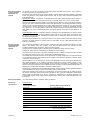
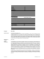
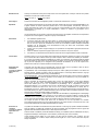
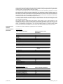
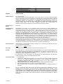
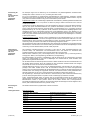
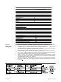
FLEX Monoclonal Mouse Anti-Human Mammaglobin Clone 304-1A5 Ready-to-Use (Dako Autostainer/Autostainer Plus) Code IS074 English Intended use For in vitro diagnostic use. FLEX Monoclonal Mouse Anti-Human Mammaglobin, Clone 304-1A5, Ready-to-Use (Dako Autostainer/Autostainer Plus), is intended for use in immunohistochemistry together with Dako Autostainer/Autostainer Plus instruments. This antibody is useful for the identification of mammaglobin expressing cells in normal and neoplastic tissues. The antibody may be useful for the identification of breast ductal carcinoma. The clinical interpretation of any staining or its absence should be complemented by morphological studies using proper controls and should be evaluated within the context of the patient's clinical history and other diagnostic tests by a qualified pathologist. Synonyms for antigen MGA, MGB1 Summary and explanation Mammaglobin is a 93 amino acid protein with homology to other secretoglobin-uteroglobin family members that was originally identified in studies of differentially expressed cDNAs from breast carcinoma cell lines (1). Mammaglobin expression has also been reported in a series of malignant and nonmalignant tissues derived from the female genital tract by RT-PCR (2). Expression of the mammaglobin gene and protein have been demonstrated in neoplastic breast tissues and in a subset of normal breast epithelial cells within the acini of type I and type II lobules and within the columnar cells of terminal ducts (3). Mammaglobin expression has been found in well, moderately, and poorly differentiated invasive ductal carcinomas by IHC and in metastatic and occult breast carcinoma by RT-PCR (3,4). Mammaglobin expression is highly restricted to breast tissue and not detected in the majority of normal tissue types evaluated, with the exception of skin (1). Refer to Dako’s General Instructions for Immunohistochemical Staining or the detection system instructions of IHC procedures for: 1) Principle of Procedure, 2) Materials Required, Not Supplied, 3) Storage, 4) Specimen Preparation, 5) Staining Procedure, 6) Quality Control, 7) Troubleshooting, 8) Interpretation of Staining, 9) General Limitations. Reagent provided Ready-to-use monoclonal mouse antibody provided in liquid form in a buffer containing stabilizing protein and 0.015 mol/L sodium azide. Clone: 304-1A5 (1). Isotype: IgG1, kappa Immunogen Purified mammaglobin recombinant protein, containing amino acids 15-93 (1). Specificity In Western blotting of lysates of breast carcinoma cell line MDA-MB-415, primary breast tumor cell line 450-17 and recombinant mammaglobin the antibody labeled bands of 14 kDa and 21 kDa. The 9 kDa form of mammaglobin was observed after enzymatic deglycosylation of MDA-MB-415 indicating glycosylation of the mammaglobin protein (1). In ELISA no reaction was found with overlapping peptides corresponding to the mammaglobin sequence indicating a conformational epitope. Precautions 1. 2. 3. 4. 5. Storage (118553-001) For professional users. This product contains sodium azide (NaN3), a chemical highly toxic in pure form. At product concentrations, though not classified as hazardous, sodium azide may react with lead and copper plumbing to form highly explosive build-ups of metal azides. Upon disposal, flush with large volumes of water to prevent metal azide build-up in plumbing. As with any product derived from biological sources, proper handling procedures should be used. Wear appropriate Personal Protective Equipment to avoid contact with eyes and skin. Unused solution should be disposed of according to local, State and Federal regulations. Store at 2-8 °C. Do not use after expiration date stamped on vial. If reagents are stored under any conditions other than those specified, the conditions must be verified by the user. There are no obvious signs to indicate instability of this product. Therefore, positive and negative controls should be run simultaneously with patient specimens. If unexpected staining is observed which cannot be explained by variations in laboratory procedures and a problem with the antibody is suspected, contact Dako Technical Support. 307696EFG_001 p. 1/8 Specimen preparation including materials required but not supplied The antibody can be used for labeling formalin-fixed, paraffin-embedded tissue sections. Tissue specimens should be cut into sections of approximately 4 µm. Pre-treatment with heat-induced epitope retrieval (HIER) is required. Optimal results are obtained by pretreating tissues using EnVision FLEX Target Retrieval Solution, High pH (10x), (Dako Autostainer/Autostainer Plus (Code K8010/K8014). Deparaffinized sections: Pre-treatment of deparaffinized formalin-fixed, paraffin-embedded tissue sections is recommended using Dako PT Link (Code PT100/PT101). For details, please refer to the PT Link User Guide. Follow the pre-treatment procedure outlined in the package insert for EnVision FLEX Target Retrieval Solution, High pH (10x), (Dako Autostainer/Autostainer Plus) (Code K8010/K8014). The following parameters should be used for PT Link: Pre-heat temperature: 65 °C; epitope retrieval temperature and time: 97 °C for 20 (±1) minutes; cool down to 65 °C. Remove Autostainer slide rack with slides from the PT Link tank and immediately dip slides into a jar/tank (e.g., PT Link Rinse Station, Code PT109) containing diluted room temperature EnVision FLEX Wash Buffer (10x), (Dako Autostainer/Autostainer Plus) (Code K8010). Leave slides in Wash Buffer for 1-5 minutes. Paraffin-embedded sections: As alternative specimen preparation, both deparaffinization and epitope retrieval can be performed in the PT Link using a modified procedure. See the PT Link User Guide for instructions. After the staining procedure has been completed, the sections must be dehydrated, cleared and mounted using permanent mounting medium. The tissue sections should not dry out during the treatment or during the following immunohistochemical staining procedure. For greater adherence of tissue sections to glass slides, the use of Dako Silanized Slides (Code S3003) is recommended. Staining procedure including materials required but not supplied The recommended visualization system is EnVision FLEX, High pH, (Dako Autostainer/Autostainer Plus) (Code K8010). The staining steps and incubation times are pre-programmed into the software of Dako Autostainer/Autostainer Plus instruments, using the following protocols: Template protocol: FLEXRTU2 (200 µL dispense volume) or FLEXRTU3 (300 µL dispense volume) Autoprogram: Mamm (without counterstaining) or MammH (with counterstaining) The Auxiliary step should be set to “rinse buffer” in staining runs with ≤10 slides. For staining runs with >10 slides the Auxiliary step should be set to “none”. This ascertains comparable wash times. All incubation steps should be performed at room temperature. For details, please refer to the Operator’s Manual for the dedicated instrument. If the protocols are not available on the used Dako Autostainer instrument, please contact Dako Technical Services. Optimal conditions may vary depending on specimen and preparation methods, and should be determined by each individual laboratory. If the evaluating pathologist should desire a different staining intensity, a Dako Application Specialist/Technical Service Specialist can be contacted for information on re-programming of the protocol. Verify that the performance of the adjusted protocol is still valid by evaluating that the staining pattern is identical to the staining pattern described in “Performance characteristics”. Counterstaining in hematoxylin is recommended using EnVision FLEX Hematoxylin, (Dako Autostainer/Autostainer Plus) (Code K8018). Non-aqueous, permanent mounting medium is recommended. Positive and negative controls should be run simultaneously using the same protocol as the patient specimens. The positive control tissue should include breast hyperplasia or skin (1) and the cells/structures should display reaction patterns as described for this tissue in “Performance characteristics” in all positive specimens. The recommended negative control reagent is FLEX Negative Control, Mouse, (Dako Autostainer/Autostainer Plus) (Code IS750). Staining interpretation The cellular staining pattern is cytoplasmic and/or membranous. Performance characteristics Normal tissues (1): (118553-001) Tissue Type (# tested) Positively Staining Tissue Elements Breast (10) 5/10 ductal epithelial cells Colon (5) 0/5 Kidney (6) 0/6 Liver (3) 0/3 Lung (4) 0/4 Brain (3) 0/3 Heart (2) 0/2 Bone Marrow (1) 0/1 Adrenal (3) 0/3 Cervix (2) 0/2 Skin (7) 7/7 epithelial cells lining the apocrine and eccrine sweat glands 307696EFG_001 p. 2/8 Duodenum (2) 0/2 Gall bladder (1) Ileum (2) 0/1 0/2 Ovary (4) 0/4 Pancreas (2) 0/2 Parotid gland (1) 0/1 Prostate (4) 0/4 Spleen (3) 0/3 Testis (1) 0/1 Skeletal muscle (1) 0/1 Endometrium (3) 0/3 Abnormal tissues (1,5,6): Tumor Type (# tested) Positively Staining Tumors Breast cancer (35) 23/35 Breast infiltrating ductal carcinoma (IDC) (96) 78/96 Endometrial carcinoma (4) 0/4 Epithelial mesothelioma (EM) (16) 1/16 Gastrointestinal carcinoma (4) 0/4 Genitourinary carcinoma (2) 0/2 Lung carcinoma (32) 0/32 Ovarian carcinoma (9) 0/9 Ovarian serous papillary carcinoma (OSPC) (21) 0/21 Pulmonary adenocarcinoma (PAC) (23) 6/23 Uterine endometrioid carcinoma (UEC) (18) 6/18 Français Réf. IS074 Utilisation prévue Pour utilisation diagnostique in vitro. L’anticorps FLEX Monoclonal Mouse Anti-Human Mammaglobin, Clone 304-1A5, Ready-to-Use (Dako Autostainer/Autostainer Plus), est destiné à être utilisé en immunohistochimie avec les appareils Dako Autostainer/Autostainer Plus. Cet anticorps est utile pour l’identification des cellules exprimant la mammaglobine dans les tissus sains et néoplasiques. Cet anticorps peut être utile pour l’identification d’un carcinome canalaire du sein. L’interprétation clinique de toute coloration ou son absence doit être complétée par des études morphologiques en utilisant des contrôles appropriés et doit être évaluée en fonction des antécédents cliniques du patient et d’autres tests diagnostiques par un pathologiste qualifié. Synonymes de l’antigène MGA, MGB1. Résumé et explication La mammaglobine est une protéine de 93 acides aminés ayant une homologie avec d’autres membres de la famille des globines sécrétoires et des utéroglobines ; elle a été, à l’origine, identifiée grâce à des études portant sur l’expression différentielle d’ADNc provenant de lignées cellulaires de carcinome mammaire (1). L’expression de la mammaglobine a également été notée dans une série de tissus malins et non malins provenant de l’appareil génital féminin par RT-PCR (2). L’expression du gène de la mammaglobine et de sa protéine a été démontrée dans des tissus mammaires néoplasiques et dans un sous-ensemble de cellules épithéliales mammaires saines à l’intérieur des acini des lobules de type I et de type II et à l’intérieur des cellules en colonnes des canaux terminaux (3). L’expression de la mammaglobine a été mise en évidence par IHC dans les carcinomes canalaires invasifs bien différenciés, moyennement différenciés et peu différenciés, et par RT-PCR dans le carcinome mammaire métastatique et occulte (3, 4). L’expression de la mammaglobine est hautement limitée au tissu mammaire et n’est pas détectée dans la majorité des types de tissus sains évalués, à l’exception de la peau (1). Se reporter aux « Instructions générales de coloration immunohistochimique » de Dako ou aux instructions du système de détection relatives aux procédures IHC pour plus d’informations concernant les points suivants : 1) Principe de procédure, 2) Matériels requis mais non fournis, 3) Conservation, 4) Préparation des échantillons, 5) Procédure de coloration, 6) Contrôle qualité, 7) Dépannage, 8) Interprétation de la coloration, 9) Limites générales. (118553-001) 307696EFG_001 p. 3/8 Réactifs fournis Anticorps monoclonal de souris prêt à l’emploi fourni sous forme liquide dans un tampon contenant une protéine stabilisante et 0,015 mol/L d’azide de sodium. Clone : 304-1A5 (1). Isotype : IgG1, kappa. Immunogène Protéine recombinante de mammaglobine purifiée, contenant les acides aminés 15 à 93 (1). Spécificité Lors de Western Blots effectués sur des lysats de la lignée cellulaire de carcinome mammaire MDA-MB-415, de la lignée cellulaire de tumeur mammaire primitive 450-17 et de mammaglobine recombinante, l’anticorps a marqué des bandes de 14 kDa et de 21 kDa. La forme de la mammaglobine de 9 kDa a été observée après déglycosylation enzymatique de la lignée MDA-MB-415, mettant en évidence la glycosylation de la mammaglobine (1). Lors de tests ELISA, aucune réaction n’a été mise en évidence avec des peptides chevauchants correspondant à la séquence de la mammaglobine, indiquant un épitope conformationnel. Précautions 1. 2. 3. 4. 5. Pour utilisateurs professionnels. Ce produit contient de l’azide de sodium (NaN3), un produit chimique hautement toxique sous sa forme pure. Aux concentrations du produit, bien que non classé comme dangereux, l’azide de sodium peut réagir avec le cuivre et le plomb des canalisations et former des accumulations d’azides métalliques hautement explosifs. Lors de l’élimination, rincer abondamment à l’eau pour éviter toute accumulation d’azide métallique dans les canalisations. Comme avec tout produit d’origine biologique, des procédures de manipulation appropriées doivent être respectées. Porter un vêtement de protection approprié pour éviter le contact avec les yeux et la peau. Les solutions non utilisées doivent être éliminées conformément aux réglementations locales et nationales. Conservation Conserver entre 2 et 8 °C. Ne pas utiliser après la date limite de péremption indiquée sur le flacon. Si les réactifs sont conservés dans des conditions autres que celles indiquées, celles-ci doivent être validées par l’utilisateur. Il n’y a aucun signe évident indiquant l’instabilité de ce produit. Par conséquent, des contrôles positifs et négatifs doivent être testés en même temps que les échantillons de patients. Si une coloration inattendue est observée, qui ne peut être expliquée par un changement des procédures du laboratoire, et en cas de suspicion d’un problème lié à l’anticorps, contacter l’assistance technique de Dako. Préparation des échantillons y compris le matériel requis mais non fourni L’anticorps peut être utilisé pour le marquage des coupes de tissus inclus en paraffine et fixés au formol. L’épaisseur des coupes d’échantillons de tissu doit être d’environ 4 µm. Le prétraitement avec un démasquage d’épitope induit par la chaleur (HIER) est nécessaire. Pour obtenir des résultats optimaux, prétraiter les tissus à l’aide du produit EnVision FLEX Target Retrieval Solution, High pH (10x), (Dako Autostainer/Autostainer Plus) (réf. K8010/K8014). Coupes déparaffinées : Il est recommandé de prétraiter les coupes de tissus fixés au formol et inclus en paraffine qui ont été déparaffinées à l’aide de l’appareil PT Link de Dako (réf. PT100/PT101). Pour plus de détails, se reporter au Guide d’utilisation du PT Link. Suivre la procédure de prétraitement indiquée dans la notice du produit EnVision FLEX Target Retrieval Solution, High pH (10x), (Dako Autostainer/Autostainer Plus) (réf. K8010/K8014). Les paramètres suivants doivent être utilisés pour le PT Link : température de préchauffage : 65 °C ; température et durée du démasquage d’épitope : 97 °C pendant 20 (±1) minutes ; refroidissement jusqu’à 65 °C. Retirer le portoir à lames Autostainer contenant les lames de la cuve du PT Link et plonger immédiatement les lames dans un récipient/une cuve (par ex., station de rinçage du PT Link, réf. PT109) contenant du tampon de lavage EnVision FLEX Wash Buffer (10x), (Dako Autostainer/Autostainer Plus) (réf. K8010) dilué à température ambiante. Laisser les lames dans le tampon de lavage pendant 1 à 5 minutes. Coupes incluses en paraffine : Comme préparation alternative des échantillons, le déparaffinage et le démasquage d’épitope peuvent être réalisés dans l’appareil PT Link en utilisant une procédure modifiée. Consulter le Guide d’utilisation du PT Link pour obtenir des instructions. Une fois que la procédure de coloration est terminée, les coupes doivent être déshydratées, éclaircies et montées à l’aide d’un milieu de montage permanent. Les coupes de tissus ne doivent pas sécher lors du traitement ni lors de la procédure de coloration immunohistochimique suivante. Pour une meilleure adhérence des coupes de tissus sur les lames de verre, il est recommandé d’utiliser des lames Dako Silanized Slides (réf. S3003). Procédure de coloration y compris le matériel requis mais non fourni Le système de visualisation recommandé est le système EnVision FLEX, High pH, (Dako Autostainer/Autostainer Plus) (réf. K8010). Les étapes de coloration et les temps d’incubation sont préprogrammés dans le logiciel des appareils Dako Autostainer/Autostainer Plus, à l’aide des protocoles suivants : Protocole modèle : FLEXRTU2 (volume de distribution de 200 µL) ou FLEXRTU3 (volume de distribution de 300 µL) Programme automatique : Mamm (sans contre-coloration) ou MammH (avec contre-coloration) L’étape Auxiliary doit être réglée sur « rinse buffer » lors des cycles de coloration avec ≤10 lames. Pour les cycles de coloration de >10 lames, l’étape Auxiliary doit être réglée sur « none ». Cela confirme des temps de lavage comparables. (118553-001) 307696EFG_001 p. 4/8 Toutes les étapes d’incubation doivent être effectuées à température ambiante. Pour plus de détails, se reporter au Manuel de l’opérateur spécifique à l'appareil. Si les protocoles ne sont pas disponibles sur l’appareil Dako Autostainer utilisé, contacter l’assistance technique de Dako. Les conditions optimales peuvent varier en fonction du prélèvement et des méthodes de préparation, et doivent être déterminées par chaque laboratoire individuellement. Si le pathologiste qui réalise l’évaluation désire une intensité de coloration différente, un ingénieur d’application/un spécialiste de l’assistance technique de Dako peut être contacté pour obtenir des informations sur la reprogrammation du protocole. Vérifier que les performances du protocole modifié sont toujours valides en vérifiant que le schéma de coloration est identique au schéma de coloration décrit à la section « Caractéristiques de performance ». Il est recommandé d’effectuer une contre-coloration à l’hématoxyline à l’aide du produit EnVision FLEX Hematoxylin, (Dako Autostainer/Autostainer Plus) (réf. K8018). L’utilisation d’un milieu de montage permanent non aqueux est recommandée. Des contrôles positifs et négatifs doivent être testés en même temps et avec le même protocole que les échantillons de patients. Le tissu de contrôle positif doit comprendre l’hyperplasie du sein et la peau (1) et les cellules/structures doivent présenter des schémas de réaction semblables à ceux décrits pour ces tissus à la section « Caractéristiques de performance » pour tous les échantillons positifs. Le réactif de contrôle négatif recommandé est le produit FLEX Negative Control, Mouse, (Dako Autostainer/Autostainer Plus) (réf. IS750). Interprétation de la coloration Le schéma de coloration cellulaire est cytoplasmique et/ou membranaire. Caractéristiques de performance Tissus sains (1) : Type de tissus (nbre testés) Éléments tissulaires colorés positivement Sein (10) 5/10 : cellules épithéliales canalaires Côlon (5) 0/5 Rein (6) 0/6 Foie (3) 0/3 Poumon (4) 0/4 Cerveau (3) 0/3 Cœur (2) 0/2 Moelle osseuse (1) 0/1 Surrénale (3) 0/3 Col de l’utérus (2) 0/2 Peau (7) 7/7 : cellules épithéliales tapissant les glandes sudoripares apocrines et eccrines Duodénum (2) 0/2 Vésicule biliaire (1) 0/1 Iléon (2) 0/2 Ovaire (4) 0/4 Pancréas (2) 0/2 Glande parotide (1) 0/1 Prostate (4) 0/4 Rate (3) 0/3 Testicule (1) 0/1 Muscle squelettique (1) 0/1 Endomètre (3) 0/3 Tissus tumoraux (1,5,6) : Type de tumeur (nbre testés) (118553-001) Tumeurs colorées positivement Cancer du sein (35) 23/35 Carcinome canalaire infiltrant mammaire (96) 78/96 Carcinome de l’endomètre (4) 0/4 Mésothéliome épithélial (16) 1/16 Carcinome gastro-intestinal (4) 0/4 Carcinome urogénital (2) 0/2 Cancer du poumon (32) 0/32 Carcinome ovarien (9) 0/9 307696EFG_001 p. 5/8 Carcinome papillaire séreux ovarien (21) 0/21 Adénocarcinome pulmonaire (23) 6/23 Carcinome de l’endomètre utérin (18) 6/18 Deutsch Code-Nr. IS074 Zweckbestimmung Zur In-vitro-Diagnostik. FLEX Monoclonal Mouse Anti-Human Mammaglobin, Clone 304-1A5, Ready-to- Use (Dako Autostainer/Autostainer Plus) ist zur Verwendung in der Immunhistochemie in Verbindung mit Dako Autostainer/Autostainer Plus-Geräten bestimmt. Dieser Antikörper dient zur Identifikation von Mammaglobin-exprimierenden Zellen in normalem und neoplastischem Gewebe. Der Antikörper kann zur Identifikation des duktalen Brustkarzinoms dienen. Die klinische Auswertung einer eventuell eintretenden Färbung sollte durch morphologische Studien mit geeigneten Kontrollen ergänzt werden und von einem qualifizierten Pathologen unter Berücksichtigung der Krankengeschichte und anderer diagnostischer Tests des Patienten vorgenommen werden. Synonyme für das Antigen MGA, MGB1 Zusammenfassung und Erklärung Mammaglobin ist ein Protein von 93 Aminosäuren Länge mit Homologie zu anderen Mitgliedern der Secretoglobin-Uteroglobin-Familie, das ursprünglich bei der Untersuchung von differenziell exprimierten cDNAs aus Brustkarzinomzelllinien nachgewiesen wurde (1). Mammaglobin-Expression wurde auch in einer Reihe von malignen und nicht-malignen Geweben aus dem weiblichen Genitaltrakt durch RT-PCR nachgewiesen (2). Expression des Mammaglobin-Gens und -Proteins wurden in neoplastischen Brustgeweben und in einer Untergruppe normaler Brustepithelzellen innerhalb der Azini von Lobuli der Typen I und II sowie innerhalb des Zylinderepithels der Milchgänge demonstriert (3). Die Mammaglobin-Expression wurde in gut, mäßig und schwach differenzierten invasiven Duktalkarzinomen durch IHC und in metastatischen und okkulten Brustkarzinomen durch RT-PCR gefunden (3, 4). Die Mammaglobin-Expression ist strikt auf das Brustgewebe beschränkt und wird in der Mehrzahl der bewerteten normalen Gewebetypen, mit Ausnahme der Haut, nicht beobachtet (1). Folgende Angaben bitte den Allgemeinen Richtlinien zur immunhistochemischen Färbung von Dako bzw. den Anweisungen des Detektionssystems für IHC-Verfahren entnehmen: 1) Verfahrensprinzip, 2) Erforderliche, aber nicht mitgelieferte Materialien, 3) Aufbewahrung, 4) Vorbereitung der Probe, 5) Färbeverfahren, 6) Qualitätskontrolle, 7) Fehlersuche und -behebung, 8) Auswertung der Färbung, 9) Allgemeine Beschränkungen. Geliefertes Reagenz Gebrauchsfertiger, monoklonaler Maus-Antikörper in flüssiger Form in einem Puffer, der stabilisierendes Protein und 0,015 mol/L Natriumazid enthält. Clone: 304-1A5 (1). Isotyp: IgG1, Kappa. Immunogen Gereinigtes rekombinantes Mammaglobin, einschließlich der Aminosäuren 15-93 (1). Spezifität Beim Western Blot von Lysaten der Brustkarzinomzelllinie MDA-MB-415, der primären Brusttumorzelllinie 450-17 und rekombinantem Mammaglobin markiert der Antikörper Banden von 14 kDa und 21 kDa. Die 9-kDa-Form von Mammaglobin wurde nach enzymatischer Deglykosylierung von MDA-MB-415 beobachtet, was auf eine Glykosylierung des Mammaglobinproteins hinweist (1). Im ELISA-Verfahren wurde keine Reaktion mit überlappenden, der Mammagobinsequenz entsprechenden Peptiden festgestellt, was auf ein konformationelles Epitop hinweist. Vorsichtsmaßnahmen 1. 2. 3. 4. 5. Lagerung (118553-001) Für Fachpersonal. Dieses Produkt enthält Natriumazid (NaN3), eine in reiner Form äußerst giftige Chemikalie. Natriumazid kann auch in als ungefährlich eingestuften Konzentrationen mit Blei- und Kupferrohren reagieren und hochexplosive Metallazide bilden. Nach der Entsorgung stets mit viel Wasser nachspülen, um Metallazidansammlungen in den Leitungen vorzubeugen. Wie alle Produkte biologischen Ursprungs müssen auch diese entsprechend gehandhabt werden. Geeignete Schutzkleidung tragen, um Augen- und Hautkontakt zu vermeiden. Nicht verwendete Lösung ist entsprechend örtlichen, bundesstaatlichen und staatlichen Richtlinien zu entsorgen. Bei 2–8 °C aufbewahren. Nach Ablauf des auf dem Fläschchen aufgedruckten Verfallsdatums nicht mehr verwenden. Werden die Reagenzien nicht entsprechend den angegebenen Bedingungen aufbewahrt, müssen die Bedingungen vom Anwender geprüft werden. Es gibt keine offensichtlichen Anzeichen für eine eventuelle Produktinstabilität. Positiv- und Negativkontrollen sollten daher zur gleichen Zeit wie die Patientenproben getestet werden. Falls es zu einer unerwarteten Färbung kommt, die sich nicht durch Unterschiede bei Laborverfahren erklären lässt und auf ein Problem mit dem Antikörper hindeutet, ist der technische Kundendienst von Dako zu verständigen. 307696EFG_001 p. 6/8 Vorbereitung der Probe und erforderliche, aber nicht mitgelieferte Materialien Der Antikörper eignet sich zur Markierung von formalinfixierten und paraffineingebetteten Gewebeschnitten. Gewebeproben sollten in Schnitte von ca. 4 µm Stärke geschnitten werden. Es ist eine Vorbehandlung mit hitzeinduzierter Epitopdemaskierung (HIER-Verfahren) erforderlich. Optimale Ergebnisse können durch Vorbehandlung der Gewebe mit EnVision FLEX Target Retrieval Solution, High pH (10x), (Dako Autostainer/Autostainer Plus) (Code-Nr. K8010/K8014) erzielt werden. Entparaffinierte Schnitte: Die Vorbehandlung entparaffinierter formalinfixierter, paraffineingebetteter Gewebeschnitte sollte mit dem System Dako PT Link (Code-Nr. PT100/PT101) erfolgen. Weitere Informationen hierzu: siehe PT Link-Benutzerhandbuch. Vorbehandlung gemäß der Beschreibung in der Packungsbeilage für EnVision FLEX Target Retrieval Solution, High pH (10x), (Dako Autostainer/Autostainer Plus) (Code-Nr. K8010/K8014) durchführen. Für das PT LinkSystem sollten die folgenden Parameter verwendet werden: Vorwärmtemperatur: 65 °C; Temperatur und Zeit für Epitopdemaskierung: 97 °C für 20 (±1) Minuten; auf 65 °C abkühlen. Das Autostainer Objektträgergestell mit den Objektträgern aus dem PT Link-Behälter herausnehmen und die Objektträger sofort in einen Behälter (z. B. PT Link Rinse Station, Code-Nr. PT109) mit verdünntem, auf Zimmertemperatur gebrachtem EnVision FLEX Wash Buffer (10x), (Dako Autostainer/Autostainer Plus) (Code-Nr. K8010) eintauchen. Die Objektträger für 1–5 Minuten im Waschpuffer belassen. Paraffineingebettete Schnitte: Alternativ können Entparaffinierung und Epitopdemaskierung im PT Link unter Verwendung eines modifizierten Verfahrens durchgeführt werden. Weitere Informationen finden Sie im PT LinkBenutzerhandbuch. Nach Abschluss des Färbeverfahrens müssen die Schnitte dehydriert, geklärt und unter Verwendung eines permanenten Fixiermittels auf die Objektträger aufgebracht werden. Die Gewebeschnitte dürfen während der Behandlung oder während des anschließenden immunhistochemischen Färbeverfahrens nicht austrocknen. Zur besseren Haftung der Gewebeschnitte an den Glasobjektträgern wird die Verwendung von Dako Silanized Slides (Code-Nr. S3003) empfohlen. Färbeverfahren und erforderliche, aber nicht mitgelieferte Materialien Das empfohlene Visualisierungssystem ist EnVision FLEX, High pH, (Dako Autostainer/Autostainer Plus) (Code-Nr. K8010). Die Färbeschritte und Inkubationszeiten sind in der Software von Autostainer Dako Autostainer/Autostainer Plus-Geräten mit den folgenden Protokollen vorprogrammiert: Matrix-Protokoll: FLEXRTU2 (200 µl Anwendungsvolumen) oder FLEXRTU3 (300 µl Anwendungsvolumen) Autoprogramm: Mamm (ohne Gegenfärbung) oder MammH (mit Gegenfärbung) Bei Färbedurchläufen mit höchstens 10 Objektträgern sollte der Zusatz-Schritt auf „Pufferspülgang“ eingestellt werden. Für Färbedurchläufe mit mehr als 10 Objektträgern den Zusatz-Schritt auf „Keine“ einstellen. Dies gewährleistet vergleichbare Waschzeiten. Alle Inkubationsschritte bei Raumtemperatur durchführen. Nähere Einzelheiten bitte dem Benutzerhandbuch für das jeweilige Gerät entnehmen. Wenn die Protokolle auf dem verwendeten Autostainer-Gerät nicht verfügbar sind, wenden Sie sich an den technischen Kundendienst von Dako. Optimale Bedingungen können je nach Probe und Präparationsverfahren unterschiedlich sein und sollten vom jeweiligen Labor selbst ermittelt werden. Falls der beurteilende Pathologe eine andere Färbungsintensität wünscht, kann ein Anwendungsspezialist oder Kundendiensttechniker von Dako bei der Neuprogrammierung des Protokolls helfen. Die Leistung des angepassten Protokolls muss verifiziert werden, indem gewährleistet wird, dass das Färbemuster mit dem unter „Leistungsmerkmale" beschriebenen Färbemuster identisch ist. Die Gegenfärbung in Hämatoxylin sollte mit EnVision FLEX Hematoxylin, (Dako Autostainer/Autostainer Plus) (Code-Nr. K8018) ausgeführt werden. Empfohlen wird ein nichtwässriges, permanentes Fixiermittel. Positiv- und Negativkontrollen sollten zur gleichen Zeit und mit demselben Protokoll wie die Patientenproben getestet werden. Als positive Kontrollen sollten eine Brusthyperplasie und Haut (1) verwendet werden, und die Zellen/Strukturen müssen in allen positiven Proben die für dieses Gewebe unter „Leistungsmerkmale" beschriebenen Reaktionsmuster aufweisen. Das empfohlene Negativ-Kontrollreagenz ist FLEX Negative Control, Mouse, (Dako Autostainer/Autostainer Plus) (Code-Nr. IS750). Auswertung der Färbung Das zelluläre Färbemuster ist zytoplasmatisch und/oder membranös. Leistungsmerkmale Normale Gewebe (1): (118553-001) Gewebetyp (Anz. getestet) Gewebeelemente mit positiver Färbung Brust (10) 5 von 10, duktale Epithelzellen Kolon (5) 0/5 Niere (6) 0/6 Leber (3) 0/3 Lunge (4) 0/4 Gehirn (3) 0/3 Herz (2) 0/2 Knochenmark (1) 0/1 Nebennieren (3) 0/3 307696EFG_001 p. 7/8 Zervix (2) 0/2 Haut (7) 7 von 7, Epithelzellen der apokrinen und ekkrinen Schweißdrüsen Zwölffingerdarm (2) 0/2 Gallenblase (1) 0/1 Ileum (2) 0/2 Ovarium (4) 0/4 Pankreas (2) 0/2 Ohrspeicheldrüsen (1) 0/1 Prostata (4) 0/4 Milz (3) 0/3 Hoden (1) 0/1 Skelettmuskel (1) 0/1 Endometrium (3) 0/3 Pathologische Gewebe (1, 5, 6): References Bibliographie Literaturangaben Tumortyp (Anz. getestet) Tumore mit positiver Färbung Brustkrebs (35) 23/35 Brust, infiltrierendes Duktalkarzinom (IDC) (96) 78/96 Endometriumkarzinom (4) 0/4 Epitheliales Mesotheliom (EM) (16) 1/16 Gastrointestinales Karzinom (4) 0/4 Urogenitales Karzinom (2) 0/2 Lungenkarzinom (32) 0/32 Ovarialkarzinom (9) 0/9 Seröses papilläres Ovarialkarzinom (OSPC) (21) 0/21 Pulmonales Adenokarzinom (PAC) (23) 6/23 Endometrioides Uteruskarzinom (UEC) (18) 6/18 1. 2. 3. 4. 5. 6. Fanger GR, Houghton RL, Retter MW, Hendrickson RC, Babcook J, Dillon DC, et al. Detection of mammaglobin in the sera of patients with breast cancer. Tumour Biol 2002; 23:212-21. Grünewald K, Haun M, Fiegl M, Urbanek M, Müller-Holzner E, Massoner A, et al. Mammaglobin expression in gynecologic malignancies and malignant effusions detected by nested reverse transcriptasepolymerase chain reaction. Laboratory Investigation 2002; 82:1147-53. Watson MA, Dintzis S, Darrow CM, Voss LE, DiPersio J, Jensen R, et al. Mammaglobin expression in primary, metastatic, and occult breast cancer. Cancer Res 1999; 59:3028-31. Min CJ, Tafra L, Verbanac KM. Identification of superior markers for polymerase chain reaction detection of breast cancer metastases in sentinel lymph nodes. Cancer Res 1998; 58:4581-4. Ciampa A, Fanger G, Khan A, Rock KL, Xu B. Mammaglobin and CRxA-01 in pleural effusion cytology: potential utility of distinguishing metastatic breast carcinomas from other cytokeratin 7-positive/cytokeratin 20-negative carcinomas. Cancer 2004; 102:368-72. Stachurski D, Fanger G, Xu B, Edminston K, Rock KL, Khan A. CRxA-01 and mammaglobin expression in breast cancer: potential role in differentiating breast cancer from other CK7+/CK20- tumors. Mod Path 2003; 16:48A. Edition 04/08 (118553-001) 307696EFG_001 p. 8/8