1
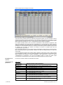
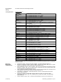
FLEX Monoclonal Rabbit Anti-Human Cyclin D1 Clone EP12 Ready-to-Use (Dako Autostainer/Autostainer Plus) English Code IS083 Intended use For in vitro diagnostic use. FLEX Monoclonal Rabbit Anti-Human Cyclin D1, Clone EP12, Ready-to-Use, (Dako Autostainer/Autostainer Plus), is intended for use in immunohistochemistry together with Dako Autostainer/Autostainer Plus instruments. Antibodies to cyclin D1 are useful for the identification of mantle cell lymphomas (1-3). The clinical interpretation of any staining or its absence should be complemented by morphological studies using proper controls and should be evaluated within the context of the patient's clinical history and other diagnostic tests by a qualified pathologist. Synonyms for antigen PRAD-1, CCND1, Bcl-1 (1). Summary and explanation Cyclin D1 is a 36 kDa protein encoded by the CCND1 (bcl-1) gene located on chromosome 11q13. Cyclin D1 associates with and activates cyclin-dependent kinases (CDKs) CDK4 and CDK6 (1,4,5). The protein functions as a CDK-dependent regulator of the cell cycle by phosphorylating and inactivating the retinoblastoma protein, thereby allowing for progression through the G1-S phase of the cell cycle (1,4,5). Cyclin D1 is also involved in cell cycle or CDK-independent functions including associating with and regulation of a variety of transcription factors and transcriptional coregulators. Cyclin D1 has also been shown to be involved in the regulation of cellular metabolism, fat cell differentiation and cell migration (1,4,5). Cyclin D1 overexpression is associated with tumorigenesis, and cyclin D1 amplification and/or overexpression have been demonstrated in a variety of human tumors, including mantle cell lymphomas, breast carcinomas, head and neck squamous cell carcinomas and esophageal cancers (1,6). Among lymphoid neoplasms a subset of chronic lymphocytic leukemia, small lymphocytic lymphoma and multiple myeloma have been reported to express Cyclin D1. The following neoplasms were reported to be negative for Cyclin D1: diffuse large B-cell lymphoma, extranodal marginal zone lymphoma of MALT, follilcular lymphoma, marginal zone lymphoma, lymphoplasmacytic lymphoma (1T and 1B lineage), extranodal NK/T-cell lymphoma, peripheral T-cell lymphoma, NOS, anaplastic large cell lymphoma, hodgkin lymphoma (7, 8). Refer to Dako’s General Instructions for Immunohistochemical Staining or the detection system instructions of IHC procedures for: 1) Principle of Procedure, 2) Materials Required, Not Supplied, 3) Storage, 4) Specimen Preparation, 5) Staining Procedure, 6) Quality Control, 7) Troubleshooting, 8) Interpretation of Staining, 9) General Limitations. Reagent provided Ready-to-use monoclonal rabbit antibody provided in liquid form in a buffer containing stabilizing protein and 0.015 mol/L sodium azide. Clone: EP12. Immunogen Synthetic peptide corresponding to residues near the C-terminus of human cyclin D1. Specificity In Western blotting of MCF7 cells lysates the antibody labels a major band of approximately 36 kDa, corresponding to the expected molecular weight of cyclin D1. Precautions 1. For professional users. 2. This product contains sodium azide (NaN3), a chemical highly toxic in pure form. At product concentrations, though not classified as hazardous, sodium azide may react with lead and copper plumbing to form highly explosive build-ups of metal azides. Upon disposal, flush with large volumes of water to prevent metal azide build-up in plumbing. 3. As with any product derived from biological sources, proper handling procedures should be used. Storage (121302-002) 4. Wear appropriate Personal Protective Equipment to avoid contact with eyes and skin. 5. Unused solution should be disposed of according to local, State and Federal regulations. Store at 2-8 °C. Do not use after expiration date s tamped on vial. If reagents are stored under any conditions other than those specified, the conditions must be verified by the user. There are no obvious signs to indicate instability of this product. Therefore, positive and negative controls should be run simultaneously with patient specimens. If unexpected staining is observed which cannot be explained by variations in laboratory procedures and a problem with the antibody is suspected, contact Dako Technical Support. P01467EFG_002_IS083/2011.04 p. 1/11 Specimen preparation including materials required but not supplied The antibody can be used for labeling formalin-fixed, paraffin-embedded tissue sections. Tissue specimens should be cut into sections of approximately 4 µm. Pre-treatment with heat-induced epitope retrieval (HIER) is required using Dako PT Link (Code PT100/PT101). For details, please refer to the PT Link User Guide. Optimal results are obtained by pretreating tissues using EnVision FLEX Target Retrieval Solution, High pH (50x) (Code K8010/K8004) for 20 minutes at 97°C followed by 5 minutes in EnVision™ FLEX Wash Buffer (Code K8007). Paraffin-embedded sections: Pre-treatment of formalin-fixed, paraffin-embedded tissue sections is recommended using the 3-in-1 specimen preparation procedure for Dako PT Link. Follow the pre-treatment procedure outlined in the package insert for EnVision FLEX Target Retrieval Solution, High pH (50x) (Code K8010/K8004). Note: After staining the sections must be dehydrated, cleared and mounted using permanent mounting medium. The tissue sections should not dry out during the treatment or during the following immunohistochemical staining procedure. For greater adherence of tissue sections to glass slides, the use of Dako Silanized Slides (Code S3003) is recommended. Staining procedure including materials required but not supplied The recommended visualization system is EnVision FLEX, High pH, (Dako Autostainer/Autostainer Plus) (Code K8010) used according to the protocol given below. Reagents must be appropriately diluted before use. All steps should be performed at room temperature (20-25 °C). 1. 2. 3. 4. 5. 6. 7. 8. 9. 10. 11. 12. 13. 14. 15. 16. Incubate tissue section with 200 µL EnVision™ FLEX Peroxidase-Blocking Reagent (DM821) for 5 (±1) minutes Rinse in EnVision™ FLEX Wash Buffer (Code K8007) for 1-5 minutes Incubate tissue section with 200 µL primary antibody (Code IS083) for 20 (±1) minutes Rinse in EnVision™ FLEX Wash Buffer (Code K8007) for 1-5 minutes Incubate tissue section with 200 µL EnVision™ FLEX/HRP (Code DM822) for 20 (±1) minutes Rinse in EnVision™ FLEX Wash Buffer (Code K8007) for 1-5 minutes Incubate tissue section with 200 µL EnVision™ FLEX Wash Buffer (Code K8007) for 5 (±1) minutes Rinse in EnVision™ FLEX Wash Buffer (Code K8007) for 1-5 minutes Incubate tissue section with 200 µL EnVision™ FLEX Substrate Working Solution (Codes DM823 and DM827) for 5 (±1) minutes Incubate tissue section with 200 µL EnVision™ FLEX Substrate Working Solution (Codes DM823 and DM827) for 5 (±1) minutes Rinse in EnVision™ FLEX Wash Buffer (Code K8007) for 1-5 minutes Counterstain slide with EnVision™ FLEX Hematoxylin (Code K8018) for 5 (±1) minutes Rinse in deionized water for 1-5 minutes Incubate tissue section with 200 µL EnVision™ FLEX Wash Buffer (Code K8007) for 5 (±1) minutes Rinse in deionized water for 1-5 minutes Mount slides with permanent mounting medium. Programming grid for recommended assay: All incubation steps should be performed at room temperature. For details, please refer to the Operator’s Manual (121302-002) P01467EFG_002_IS083/2011.04 p. 2/11 for the dedicated instrument. The Auxiliary step should be set to “rinse buffer” in staining runs with ≤10 slides. For staining runs with >10 slides the Auxiliary step should be set to “none”. This ascertains comparable wash times. Optimal conditions may vary depending on specimen and preparation methods, and should be determined by each individual laboratory. If the evaluating pathologist should desire a different staining intensity, a Dako Application Specialist/Technical Service Specialist can be contacted for information on re-programming of the protocol. Verify that the performance of the adjusted protocol is still valid by evaluating that the staining pattern is identical to the staining pattern described in “Performance characteristics”. Counterstaining in hematoxylin is recommended using EnVision FLEX Hematoxylin, (Dako Autostainer/Autostainer Plus) (Code K8018). Non-aqueous, permanent mounting medium is recommended. It is recommended that positive and negative controls be run simultaneously using the same protocol as the patient specimens. The positive control tissue should include tonsil and the cells/structures should display reaction patterns as described for this tissue in “Performance characteristics” in all positive specimens. The recommended negative control reagent is FLEX Negative Control, Rabbit, (Dako Autostainer/Autostainer Plus) (Code IS600). Staining interpretation The cellular staining pattern is predominantly nuclear. Performance characteristics Normal tissues: (9) Tissue Type (# tested) Adrenal (3) Bone marrow (3) Breast (2) Cerebellum (3) Cerebrum (3) Cervix (2) Colon (3) Esophagus (3) Kidney (3) Liver (3) Lung (3) Mesothelial cells (3) Muscle, cardiac (3) Muscle, skeletal (3) Nerve, peripheral (3) Ovary (3) Pancreas (3) Parathyroid (3) Pituitary (3) Prostate (3) Salivary gland (3) Skin (3) Small intestine (3) Spleen (3) Stomach (3) Testis (3) Thymus (3) Thyroid (3) Tonsil (3) Uterus (3) Positively Staining Tissue Elements 1/3 Epithelial cells (<1%), nuclear 1/3 Myeloid cells (<1%), nuclear 1/2 Ductal epithelial cells (<10%), nuclear 1/2 Ductal epithelial cells (<1%), nuclear 2/3 Astrocytes (<1%), nuclear 2/3 Rare positive cells (<1%), nuclear 1/2 Basal epithelial cells (10%), nuclear 1/2 Basal epithelial cells (40%), nuclear 3/3 Epithelial cells (20-30%), nuclear, stromal cells (rare), nuclear 3/3 Epithelial cells (20-30%), nuclear, stromal cells (rare), nuclear 1/3 Epithelial cells (<5%), nuclear 2/3 Epithelial cells (10-20%), nuclear 1/3 Parenchymal epithelial cells (20%), nuclear 2/3 Parenchymal epithelial cells (<1%), nuclear 1/3 Alveolar epithelial cells (30%) 2/3 Alveolar epithelial cells (10-20%) 0/3 1/3 Cardiac muscle cells (<10%), nuclear, connective tissue and stromal cells (<5%), nuclear 1/3 Connective tissue (<5%), nuclear 0/3 1/3 Mesenchymal epithelial cells (<1%), nuclear 1/3 Schwann cells (<5%), nuclear 2/3 Ovarian stromal cells (rare), nuclear 3/3 Epithelial cells (25-30%), nuclear 1/3 Epithelial cells (<1%), nuclear 1/3 Epithelial cells (<5%), nuclear 1/3 Epithelial cells (30%), nuclear 1/3 Epithelial cells, (<10%), nuclear 2/3 Epithelial cells, (40%), nuclear 1/3 Glandular epithelial cells and stromal cells (10%), nuclear 1/3 Glandular epithelial cells and stromal cells (<5%), nuclear 1/3 Glandular epithelial cells (<10%), stromal cells (<5%), nuclear 2/3 Glandular epithelial cells, (<5%), nuclear 1/3 Glandular epithelial cells, (10%), nuclear and cytoplasmic 1/3 Squamous epithelial cells (30-40%), nuclear 1/3 Squamous epithelial cells (<5%), nuclear 3/3 Crypt epithelial cells (40-50%), nuclear 1/3 Lymphocytes and littoral cells (10%), nuclear 2/3 Lymphocytes and littoral cells (5%), nuclear 2/3 Epithelial cells (<5%), nuclear 3/3 Stromal cells (<1%), nuclear 3/3 Lymphoid cells (<1%), nuclear 2/3 Follicular epithelial cells and stromal cells (<1%), nuclear 3/3 Epithelial cells (30%), nuclear, lymphocytes (rare), nuclear 3/3 Endometrial epithelial cells and stromal cells(<5%) nuclear Note: Positive staining of endothelial cells was observed in the majority of normal tissues tested. (121302-002) P01467EFG_002_IS083/2011.04 p. 3/11 Français Réf. IS083 Utilisation prévue Pour utilisation diagnostique in vitro. FLEX Monoclonal Rabbit Anti-Human Cyclin D1, Clone EP12, prêt à l'emploi (Dako Autostainer/Autostainer Plus), est destiné à être utilisé en immunohistochimie conjointement avec des instruments Dako Autostainer/Autostainer Plus. Les anticorps dirigés contre la cycline D1 sont utiles pour l'identification des lymphomes du manteau (1-3). L'interprétation clinique de toute coloration ou son absence doit être complétée par des études morphologiques en utilisant des contrôles appropriés et doit être évaluée en fonction des antécédents cliniques du patient et d'autres tests diagnostiques par un pathologiste qualifié. Synonymes de l'antigène PRAD-1, CCND1, Bcl-1 (1). Résumé et explication La cycline D1 est une protéine de 36 kDa codée par le gène CCND1 (bcl-1) situé sur la bande chromosomique 11q13. La cycline D1 s'associe et active les kinases dépendantes des cyclines (CyclinDependent Kinases, CDK) CDK4 et CDK6 (1,4,5). La protéine fonctionne comme un régulateur du cycle cellulaire dépendant des CDK en phosphorylant et en inactivant la protéine du rétinoblastome, permettant ainsi la progression à travers la phase G1-S du cycle cellulaire (1,4,5). La cycline D1 est également impliquée dans le cycle cellulaire ou dans des fonctions indépendantes des CDK, notamment l'association avec divers facteurs de transcription et co-régulateurs transcriptionnels ainsi que la régulation desdits facteurs et co-régulateurs. Il a également été montré que la cycline D1 est impliquée dans la régulation du métabolisme cellulaire, la différenciation des cellules graisseuses et la migration cellulaire (1,4,5). La surexpression de la cycline D1 est associée à une tumorigenèse. Une amplification et/ou une surexpression de la cycline D1 ont été mises en évidence dans diverses tumeurs humaines, y compris les lymphomes du manteau, les carcinomes mammaires, les carcinomes spinocellulaires de la tête et du cou et les cancers de l'œsophage (1,6). Parmi les néoplasmes lymphoïdes, il a été démontré qu’un sous-ensemble comprenant la leucémie lymphocytique chronique, un petit lymphome lymphocytique et des myélomes multiples expriment la cycline D1. Il a été aussi démontré que les néoplasmes suivants sont négatifs à la cycline D1 : lymphome diffus à grandes cellules B, lymphome de la zone marginale extranodal du type MALT, lymphome folliculaire, lymphome de la zone marginale, lymphome lymphoplasmocytique (lignées 1T et 1B), lymphome à cellules NK/T extranodal, lymphome à cellules T périphérique, NOS, lymphome à grandes cellules anaplasique, lymphome de Hodgkin (7, 8). Se reporter aux « Instructions générales de coloration immunohistochimique » de Dako ou aux instructions du système de détection relatives aux procédures IHC pour plus d'informations concernant les points suivants : 1) Principe de procédure, 2) Matériels requis mais non fournis, 3) Conservation, 4) Préparation des échantillons, 5) Procédure de coloration, 6) Contrôle qualité, 7) Dépannage, 8) Interprétation de la coloration, 9) Limites générales. Réactifs fournis Anticorps monoclonal de lapin prêt à l'emploi, fourni sous forme liquide dans un tampon contenant une protéine stabilisante et 0,015 mol/L d'azide de sodium. Clone : EP12. Immunogène Peptide de synthèse correspondant à des résidus à proximité de la partie C-terminale de la cycline D1 humaine. Spécificité Par Western Blot de lysats de cellules MCF7, l'anticorps marque une bande principale d'environ 36 kDa correspondant au poids moléculaire attendu de la cycline D1. Précautions 1. Pour utilisateurs professionnels. 2. Ce produit contient de l'azide de sodium (NaN3), un produit chimique hautement toxique sous sa forme pure. Aux concentrations du produit, bien que non classé comme dangereux, l'azide de sodium peut réagir avec le cuivre et le plomb des canalisations et former des accumulations d'azides métalliques hautement explosifs. Lors de l'élimination, rincer abondamment à l'eau pour éviter toute accumulation d'azide métallique dans les canalisations. 3. Comme avec tout produit d'origine biologique, des procédures de manipulation appropriées doivent être respectées. 4. Porter un vêtement de protection approprié pour éviter le contact avec les yeux et la peau. 5. Les solutions non utilisées doivent être éliminées conformément aux réglementations locales et nationales. Conservation Conserver entre 2 et 8 °C. Ne pas utiliser après la date de péremption imprimée sur le flacon. Si les réactifs sont conservés dans des conditions autres que celles indiquées, celles-ci doivent être validées par l'utilisateur. Il n'y a aucun signe évident indiquant l'instabilité de ce produit. Par conséquent, des contrôles positifs et négatifs doivent être testés en même temps que les échantillons de patients. Si une coloration inattendue est observée, qui ne peut être expliquée par des différences dans les procédures du laboratoire et si un problème lié à l'anticorps est suspecté, contacter l'assistance technique de Dako. Préparation des échantillons y compris le matériel requis mais non fourni L'anticorps peut être utilisé pour le marquage des coupes de tissus inclus en paraffine et fixés au formol. L'épaisseur des coupes d'échantillons de tissu doit être d'environ 4 µm. (121302-002) Le prétraitement avec une restauration des épitopes par la chaleur (HIER) est nécessaire à l'aide du Dako PT Link (Réf. PT100/PT101). Pour plus de détails, se reporter au Guide d'utilisation du PT Link. Des résultats optimaux sont P01467EFG_002_IS083/2011.04 p. 4/11 obtenus en prétraitant les tissus à l'aide de l'EnVision FLEX Target Retrieval Solution, High pH (50x) (Réf. K8010/K8004) pendant 20 minutes à 97 °C, suiv ies de 5 minutes dans de l'EnVision FLEX Wash Buffer (Réf. K8007). Coupes incluses en paraffine : Le prétraitement des coupes de tissus fixés au formol et inclus en paraffine est recommandé à l'aide de la procédure 3 en 1 de préparation de l'échantillon pour Dako PT Link. Suivre la procédure de prétraitement indiquée dans la notice de l'EnVision FLEX Target Retrieval Solution, High pH (50x) (Réf. K8010/K8004). Remarque : Après la coloration, les sections doivent être déshydratées, éclaircies et montées à l'aide d'un milieu de montage permanent. Les coupes de tissus ne doivent pas sécher lors du traitement ni lors de la procédure de coloration immunohistochimique qui suit. Pour une meilleure adhérence des coupes de tissus sur les lames de verre, il est recommandé d'utiliser des lames Dako Silanized Slides (Réf. S3003). Procédure de coloration y compris le matériel requis mais non fourni Le système de visualisation recommandé est le suivant : EnVision FLEX, High pH (Dako Autostainer/Autostainer Plus) (Réf. K8010), utilisé conformément au protocole proposé ci-après. Les réactifs doivent être dilués de manière appropriée avant utilisation. Toutes les étapes doivent être réalisées à température ambiante (20-25 °C). 1. 2. 3. 4. 5. 6. 7. 8. 9. 10. 11. 12. 13. 14. 15. 16. (121302-002) Incuber les coupes de tissu avec 200 µL d'EnVision FLEX Peroxidase-Blocking Reagent (DM821) pendant 5 (±1) minutes Rincer dans de l'EnVision™ FLEX Wash Buffer (Réf. K8007) pendant 1-5 minutes Incuber les coupes de tissu avec 200 µL de Primary Antibody (Réf. IS083) pendant 20 (±1) minutes Rincer dans de l'EnVision™ FLEX Wash Buffer (Réf. K8007) pendant 1-5 minutes Incuber les coupes de tissu avec 200 µL d'EnVision™ FLEX/HRP (Réf. DM822) pendant 20 (±1) minutes Rincer dans de l'EnVision™ FLEX Wash Buffer (Réf. K8007) pendant 1-5 minutes Incuber les coupes de tissu avec 200 µL d'EnVision™ FLEX Wash Buffer (Réf. K8007) pendant 5 (±1) minutes Rincer dans de l'EnVision™ FLEX Wash Buffer (Réf. K8007) pendant 1-5 minutes Incuber les coupes de tissu avec 200 µL d'EnVision™ FLEX Substrate Working Solution (Réf. DM823 et DM827) pendant 5 (±1) minutes Incuber les coupes de tissu avec 200 µL d'EnVision™ FLEX Substrate Working Solution (Réf. DM823 et DM827) pendant 5 (±1) minutes Rincer dans de l'EnVision™ FLEX Wash Buffer (Réf. K8007) pendant 1-5 minutes Procéder à une contre-coloration des lames avec de l'EnVision™ FLEX Hematoxylin (Réf. K8018) pendant 5 (±1) minutes Rincer dans de l'eau déionisée pendant 1-5 minutes Incuber les coupes de tissu avec 200 µL d'EnVision™ FLEX Wash Buffer (Réf. K8007) pendant 5 (±1) minutes Rincer dans de l'eau déionisée pendant 1-5 minutes Monter les lames avec un milieu de montage permanent P01467EFG_002_IS083/2011.04 p. 5/11 Grille de programmation du dosage recommandé : Toutes les étapes d'incubation doivent être effectuées à température ambiante. Pour plus de détails, se référer au Manuel de l'opérateur spécifique à l'instrument. L'étape Auxiliary doit être réglée sur « rinse buffer » lors des cycles de coloration comportant 10 lames ou moins. Pour les cycles de coloration comportant plus de 10 lames, l'étape Auxiliary doit être réglée sur « none ». Cela garantit des temps de lavage comparables. Les conditions optimales peuvent varier en fonction du prélèvement et des méthodes de préparation. Elles doivent être déterminées par chaque laboratoire individuellement. Si le pathologiste qui réalise l'évaluation souhaite une intensité de coloration différente, un spécialiste technicien Dako peut être contacté pour obtenir des informations sur la reprogrammation du protocole. Vérifier que l'exécution du protocole modifié est toujours valide en s'assurant que le schéma de coloration est identique au schéma de coloration décrit dans les « Caractéristiques de performance ». Il est recommandé d'effectuer une contre-coloration dans de l'hématoxyline à l'aide d'EnVision FLEX Hematoxylin (Dako Autostainer/Autostainer Plus) (Réf. K8018). L'utilisation d'un milieu de montage permanent non aqueux est recommandée. Il est recommandé de procéder à des contrôles positifs et négatifs en même temps et avec le même protocole que les échantillons du patient. Le tissu de contrôle positif doit comprendre l'amygdale et les cellules/structures doivent présenter des schémas de réaction semblables à ceux décrits pour ce tissu à la section « Caractéristiques de performance » pour tous les échantillons positifs. Le réactif de contrôle négatif recommandé est le FLEX Negative Control, Rabbit (Dako Autostainer/Autostainer Plus) (Réf. IS600). Interprétation de la coloration Le schéma de coloration cellulaire est essentiellement nucléaire. Caractéristiques de performance Tissus sains : (9) Type de tissus (nombre testé) Amygdale (3) Cellules mésothéliales (3) Cerveau (3) Cervelet (3) Col de l'utérus (2) Côlon (3) Estomac (3) Foie (3) Glande salivaire (3) Hypophyse (3) (121302-002) Éléments tissulaires colorés positivement 3/3 : cellules épithéliales (30 %), noyau, lymphocytes (rares), noyau 0/3 2/3 : rares cellules positives (< 1 %), noyau 2/3 : astrocytes (< 1 %), noyau 1/2 : cellules épithéliales basales (10 %), noyau 1/2 : cellules épithéliales basales (40 %), noyau 3/3 : cellules épithéliales (20-30 %), noyau, cellules stromales (rares), noyau 2/3 : cellules épithéliales (< 5 %), noyau 1/3 : cellules épithéliales parenchymateuses (20 %), noyau 2/3 : cellules épithéliales parenchymateuses (< 1 %), noyau 2/3 : cellules épithéliales glandulaires (< 5 %), noyau 1/3 : cellules épithéliales glandulaires (10 %), noyau et cytoplasme 1/3 : cellules épithéliales (< 10 %), noyau 2/3 : cellules épithéliales (40 %), noyau P01467EFG_002_IS083/2011.04 p. 6/11 Intestin grêle (3) Moelle osseuse (3) Muscle, cardiaque (3) Muscle, squelettique (3) Nerf périphérique (3) Œsophage (3) Ovaire (3) Pancréas (3) Parathyroïde (3) Peau (3) Poumon (3) Prostate (3) Prostate (3) Rate (3) Rein (3) Sein (2) Surrénale (3) Testicule (3) Thymus (3) Thyroïde (3) Utérus (3) 3/3 : cellules épithéliales de la crypte (40-50 %), noyau 1/3 : cellules myéloïdes (< 1 %), noyau 1/3 : cellules musculaires cardiaques (< 10 %), noyau, tissu conjonctif et cellules stromales (< 5 %), noyau 1/3 : tissu conjonctif (< 5 %), nucléaire 0/3 1/3 : cellules épithéliales mésenchymateuses (< 1 %), noyau 1/3 : cellules de Schwann (< 5 %), noyau 3/3 : cellules épithéliales (20-30 %), noyau, cellules stromales (rares), noyau 2/3 : cellules stromales ovariennes (rares), noyau 3/3 : cellules épithéliales (25-30 %), noyau 1/3 : cellules épithéliales (< 1 %), noyau 1/3 : cellules épithéliales (< 5 %), noyau 1/3 : cellules épithéliales (30 %), noyau 1/3 : cellules épithéliales squameuses (30-40 %), noyau 1/3 : cellules épithéliales squameuses (< 5 %), noyau 1/3 : cellules épithéliales alvéolaires (30 %) 2/3 : cellules épithéliales alvéolaires (10-20 %) 1/3 : cellules épithéliales glandulaires et cellules stromales (10 %), noyau 1/3 : cellules épithéliales glandulaires et cellules stromales (< 5 %), noyau 1/3 : cellules épithéliales glandulaires (< 10 %), cellules stromales (< 5 %), noyau 1/3 : lymphocytes et cellules littorales (10 %), noyau 2/3 : lymphocytes et cellules littorales (5 %), noyau 1/3 : cellules épithéliales (< 5 %), noyau 2/3 : cellules épithéliales (10-20 %), noyau 1/2 : cellules épithéliales canalaires (< 10 %), noyau 1/2 : cellules épithéliales canalaires (< 1 %), noyau 1/3 : cellules épithéliales (< 1 %), noyau 3/3 : cellules stromales (< 1 %), noyau 3/3 : cellules lymphoïdes (< 1 %), noyau 2/3 : cellules épithéliales folliculaires et cellules stromales (< 1 %), noyau 3/3 : cellules épithéliales de l'endomètre et cellules stromales (< 5 %), noyau Remarque : Une coloration positive des cellules endothéliales a été observée sur la majorité des tissus sains testés. Deutsch Code-Nr. IS083 Verwendungszweck Zur In-vitro-Diagnostik. FLEX Monoclonal Rabbit Anti-Human Cyclin D1, Klon EP12, Ready-to-Use, (Dako Autostainer/Autostainer Plus), ist zur Verwendung mit den Geräten Dako Autostainer/Autostainer Plus in der Immunhistochemie bestimmt. Antikörper gegen Cyclin D1 dienen zur Identifikation von Mantelzelllymphomen (1–3). Die klinische Auswertung einer eventuell eintretenden Färbung sollte durch morphologische Untersuchungen mit geeigneten Kontrollen ergänzt und von einem qualifizierten Pathologen unter Berücksichtigung der Krankengeschichte und anderer diagnostischer Tests des Patienten vorgenommen werden. Synonyme für das Antigen PRAD-1, CCND1, Bcl-1 (1). Zusammenfassung und Erklärung Cyclin D1 ist ein 36-kDa-Protein, das durch das Gen CCND1 (bcl-1) kodiert ist, welches auf Chromosom 11q13 liegt. Cyclin D1 verbindet sich mit den cyclinabhängigen Kinasen (CDKs) CDK4 und CDK6 und aktiviert diese (1,4,5). Das Protein fungiert als CDK-abhängiger Regulator des Zellzyklus, indem es das Retinoblastom-Protein phosphoryliert und deaktiviert und dadurch die Progression durch die G1-S-Phase des Zellzyklus ermöglicht (1,4,5). Cyclin D1 ist auch an Zellzyklus- oder CDK-unabhängigen Prozessen beteiligt, unter anderem an der Verbindung mit und Regulierung von verschiedenen Transkriptionsfaktoren und Transkriptionskoregulatoren. Es hat sich gezeigt, dass Cyclin D1 auch an der Regulierung des Zellmetabolismus, der Fettzellendifferenzierung und der Zellmigration beteiligt ist (1,4,5). Die Überexpression von Cyclin D1 wird mit der Tumorentstehung in Verbindung gebracht, und Cyclin-D1-Amplifizierung und/oder -Überexpression wurde in verschiedenen menschlichen Tumoren nachgewiesen, darunter Mantelzelllymphome, Brustkarzinome, Plattenzellkarzinome an Kopf und Hals sowie Speiseröhrenkrebs (1,6). Unter den lymphoiden Neoplasmen wurde für eine Untergruppe von chronischer lymphatischer Leukämie, kleinzelligem lymphozytischen Lymphom und multiplem Myelom eine Expression von Cyclin D1 berichtet. Die folgenden Neoplasmen wurden als negativ für Cyclin D1 berichtet: diffuses Groß-B-Zelllymphom, extranodales Marginalzonenlymphom von MALT, follikuläres Lymphom, Marginalzonenlymphom , lymphoplasmazytisches Lymphom (1T- und 1B-Linie), extranodales NK/T-ZellLymphom, peripheres T-Zelllymphom, NOS, anaplastisches Großzell-Lymphom, Hodgkin-Lymphom (7, 8). Folgende Angaben bitte den Allgemeinen Richtlinien zur immunhistochemischen Färbung von Dako oder den Anweisungen des Detektionssystems für IHC-Verfahren entnehmen: 1) Verfahrensprinzip, 2) Erforderliche, aber nicht mitgelieferte Materialien, 3) Aufbewahrung, 4) Vorbereitung der Probe, 5) Färbeverfahren, 6) Qualitätskontrolle, 7) Fehlersuche und -behebung, 8) Auswertung der Färbung, 9) Allgemeine Beschränkungen. (121302-002) P01467EFG_002_IS083/2011.04 p. 7/11 Geliefertes Reagenz Gebrauchsfertiger, monoklonaler Kaninchen-Antikörper in flüssiger Form in einem Puffer, der stabilisierendes Protein und 0,015 mol/L Natriumazid enthält. Klon: EP12. Immunogen Synthetisches Peptid, das Rückständen nahe dem C-Terminus von humanem Cyclin D1 entspricht. Spezifität Beim Western-Blot von MCF7-Zelllysaten markiert der Antikörper eine starke Bande bei ungefähr 36 kDa, was mit dem erwarteten Molekulargewicht von Cyclin D1 übereinstimmt. Vorsichtsmaßnahmen 1. Für Fachpersonal. 2. Dieses Produkt enthält Natriumazid (NaN3), eine in reiner Form äußerst giftige Chemikalie. Natriumazid kann auch in als ungefährlich eingestuften Konzentrationen mit Blei- und Kupferrohren reagieren und hochexplosive Metallazide bilden. Nach der Entsorgung stets mit viel Wasser nachspülen, um Metallazidansammlungen in den Leitungen vorzubeugen. 3. Wie alle Produkte biologischen Ursprungs müssen auch diese entsprechend gehandhabt werden. 4. Geeignete Schutzkleidung tragen, um Augen- und Hautkontakt zu vermeiden. 5. Nicht verwendete Lösung ist entsprechend den einschlägigen gesetzlichen Regelungen von Bund, Ländern und Gemeinden zu entsorgen. Lagerung Bei 2–8 °C aufbewahren. Nach Ablauf des auf dem Flä schchen aufgedruckten Verfalldatums nicht mehr verwenden. Werden die Reagenzien nicht entsprechend den angegebenen Bedingungen aufbewahrt, müssen die Bedingungen vom Anwender geprüft werden. Es gibt keine offensichtlichen Anzeichen für eine eventuelle Produktinstabilität. Positiv- und Negativkontrollen sollten daher zur gleichen Zeit wie die Patientenproben getestet werden. Falls es zu einer unerwarteten Färbung kommt, die sich nicht durch Unterschiede bei Laborverfahren erklären lässt und auf ein Problem mit dem Antikörper hindeutet, ist der technische Kundendienst von Dako zu verständigen. Vorbereitung der Probe und erforderliche, aber nicht mitgelieferte Materialien Der Antikörper eignet sich zur Markierung von formalinfixierten und paraffineingebetteten Gewebeschnitten. Gewebeproben sollten in Schnitte von ca. 4 µm Stärke geschnitten werden. Es ist eine Vorbehandlung durch hitzeinduzierte Epitopdemaskierung (HIER-Verfahren) mit dem Dako PT Link (Code-Nr. PT100/PT101) erforderlich. Weitere Informationen hierzu siehe PT Link-Benutzerhandbuch. Optimale Ergebnisse können erzielt werden durch 20-minütige Vorbehandlung der Gewebe bei 97 °C mit EnVision FLEX Target Retrieval Solution, High pH (50x) (Code-Nr. K8010/K8004), gefolgt von 5 Minuten in EnVision FLEX Wash Buffer (Code-Nr. K8007). Paraffineingebettete Schnitte: Die Vorbehandlung der formalinfixierten, paraffineingebetteten Gewebeschnitte mit dem 3-in-1-Probenvorbereitungsverfahren für Dako PT Link wird empfohlen. Vorbehandlung gemäß der Beschreibung in der Packungsbeilage für EnVision FLEX Target Retrieval Solution, High pH (50x) (Code-Nr. K8010/K8004) durchführen. Hinweis: Nach dem Färben müssen die Schnitte dehydriert, geklärt und mit permanentem Einbettmedium auf den Objektträger aufgebracht werden. Die Gewebeschnitte dürfen während der Behandlung oder des anschließenden immunhistochemischen Färbeverfahrens nicht austrocknen. Zur besseren Haftung der Gewebeschnitte an den Glasobjektträgern wird die Verwendung von Dako Silanized Slides (Code-Nr. S3003) empfohlen. (121302-002) P01467EFG_002_IS083/2011.04 p. 8/11 Färbeverfahren und erforderliche, aber nicht mitgelieferte Materialien Als Visualisierungssystem wird EnVision FLEX, High pH, (Dako Autostainer/Autostainer Plus) (Code-Nr. K8010) empfohlen. Zu verwenden gemäß dem unten aufgeführten Protokoll. Die Reagenzien müssen vor der Verwendung entsprechend verdünnt werden. Alle Schritte sollten bei Zimmertemperatur (20–25 °C) durchgeführt werde n. 1. 2. 3. 4. 5. 6. 7. 8. 9. 10. 11. 12. 13. 14. 15. 16. Gewebeschnitt mit 200 µL EnVision FLEX Peroxidase-Blocking Reagent (Code-Nr. DM821) 5 (±1) Minuten inkubieren. Mit EnVision™ FLEX Wash Buffer (Code-Nr. K8007) 1–5 Minuten abspülen. Gewebeschnitt mit 200 µL Primary Antibody (Code-Nr. IS083) 20 (±1) Minuten inkubieren. Mit EnVision™ FLEX Wash Buffer (Code-Nr. K8007) 1–5 Minuten abspülen. Gewebeschnitt mit 200 µL EnVision™ FLEX/HRP (Code-Nr. DM822) 20 (±1) Minuten inkubieren. Mit EnVision™ FLEX Wash Buffer (Code-Nr. K8007) 1–5 Minuten abspülen. Gewebeschnitt mit 200 µL EnVision™ FLEX Wash Buffer (Code-Nr. K8007) 5 (±1) Minuten inkubieren. Mit EnVision™ FLEX Wash Buffer (Code-Nr. K8007) 1–5 Minuten abspülen. Gewebeschnitt mit 200 µL EnVision™ FLEX Substrate Working Solution (Code-Nr. DM823 und DM827) 5 (±1) Minuten inkubieren. Gewebeschnitt mit 200 µL EnVision™ FLEX Substrate Working Solution (Code-Nr. DM823 und DM827) 5 (±1) Minuten inkubieren. Mit EnVision™ FLEX Wash Buffer (Code-Nr. K8007) 1–5 Minuten abspülen. Objektträger mit EnVision™ FLEX Hematoxylin (Code-Nr. K8018) 5 (±1) Minuten gegenfärben. Mit entionisiertem Wasser 1–5 Minuten abspülen. Gewebeschnitt mit 200 µL EnVision™ FLEX Wash Buffer (Code-Nr. K8007) 5 (±1) Minuten inkubieren. Mit entionisiertem Wasser 1–5 Minuten abspülen. Objektträger mit permanentem Fixiermittel fixieren. Programmierraster für empfohlenes Assay: Alle Inkubationsschritte sollten bei Zimmertemperatur durchgeführt werden. Nähere Einzelheiten bitte dem Benutzerhandbuch für das jeweilige Gerät entnehmen. Bei Färbedurchläufen mit 10 oder weniger Objektträgern sollte der Zusatz-Schritt auf „Pufferspülgang“ eingestellt werden. Für Färbedurchläufe mit mehr als 10 Objektträgern den Zusatz-Schritt auf „Keine“ einstellen. Dies sorgt für vergleichbare Waschzeiten. Optimale Bedingungen können je nach Probe und Präparationsverfahren unterschiedlich sein und sollten vom jeweiligen Labor selbst ermittelt werden. Falls der beurteilende Pathologe eine andere Färbungsintensität wünscht, kann ein Anwendungsspezialist oder Kundendiensttechniker von Dako bei der Neuprogrammierung des Protokolls helfen. Die Leistung des angepassten Protokolls muss verifiziert werden, indem überprüft wird, ob dass das Färbemuster mit dem unter „Leistungsmerkmale“ beschriebenen Färbemuster identisch ist. Die Gegenfärbung in Hämatoxylin sollte mit EnVision FLEX Hematoxylin (Code-Nr. K8008) Autostainer/Autostainer Plus) ausgeführt werden. Empfohlen wird ein nichtwässriges, permanentes Fixiermittel. (Dako Es wird empfohlen, Positiv- und Negativkontrollen zur gleichen Zeit und mit demselben Protokoll wie die Patientenproben zu testen. Als positive Kontrolle sollte Mandelgewebe verwendet werden, und die Zellen/Strukturen müssen in allen positiven Proben die für dieses Gewebe unter „Leistungsmerkmale“ beschriebenen Reaktionsmuster aufweisen. Als Negativkontrolle wird FLEX Negative Control, Rabbit, (Dako Autostainer/Autostainer Plus) (Code-Nr. IS600) empfohlen. (121302-002) P01467EFG_002_IS083/2011.04 p. 9/11 Auswertung der Färbung Das zelluläre Färbemuster ist überwiegend nukleär. Leistungsmerkmale Gesunde Gewebe: (9) Gewebetyp (Anz. getestet) Brust (2) Darm (3) Dünndarm (3) Eierstock (3) Haut (3) Herzmuskel (3) Hoden (3) Hypophyse (3) Knochenmark (3) Leber (3) Lunge (3) Magen (3) Mandel (3) Mesothelzellen (3) Milz (3) Nebennieren (3) Nebenschilddrüse (3) Nerv, peripher (3) Niere (3) Pankreas (3) Prostata (3) Schilddrüse (3) Skelettmuskulatur (3) Speicheldrüsen (3) Speiseröhre (3) Thymus (3) Uterus (3) Zerebellum (3) Zerebrum (3) Zervix (2) Gewebeelemente mit positiver Färbung 1 von 2 duktalen Epithelzellen (<10 %), nukleär 1 von 2 duktalen Epithelzellen (<1 %), nukleär 3 von 3 Epithelzellen (20–30 %), nukleär, Stromazellen (selten), nukleär 3 von 3 Kryptenepithelzellen (40–50 %), nukleär 2 von 3 ovarielle Stromazellen (selten), nukleär 1 von 3 Plattenepithelzellen (30–40 %), nukleär 1 von 3 Plattenepithelzellen (<5 %), nukleär 1 von 3 Herzmuskelzellen (<10 %), nukleär, Bindegewebe und Stromazellen (<5 %), nukleär 1 von 3 Bindegewebszellen (<5 %), nukleär 3 von 3 Stromazellen (<1 %), nukleär 1 von 3 Epithelzellen (<10 %), nukleär 2 von 3 Epithelzellen (40 %), nukleär 1 von 3 Myeloidzellen (<1 %), nukleär 1 von 3 parenchymalen Epithelzellen (20 %), nukleär 2 von 3 parenchymalen Epithelzellen (<1 %), nukleär 1 von 3 alveolaren Epithelzellen (30 %) 2 von 3 alveolaren Epithelzellen (10–20 %) 2 von 3 Epithelzellen (<5 %), nukleär 3 von 3 Epithelzellen (30 %), nukleär, Lymphozyten (selten), nukleär 0 von 3 1 von 3 Lymphozyten und wandständigen Zellen (10 %), nukleär 2 von 3 Lymphozyten und wandständigen Zellen (5 %), nukleär 1 von 3 Epithelzellen (<1 %), nukleär 1 von 3 Epithelzellen (<1 %), nukleär 1 von 3 Epithelzellen (<5 %), nukleär 1 von 3 Epithelzellen (30 %), nukleär 1 von 3 mesenchymale Epithelzellen (<1 %), nukleär 1 von 3 Schwann-Zellen (<5 %), nukleär 1 von 3 Epithelzellen (<5 %), nukleär 2 von 3 Epithelzellen (10–20 %), nukleär 3 von 3 Epithelzellen (25–30 %), nukleär 1 von 3 Drüsenepithelzellen und Stromazellen (10 %), nukleär 1 von 3 Drüsenepithelzellen und Stromazellen (<5 %), nukleär 1 von 3 Drüsenepithelzellen (<10 %), Stromazellen (<5 %), nukleär 2 von 3 Follikelepithelzellen und Stromazellen (<1 %), nukleär 0 von 3 2 von 3 Drüsenepithelzellen (<5 %), nukleär 1 von 3 Drüsenepithelzellen (10 %), nukleär und zytoplasmatisch 3 von 3 Epithelzellen (20-30 %), nukleär, Stromazellen (selten), nukleär 3 von 3 lymphoide Zellen (<1 %), nukleär 3 von 3 endometrialen Epithelzellen und Stromazellen (<5 %), nukleär 2 von 3 Astrozyten (<1 %), nukleär 2 von 3 selten positive Zellen (<1 %), nukleär 1 von 2 Basalepithelzellen (10 %), nukleär 1 von 2 Basalepithelzellen (40 %), nukleär Hinweis: Eine positive Färbung von Endothelzellen wurde bei der Mehrheit der getesteten Proben von gesundem Gewebe beobachtet. References/ Bibliographie/ Literaturhinweise 1. 2. 3. 4. 5. 6. 7. (121302-002) Donnellan R, Chetty R. Cyclin D1 and human neoplasia. J Clin Pathol: Mol Pathol 1998;51:1-7. De Boer CJ, Schuuring E, Dreef E, Peters G, Bartek J, Kluin PM, van Krieken HJM. Cyclin D1 protein analysis in the diagnosis of mantle cell lymphoma. Blood 1995;86:2715-23. Falini B, Martelli MP, Tiacci E, Ascani S, Thiede C, Pileri SA. Immunohistochemical surrogates for genetic alterations of CCDN1, PML, ALK, and NPM1 genes in lymphomas and acute myeloid leukemia. Best Practice & Research Clin Haematol 2010:23:417-31. Lukas J, Pagano M, Staskova Z, Draetta G, Bartek J. Cyclin D1 protein oscillates and is essential for cell cycle progression in human tumour cell lines. Oncogene 1994;9:707-18. Fu M, Wang C, Li Z, Sakamaki T, Pestell RG. Minireview: Cyclin D1: normal and abnormal functions. Endocrin 2004;145:5439-47. Reis-Filho JS, Savage K, Lambros MBK, James M, Steele D, Jones RL, et al. Cyclin D1 protein overexpression and CCND1, PML, ALK, and NPM1 genes in lymphomas and AML. Clin Haematology. 2006:19:999-1009. Cheuk W, Wong K, Wong C, Chan J. Consistent immunostaining for Cyclin D1 can be achieved on a P01467EFG_002_IS083/2011.04 p. 10/11 8. 9. routine basis using a newly available rabbit monoclonal antibody. Am J Surg Pathol 2004;28:801-7. Rossi S, Laurino L, Furlanetto A, Chinellato S, Orvieto E, Canal F, et al. Rabbit monoclonal antibodies: A comparative study between a novel category of immunoreagents and the corresponding mouse monoclonal antibodies. Am J Clin Pathol 2005;124:1-8. M3642 (IHC003-D03559). ENGLISH: Monoclonal Rabbit Anti-Human Cyclin D1, Clone EP12 has been created by Epitomics Inc., using Epitomics’ proprietary rabbit monoclonal antibody technology covered under Patent No.’s 5,675,063 and 7,402,409. FRANÇAIS : Monoclonal Rabbit Anti-Human Cyclin D1, Clone EP12, a été créé par Epitomics Inc., en utilisant la technologie exclusive d'anticorps monoclonal de lapin d'Epitomics couverte par les numéros de brevet suivants : 5,675,063 et 7,402,409. DEUTSCH: Monoclonal Rabbit Anti-Human Cyclin D1, Klon EP12 wurde von Epitomics Inc. unter Verwendung proprietärer Technologie mit monoklonalen Kaninchen-Antikörpern entwickelt und ist durch die Patente Nr. 5,675,063 und 7,402,409 geschützt. Edition 05/11 (121302-002) P01467EFG_002_IS083/2011.04 p. 11/11