1
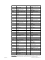
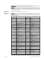
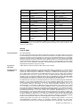
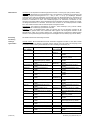
FLEX Monoclonal Rabbit Anti-Human Postmeiotic Segregation Increased 2 Clone EP51 Ready-to-Use (Link) English Code IR087 Intended use For in vitro diagnostic use. FLEX Monoclonal Rabbit Anti-Human Postmeiotic Segregation Increased 2, Clone EP51, Ready-to-Use (Link) is intended for use in immunohistochemistry together with Autostainer Link instruments. Antibodies to postmeiotic segregation increased 2 (PMS2) may be useful for the differential identification of colorectal carcinomas. When deficient, PMS2 is associated with the onset of hereditary non-polyposis colorectal cancer (HNPCC) (1, 2). The clinical interpretation of any staining or its absence should be complemented by morphological studies using proper controls and should be evaluated within the context of the patient's clinical history and other diagnostic tests by a qualified pathologist. Synonyms for antigen hPMS2, mismatch repair endonuclease PMS2, PMS1 protein homolog 2 Summary and explanation PMS2 is a ubiquitously expressed gene encoding the protein PMS2, an ~100 kDa nuclear protein involved in the DNA mismatch repair (MMR) pathway. The MMR pathway is utilized by normal proliferating cells to repair mutations that may occur during DNA replication. PMS2 forms a heterodimer with the mismatch repair protein MutL protein homolog 1 (MLH1), and the MLH1-PMS2 heterodimeric complex is recruited to the mismatch DNA sequence by a MutS MMR heterodimer consisting of MSH2 and MSH6, which binds directly to the base mismatch. The MLH1-PMS2 complex then initiates downstream functions including the excision of the mismatched DNA strands and repair through the recruitment of nucleases, polymerases, and other proteins (1). PMS2 deficiency is often the result of germline mutations in MMR deficient individuals (1). PMS2 loss of function results in an increased mutation rate and is associated with the development of HNPCC (also called Lynch syndrome). HNPCC, which accounts for 1-5% of colorectal cancers, is an autosomal dominant condition that results from germline mutations in any of at least four MMR genes including MSH2 (~40%), MLH1 (~50%), MSH6 (~5%) and PMS2 (<5%) and is associated with increased susceptibility to colon and endometrial cancer and to a lesser degree other types of neoplasms (1-3). A hallmark feature of cancers with defects in the MMR pathway is microsatellite instability (MSI), alterations in the number of microsatellite repeats in the DNA as a result of the faulty repair process (4, 5). A germline mutation in a single MMR gene copy predisposes patients to HNPCC, however, cancer development requires loss of function of the remaining wildtype copy. Mutations in PMS2 account for approximately 5% of all cases of HNPCC (6), however PMS2 is dependent upon MLH1 for stable expression, thus at the protein level, both sporadic and hereditary colorectal cancers deficient in MLH1 generally exhibit concomitant loss of PMS2 (7). Antibodies to PMS2 are useful for identifying mismatch repair deficiencies in tumors of the gastrointestinal tract including HNPCC and associated extracolonic cancers by immunohistochemistry (IHC). PMS2 deficiency as determined by IHC has been reported in endometrial, ovarian, breast, esophageal, and ampullary carcinomas, and melanomas (8-13). Refer to Dako’s General Instructions for Immunohistochemical Staining or the detection system instructions of IHC procedures. Reagent provided Ready-to-use monoclonal rabbit antibody provided in liquid form in a buffer containing stabilizing protein and 0.015 mol/L sodium azide. Clone: EP51. Isotype: Rabbit IgG. Immunogen Synthetic peptide corresponding to residues in human PMS2 protein. Specificity In Western blotting of SHSY5Y cells, anti-PMS2, clone EP51, recognizes a major band at 110 kDa corresponding to the expected molecular weight of PMS2. Precautions 1. For professional users. 2. This product contains sodium azide (NaN3), a chemical highly toxic in pure form. At product concentrations, though not classified as hazardous, sodium azide may react with lead and copper plumbing to form highly explosive build-ups of metal azides. Upon disposal, flush with large volumes of water to prevent metal azide build-up in plumbing. 3. As with any product derived from biological sources, proper handling procedures should be used. 4. Wear appropriate Personal Protective Equipment to avoid contact with eyes and skin. (122463-001) P01935EFG_001_IR087/2012.05 p. 1/4 5. Storage Quick guide Unused solution should be disposed of according to local, State and Federal regulations. Store at 2-8 °C. Do not use after expiration date s tamped on vial. If reagents are stored under any conditions other than those specified, the conditions must be verified by the user. There are no obvious signs to indicate instability of this product. Therefore, positive and negative controls should be run simultaneously with patient specimens. If unexpected staining is observed which cannot be explained by variations in laboratory procedures and a problem with the antibody is suspected, contact Dako Technical Support. Step Comments Fixation Pre-treatment Formalin EnVision™ FLEX, High pH (Code K8004) Dilution Dilution buffer Negative Control Visualization Counterstain Control Tissue Slides Ready-to-use Pre-diluted FLEX Negative Control, Rabbit (Code IR600) EnVision™ FLEX, High pH (Code K8000) EnVision™ FLEX Hematoxylin (Code K8008) Appendix, colon FLEX IHC Microscope Slides (Code K8020) Mounting Non-aqueous, permanent mounting required After staining, the sections must be dehydrated, cleared and mounted using permanent mounting medium. Instrumentation Autostainer Link 48 Use instrument-specific vials (Code SK201-SK203) 20 min HIER, 3-in-1 using PT Link and PT Link Rinse Station 30 min incubation 30 min incubation 30 min incubation, 2x5 min DAB+ 5 min incubation Nuclear staining Recommended for greater adherence of tissue sections to glass slides *The user must always read the package insert for detailed instructions of the staining procedure and handling of the product. Specimen preparation Paraffin sections: The antibody can be used for labeling formalin-fixed, paraffin-embedded tissue sections. Tissue specimens should be cut into sections of approximately 4 µm. Pre-treatment: Pre-treatment of formalin-fixed, paraffin-embedded tissue sections with heat-induced epitope retrieval (HIER) is required. Pretreating tissues with HIER using diluted EnVision™ FLEX Target Retrieval Solution, High pH (50x) (Code K8004) is recommended. Deparaffinization, rehydration and epitope retrieval can be performed in Dako PT Link (Code PT100/PT101). For details, please refer to PT Link User Guide. The following parameters should be used for PT Link: Pre-heat temperature: 65 °C; epitope retrieval temper ature and time: 97 °C for 20 (±1) minutes; cool down to 65 °C . Remove slide rack from PT tank and immediately dip slides in jar/tank (e.g., PT Link Rinse Station (Code PT109)) containing diluted room temperature EnVision™ FLEX Wash Buffer (20x) (Code K8007). Leave slides in Wash Buffer for 1-5 minutes. The tissue sections should not dry out during the treatment or during the following immunohistochemical staining procedure. For greater adherence of tissue sections to glass slides, the use of FLEX IHC Microscope Slides (Code K8020) is recommended. After staining the sections must be dehydrated, cleared and mounted using permanent mounting medium. Staining procedure Visualization: The recommended visualization system is EnVision FLEX, High pH (Link) (Code K8000). Program: The staining steps and incubation times are pre-programmed into the Autostainer Link software. The recommended reagent application volume is 1 x 200 µL or 2 x 150 µL per slide. Please refer to the proper Autostainer Link User Guide for detailed instructions on loading slides and reagents. If the protocols are not available on the Autostainer Link platform, please contact Dako Technical Services. An Autostainer Link software installer can be found at www.dako.com\Installer. This installer will update DakoLink software with protocol and reagent information for Anti-PMS2, Clone EP51. The short name for the antibody protocol is PMS2 EP51. All incubation steps should be performed at room temperature. Counterstaining: The recommended counterstain is EnVision FLEX Hematoxylin (Link) (Code K8008). Nonaqueous, permanent mounting medium is recommended. Controls: Positive and negative controls should be run simultaneously using the same protocol as the patient specimens. The positive control tissue should include appendix or colon and the cells/structures should display reaction patterns as described for this tissue in the “Performance characteristics” section. The recommended negative control reagent is FLEX Negative Control, Rabbit (Link) (Code IR600). Staining interpretation The cellular staining pattern is predominantly nuclear. Performance characteristics Normal tissues: Monoclonal Rabbit Anti-Human Postmeiotic Segregation Increased 2, Clone EP51 positively labels the nuclei of a variety of cell types in normal tissue. Although not noted in the table below, positive nuclear staining was observed in endothelial cells in many tissue types. (14) Tissue Type (# tested) Adrenal (3) Positive Tissue Elements (3/3) Cortex (10-50%), nuclear (2/3) Medulla (10-50%), nuclear (122463-001) Tissue Type (# tested) Parathyroid (3) Pituitary (3) Positive Tissue Elements (3/3) Epithelium (20-80%), nuclear (3/3) Endocrine cells (50-90%), nuclear P01935EFG_001_IR087/2012.05 p. 2/4 Appendix (1) Bone marrow (2) Breast (3) Cerebellum (3) (1/1) Epithelium (80%), nuclear (1/1) Extra-follicular lymphocytes (100%), nuclear (1/1) Smooth muscle (50%), nuclear (2/2) Blast cells (10-20%), nuclear (1/1) Glandular epithelium (80%), nuclear (2/2) Ductal epithelium (80%), nuclear (1/1) Connective Tissue (30%), nuclear (3/3) Purkinje cells (80-90%), nuclear (3/3) Glial cells (50%), nuclear Cerebrum (3) (3/3) Glial cells (20-60%), nuclear Cervix (3) (3/3) Basal epithelium (80-100%), nuclear (3/3) Stromal cells (50%), nuclear Colon (3) Esophagus (3) Heart (3) Kidney (3) Liver (3) Lung (3) Mesothelial cells (3) Nerve, peripheral (3) Ovary (3) Pancreas (3) (3/3) Epithelium (50-80%), nuclear (3/3) Lymphocytes (20-40%), nuclear (3/3) Connective tissue (10-40%), nuclear (3/3) Squamous epithelium (1050%), nuclear (1/1) Connective tissue (30%), nuclear (0/1) Lymphocytes (3/3) Cardiac cells (50-60%), nuclear (3/3) Connective tissue (30-40%), nuclear (3/3) Tubule (50-60%), nuclear (3/3) Bowman’s capsule (1020%), nuclear (3/3) Glomerulus (10%), nuclear (0/3) Hepatocytes (1/3) Bile duct (10%), nuclear (2/2) Lymphocytes (80%), nuclear (3/3) Alveolar epithelium (5060%), nuclear (2/2) Mesothelium (20-50%), nuclear (3/3) Connective tissue (50%), nuclear (3/3) Schwann cells (10-30%), nuclear (3/3) Connective tissue (30-50%), nuclear (3/3) Stromal cells (20-40%), nuclear (3/3) Follicular epithelium (1050%), nuclear (3/3) Acinar cells (50-60%), nuclear Prostate (3) Salivary gland (3) Skeletal muscle (3) Skin (3) Small intestine (3) Spleen (3) Stomach (3) Testis (3) Thymus (3) Thyroid (3) Tonsil (3) Uterus (3) (3/3) Glandular and ductal epithelium (40-90%), nuclear (3/3) Stromal cells (50-70%), nuclear (3/3) Ductal epithelium (60-80%), nuclear (3/3) Glandular epithelium (2080%), nuclear (1/1) Smooth muscle (30%), nuclear (1/1) Lymphoid cells (50%), nuclear (2/2) Connective tissue cells (20%), nuclear (0/3) Muscle cells (3/3) Connective tissue (10-20%), nuclear (2/2) Basal epithelium (50-80%), nuclear (3/3) Sweat gland (30-90%), nuclear (3/3) Epithelium (60-80%), nuclear (3/3) Smooth muscle (30-40%), nuclear (3/3) Resting lymphocytes (2070%), nuclear (1/1) Reactive lymphocytes (80%), nuclear (3/3) Sinus lining (red pulp) (3050%), nuclear (1/1) Connective tissue (30%), nuclear (1/2) Epithelium (10% ), nuclear (1/1) Gastric gland (10%), nuclear (2/2) Smooth muscle (20-60%), nuclear (1/1) Lymphocytes (60%), nuclear (3/3) Leydig cells (60-80%), nuclear (3/3) Tubular cells (10-20%), nuclear (3/3) Cortex (10-30%), nuclear (3/3) Medulla (10%), nuclear (2/3) Follicular epithelium (50%), nuclear (3/3) Parafollicular C cells (1030%), nuclear (3/3) Follicular lymphocytes (4060%), nuclear (3/3) Extra-follicular lymphocytes (40-50%), nuclear (3/3) Epithelium (60-80%), nuclear (3/3) Glandular epithelium (3050%), nuclear (3/3) Stromal cells (50-60%), nuclear (3/3) Myometrium (50%), nuclear (3/3) Islet cells (10-30%), nuclear (3/3) Pancreatic ductal cells (1040%), nuclear Abnormal tissues: In PMS2 deficient tumors, no nuclear staining should be observed in the neoplastic cells. A population of benign cells should demonstrate nuclear staining and serve as an internal tissue control. (122463-001) P01935EFG_001_IR087/2012.05 p. 3/4 Français Réf. IR087 Intérêt Pour diagnostic in vitro. L’anticorps FLEX Monoclonal Rabbit Anti-Human Postmeiotic Segregation Increased 2, Clone EP51, Ready-toUse (Link), est destiné à être utilisé en immunohistochimie avec les appareils Autostainer Link. Les anticorps dirigés contre la protéine PMS2 (postmeiotic segregation increased 2) peuvent être utiles pour l’identification différentielle des cancers colorectaux. Lorsqu’elle est déficiente, PMS2 est associée à l’apparition d’un cancer colorectal héréditaire sans polypose ou syndrome HNPCC (hereditary non-polyposis colorectal cancer) (1, 2). L’interprétation clinique de toute coloration ou son absence doit être complétée par des études morphologiques en utilisant des contrôles appropriés et doit être évaluée en fonction des antécédents cliniques du patient et d’autres tests diagnostiques par un pathologiste qualifié. Synonymes de l’antigène hPMS2, endonucléase PMS2 de réparation des mésappariements, homologue 2 de la protéine PMS1 Résumé et explication PMS2 est un gène exprimé de manière ubiquitaire codant pour la protéine PMS2, une protéine nucléaire d’environ 100 kDa impliquée dans la voie de réparation des mésappariements (mismatch repair, MMR) de l’ADN. La voie MMR est utilisée par les cellules saines en prolifération afin de réparer les mutations pouvant survenir au cours de la réplication de l’ADN. PMS2 forme un hétérodimère avec la protéine de réparation des mésappariements MLH1 (MutL protein homolog 1), et le complexe hétérodimérique MLH1-PMS2 est recruté pour la séquence ADN mésappariée par un complexe hétérodimérique de réparation des mésappariements MutS composé de MSH2 et de MSH6, qui se lie directement là où se trouve le mésappariement de la base. Le complexe MLH1-PMS2 initie ensuite des fonctions en aval, y compris l’excision des brins d’ADN mésappariés et la réparation grâce au recrutement de nucléases, de polymérases et d’autres protéines (1). Une déficience en PMS2 est souvent le résultat de mutations au niveau des lignées germinales chez des individus présentant une déficience de la réparation des mésappariements (1). La perte de fonction de PMS2 se traduit par une augmentation du taux de mutations et est associée au développement d’un syndrome HNPCC (appelé aussi syndrome de Lynch). Le syndrome HNPCC, qui représente 1 à 5 % des cancers colorectaux, est une maladie autosomique dominante qui résulte de mutations au niveau des lignées germinales dans au moins un des quatre gènes MMR, y compris MSH2 (env. 40 %), MLH1 (env. 50 %), MSH6 (env. 5 %) et PMS2 (<5 %), et il est associé à une augmentation de la susceptibilité au cancer du côlon et de l’endomètre et à un moindre degré aux autres types de cancers (1-3). Une caractéristique de marque des cancers présentant des erreurs dans la voie MMR est l’instabilité des microsatellites (MSI), c’est-à-dire des altérations du nombre de répétitions des microsatellites dans l’ADN à cause du processus de réparation défectueux (4, 5). Une mutation au niveau des lignées germinales dans une seule copie d’un gène MMR prédispose les patients à un syndrome HNPCC ; toutefois, le développement d’un cancer nécessite la perte de fonction de la copie du type sauvage restante. Les mutations de PMS2 représentent environ 5 % de l’ensemble des cas de syndrome HNPCC (6) ; toutefois, la stabilité de l’expression de PMS2 dépend de MLH1, ainsi au niveau protéique, les cancers colorectaux sporadiques ainsi que les cancers colorectaux héréditaires ayant une déficience en MLH1 présentent en général une perte concomitante de PMS2 (7). Les anticorps dirigés contre PMS2 sont utiles pour identifier par immunohistochimie (IHC) les déficiences de la réparation des mésappariements dans les tumeurs de l’appareil digestif, y compris le syndrome HNPCC et les cancers extra-coliques associés. Une déficience en PMS2 déterminée par IHC a été rapportée dans les cancers de l’endomètre, de l’ovaire, du sein, de l’œsophage et de l’ampoule de Vater, ainsi que dans les mélanomes (8-13). Se reporter aux Instructions générales de coloration immunohistochimique de Dako ou aux instructions du système de détection relatives aux procédures IHC. Réactif fourni Anticorps monoclonal de lapin prêt à l’emploi fourni sous forme liquide dans un tampon contenant une protéine stabilisante et 0,015 mol/L d’azide de sodium. Clone : EP51. Isotype : IgG de lapin. Immunogène Peptide synthétique correspondant aux résidus de la protéine PMS2 humaine. Spécificité Lors de Western Blots effectués sur des cellules SHSY5Y, l’anticorps anti-PMS2, clone EP51 reconnaît une bande principale à 110 kDa qui correspond au poids moléculaire attendu de PMS2. (122463-001) P01935EFG_001_IR087/2012.05 p. 4/4 Précautions d’emploi Conservation Guide rapide* 1. Pour utilisateurs professionnels. 2. Ce produit contient de l’azide de sodium (NaN3), produit chimique hautement toxique dans sa forme pure. Aux concentrations du produit, bien que non classé comme dangereux, l’azide de sodium peut réagir avec le cuivre et le plomb des canalisations et former des accumulations d’azides métalliques hautement explosives. Lors de l’élimination, rincer abondamment à l’eau pour éviter toute accumulation d’azide métallique dans les canalisations. 3. Comme avec tout produit d’origine biologique, respecter les procédures de manipulation appropriées. 4. Porter un équipement de protection individuelle approprié pour éviter tout contact avec les yeux et la peau. 5. Les solutions non utilisées doivent être éliminées conformément aux réglementations locales et nationales. Conserver entre 2 et 8 °C. Ne pas utiliser après la date de péremption indiquée sur le flacon. Si les réactifs sont conservés dans des conditions autres que celles indiquées, celles-ci doivent être validées par l’utilisateur. Il n’existe pas de signe particulier pour indiquer l’instabilité de ce produit. Par conséquent, des contrôles positifs et négatifs doivent être testés en même temps que les échantillons de patients. Si une coloration inattendue est observée, qui ne peut être expliquée par un changement des procédures du laboratoire, et en cas de suspicion d’un problème lié à l’anticorps, contacter l’assistance technique de Dako. Étape Commentaires Fixation Prétraitement Formol EnVision™ FLEX, High pH (réf. K8004) Dilution Tampon de dilution Contrôle négatif Visualisation Prêt à l’emploi Prédilué FLEX Negative Control, Rabbit (réf. IR600) EnVision™ FLEX, High pH (réf. K8000) Contre-coloration Tissu de contrôle Lames EnVision™ FLEX Hematoxylin (réf. K8008) Appendice, côlon FLEX IHC Microscope Slides (réf. K8020) Montage Milieu de montage permanent non aqueux requis Une fois la procédure de coloration terminée, les coupes doivent être déshydratées, éclaircies et montées à l’aide d’un milieu de montage permanent. Appareillage Autostainer Link 48 Utiliser les flacons spécifiques à l’appareil (réf. SK201 à SK203) Démasquage des épitopes induit par la chaleur (HIER) de 20 minutes, procédure 3 en 1 à l’aide de l’appareil PT Link et de la station de rinçage du PT Link Incubation de 30 minutes Incubation de 30 minutes Incubation de 30 minutes, 2 incubations de 5 minutes avec le DAB+ Incubation de 5 minutes Coloration nucléaire Recommandées pour une meilleure adhérence des coupes de tissu sur les lames de verre *L’utilisateur doit toujours lire la notice pour obtenir des instructions détaillées sur la procédure de coloration et sur la manipulation du produit. Préparation des échantillons Coupes en paraffine : l’anticorps peut être utilisé pour le marquage des coupes de tissu fixées au formol et incluses en paraffine. L’épaisseur des coupes d’échantillons de tissu doit être d’environ 4 µm. Prétraitement : le prétraitement des coupes de tissu fixées au formol et incluses en paraffine par démasquage des épitopes induit par la chaleur (HIER) est nécessaire. Il est recommandé de prétraiter les tissus par la méthode HIER à l’aide de la solution de démasquage des cibles EnVision™ FLEX Target Retrieval Solution, High pH (50x) (réf. K8004) diluée. Le déparaffinage, la réhydratation et le démasquage des épitopes peuvent être réalisés dans l’appareil PT Link de Dako (réf. PT100/PT101). Pour plus de détails, se reporter au Manuel d’utilisation du PT Link. Les paramètres suivants doivent être utilisés pour le PT Link : température de préchauffage : 65 °C ; température et durée du démasquage des épitopes : 97 °C pendant 20 minutes (±1 minute) ; laisser refroidir jusqu’à 65 °C. Reti rer le portoir de lames de la cuve PT et plonger immédiatement les lames dans une jarre/cuve (par ex. station de rinçage du PT Link, réf. PT109) contenant du tampon de lavage EnVision™ FLEX Wash Buffer (20x) (réf. K8007) dilué à température ambiante. Laisser les lames dans le tampon de lavage pendant 1 à 5 minutes. Les coupes de tissu ne doivent pas sécher lors du traitement ni lors de la procédure de coloration immunohistochimique qui suit. Pour une meilleure adhérence des coupes de tissu sur les lames de verre, il est recommandé d’utiliser les lames FLEX IHC Microscope Slides (réf. K8020). Une fois la procédure de coloration terminée, les coupes doivent être déshydratées, éclaircies et montées à l’aide d’un milieu de montage permanent. Procédure de coloration (122463-001) Visualisation : le système de visualisation recommandé est le système EnVision FLEX, High pH (Link) (réf. K8000). Programme : les étapes de coloration et les temps d’incubation sont préprogrammés dans le logiciel Autostainer Link. Le volume de réactif recommandé devant être appliqué est de 1 x 200 µL ou 2 x 150 µL par lame. Se reporter au Manuel d’utilisation de l’Autostainer Link correspondant pour plus de détails sur le chargement des lames et des réactifs. Si les protocoles ne sont pas disponibles sur la plate-forme Autostainer Link, contacter l’assistance technique de Dako. Un programme d’installation du logiciel Autostainer Link peut être obtenu sur www.dako.com\Installer. Ce programme d’installation mettra le logiciel DakoLink à jour avec les informations relatives au protocole et aux réactifs pour l’anticorps anti-PMS2, clone EP51. Le nom abrégé pour le protocole de l’anticorps est PMS2 EP51. Toutes les étapes d’incubation doivent être effectuées à température ambiante. P01935EFG_001_IR087/2012.05 p. 5/4 Contre-coloration : le contre-colorant recommandé est le produit EnVision FLEX Hematoxylin (Link) (réf. K8008). L’utilisation d’un milieu de montage permanent non aqueux est recommandée. Contrôles : des contrôles positifs et négatifs doivent être testés en même temps et en suivant le même protocole que les échantillons de patients. Le tissu de contrôle positif doit comprendre l’appendice ou le côlon et les cellules/structures doivent présenter les schémas de réaction décrits pour ces tissus dans la section « Caractéristiques de performance ». Le réactif de contrôle négatif recommandé est le produit FLEX Negative Control, Rabbit, (Link) (réf. IR600). Interprétation de la coloration Le schéma de coloration cellulaire est principalement nucléaire. Caractéristiques de performance Tissus sains : l’anticorps Monoclonal Rabbit Anti-Human Postmeiotic Segregation Increased 2, Clone EP51, marque de façon positive les noyaux de divers types cellulaires dans les tissus sains. Bien que cela ne soit pas noté dans le tableau ci-après, une coloration nucléaire positive a été observée dans les cellules endothéliales de nombreux types de tissu (14). Type de tissu (nb. testés) Surrénale (3) Appendice (1) Moelle osseuse (2) Sein (3) Cervelet (3) Cerveau (3) Col de l’utérus (3) Côlon (3) Œsophage (3) Cœur (3) Rein (3) Foie (3) Éléments tissulaires positifs (2/3) Moelle (10 à 50 %), nucléaire Type de tissu (nb. testés) Parathyroïde (3) Pituitaire (3) (1/1) Épithélium (80 %), nucléaire Prostate (3) (3/3) Cortex (10 à 50 %), nucléaire (1/1) Lymphocytes extrafolliculaires (100 %), nucléaire (1/1) Muscle lisse (50 %), nucléaire (2/2) Blastes (10 à 20 %), nucléaire (1/1) Épithélium glandulaire (80 %), nucléaire (2/2) Épithélium canalaire (80 %), nucléaire (1/1) Tissu conjonctif (30 %), nucléaire (3/3) Cellules de Purkinje (80 à 90 %), nucléaire (3/3) Cellules gliales (50 %), nucléaire (3/3) Cellules gliales (20 à 60 %), nucléaire (3/3) Épithélium basal (80 à 100 %), nucléaire (3/3) Cellules stromales (50 %), nucléaire (3/3) Épithélium (50 à 80 %), nucléaire (3/3) Lymphocytes (20 à 40 %), nucléaire (3/3) Tissu conjonctif (10 à 40 %), nucléaire (3/3) Épithélium épidermoïde (10 à 50 %), nucléaire (1/1) Tissu conjonctif (30 %), nucléaire (0/1) Lymphocytes (3/3) Cellules cardiaques (50 à 60 %), nucléaire (3/3) Tissu conjonctif (30 à 40 %), nucléaire (3/3) Tubule (50 à 60 %), nucléaire (3/3) Capsule de Bowman (10 à 20 %), nucléaire (3/3) Glomérule (10 %), nucléaire (0/3) Hépatocytes (1/3) Canal biliaire (10 %), nucléaire (122463-001) Glande salivaire (3) Muscle squelettique (3) Peau (3) Intestin grêle (3) Rate (3) Estomac (3) Testicule (3) Thymus (3) Éléments tissulaires positifs (3/3) Épithélium (20 à 80 %), nucléaire (3/3) Cellules endocrines (50 à 90 %), nucléaire (3/3) Épithélium glandulaire et canalaire (40 à 90 %), nucléaire (3/3) Cellules stromales (50 à 70 %), nucléaire (3/3) Épithélium canalaire (60 à 80 %), nucléaire (3/3) Épithélium glandulaire (20 à 80 %), nucléaire (1/1) Muscle lisse (30 %), nucléaire (1/1) Cellules lymphoïdes (50 %), nucléaire (2/2) Cellules du tissu conjonctif (20 %), nucléaire (0/3) Cellules musculaires (3/3) Tissu conjonctif (10 à 20 %), nucléaire (2/2) Épithélium basal (50 à 80 %), nucléaire (3/3) Glande sudoripare (30 à 90 %), nucléaire (3/3) Épithélium (60 à 80 %), nucléaire (3/3) Muscle lisse (30 à 40 %), nucléaire (3/3) Lymphocytes au repos (20 à 70 %), nucléaire (1/1) Lymphocytes réactifs (80 %), nucléaire (3/3) Tapis de sinus (pulpe rouge) (30 à 50 %), nucléaire (1/1) Tissu conjonctif (30 %), nucléaire (1/2) Épithélium (10 %), nucléaire (1/1) Glande gastrique (10 %), nucléaire (2/2) Muscle lisse (20 à 60 %), nucléaire (1/1) Lymphocytes (60 %), nucléaire (3/3) Cellules de Leydig (60 à 80 %), nucléaire (3/3) Cellules tubulaires (10 à 20 %), nucléaire (3/3) Cortex (10 à 30 %), nucléaire (3/3) Moelle (10 %), nucléaire P01935EFG_001_IR087/2012.05 p. 6/4 Poumon (3) Cellules mésothéliales (3) Nerf périphérique (3) Ovaire (3) Pancréas (3) (2/2) Lymphocytes (80 %), nucléaire (3/3) Épithélium alvéolaire (50 à 60 %), nucléaire (2/2) Mésothélium (20 à 50 %), nucléaire (3/3) Tissu conjonctif (50 %), nucléaire (3/3) Cellules de Schwann (10 à 30 %), nucléaire (3/3) Tissu conjonctif (30 à 50 %), nucléaire (3/3) Cellules stromales (20 à 40 %), nucléaire (3/3) Épithélium folliculaire (10 à 50 %), nucléaire (3/3) Cellules acinaires (50 à 60 %), nucléaire Thyroïde (3) Amygdale (3) Utérus (3) (2/3) Épithélium folliculaire (50 %), nucléaire (3/3) Cellules C parafolliculaires (10 à 30 %), nucléaire (3/3) Lymphocytes folliculaires (40 à 60 %), nucléaire (3/3) Lymphocytes extrafolliculaires (40 à 50 %), nucléaire (3/3) Épithélium (60 à 80 %), nucléaire (3/3) Épithélium glandulaire (30 à 50 %), nucléaire (3/3) Cellules stromales (50 à 60 %), nucléaire (3/3) Myomètre (50 %), nucléaire (3/3) Cellules des îlots de Langerhans (10 à 30 %), nucléaire (3/3) Cellules canalaires pancréatiques (10 à 40 %), nucléaire Tissus tumoraux : dans les tumeurs déficientes en PMS2, aucune coloration nucléaire ne doit être observée dans les cellules néoplasiques. Une population de cellules bénignes doit présenter une coloration nucléaire et servir de tissu de contrôle interne. Deutsch Code-Nr. IR087 Verwendungszweck Zur In-vitro-Diagnostik FLEX Monoclonal Rabbit Anti-Human Postmeiotic Segregation Increased 2, Clone EP51, Ready-to-Use (Link) ist für die Verwendung in der Immunhistochemie zusammen mit Autostainer Link-Geräten bestimmt. Antikörper für Postmeiotic segregation increased 2 (PSM2) können hilfreich bei der Differentialidentifizierung von kolorektalen Karzinomen sein. Ein Defekt von PMS2 wird mit dem Auftreten des erblichen, nicht polypösen kolorektalen Karzinoms (hereditary non-polyposis colorectal cancer, HNPCC) assoziiert (1, 2). Die klinische Auswertung einer eventuell eintretenden Färbung sollte durch morphologische Studien mit ordnungsgemäßen Kontrollen ergänzt werden und von einem qualifizierten Pathologen unter Berücksichtigung der Krankengeschichte und anderer Diagnostiktests des Patienten vorgenommen werden. Synonyme für das Antigen hPMS2, Mismatch-Repair-Endonuklease PMS2, PMS1 Protein Homolog 2 Zusammenfassung und Erklärung PMS2 ist ein ubiquitär exprimiertes Gen, das das Protein PMS2 kodiert, ein Kernprotein von 100 kDa, welches wiederum am DNA-Mismatch-Repair (MMR)-Pfad beteiligt ist. Der MMR-Mechanismus wird von sich normal vermehrenden Zellen verwendet, um Mutationen zu reparieren, die während der DNA-Replikation auftreten können. PMS2 bildet ein Heterodimer mit dem MMR-Protein MutL Protein Homolog 1 (MLH1). Der heterodimere MLH1-PMS2-Komplex wird durch ein weiteres, aus MSH2 und MSH6 bestehendem MutS-MMR-Heterodimer, das direkt an den zu reparierenden Basen-Mismatch bindet, zum DNA-Mismatch gebracht. Der MLH1-PMS2Komplex löst dann eine Reihe von Funktionen aus, die das Ausschneiden des falsch gepaarten DNA-Strangs und die Reparatur durch Nukleasen, Polymerasen und weitere spezielle Proteine einschließt (1). Ein PMS2-Defekt ist oft die Folge von Keimbahnmutationen bei Personen mit Beeinträchtigungen der MismatchReparatur (1). Der Verlust der PMS2-Funktion führt zu einer gesteigerten Mutationsrate und wird mit dem Entstehen von HNPCC (auch bekannt als Lynch-Syndrom) in Zusammenhang gebracht. HNPCC, das für 1–5 % der kolorektalen Karzinome verantwortlich ist, ist eine autosomal-dominante Erkrankung, die aus der Keimbahnmutation von mindestens vier MMR-Genen entsteht, darunter MSH2 (~40 %), MLH1 (~50 %), MSH6 (~5 %) und PMS2 (<5 %); sie wird mit einer größeren Anfälligkeit für Kolon- und Endometriumkarzinome und zu einem geringeren Grade für andere Arten von Neoplasien in Verbindung gebracht (1-3). Deutliches Kennzeichen für Karzinome mit Defekten im MMR-Pfad ist die Mikrosatelliteninstabilität (MSI), Änderungen in der Anzahl von Mikrosatellitenwiederholungen in der DNA aufgrund des fehlerhaften Reparaturprozesses (4, 5). Eine Keimbahnmutation in einer einzelnen MMR-Gen-Kopie stellt eine Prädisposition des Patienten für HNPCC dar, für die Entwicklung des Karzinoms ist allerdings der Funktionsverlust der verbleibenden Wildtyp-Kopie erforderlich. Mutationen in PMS2 sind für ca. 5 % aller Fälle von HNPCC verantwortlich (6), PMS2 hängt jedoch von MLH1 für eine stabile Expression ab, sodass auf der Proteinebene sporadische und hereditäre kolorektale Karzinome mit einer Defizienz an MLH1 den damit einhergehenden Verlust von PMS2 aufweisen (7). Antikörper gegen PMS2 sind hilfreich bei der Identifizierung von Mismatch-Repair-Defekten in Tumoren des Gastrointestinaltrakts einschließlich HNPCC und assoziierter Krebsarten außerhalb des Kolons durch Immunhistochemie (IHC). Die durch IHC definierte PMS2-Defizienz wurde in Endometrium-, Ovarial-, Mamma-, (122463-001) P01935EFG_001_IR087/2012.05 p. 7/4 Ösophagus- und Papillenkarzinomen und -melanomen beobachtet (8-13). Siehe Allgemeine Richtlinien zur immunhistochemischen Färbung von Dako oder die Anweisungen des Detektionssystems von IHC-Verfahren. Geliefertes Reagenz Gebrauchsfertiger, monoklonaler Kaninchen-Antikörper in flüssiger Form in einem Puffer, der stabilisierendes Protein und 0,015 mol/L Natriumazid enthält. Klon: EP51. Isotyp: Kaninchen-IgG. Immunogen Synthetisches Peptid, das den Rückständen in humanem PMS2-Protein entspricht. Spezifität Beim Western-Blot von SHSY5Y-Zellen erkannte der Anti-PMS2, Clone EP51 eine starke Bande bei 110 kDa, die mit dem erwarteten Molekulargewicht von PMS2 übereinstimmt. Vorsichtsmaßnahmen 1. Für Fachpersonal. 2. Dieses Produkt enthält Natriumazid (NaN3), eine in reiner Form äußerst giftige Chemikalie. Bei den in diesem Produkt verwendeten Konzentrationen kann Natriumazid, obwohl nicht als gefährlich klassifiziert, mit Blei- und Kupferabflussrohren reagieren und hochexplosive Metallazide bilden. Nach der Entsorgung stets mit viel Wasser nachspülen, um Ansammlungen von Metallazid in den Leitungen vorzubeugen. 3. Wie alle Produkte biologischen Ursprungs müssen auch diese entsprechend gehandhabt werden. 4. Geeignete Schutzkleidung tragen, um Augen- und Hautkontakt zu vermeiden. 5. Nicht verwendete Lösung ist entsprechend örtlichen, bundesstaatlichen und staatlichen Richtlinien zu entsorgen. Lagerung Kurzanleitung* Bei 2–8 °C aufbewahren. Nach Ablauf des auf dem Flä schchen aufgedruckten Verfalldatums nicht mehr verwenden. Werden die Reagenzien nicht entsprechend den angegebenen Bedingungen aufbewahrt, müssen die Bedingungen vom Anwender geprüft werden. Es gibt keine offensichtlichen Anzeichen für eine eventuelle Produktinstabilität. Positiv- und Negativkontrollen sollten daher zur gleichen Zeit wie die Patientenproben durchgeführt werden. Falls es zu einer unerwarteten Färbung kommt, die sich nicht durch Unterschiede bei Laborverfahren erklären lässt und auf ein Problem mit dem Antikörper hindeutet, ist der technische Kundendienst von Dako zu verständigen. Schritt Anmerkungen Fixierung Vorbehandlung Formalin EnVision™ FLEX, High pH (Code-Nr. K8004) Verdünnung Verdünnungspuffer Negativkontrolle Visualisierung Gegenfärbung Kontrollgewebe Objektträger Gebrauchsfertig Vorverdünnt FLEX Negative Control, Rabbit (Code-Nr. IR600) EnVision™ FLEX, High pH (Code-Nr. K8000) EnVision™ FLEX Hematoxylin (Code-Nr. K8008) Appendix, Kolon FLEX IHC Microscope Slides (Code-Nr. K8020) 30 Min. Inkubation 30 Min. Inkubation, 2 x 5 Min. DAB+ 5 Min. Inkubation Nukleäre Färbung Zur besseren Haftung der Gewebeschnitte an Glasobjektträgern Fixierung Nichtwässrige, permanente Fixierung erforderlich Nach dem Färben müssen die Schnitte dehydriert, geklärt und unter Verwendung eines permanenten Fixiermittels fixiert werden. Geräte Autostainer Link 48 Gerätespezifische Fläschchen verwenden (Code-Nr. SK201-SK203) 20 Min. HIER, 3-in-1 mit PT Link und PT Link Rinse Station 30 Min. Inkubation *Der Anwender muss stets die Packungsbeilage lesen, um sich über die detaillierten Anweisungen für das Färbeverfahren und die Handhabung des Produkts zu informieren. Probenvorbereitung Paraffinschnitte: Der Antikörper eignet sich zur Markierung von formalinfixierten, paraffineingebetteten Gewebeschnitten. Gewebeproben sollten in Schnitte von ca. 4 µm Stärke geschnitten werden. Vorbehandlung: Es ist eine Vorbehandlung der formalinfixierten und paraffineingebetteten Gewebeschnitte durch hitzeinduzierte Epitopdemaskierung (HIER) erforderlich. Die Vorbehandlung des Gewebes mit HIER unter Verwendung von verdünnter EnVision™ FLEX Target Retrieval Solution, High pH (50x) (Code-Nr. K8004) wird empfohlen. Entparaffinierung, Rehydrierung und Epitopdemaskierung können im Dako PT Link (Code-Nr. PT100/PT101) vorgenommen werden. Weitere Informationen hierzu siehe PT Link-Benutzerhandbuch. Für PT Link sollten die folgenden Parameter verwendet werden: Vorwärmtemperatur: 65 °C; Temperatur und Zeit f ür Epitopdemaskierung: 97 °C für 20 (±1) Minuten; auf 65 °C abkühlen. Das Objektträgergestell aus dem PT LinkBehälter herausnehmen und die Objektträger sofort in ein Gefäß/einen Behälter (z. B. PT Link Rinse Station, Code-Nr. PT109) mit verdünntem, auf Zimmertemperatur gebrachtem EnVision™ FLEX Wash Buffer (20x) (Code-Nr. K8007) eintauchen. Die Objektträger für 1–5 Minuten im Wash Buffer belassen. Die Gewebeschnitte dürfen während der Behandlung oder des anschließenden immunhistochemischen Färbeverfahrens nicht austrocknen. Für eine bessere Haftung der Gewebeschnitte an den Glas-Objektträgern werden FLEX IHC Microscope Slides (Code-Nr. K8020) empfohlen. Nach dem Färben müssen die Schnitte dehydriert, geklärt und unter Verwendung eines permanenten Fixiermittels fixiert werden. (122463-001) P01935EFG_001_IR087/2012.05 p. 8/4 Färbeverfahren Visualisierung: Das empfohlene Visualisierungssystem ist EnVision FLEX, High pH (Link) (Code-Nr. K8000). Programm: Die Färbeschritte und Inkubationszeiten sind in der Autostainer Link-Software vorprogrammiert. Das empfohlene Reagenz-Anwendungsvolumen ist 1 x 200 µL oder 2 x 150 µL pro Objektträger. Detaillierte Anweisungen zum Laden der Objektträger und Reagenzien bitte dem Benutzerhandbuch zum Autostainer Link entnehmen. Wenn die Protokolle auf der Autostainer Link-Plattform nicht verfügbar sind, wenden Sie sich an den technischen Kundendienst von Dako. Das Installationsprogramm der Autostainer Link-Software ist unter www.dako.com\Installer erhältlich. Dieses Installationsprogramm aktualisiert die DakoLink Software mit Protokollund Reagenzinformationen für Anti-PMS2, Clone EP51. Der Kurzname für das Antikörperprotokoll ist PMS2 EP51. Alle Inkubationsschritte sollten bei Raumtemperatur durchgeführt werden. Gegenfärbung: Die empfohlene Gegenfärbung ist EnVision FLEX Hematoxylin (Link) (Code-Nr. K8008). Empfohlen wird ein nichtwässriges, permanentes Fixiermittel. Kontrollen: Positiv- und Negativkontrollen sollten zur gleichen Zeit und mit demselben Protokoll wie die Patientenproben getestet werden. Die Positivkontrolle sollte Appendix- oder Kolongewebe enthalten und die Zellen/Strukturen sollten die für dieses Gewebe unter „Leistungsmerkmale“ beschriebenen Reaktionsmuster aufweisen. Das empfohlene Negativ-Kontrollreagenz ist FLEX Negative Control, Rabbit (Link) (Code-Nr. IR600). Auswertung der Färbung Das zelluläre Färbemuster ist überwiegend nukleär. Leistungseigenschaften Gesunde Gewebe: Monoclonal Rabbit Anti-Human Postmeiotic Segregation Increased 2, Clone EP51 markiert die Kerne einer Reihe von Zelltypen in gesundem Gewebe. Obwohl nicht in der Tabelle unten aufgezeigt, wurde eine positive nukleäre Färbung in Endothelzellen bei vielen Gewebetypen beobachtet. (14) Gewebetyp (Anz. getestet) Nebennieren (3) (2/3) Medulla (10–50 %), nukleär Gewebetyp (Anz. getestet) Nebenschilddrüse (3) Hypophyse (3) Appendix (1) (1/1) Epithel (80 %), nukleär Prostata (3) Knochenmark (2) (1/1) Extrafollikuläre Lymphozyten (100 %), nukleär (1/1) Glatte Muskelzellen (50 %), nukleär (2/2) Blastzellen (10–20 %), nukleär (1/1) Drüsenepithel (80 %), nukleär (2/2) Duktusepitel (80 %), nukleär Mamma (3) Zerebellum (3) Zerebrum (3 (3/3) Cortex (10–50 %), nukleär (1/1) Bindegewebe (30 %), nukleär (3/3) Purkinje-Zellen (80–90 %), nukleär (3/3) Gliazellen (50 %), nukleär Speicheldrüsen (3) Skelettmuskulatur (3) (3/3) Gliazellen (20–60 %), nukleär (3/3) Basalepithel (80–100 %), nukleär (3/3) Stromazellen (50 %), nukleär (3/3) Epithel (50–80 %), nukleär Haut (3) Milz (3) Herz (3) (3/3) Lymphozyten (20–40 %), nukleär (3/3) Bindegewebe (10–40 %), nukleär (3/3) Plattenepithel (10–50 %), nukleär (1/1) Bindegewebe (30 %), nukleär (0/1) Lymphozyten (3/3) Herzzellen (50–60 %), nukleär Niere (3) (3/3) Bindegewebe (30–40 %), nukleär (3/3) Tubuli (50–60 %), nukleär Zervix (3) Kolon (3) Speiseröhre (3) (122463-001) Positive Gewebeelemente Dünndarm (3) Magen (3) Positive Gewebeelemente (3/3) Epithel (20–80 %), nukleär (3/3) Endokrine Zellen (50–90 %), nukleär (3/3) Drüsen- und Duktusepithel (40–90 %), nukleär (3/3) Stromazellen (50–70 %), nukleär (3/3) Duktusepithel (60–80 %), nukleär (3/3) Drüsenepithel (20–80 %), nukleär (1/1) Glatte Muskelzellen (30 %), nukleär (1/1) Lymphoide Zellen (50 %), nukleär (2/2) Bindegewebszellen (20 %), nukleär (0/3) Muskelzellen (3/3) Bindegewebe (10–20 %), nukleär (2/2) Basalepithel (50–80 %), nukleär (3/3) Schweißdrüsen (30–90 %), nukleär (3/3) Epithel (60–80 %), nukleär (3/3) Glatte Muskelzellen (30–40 %), nukleär (3/3) Ruhende Lymphozyten (20–70 %), nukleär (1/1) Reaktive Lymphozyten (80 %), nukleär (3/3) Sinus auskleidende Zellen (rote Pulpa) (30–50 %), nukleär (1/1) Bindegewebe (30 %), nukleär (1/2) Epithel (10 %), nukleär (1/1) Magendrüse (10 %), nukleär (2/2) Glatte Muskelzellen (20–60 %), nukleär (1/1) Lymphozyten (60 %), nukleär P01935EFG_001_IR087/2012.05 p. 9/4 Leber (3) (3/3) Bowman-Kapsel (10–20 %), nukleär (3/3) Glomerulus (10 %), nukleär Hoden (3) (0/3) Hepatozyten Thymus (3) (3/3) Leydig-Zellen (60–80 %), nukleär (3/3) Tubuluszellen (10–20 %), nukleär (3/3) Cortex (10–30 %), nukleär Schilddrüse (3) (3/3) Medulla (10 %), nukleär (2/3) Follikelepithel (50 %), nukleär (1/3) Gallengang (10 %), nukleär (2/2) Lymphozyten (80 %), nukleär (3/3) Alveolarepithel (50–60 %), nukleär Mesothelzellen (3) (2/2) Mesothel (20–50 %), nukleär (3/3) Bindegewebe (50 %), nukleär Nerv, peripher (3) (3/3) Schwann-Zellen (10–30 %), nukleär (3/3) Bindegewebe (30–50 %), nukleär Ovarium (3) (3/3) Stromazellen (20–40 %), nukleär (3/3) Follikelepithel (10–50 %), nukleär Pankreas (3) (3/3) Azinuszellen (50–60 %), nukleär Lunge (3) Mandel (3) Uterus (3) (3/3) Parafollikuläre C-Zellen (10–30 %), nukleär (3/3) Follikuläre Lymphozyten (40–60 %), nukleär (3/3) Extrafollikuläre Lymphozyten (40–50 %), nukleär (3/3) Epithel (60–80 %), nukleär (3/3) Drüsenepithel (30–50 %), nukleär (3/3) Stromazellen (50–60 %), nukleär (3/3) Myometrium (50 %), nukleär (3/3) Inselzellen (10–30 %), nukleär (3/3) Zellen des Pankreasduktus (10–40 %), nukleär Pathologische Gewebe: Bei Tumoren mit PMS2-Defekt sollte in den neoplastischen Zellen keine nukleäre Färbung zu beobachten sein. Eine Population aus benignen Zellen sollte nukleäre Färbung aufweisen und als interne Gewebekontrolle dienen. Literaturangaben 1. 2. 3. Peltomäki P. Role of DNA mismatch repair defects in the pathogenesis of human cancer. J Clin Oncol 2003;21:1174-79. Lynch H. Smyrk, T. Hereditary nonpolyposis colorectal cancer (Lynch syndrome). An updated review. Cancer 1996;78:1149-67. Marcus VA, Madlensky L, Gryfe R, Kim H, So K, Millar A, et al. Immunohistochemistry for hMLH1 and hMSH2: A practical test for DNA mismatch repair-deficient tumors. Am J Surg Pathol 1999;23:1248-55. 4. Lanza G, Gafà R, Maestri I, Santini A, Matteuzzi M, Cavazzini, L. Immunohistochemical pattern of MLH1/MSH2 expression is related to clinical and pathological features in colorectal adenocarcinomas with microsatellite instability. Mod Pathol 2002;15:741-9. 5. Okuda T, Sekizawa A, Purwosunu Y, Nagatsuka M, Morioka M, Hayashi M, Okai T. Genetics of endometrial cancers. Obstet Gynecol Int 2010;984013. 6. Burgart, L.J. Testing for defective DNA mismatch repair in colorectal carcinoma. A practical guide. Arch Pathol Lab Med 2005;129:1385-9. 7. Lynch HT, Lynch PM, Lanspa SJ, Snyder CL, Lynch JF, Boland CR. Review of the Lynch syndrome: history, molecular genetics, screening, differential diagnosis, and medicolegal ramifications. Clin Genet 2009; 76:1-18. 8. Garg K, Shih K, Barakat R, Zhou Q, Iasonos A, Soslow RA. Endometrial carcinomas in women aged 40 years and younger: tumors associated with loss of DNA mismatch repair proteins comprise a distinct clinicopathologic subset. Am J Surg Pathol 2009;33:1869-77. 9. Pal T, Permuth-Wey J, Sellers TA. A review of the clinical relevance of mismatch-repair deficiency in ovarian cancer. Cancer 2008;113:733-42. 10. Buerki N, Gautier L, Kovac M, Marra G, Buser M, Heinimann K. Evidence for breast cancer as an integral part of Lynch syndrome. Gene Chrom Cancer 2012;51:83-91. 11. Agaram NP, Shia J, Tang LH, Klimstra DS. DNA mismatch repair deficiency in ampullary carcinoma: a morphologic and immunohistochemical study of 54 cases. Am J Clin Pathol 2010;133:772-80. 12. Farris AB 3rd, Demicco EG, Le LP, Finberg KE, Miller J, Mandal R, et al. Clinicopathologic and molecular profiles of microsatellite unstable Barrett Esophagus-associated adenocarcinoma. Am J Surg Pathol 2011;35:647-55. 13. Alvino E, Marra G, Pagani E, Falcinelli S, Pepponi R, Perrera C, et al. High-frequency microsatellite instability is associated with defective DNA mismatch repair in human melanoma. J Invest Dermatol 2002;118:79-86. 14. PMS2 clone EP51 M3647.IR087 30 Normal Report D08987 Monoclonal Rabbit Anti-Human Postmeiotic Segregation Increased 2, Clone EP51 has been created by Epitomics Inc., using Epitomics’ proprietary rabbit monoclonal antibody technology covered under Patent Nos 5,675,063 and 7,402,409. (122463-001) P01935EFG_001_IR087/2012.05 p. 10/4 Monoclonal Rabbit Anti-Human Postmeiotic Segregation Increased 2, Clone EP51, a été créé par Epitomics Inc.,suivant la technologie d’anticorps monoconal de lapin déposée de Epitomics, protégée par les brevets No 5,675,063 et 7,402,409. Monoclonal Rabbit Anti-Human Postmeiotic Segregation Increased 2, Clone EP51, wurde von Epitomics Inc. unter Verwendung der firmeneigenen Technik für monoklonale Kaninchen-Antikörper von Epitomics hergestellt und ist durch die Patente mit den Nummern 5,675,063 und 7,402,409 geschützt Edition 05/12 (122463-001) P01935EFG_001_IR087/2012.05 p. 11/4


![Human Tau [pT181]Singleplex Bead Kit](http://vs1.manualzilla.com/store/data/005836208_1-535995cf2d5d3bddb8b2b8afcdb790d7-150x150.png)